Lucrări ştiinţifice Zootehnie şi Biotehnologii, vol. 42 (1) (2009), Timişoara
NEOMYCIN INDUCES APOPTOSIS IN THE INNER EAR
NEOMICINA INDUCE APOPTOZA ÎN URECHEA INTERN
Ă
ROXANA D. VINTILA1, STAN COTULBEA1, KARINA GÜLTIG2, ANDREA MÜLLER2, CORNELIA VINTILA3, BERNHARDT HIRT2, HUBERT
LÖWENHEIM2
, H. P. ZENNER
1 Department of ENT, University of Medicine and Pharmacy Timisoara, Romania 2 Department of Otorhinolaryngology, University of Tübingen, E. Aulhornstr. 5, Germany
(Tübingen Hearing Research Center), Marie Curie Program 3Banat University of Agriculture and Veterinary Medicine Timisoara
Objectives: to study the apoptotic response of auditory hair cells in the organ of Corti after exposure to neomycin within the first 6 hours. Materials and methods: auditory hair cells harvested on the postnatal day 7 from the cochleae of C57BL6 wild type mice were cultured in vitro in the presence (neomycin group, n= 59 cochleas) or absence of neomycin (control group, n= 31 cochleas). Apoptotic cells were evaluated within 6 hours of incubation for the expression of active caspases (8, 9 or 3) in the cytoplasm and counterstained using propidium iodide to highlight apoptotic nuclei. Cells were statistically quantified using average and standard deviation. The differences between the averages were tested with the help of Student T test (threshold p< 0,05). Results: The statistic comparison between the different caspase types (caspase 3, 8 and 9), the average and dispersion inside the cell groups, permitted us to detect the first step of apoptosis taking place in the cytoplasm. The comparison of the cell group where propidium iodide was accumulated in the nucleus with the cell group where both the caspase and propidium iodide appeared, permitted us to detect the initiation and development of the second step of apoptosis, the one where the nucleus has already been affected. Conclusions: Neomycin is initiating, right after 6 hours of incubation, the process of apoptosis in auditory hair cells, determining the activation of cytoplasm caspases.
Keywords: Apoptosis, auditory hair cells, neomycin
Introduction
Many in vivo and in vitro studies have suggested that aminoglycosides determine hair
cell death in the organ of Corti.
recognized in tissues, necrosis and apoptosis. The cytologic features of these are quite distinct [6].
In recent years, specific intracellular proteases belonging to the caspase family have surfaced as crucial effectors of apoptosis [1,4]. Caspases are expressed as pro- enzymes and are activated by upstream stimuli, determining the beginning of the apoptotic process. There are data that indicate that undamaged mouse vestibular hair cells express pro- caspases 3, 7, 8 and 9, and that activation of caspase 9 may be crucial for neomycin- induced apoptosis [2,3].
Laura Zöllner, H. Löwenheim, 2005 [7], Tubingen Hearing Research center, have registered the apoptotic changes that occur in the Organ of Corti under neomycin influence after 24 and 12 hours of incubating the inner ears in bioreactors, in a culture medium with neomycine. We had the mission to establish the cytotoxic effect of neomycin upon auditory hair cells after 6 hours of in vitro contact of the cochleas with
the antibiotic.
Objectives
Our research had the following purpose:
1) To establish if after a 6 hour contact of the cochleas with 1 mM neomycin we can find death between sensory hair cells in the organ of Corti
2) To establish if cell death is realized through necrosis or apoptosis
3) To know if cells in the three cochlear segments (basal, middle and apical) are equally sensitive to the cytotoxic effect of neomycin
In order to obtain correct responses to these questions, we organized a number of experiments on cochleas cultivated in vitro under same conditions, having as variable
factor the presence or absence of neomycin from the culture medium. In the following chapter we will describe in detail the working tehniques.
Materials and Methods
Our study took place in Tuebingen Hearing Research Center, Marie Curie training site, Germany, during 1 year, in the departement of Molecular Otology, leaded by Dr. H. Löwenheim. Our work is included in a research program which has the purpose to understand the modifications which are produced in the structure of the hearing cells under the influence of ototoxic aminoglycosides treatment.
Organisation of experiments
to analyze caspase 3 we treated 21 cochleas with neomycin. For highlighting caspase 8, 16 cochleas were treated with neomycin. For caspase 9, 22 cochleas were treated with neomycin. Another group of cochleae was cultivated under the same conditions but in the absence of neomycin (caspase 3 with 10 control cochleas; caspase 8 with 7 control cochleas and caspase 9 with 14 control cochleas ). In order to obtain a good response to the question if hair cell death due to neomycin is realized by necrosis or apoptosis, the two cochlear groups were sacrificed, fixed and specifically stained, after 6 hours of cultivation in bioreactors. We performed specific staining in order to evidence active caspases in hair cells (3, 8 and 9) as well as to evidence the level of nuclear degeneration by propidium iodide staining. Both the presence of active caspases in cellular cytoplasm as well as the appearance of propidium iodide in the nucleus, assures us that cell death occurs through apoptosis and not necrosis. The quantification of hair cell death due to both apoptosis and necrosis was evidenced with help of phalloidin staining, a specific marker for F- actin in the stereocillia.
Cochlear culture preparation
The study was performed in the Tubingen Hearing Research Center (Marie Curie Program).
Treatment of cultures and micro dissection
After the initial culture period of 6 hours, the cochleas were removed from the rotation vessels, and incubated for 40 minutes at 37°C in 48- wellplates (Corning Incorporated, NY) in the caspase inhibitor solution, depending on the caspase we wanted to check: caspase 8, 9 or 3. In order to visualize the localization of the caspases, we used cell permeable fluorochrominhibitors (FLICA) from CHEMICON caspase kits, for each caspase: caspase 3: FAM-DEVD-FMK (APT 403); caspase 8: FAM-LETD-FMK(APT 428)and for caspase 9: FAM-LEHD-FMK (APT 429), Chemicon. The lyophilized FLICA reagent was mixed with 50 ul DMSO (SIGMA) and then, immediately before use, 1:5 diluted in PBS (pH= 7,4) in order to obtain a 30X solution. All the procedures were done under light protection. After that, the cochleae were washed 3 times for 10 minutes in wash buffer (diluted 1:10 in distilled water). The next step was to stain the apoptotic nuclei with propidium iodid (CHEMICON)(4,5 ul) for 4 minutes on ice. The cochleae were then fixed with 50 µl fixing solution (CHEMICON) for 2 hours on ice. After that we washed the cochleae 2 times in PBS and maintained them overnight in 1 % PFA at 4ºC. The next day, the cochleas were mycrodissected in PBS under a ZEISS microscope by entirely removing the bony labyrinth and the modiolus. The membranous cochlea was split in three parts: a basal, medial and apical part. From each segment we removed the Reissner and tectorial membrane. The cochlea parts were then placed on Superfrost thin glass in Vectashield (Vector laboratories) .
Fluorescence microscopy
Once the caspase inhibitors have entered the cell, they bind covalent on the active center of the caspase and the prosthetic chromophore group permits a green fluorescent light emission of 520 nm. Propidium iodid can be excited with a 488 line of an argon- ion laser and its absorption maximum is 535 nm.
The organs were analyzed under a Confocal Laser Scanning Microscope (LSM) Pascal system (ZEISS) with a Neon- Helium- Laser (543 nm, Lasos) and an Argon- Laser (488 nm, Lasos). We used an LSM software (EMBL, Heidelberg) and a AxioCam MR c5 camera. The photos were taken with the 25- th objective under immersion oil. We measured the length of the entire Corti tunnel, the length of the caspase staining , the one of the propidium iodide (PI) staining and the length of Caspase- PI staining. After that, we counted each stained hair cell (inner hair cells and the three rows of outer hair cells) for caspase (green), PI (red) and caspase- PI (yellow). Each curve needed a number of shots: the apical part 8 photos, the medial part 5-8 and the basal part 3-5.
Statistic analysis
obtain a significance level. For T- test, we took as significant the values p< 0,05 (*), p< 0,01 (**), p< 0,005 (***). In general we performed 3-4 experiments x 14- 16 Corti organs per each caspase. For caspase 9 we used 36 cochleae from which 22 cochleas were treated with neomycin and 14 were controls. For caspase 8 we used 16 neomycin treated cochleas and 7 controls. Caspase 3 experiments included 20 neomycin treated cochleae and 10 controls. We analyzed the caspase expression pro mm Corti organ (controls, neomycin), caspase expression depending on the segment (controls and neomycin) and caspase expression depending on the cell type (controls, neomycin).
Results and Discussion
1. Regarding the cytotoxic effect of neomycin upon sensorial hair cells in the organ of Corti
Cell death occurs by two ways. One is necrosis and the other one is apoptosis. Apoptosis can be detected by the presence of active caspases and propidium iodide in the cells. Hair cell death in the organ of Corti, not depending on the cause, can be evidenced by the presence or absence of stereocillia with the help of phalloidin, a marker for F- actin in hair cell stereocillia.
Figure 1. Neomycin induced sensorial cell degeneration and cellular death after 6 hours.
Phalloidin staining characteristic to F-actin in the stereocillia. After neomycin treatment we can observe the disappearance of stereocillia and the appearance of the characteristic scar.
Evidencing hair cell death due to both apoptosis and necrosis
In figure 1 we expose the obtained results from our experiments regarding auditory cell death in general in cochleae treated and not treated with neomycin.
A- 6 ore bazal
Cont rol
medial
Neo
micin OHC
In figure 1 we can observe that after 6 hours of cochlear contact with neomycin, cellular death occurs only in the basal and middle cochlear segment. Cells which have lost their stereocillia and which present instead the specific scar, are more frequent in cochleae treated with neomycin than in the control group. In the organs exposed above we can observe a structural degradation of external auditory hair cells compared to internal auditory hair cells. In order to sustain this idea, we expose in the figure below (figure 2), the proportion of dead hair cells by apoptosis and necrosis in the three cochlear segments belonging to the control and neomycin group.
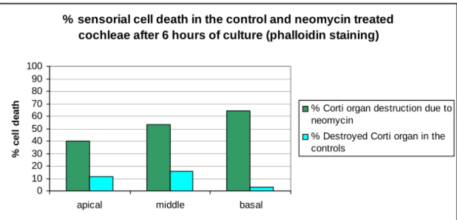
% sensorial cell death in the control and neomycin treated cochleae after 6 hours of culture (phalloidin staining)
0 10 20 30 40 50 60 70 80 90 100
apical middle basal
%
ce
ll
d
e
at
h % Corti organ destruction due to
neomycin
% Destroyed Corti organ in the controls
Figure 2. Neomycin induces cell death
Data exposed in figure 2 suggest that neomycin induces stereocilliar loss first in the basal segment of the cochlea, followed by the middle one and finally by the apical one. Hair cell death after 6 hours from the beginning of the treatment reaches in the basal segment more than 60% of the cochlear length, followed by 52% in the middle segment and 40% in the apical one. It is interesting to observe that a certain amount of hair cells lose their stereocillia even outside neomycin treatment. This phenomenon seems to appear mostly in the middle segment and less in the basal one.
2. Regrading apoptotic neomycin induced hair cell death
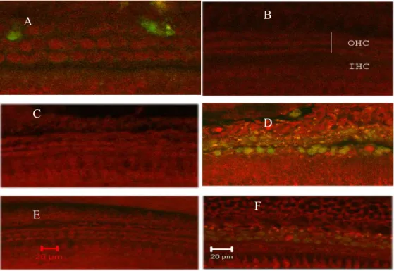
Because apoptotic hair cell death due to neomycin was similar in all analyzed slides marked for caspase 3, 8 and 9, we will present only the results for caspase 8 and counterstained with propidium iodide.
Figure 3. Activation of caspase 8 (green) and PI nuclear staining (red). Nuclei of apoptotic
cells appear bright red and round and the cytoplasm stained green. We can remark also the presence of apoptotic bodies stained yellow.
IHC: outer hair cells; IHC: inner hair cells
Left (A,C,E)- controls without neomycin; right (B,D,F)- after 6 hours neomycin treatment. After segments: apical (A,B), medial (C,D) and basal (E,F). Scale bar: 20 µm
Figure 3 reveals the frequency of cells stained for caspase 3, 8 and 9 (green color of the cytoplasm) as well as with propidium iodide (red color of nuclei), from the in vitro
cultivated cochleae, in the presence or absence of neomycin. From microscopic analyzing of the two groups of cochleae we can observe that the frequency of cells stained green, with red nuclei, or green cytoplasm with red nuclei, present in the neomycin treated group, is superior to the one in the control group. The fact that the amplitude of caspase and propidium iodide stained cell frequency in cochleae treated with neomycin is similar to the amplitude of cell death evidenced with phalloidin (fig.
B
A
B
B
C
E
D
2), suggests that hair cell death in the organ of Corti is realized mostly by apoptosis and less by necrosis.
The fact that in the basal and middle segments of the cochleae belonging to the neomycin group we can observe a larger number of cells stained for the three active caspases (3, 8 and 9) in the cytoplasm, compared to control cochleae, is an argument which sustains the idea that neomycin induces apoptosis in auditory hair cells.
3. Regarding neomycin induced apoptotic hair cell death quantification
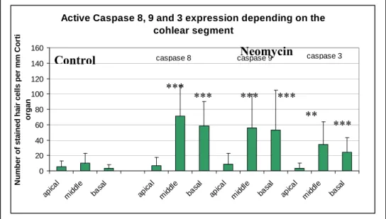
In order to establish if qualitative observations specified above are real, we tried to statistically quantify the apoptotic process. On this purpose we counted all cells with green cytoplasm, marking the presence of active cytoplasmic caspases 3, 8 and 9 in all cochleae belonging to the two groups (control and neomycin). Obtained data were then statistically calculated obtaining the media and standard deviation for each group and segment of cultivated cochlea. Results are exposed in figure 4.
Figure 4. The average number of hair cells in the segments of the Corti organ marked for
caspase 3, 8 and 9 after 6 hours of neomycin contact, compared to the stained cells in the control group.
In figure 4 we showed as columns the average number of stained cells for each of the three active caspases (8, 9 and 3). The vertical line stays for average standard deviation. The significance of the differences between the average of apoptotic cells in the three cochlear segments from the neomycin treated group compared to the control
Active Caspase 8, 9 and 3 expression depending on the cohlear segment 0 20 40 60 80 100 120 140 160 apica l middl e basa l apic al middl e basa l apic al middl e basa l apic al midd le basa l N u m b e r of s ta ine d ha ir c e lls pe r m m C o rt i or ga n
caspase 8 caspase 9 caspase 3
one is symbolized with (p<0,05) or (p<0,005). The data exposed in figure 4 suggest that under 6 hours of neomycin influence, many phenomenon take place. These are the following:
1. The average value of hair cells where we identified the three types of active caspases being significantly bigger in the cochleae cultivated in the presence of neomycin compared to the control cochleae, lets us conclude that neomycin induces and sustains the apoptotic process of self distruction in auditory hair cells. The process starts to manifest itself after 6 hours of neomycin contact.
2. Simultaneous active caspase 8, 9 and 3 presence in cell cytoplasm (around 20/ 60 cells per mm Corti organ) shows that they participate together in this process and in a certain way in inducing and realizing the apoptotic process in the organ of Corti
3. Neomycin ototoxic activity followed by apoptotic unleash at 6 hours from neomycin contact of the cochleae, are manifested only in the basal and middle segments of the Corti organ. Cells in the apical area of the Corti organ are almost not affected by the ototoxic neomycin effect.
4. High standard deviation values in all neomycin treated cochlear groups suggest that the sensitivity of hearing cells in the organ of Corti towards neomycin is very variable and different from one individual to another.
5. The fact that the average number of cells marked for active caspases is the biggest in the group marked for caspase 8 followed in a decreasing order by other caspases, suggests that there is a hierarchy in the participation of the three caspases in initiating and sustaining the apoptotic process.
Propidium iodid expression depending on the cochlear segment 0 10 20 30 40 50 60 70 80 90 apica l mid dle basa l apica l med ial basa l apica l mid dle basa l apika l med ial bas al N u m b e r o f s ta in e d h a ir c e lls p e r m m C o rt i or ga n
PI (caspase 8) PI(caspase 9)
Figure 5. The average number of cells where apoptosis has involved also their nuclei in
While the apoptotic process can be identified at the beginning by evidencing cytoplasmic caspase 3, 8 and 9, the advancement of the process towards the nucleus, can be identified by propidium iodide nuclear incorporation and the red colour of the nucleus. In order to know if after 6 hours of neomycin treatment, apoptosis is at its debut or if the process has started much earlier, we have counted all red coloured, round nuclei within the 3 experiments concerning caspase 3, 8 and 9 and processed them statistically. Results are shown in figure 5.
The average number of cells which have incorporated propidium iodide in their nuclei and their standard deviations, in the three cochlear groups stained for active caspases, shown in the figure above, suggests that in auditory hair cells, at 6 hours from the contact with neomycin, following processes take place:
1. The fact that auditory hair cell number where propidium iodide has accumulated is significantly higher within the experiments concerning activated caspase 3, 8 and 9 compared to the control group, suggests that the apoptotic process doesn’t start only at 6 hours after neomycin treatment, but probably much earlier.
2. The fact that the average number of cells which have incorporated propidium iodid in their nuclei (red nuclei) is significantly high (p<0,005), in cochleae treated with neomycin compared to the control group, is a solid argument which sustains the idea that neomycin initiates the process of apoptosis by activating the three procaspases. On their turn, these are sustaining further the process of nuclear destruction (propidium iodide penetrates the nucleus only if its membrane is affected).
3. Apoptosis is present also in hair cell nuclei, especially in the basal and middle cochlear segment and is almost absent in the apical segment.
4. Although the cell number where apoptosis has reached the nucleus is higher in cochleae stained for caspase 9 compared to other experiments, the differences are not significant because of high standard deviation.
5. High deviation standard values suggest that hair cell sensitivity towards cytotoxic action of neomycin is very different from one individual to another because of genetic differences between each individual.
Conclusions
Experiments done on mice cochleae cultivated for 6 hours in the presence or absence of neomycin and specifically stained for apoptosis by cytoplasm presence of active caspase 8, 9 and 3 and nuclear propidium iodide presence let us conclude the following:
2. Hair cell death in the organ of Corti is produced especially by apoptosis and less by necrosis (in dead cells active caspase 3, 8 and 9 are present in the cytoplasm and propidium iodide in the nucleus).
3. Initiation and finalizing of hair cell apoptosis due to neomycin, takes place only in the basal and middle segment of the Corti organ. Hair cells in the apical segment are almost not affected by ototoxic effect of neomycin.
4. Auditory cell sensitivity towards neomycin is very different from one individual to another.
References
1. Adams JM, Cory S., 1998, The Bcl-2 protein family: arbiters of cell survival.
Science, 281(5381), 1322-6.
2. Cheng AG, Cunningham LL, Rubel EW., 2003, Hair cell death in the avian basilar papilla: characterization of the in vitro model and caspase activation. J Assoc
Res Otolaryngol., 4(1), 91-105.
3. Cunningham LL, Cheng AG, Rubel EW. 2002, Caspase activation in hair cells of the mouse utricle exposed to neomycin. J Neurosci., 22(19):8532-40.
4. Devarajan P, Savoca M, Castaneda MP, et al., 2002, Cisplatin-induced apoptosis in auditory cells: role of death receptor and mitochondrial pathways. Hear Res.; 174(1-2), 45-54.
5. Forge A, Li L., 2000, Apoptotic death of hair cells in mammalian vestibular sensory epithelia. Hear Res. 139(1-2), 97-115.
6. Lang H, Liu C., 1997, Apoptosis and hair cell degeneration in the vestibular sensory epithelia of the guinea pig following a gentamicin insult. Hear Res.
111(1-2),177-84.
7. Löwenheim H, Kil J, Gultig K., Zenner HP , 1999, Determination of hair cell degeneration and hair cell death in neomycin treated cultures of neonatal rat cochlea
Hearing Research 128, 16-19
8. Matsui JI, Gale JE, Warchol ME., 2004, Critical signaling events during the aminoglycoside-induced death of sensory hair cells in vitro. J Neurobiol.,