Medullary Norepinephrine Projections Release Norepinephrine into
the Contralateral Bed Nucleus of the Stria Terminalis
Megan E. Fox, Elizabeth S. Bucher, Justin A. Johnson, and R. Mark Wightman
*
Department of Chemistry, Neuroscience Center, University of North Carolina at Chapel Hill, Chapel Hill, North Carolina
27599-3290, United States
*
S Supporting InformationABSTRACT:
Central norepinephrine signaling in
fl
uences a wide
range of behavioral and physiological processes, and the ventral bed
nucleus of the stria terminalis (vBNST) receives some of the densest
norepinephrine innervation in the brain. Previous work describes
norepinephrine neurons as projecting primarily unilaterally; however,
recent evidence for cross-hemispheric catecholamine signaling
challenges this idea. Here, we use fast-scan cyclic voltammetry and
retrograde tracing to characterize cross-hemispheric norepinephrine
signaling in the vBNST. We delivered stimulations to noradrenergic
pathways originating in the A1/A2 and locus coeruleus and found hemispherically equivalent norepinephrine release in the
vBNST regardless of stimulated hemisphere. Unilateral retrograde tracing revealed that medullary, but not locus coeruleus
norepinephrine neurons send cross-hemispheric projections to the vBNST. Further characterization with pharmacological lesions
revealed that stimulations of the locus coeruleus and its axon bundles likely elicit vBNST norepinephrine release through indirect
activation. These experiments are the
fi
rst to demonstrate contralateral norepinephrine release and establish that medullary, but
not coerulean neurons are responsible for norepinephrine release in the vBNST.
KEYWORDS:
Norepinephrine, fast-scan cyclic voltammetry, cross-hemispheric, locus coeruleus, nucleus of the solitary tract,
ventral bed nucleus of the stria terminalis
C
entral norepinephrine signaling mediates a variety of
processes including learning and memory, drug reward
and withdrawal, and the behavioral and physiological responses
to stress.
1−5Dysregulation of noradrenergic signaling is
implicated in disorders ranging from drug addiction
6to
Alzheimer
’
s disease,
7and the ventral bed nucleus of the stria
terminalis (vBNST) is a site of some of the densest
noradrenergic innervation in the brain.
8Limbic, forebrain,
and brainstem inputs converge in the BNST to relay
information about stressors and generate an appropriate
physiological response through regulation of the
hypothala-mic-pituitary adrenal (HPA) axis.
1The vBNST receives
noradrenergic input primarily from medullary neurons (A1/
A2) coursing through the ventral noradrenergic bundle (VNB)
and, to a lesser extent, from the neurons of the locus coeruleus
(LC) through the dorsal noradrenergic bundle (DNB).
1,9−12Norepinephrine is released in the vBNST during presentation
of an aversive tastant, omission of an expected reward, and
delivery of a noxious stimulus.
13−15Furthermore, the vBNST is
an important structure in mediating the aversive components of
drug-withdrawal,
4,16,17and norepinephrine signaling in the
vBNST undergoes robust plasticity following stress or
drug-withdrawal dependent on HPA axis function.
18,19Norepinephr-ine signaling in the vBNST can integrate information about
aversive and stressful stimuli to generate an appropriate
physiological response, thus how it is regulated is an important
topic of investigation.
Early anatomical studies described catecholamine neurons as
projecting solely to one hemisphere in rodents.
20,21However,
more modern tracing studies challenge the exclusively unilateral
nature of catecholaminergic projections, and provide evidence
for crossing projections originating in cell groups ranging from
the ventral tegmental area (VTA)
22,23to the LC.
24,25We
recently asked to what extent cross-hemispheric projections
contribute to striatal dopamine release in rats.
26Despite the
reportedly small number of contralateral dopamine
projec-tions,
22,23we found that stimulating these projections resulted
in physiologically relevant striatal dopamine release, which may
confound the interpretation of unilateral manipulations.
26Since
some norepinephrine neurons also exhibit crossing projections
in rats,
24,25,27,28and primates,
29,30we hypothesized that, like
dopamine, bilateral norepinephrine projections would in
fl
uence
measured norepinephrine release. In this work, we used
fast-scan cyclic voltammetry in anesthetized rats to measure
cross-hemispheric norepinephrine release in the vBNST. We found
noradrenergic axon pathways can release norepinephrine in the
contralateral vBNST, and we identi
fi
ed contralaterally
projec-ting norepinephrine neurons using retrograde tracing. As a
consequence of these investigations, we serendipitously
discovered that LC and DNB stimulations produce
norepi-Received: July 13, 2016
Accepted: September 12, 2016
Published: September 12, 2016
Research Article
pubs.acs.org/chemneuro
■
RESULTS AND DISCUSSION
Stimulation of Noradrenergic Axons Elicits Release in
the Contralateral vBNST.
Early anatomical studies describe
catecholamine neurons as projecting primarily unilaterally in
rodents;
20,21however, more modern tracing studies have
revealed some catecholamine neurons project contralateral to
their origin.
22−25We recently showed crossing dopaminergic
projections support dopamine release in the contralateral
striatum and may contribute to interhemispheric signaling.
26To determine if norepinephrine neurons might also exhibit
cross-hemispheric functionality and in
fl
uence release in the
vBNST, we used fast-scan cyclic voltammetry at dual
carbon-fi
ber electrodes
32to measure norepinephrine release in
anesthetized rats. We
fi
rst lowered two carbon-
fi
ber electrodes
bilaterally into the vBNST, and a stimulating electrode
unilaterally into the VNB (schematic in
Figure 1
a). Unilateral
VNB stimulations produced norepinephrine release at both
electrodes (examples in
Figure 1
b), supporting our hypothesis
that norepinephrine projections exhibit cross-hemispheric
functionality.
To map the location of contralateral projections, we next
held the carbon-
fi
ber electrode at a
fi
xed depth in the vBNST
(
−
7.2 mm DV), and lowered the stimulating electrode ventrally
through the contralateral noradrenergic bundles. We measured
contralaterally evoked norepinephrine release over a large
dorsal-ventral range, peaking at locations corresponding to the
DNB (
−
6.4 mm DV) and VNB (
−
8.0 mm DV,
Figure 1
c), in
agreement with previous ipsilateral characterization.
31Nor-epinephrine release evoked by contralateral DNB and VNB
stimulation was linear with increasing stimulation duration
(DNB slope = 0.146
±
0.007
r
2= 0.97; VNB slope = 0.170
±
0.005,
r
2= 0.98;
Figure 1
d), similar to our previous report with
ipsilateral stimulations.
18On average, maximal norepinephrine
release in the vBNST was of comparable magnitude following
stimulations of the contralateral DNB or VNB (DNB: 0.214
±
0.066
μ
M; VNB: 0.192
±
0.038
μ
M,
n
= 10, respectively), and
ipsilateral DNB and VNB (DNB: 0.228
±
0.063
μ
M; VNB:
0.191
±
0.029
μ
M,
n
= 10, respectively). In a subset of animals,
we performed within-animal comparisons of ipsilateral vs
contralateral release by lowering the stimulating electrode
ventrally through the ipsilateral, then contralateral hemisphere.
Strikingly, the ratio of ipsilateral to contralateral release was
equal between hemispheres following either DNB or VNB
stimulations (Ipsi/Contra, VNB: 1.3
±
0.20; DNB: 1.0
±
0.08,
n
= 6 animals,
P
> 0.05,
Figure 1
e). In this way,
cross-hemispheric norepinephrine in the vBNST closely resembled
dopamine in the dorsomedial striatum, in that it exhibited
equivalent release regardless of stimulated hemisphere.
26DSP-4 Treatment Does Not Attenuate vBNST
Nor-epinephrine Release.
The vBNST is thought to receive a
small input from the LC through the DNB.
1,9Since DNB
stimulations produced hemispherically equivalent release
amplitudes, we next placed our stimulating electrode in the
contralateral LC to ascertain its contribution to vBNST
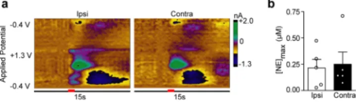
norepinephrine release. Stimulations of the LC elicited robust
norepinephrine regardless of stimulated hemisphere (Ipsi,
0.163
±
0.082
μ
M; Contra, 0.250
±
0.115
μ
M,
n
= 5,
P
>
0.05,
Figure 2
), and of similar magnitude to previously reported
LC-evoked norepinephrine in the vBNST.
31However,
coerulean inputs to the vBNST are sparse;
9thus, we next
Figure 1. Stimulation of noradrenergic axon bundles produces hemispherically equivalent norepinephrine release in the ventral bed nucleus of the stria terminalis (vBNST). (a) Schematic of dualcarbon-fiber electrodes in the vBNST (gray) with unilateral stimulating electrode (red) in the ventral noradrenergic bundle (VNB). (b) Representative color plots demonstrating norepinephrine release to a 1s electrical stimulation (red bar) to the VNB recording ipsilateral (Ipsi) and contralateral (Contra) to the stimulation. Applied potential is plotted on the abiscca, recording time on the ordinate, and current is encoded in false color. (c) Effect of stimulation electrode placement on contralateral norepinephrine release. Data are plotted as norepinephrine release [NE]conover maximal norepinephrine release
[NE]con‑maxas elicted by contralateral stimulations and are presented as
asked if o
ff
-target e
ff
ects could contribute to robust
norepinephrine release described here and in the previous
study.
31We employed the selective neurotoxin DSP-4 to lesion
norepinephrine terminals from the LC.
33Tissue content
analysis revealed the treatment was e
ff
ective at eliminating a
signi
fi
cant degree of LC innervation, as concentrations were
markedly reduced in the anteroventral thalamus (AV), a brain
region receiving exclusively coerulean input (
Table 1
). Control
values for the vBNST and the AV are similar to those
previously reported from our lab
18,31and others.
8,34DSP-4
treatment signi
fi
cantly reduced norepinephrine and dopamine
in the AV (unpaired
t
test, norepinephrine:
t
(9)= 3.579,
P
=
0.006; dopamine:
t
(9)= 2.586,
P
= 0.029), but did not exhibit an
e
ff
ect on the catecholamine content of the vBNST
(norepinephrine:
t
(9)= 0.959,
P
= 0.363; dopamine:
t
(9)=
0.371,
P
= 0.719), in agreement with sparse coerulean
innervation of the latter.
9As expected from the tissue content
fi
ndings, DSP-4
treatment signi
fi
cantly attenuated DNB-evoked norepinephrine
release in the AV (control vs DSP-4, 0.216
±
0.035 vs 0.0950
±
0.0376
μ
M,
n
= 5, respectively, unpaired
t
test:
t
(8)= 2.363,
p
<
0.05,
Figure S1
). DSP-4 treatment did not a
ff
ect vBNST release
with ipsilateral VNB stimulations (control vs DSP-4, 0.281
±
0.046 vs 0.408
±
0 0.104
μ
M,
n
= 5 rats, respectively,
Figure
3
a) in agreement with our previous work.
19Surprisingly, DSP-4
treatment did not reduce vBNST norepinephrine evoked by
ipsilateral LC or DNB stimulations (control vs DSP-4, DNB:
0.288
±
0.046 vs 0.386
±
0.154; LC 0.163
±
0.032 vs 0.237
±
0.073
μ
M,
n
= 5 rats, respectively,
P
> 0.05,
Figure 3
a),
suggesting LC inputs to the vBNST were spared by DSP-4, or
that release arises though an indirect e
ff
ect.
Physical, but Not 6-OHDA LC Lesions Attenuate
vBNST Norepinephrine Release.
Since DSP-4 does not
lesion the LC with 100% e
ffi
cacy,
35and its actions on the LC
noradrenergic system have been called into question,
36we next
used bilateral 6-hydroxydopamine (6-OHDA) lesions targeted
to the LC to corroborate the DSP-4
fi
ndings. Two weeks after
6-OHDA treatment, we anesthetized animals and recorded
norepinephrine evoked from ipsilateral electrical stimulations.
Similar to DSP-4 treatment, 6-OHDA lesions of the LC had no
measurable e
ff
ect on norepinephrine release in the vBNST
(Sham vs 6-OHDA; DNB: 0.364
±
0.092 vs 0.342
±
0.054
μ
M;
VNB: 0.339
±
0.065 vs 0.457
±
0.072
μ
M; LC 0.213
±
0.082
vs 0.278
±
0.060
μ
M,
n
= 5 rats, respectively,
P
> 0.05,
Figure
3
b). In our previous report of LC-evoked norepinephrine in the
vBNST, release was suppressed after delivery of lidocaine to the
stimulation site.
31Given that selective chemical ablations did
not reduce norepinephrine release, we turned to a physical
disconnection approach by performing a knife-cut at the level
of the LC. Cutting the ipsilateral LC markedly reduced
LC-evoked norepinephrine release in the vBNST (9.0
±
6.1%,
n
=
5 rats,
Figure 4
), similar to lidocaine.
31A2, but Not LC Neurons Project Bilaterally to the
vBNST.
Since a physical, but not selective pharmacological
lesion of the LC reduced norepinephrine over
fl
ow, we asked if
the LC sends projections to the vBNST that might be spared
by chemical treatments. We unilaterally injected
fl
uorogold into
the vBNST, and looked for retrograde labeling in the LC and
the nucleus of the solitary tract (A2) (Schematic in
Figure 5
a,
Representative injection site
Figure 5
b). Even after signal
ampli
fi
cation with an antibody against
fl
uorogold, we did not
fi
nd any retrogradely labeled cells in the LC (
Figure 5
c,d).
Instead, we found bilateral
fl
uorogold labeling in the A2 (
Figure
5
e
−
g). A1 norepinephrine neurons also innervate the
vBNST,
1,7,9−11which may provide an additional source of
cross-hemispheric projections. However, further work is needed
to assess if the A1 sends bilateral projections to the vBNST, and
Figure 2. Locus coeruleus stimulations produce equivalentnorepi-nephrine release in the ventral bed nucleus of the stria terminalis independent of stimulated hemisphere. (a) Representative color plots demonstrating norepinephrine release in the vBNST following a 1 s electrical stimulation (red bar) of the ipsilateral (Ipsi) and contralateral (Contra) locus coeruleus (LC). (b) Maximal norepinephrine release (NEmax) in the vBNST following Ipsi and Contra LC stimulations in
all subjects.
Table 1. Catecholamine Tissue Content in Target Regions for Untreated and DSP-4-Treated Animals
aNE (μg/g tissue) DA (μg/g tissue)
untreated DSP-4 untreated DSP-4
vBNST 2.98±0.80 2.14±0.58 0.82±0.25 0.84±0.17
AV 1.82±0.50 0.18±0.10** 1.62±0.52 0.34±0.20*
aValues are shown as mean±SEM.*P< 0.05,**P< 0.01, compared to untreated values. Abbreviations: NE, norepinephrine; DA, dopamine.
Figure 3. Chemical lesions of the LC do not impact vBNST norepinephrine release. (a) Maximal norepinephrine concentrations elicited by ipsilateral DNB, VNB, and LC stimulations in control (white) and DSP-4 treated rats (black). (b) Maximal norepinephrine concentrations elicted by ipsilateral DNB, VNB, and LC stimulations in sham (white), and 6-OHDA lesioned rats (black). Average±SEM with individual experiments overlaid.
if they contribute to contralateral norepinephrine release.
Nevertheless, we found strong bilateral retrograde labeling in
the A2. The number of labeled cells ipsilateral to the tracer
infusion was greater than those in the contralateral hemisphere
(27
±
1.7 vs 5.3
±
1.2 cells,
n
= 3 rats), which was surprising
since we measured similar concentrations of norepinephrine
with ipsilateral and contralateral VNB stimulations.
Hemispherically equivalent release could arise from a
number of mechanisms. First, norepinephrine concentrations
released from contralateral projections were linear with respect
to stimulation duration, in a similar manner as ipsilaterally
evoked norepinephrine.
18This might indicate norepinephrine
release is
“
capped
”
from ipsilateral projections by regulation
mechanisms. In our previous characterization of
cross-hemi-spheric dopamine, we found the ratio of ipsilateral to
contralateral dopamine release was at least partially dependent
on D2 autoreceptor control.
26Thus, autoreceptors may play a
role in balancing norepinephrine concentrations between
hemispheres as they do for dopamine. Alternatively,
norepi-nephrine neurons may co-release glutamate, which was recently
demonstrated to occur from some dopamine terminals.
37Co-release of other neurotransmitters may depolarize
norepinephr-ine terminals in the vBNST and partially explain the apparent
hemispheric equivalence. Indeed, bath application of excitatory
amino acids can stimulate norepinephrine release in brain
slices.
38,39It is also worth considering that stimulations of the
VNB also target the VTA,
32which receives inputs from both
the A2 group
40and the BNST.
41Activation of these projections
may also contribute to electrically evoked norepinephrine
release and hemispheric equivalence. Although the mechanism
underlying hemispherically equivalent norepinephrine release is
unknown, these data reveal that norepinephrine in the vBNST
is similar to dopamine release in the dorsomedial striatum,
26in
that it is of similar magnitude with ipsilateral or contralateral
stimulations.
DNB Stimulations Produce vBNST Norepinephrine
Indirectly.
The LC sends most of its forebrain projections
through the DNB.
21Since the LC was not labeled with
fl
uorogold, we hypothesized DNB stimulations produced
norepinephrine release through an indirect mechanism. To
test the possibility that another midbrain structure was
mediating norepinephrine release, we delivered ibotenic acid
(IBA) to the DNB (schematic in
Figure 6
a). IBA is a glutamate
analog that, through excitotoxicity, selectively inactivates cell
region, release is mediated by direct activation of the DNB.
However, IBA infusions in the DNB signi
fi
cantly reduced
norepinephrine release in the vBNST (saline 77.9
±
3.3% vs
IBA 20.3
±
3.8%,
n
= 5 rats, two-way repeated measures
ANOVA: drug
×
region interaction
F
(2,16)= 16.52, e
ff
ect of
region
F
(2,16)= 21.47, e
ff
ect of drug
F
(2,16)= 46.67, Bonferroni
post hoc,
p
< 0.001,
Figure 6
b,c), suggesting DNB evoked
release in the vBNST arises through an indirect mechanism.
Possible Mechanisms for Coerulean Evoked
Norepi-nephrine in the vBNST.
Based on the data obtained after
selective chemical inactivation, we propose that norepinephrine
release in the vBNST evoked from DNB and LC stimulations
arises through an indirect mechanism. First, vBNST
norepi-nephrine release was attenuated following IBA infusions in the
DNB. Since IBA does not a
ff
ect
fi
bers of passage,
42the results
from this experiment suggest that other midbrain nuclei are
mediating release following DNB stimulation. At the
coordinates used in this study, the DNB courses by several
structures including the periaqueductal gray (PAG). Fluorogold
infusions into the ventrolateral portion of the PAG bilaterally
labels neurons in the A2 region.
43Thus, PAG activation may
antidromically activate the A2 group to elicit vBNST release, or
this release could arise from another unidenti
fi
ed mechanism.
Second, selective chemical inactivation with DSP-4 and
6-OHDA strongly suggest that noradrenergic neurons of the LC
are not responsible for release in the vBNST. However, both
lidocaine and knife-cut would prevent the propogation of action
potentials traveling down axons near the LC. It is therefore
possible that projections from medullary noradrenergic neurons
close to the LC are responsible for this release. Indeed, an
anterograde tracing study revealed A2 neurons innervate
regions proximal to the LC.
44Alternatively, the A2 projects to the nucleus
paragiganto-cellularis (PGi),
45which in turn, sends projections to the LC.
46Therefore, LC stimulation may antidromically activate the PGi
and subsequently the A2 to produce norepinephrine release in
the vBNST. Furthermore, cross-talk between coerulean and
medullary cell groups is supported by work demonstrating both
norepinephrine cell groups contribute to opiate withdrawal
syndrome,
4,47and coerulean lesions produce adaptations in
norepinephrine signaling originating from A1/A2 neurons.
19Regardless of mechanism, the selective chemical lesions reveal
that, although the DNB and LC can produce release in both
ipsilateral and contralateral vBNST, it likely does so indirectly.
Interestingly, the nonspeci
fi
c activation we propose here may
be responsible for the disparity in reports describing electrical
self-stimulations of the LC or its pathways.
48−51Indeed, in
animals trained to self-stimulate the LC, neither 6-OHDA
lesions, nor electrolytic lesions of the DNB attenuate
self-stimulation behavior,
52,53providing further support that
stimulations of the LC/DNB activate other pathways.
There are two obvious approaches to address the speci
fi
c
neuronal populations contributing to vBNST norepinephrine
release. The
fi
rst would be to inactivate medullary
norepi-nephrine neurons to con
fi
rm they are the sole population
responsible for release in the vBNST. We have made repeated
attempts to chemically ablate the A2 group, however these
Figure 4. Knife-cut of the LC reduces LC-evoked vBNSTmanipulations in the brainstem typically result in ceased
respiratory activity and subsequent death of the subject. The
second approach would rely on a more selective stimulation
method, such as optogentics. Although optogenetic approaches
have been widely successful for dopamine measurements,
54thus far we have been unable to measure norepinephrine
release from optogenetic stimulations in intact rats. For the
time being, electrical stimulations must su
ffi
ce for studying
vBNST norepinephrine release in anesthetized animals. It is in
this context that we place our
fi
ndings regarding LC and DNB
evoked norepinephrine. Although this release likely arises
through an indirect mechanism, we can not provide a de
fi
nitive
source. However, it is clear that care must be taken when
interpreting data obtained with electrical stimulations of
norepinephrine neurons and their projections.
Cross-Hemispheric Projections in the Context of
Unilateral Manipulations.
Neurodegenerative conditions
such as Parkinson
’
s disease are often modeled with unilateral
lesions of catecholaminergic neurons.
55However, the
cross-hemispheric nature of catecholamine projections may confound
the interpretation of data obtained following unilateral
manipulations in rats. Indeed, stimulating intact, contralateral
dopamine projections can release dopamine into an otherwise
depleted hemisphere.
26Furthermore, in a recent report,
Figure 5.Unilateralfluorogold tracing in the vBNST. (a,b) Schematic and representative infusion site offluorogold into the vBNST. (c,d) Apparent lack offluorogold-positive cells in the ipsilateral LC and atlas section corresponding to the photomicrograph. (e,f) Bilateralfluorogold-positive cells in the A2 and camera lucida drawing of labeled cells in the atlas section corresponding to the photomicrograph. (g) Higher magnification image ofresearchers used a unilateral knife-cut of the DNB to rule out
contributions of LC norepinephrine to catecholamine release
measured in the medial prefrontal cortex.
56However, LC
neurons project bilaterally to some regions
24,25and these
contralateral projections may release physiologically relevant
norepinephrine concentrations. It is clear from these
fi
ndings
that care must be taken when performing unilateral
disconnection studies, since the unilateral nature of the
monoamine projections originally described by Ungerstedt
20,21is now being called into question. Finally, exploiting the
cross-hemispheric nature of catecholamine projections may prove
useful in therapies such as deep-brain stimulation for restoring
catecholamine concentrations in the brain.
■
CONCLUSIONS
In summary, we found norepinephrine release was elicited in
the vBNST contralateral to the stimulation location.
Stimulations of the DNB, VNB, and LC evoked norepinephrine
of equal magnitude in both hemispheres. Norepinephrine
evoked from LC stimulations occurred via nonspeci
fi
c
activation, as only physical, but not selective pharmacological
lesions of the LC attenuated release. DNB stimulations also
elicited norepinephrine in a nonspeci
fi
c way, since inactivation
of cells proximal to the DNB reduced evoked vBNST
norepinephrine. Furthermore,
fl
uorogold tracing revealed
medullary, but not LC neurons, send bilateral projections to
the vBNST. Taken together, these data show that, although
norepinephrine is released in both hemispheres with unilateral
activation, only medullary norepinephrine neurons are directly
responsible for cross-hemispheric release in the vBNST. This
previously undescribed property of norepinephrine neurons
should be taken into account when performing unilateral
manipulations.
a 12:12 h light:dark cycle.
Voltammetric Norepinephrine Measurements. Norepinephr-ine release was measured in anesthetized rats as described previously19 using HDCV.57An average in vitro calibration factor of 6 nA/μM was used to convert norepinephrine current to concentration following principal component analysis.58For bilateral norepinephrine measure-ments, rats were anesthetized with urethane (1.5 g/kg) and placed in a stereotaxic frame (Kopf. Tujunga, CA). Holes were drilled for the vBNST (AP 0 mm, ML±1.2 mm), the DNB/VNB (AP−5.2 mm, ML +1.2 mm), and the LC (AP−9.8 mm, ML +1.3 mm), referenced from bregma and based on the atlas of Paxinos and Watson.59A Ag/ AgCl reference electrode was placed in the left hemisphere and secured with a jeweler’s screw. A carbonfiber microelectrode (∼100 μm active length) was lowered into the right vBNST (−7 to−7.5 mm DV) and a bipolar stimulating electrode (Plastics One, Roanoke, VA) was lowered ipsilateral to the carbon-fiber electrode in the LC (∼ −7.0 mm DV), DNB (∼ −6.5 mm DV), or VNB (∼ −8.0 mm DV) until maximal norepinephrine release was attained. Both stimulating and carbon-fiber electrodes were subsequently secured with dental cement. A second carbon fiber microelectrode was lowered into the left, contralateral vBNST (−7 to 7.5 mm DV) until maximal norepinephrine was achieved. A total of 10 rats were used for these studies.
For mapping experiments and stimulation duration studies,first a carbon-fiber electrode was lowered into the right vBNST until maximal release with ipsilateral VNB stimulations was attained. Next, the stimulating electrode was raised to the DNB to determine maximum ipsilateral release. Then, the stimulating electrode was lowered through the contralateral hemisphere in 200μm increments to map the effect of contralateral stimulating electrode placement in the vBNST. Sixty Hz stimulations of varying duration (20−120 pulses) were delivered at depths corresponding to maximal release from contralateral DNB and VNB stimulations and plotted vs stimulation duration. We compared maximal norepinephrine evoked by ipsilateral and contralateral DNB and VNB stimulations at the same recording electrode location. A total of 6 rats were used for these experiments. In another group of rats (n= 5), we compared maximal norepinephrine release in the vBNST following ipsilateral and contralateral LC stimulations.
DSP-4 Treatment. Adolescent rats (150−200 g) were adminis-tered DSP-4 (N-(2-chloroethyl)-N-ethyl-2-bromobenzylamine) in two doses (0.5 mL, 50 mg/kg, i.p.) provided 3 days apart.19Voltammetric (n= 5 DSP-4,n= 5 control) and tissue content (n= 6 DSP-4,n= 5 control) experiments were conducted 10−15 days after the last dose. Tissue Content Analysis. A separate group of rats was anesthetized with urethane (1.5 g/kg), and their brains were rapidly removed and placed in ice-cold artificial cerebral spinalfluid (aCSF). Coronal sections (300μm thick) containing the BNST or AV were collected with a VF-200 Compresstome (Precisionary Instruments Greenville, NC) in ice cold aCSF. The aCSF contained (in mM) 126 NaCl, 25 NaHCO3, 2.45 KCl, 12 NaH2PO4, 1.2 MgCl2, 2.4 CaCl2, 20
HEPES, and 11 glucose, and was adjusted to pH 7.4 and saturated with 95% O2 /5% CO2. Tissue containing the vBNST or AV was
excised bilaterally with a 1 mm punch, and collected into preweighed tubes. The samples were mixed with 200 μL of 0.1 N HClO4
containing 1 μM hydroquinone, the internal standard, and subsequently homogenized using a sonic dismembrator (Fisher Scientific, Model 60, Pittsburgh, PA). The homogenate was spun down at 6000 rpm for 10 min, and the supernatant was removed and
filtered using a 0.2 μm syringe filter. High performance liquid chromatography was performed as described previously.18,31Briefly, 20 μL injections were made onto a reversed-phase column (5μm, 4.6×5 mm, Waters Atlantis, Milford, MA). The mobile phase consisted of 0.1 Figure 6.Ibotenic acid infusions in the DNB attenuate norepinephrine
M citric acid, 1 mM sodium hexylsulfate, 0.1 mM EDTA (pH = 3), and 10% methanol organic modifier at a flow rate of 1.0 mL/min. Norepinephrine and dopamine were detected with a thin layer radial electrochemical cell (BASi, West Lafayette, IN) at a potential of +800 mV vs Ag/AgCl. Data were collected at 60 Hz using a LabVIEW stripchart recorder program (Jorgenson Lab, UNC) and custom-built electronics. Concentration was determined by a ratio of analyte peak area to internal standard peak area, and normalized to wet tissue weight.
6-Hydroxydopamine Lesions. Rats underwent stereotaxic surgery under isoflurane anesthesia (4% induction, 1.5% maintenance), and an incision was made in the scalp to drill bilateral holes targeting the LC (AP−9.8 mm, ML±1.4). An infusion cannula (Plastics One) was lowered to a depth of 7.0 mm from brain surface, and 1μL of 10 mM 6-hydroxydopamine hydrobromide (6-OHDA)/ 0.01% w/v ascorbic acid (Sigma-Aldrich) in sterile saline (0.9%), or saline (sham-lesioned) was infused into each hemisphere with an infusion needle (33 ga, 10 mm, Plastics One) over 5 min. The scalp was closed with Vet Bond (3M, St Paul, MN) and rats were allowed to recover for 2 weeks before being anesthetized with urethane for voltammetric norepinephrine measurements.
Knife-Cut Experiments.Rat were anesthetized with urethane (1.5 g/kg) and affixed in a stereotaxic frame. Holes were drilled for the vBNST, VNB, and LC as described above. Once maximal release was attained with ipsilateral VNB stimulations the stimulating electrode was moved to the ipsilateral LC and adjusted for maximal release. The stimulating electrode was subsequently removed, and a surgical blade was lowered 0.2 mm past the depth of maximal LC release. The stimulating electrode was repositioned in the LC, and norepinephrine release was measured after subsequent LC stimulations. A total of 5 animals were used for these experiments.
FluoroGold Tracing. Rats underwent stereotaxic surgery under isoflurane anesthesia (4% induction, 1.5% maintenance). A small incision was made in the scalp, a hole was drilled in the skull to unilaterally target the BNST (AP 0.0 mm, ML 1.2 mm), and a 2μL Hamilton syringe was lowered to a depth of 7.2 mm from brain surface. 200 nL of FluoroGold (4% w/v in 0.9% saline, Fluorochrome, Denver, CO) was infused slowly over 5 min using a microinjection unit (model 500, Kopf, Tujunga, CA). The syringe was left in place for an additional 5 min to minimize spread up the tract. The scalp was closed with vet bond (3M), and rats were allowed to recover for 2 weeks. Rats were then anesthetized with urethane (1.5 g/kg) and transcardially perfused with 0.1 M phosphate buffered saline (PBS, pH 7.4), followed by 4% paraformaldehyde dissolved in PBS. Brains were subsequently removed and postfixed for >24 h in 4% paraformalde-hyde. The fixed tissue was cryoprotected for >24 h in 30% sucrose before 30 μm sections were collected in PBS using a freezing microtome (Leica, Germany).
Free-floating sections were incubated in 1%NaBH4/0.1 M PBS for
15 min to quench endogenousfluorescence, and then rinsed in PBS 3× 15 min. Sections were blocked in 10% Normal Goat Serum (NGS)/ 0.3% Triton x-100 for 2 h at room temperature. After blocking, sections were incubated in 1:1000 rabbit-anti-FluoroGold (Fluorochrome) in 3% NGS 0.1%Triton x-100 overnight at 4 °C. Sections were washed in PBS before being incubated for 2 h in 1:500 goat anti-rabbit IgG FITC conjugate (Millipore) in 2% bovine serum albumin/0.1 M PBS at room temperature. Sections were rinsed 3×in PBS, then mounted, dried, and coverslipped withfluoromount (Sigma-Aldrich) for imaging on an Olympus FV1000 confocal microscope.
Ibotenic Acid Infusion. Electrical stimulation of the DNB was repeated every 3 min over a 1 h period to establish a baseline for norepinephrine release. Thereafter the stimulating electrode was removed, and the tip of a 2μL Hamilton syringe containing sterile saline was positioned 500μm dorsal to the original stimulation depth. The saline was infused manually with a microinjection unit (Model 500, Kopf, Tujunga, CA) over a 20 min period, and the syringe was removed for reinsertion of the stimulating electrode. Stimulations commenced for another 1 h period before the infusion procedure was repeated with 2μL ibotenic acid (130 mM in 2% Chicago Sky Blue prepared in sterile saline, Abcam, Cambridge, MA). The last 15 min of
data collected for baseline, post-saline and post-IBA were used in analysis
Statistics.All statistical tests were performed in Graph Pad Prism. Two-tailed t tests were used to assess differences in ipsilateral vs contralateral release. Two-way analysis of variance (ANOVA) with Bonferroni post hoc was used to assess differences in release following DSP-4 or 6-OHDA treatment. Two-way repeated-measures ANOVA with Bonferroni post hoc was used to assess differences in norepinephrine release following IBA lesions.
■
ASSOCIATED CONTENT
*
S Supporting InformationThe Supporting Information is available free of charge on the
ACS Publications website
at DOI:
10.1021/acschemneur-o.6b00210
.
Figure demonstrating norepinephrine release in the AV
after DSP-4 treatment (
)
■
AUTHOR INFORMATION
Corresponding Author
*
Mailing address: Department of Chemistry, Caudill Labs, Rm
339, 131 South Road, University of North Carolina, Chapel
Hill, NC 27599-3290, USA. Tel: 1472. Fax:
919-962-2388. E-mail:
[email protected]
.
Author Contributions
M.E.F. and E.S.B. performed and analyzed voltammetry
experiments. J.A.J. assisted with DSP-4 experiments. M.E.F.
performed HPLC analysis of catecholamine content and
fl
uorogold tracing. M.E.F., E.S.B., and R.M.W. wrote the
manuscript.
Notes
The authors declare no competing
fi
nancial interest.
■
ACKNOWLEDGMENTS
This work was funded by NIH (DA 10900 to R.M.W.). The
authors acknowledge Dr. Vladimir Ghukasyan at the University
of North Carolina Confocal and Multiphoton Imaging Core for
technical assistance (P30NS045892).
■
ABBREVIATIONS
A2, nucleus of the solitary tract; DNB, dorsal noradrenergic
bundle; DSP-4,
N-(2-chloroethyl)-N-ethyl-2-bromobenzyl-amine; IBA, ibotenic acid; LC, locus coeruleus; vBNST, ventral
bed nucleus of the stria terminalis; VNB, ventral noradrenergic
bundle
■
REFERENCES
(1) Forray, M. I., and Gysling, K. (2004) Role of noradrenergic projections to the bed nucleus of the stria terminalis in the regulation of the hypothalamic-pituitary-adrenal axis.Brain Res. Rev. 47, 145−160. (2) Cecchi, M., Khoshbouei, H., Javors, M., and Morilak, D. A. (2002) Modulatory effects of norepinephrine in the lateral bed nucleus of the stria terminalis on behavioral and neuroendocrine responses to acute stress.Neuroscience 112, 13−21.
(3) Berridge, C. W., and Waterhouse, B. D. (2003) The locus coeruleus-noradrenergic system: modulation of behavioral state and state-dependent cognitive processes.Brain Res. Rev. 42, 33−84.
(4) Delfs, J. M., Zhu, Y., Druhan, J. P., and Aston-Jones, G. (2000) Noradrenaline in the ventral forebrain is critical for opiate withdrawal-induced aversion.Nature 403, 430−434.
(5) Olson, V. G., Heusner, C. L., Bland, R. J., During, M. J., Weinshenker, D., and Palmiter, R. D. (2006) Role of noradrenergic signaling by the nucleus tractus solitarius in mediating opiate reward.
micropunched rat brain nuclei by on-line trace enrichment HPLC with electrochemical detection: Distribution of catecholamines in the limbic system.Neurochem. Int. 9, 437−445.
(9) Robertson, S. D., Plummer, N. W., de Marchena, J., and Jensen, P. (2013) Developmental origins of central norepinephrine neuron diversity.Nat. Neurosci. 16, 1016−1023.
(10) Banihashemi, L., and Rinaman, L. (2006) Noradrenergic inputs to the bed nucleus of the stria terminalis and paraventricular nucleus of the hypothalamus underlie hypothalamic-pituitary-adrenal axis but not hypophagic or conditioned avoidance responses to systemic yohimbine.J. Neurosci. 26, 11442−11453.
(11) Forray, M. I., Gysling, K., Andres, M. E., Bustos, G., and Araneda, S. (2000) Medullary noradrenergic neurons projecting to the bed nucleus of the stria terminalis express mRNA for the NMDA-NR1 receptor.Brain Res. Bull. 52, 163−169.
(12) Shin, J. W., Geerling, J. C., and Loewy, A. D. (2008) Inputs to the ventrolateral bed nucleus of the stria terminalis.J. Comp. Neurol. 511, 628−657.
(13) Park, J., Wheeler, R. A., Fontillas, K., Keithley, R. B., Carelli, R. M., and Wightman, R. M. (2012) Catecholamines in the bed nucleus of the stria terminalis reciprocally respond to reward and aversion.Biol. Psychiatry 71, 327−334.
(14) Park, J., Bucher, E. S., Budygin, E. A., and Wightman, R. M. (2015) Norepinephrine and dopamine transmission in 2 limbic regions differentially respond to acute noxious stimulation.Pain 156, 318−327.
(15) Park, J., Bucher, E. S., Fontillas, K., Owesson-White, C., Ariansen, J. L., Carelli, R. M., and Wightman, R. M. (2013) Opposing catecholamine changes in the bed nucleus of the stria terminalis during intracranial self-stimulation and its extinction.Biol. Psychiatry 74, 69− 76.
(16) Aston-Jones, G., Delfs, J. M., Druhan, J., and Zhu, Y. A. N. (1999) The Bed Nucleus of the Stria Terminalis: A Target Site for Noradrenergic Actions in Opiate Withdrawal.Ann. N. Y. Acad. Sci. 877, 486−498.
(17) Fox, M. E., Rodeberg, N. T., and Wightman, R. M. (2016) Reciprocal Catecholamine Changes during Opiate Exposure and Withdrawal.Neuropsychopharmacology,DOI: 10.1038/npp.2016.135.
(18) McElligott, Z. A., Fox, M. E., Walsh, P. L., Urban, D. J., Ferrel, M. S., Roth, B. L., and Wightman, R. M. (2013) Noradrenergic Synaptic Function in the Bed Nucleus of the Stria Terminalis Varies in Animal Models of Anxiety and Addiction. Neuropsychopharmacology 38, 1665−1673.
(19) Fox, M. E., Studebaker, R. I., Swofford, N. J., and Wightman, R. M. (2015) Stress and Drug Dependence Differentially Modulate Norepinephrine Signaling in Animals with Varied HPA Axis Function.
Neuropsychopharmacology 40, 1752−1761.
(20) Anden, N. E., Dahlstrom, A., Fuxe, K., Larsson, K., Olson, L.,́ and Ungerstedt, U. (1966) Ascending Monoamine Neurons to the Telencephalon and Diencephalon.Acta Physiol. Scand. 67, 313−326.
(21) Ungerstedt, U. (1971) Stereotaxic mapping of the monoamine pathways in the rat brain.Acta Physiol. Scand. 82, 1−48.
(22) Geisler, S., and Zahm, D. S. (2005) Afferents of the ventral tegmental area in the rat-anatomical substratum for integrative functions.J. Comp. Neurol. 490, 270−294.
(23) Jaeger, C. B., Joh, T. H., and Reis, D. J. (1983) The effect of forebrain lesions in the neonatal rat: survival of midbrain dopaminergic neurons and the crossed nigrostriatal projection.J. Comp. Neurol. 218, 74−90.
(24) Simpson, K. L., Altman, D. W., Wang, L., Kirifides, M. L., Lin, R. C., and Waterhouse, B. D. (1997) Lateralization and functional
Gainetdinov, R. R., Budygin, E. A., and Wightman, R. M. (2016) Cross-hemispheric dopamine projections have functional significance.
Proc. Natl. Acad. Sci. U. S. A. 113, 6985−6990.
(27) Kobayashi, R. M., Palkovits, M., Jacobowitz, D. M., and Kopin, I. J. (1975) Biochemical mapping of the noradrenergic projection from the locus coeruleus. A model for studies of brain neuronal pathways.
Neurology 25, 223−233.
(28) Ader, J. P., Room, P., Postema, F., and Korf, J. (1980) Bilaterally diverging axon collaterals and contralateral projections from rat locus coeruleus neurons, demonstrated by fluorescent retrograde double labeling and norepinephrine metabolism.J. Neural Transm 49, 207− 208.
(29) Porrino, L. J., and Goldman-Rakic, P. S. (1982) Brainstem innervation of prefrontal and anterior cingulate cortex in the rhesus monkey revealed by retrograde transport of HRP.J. Comp. Neurol. 205, 63−76.
(30) Felten, D. L., and Sladek, J. R., Jr (1983) Monoamine distribution in primate brain V. Monoaminergic nuclei: Anatomy, pathways and local organization.Brain Res. Bull. 10, 171−284.
(31) Park, J., Kile, B. M., and Wightman, R. M. (2009) In vivo voltammetric monitoring of norepinephrine release in the rat ventral bed nucleus of the stria terminalis and anteroventral thalamic nucleus.
Eur. J. Neurosci. 30, 2121−2133.
(32) Park, J., Takmakov, P., and Wightman, R. M. (2011) In vivo comparison of norepinephrine and dopamine release in rat brain by simultaneous measurements with fast-scan cyclic voltammetry. J. Neurochem. 119, 932−944.
(33) Fritschy, J. M., and Grzanna, R. (1989) Immunohistochemical analysis of the neurotoxic effects of DSP-4 identifies two populations of noradrenergic axon terminals.Neuroscience 30, 181−197.
(34) Oke, A., Solnick, J., and Adams, R. N. (1983) Catecholamine distribution patterns in rat thalamus.Brain Res. 269, 180−183.
(35) Bortel, A. (2014) Nature of DSP-4-Induced Neurotoxicity, In
Handbook of Neurotoxicity (Kostrzewa, M. R., Ed.), pp 219−236, Springer, New York.
(36) Szot, P., Miguelez, C., White, S. S., Franklin, A., Sikkema, C., Wilkinson, C. W., Ugedo, L., and Raskind, M. A. (2010) A Comprehensive Analysis of the Effect of DSP4 on the Locus Coeruleus Noradrenergic System in the Rat.Neuroscience 166, 279− 291.
(37) Zhang, S., Qi, J., Li, X., Wang, H. L., Britt, J. P., Hoffman, A. F., Bonci, A., Lupica, C. R., and Morales, M. (2015) Dopaminergic and glutamatergic microdomains in a subset of rodent mesoaccumbens axons.Nat. Neurosci. 18, 386−392.
(38) Jones, S. M., Snell, L. D., and Johnson, K. M. (1987) Phencyclidine selectively inhibitsN-methyl-D-aspartate-induced hippo-campal [3H]norepinephrine release. J. Pharmacol. Exp. Ther. 240, 492−497.
(39) Vezzani, A., Wu, H. Q., and Samanin, R. (1987) [3H]-norepinephrine release from hippocampal slices is an in vitro biochemical tool for investigating the pharmacological properties of excitatory amino acid receptors.J. Neurochem. 49, 1438−1442.
(40) Moore, R. Y., and Bloom, F. E. (1979) Central catecholamine neuron systems: anatomy and physiology of the norepinephrine and epinephrine systems.Annu. Rev. Neurosci. 2, 113−168.
(41) Georges, F., and Aston-Jones, G. (2002) Activation of ventral tegmental area cells by the bed nucleus of the stria terminalis: a novel excitatory amino acid input to midbrain dopamine neurons.J. Neurosci. 22, 5173−5187.
(43) Chang, Z., Okamoto, K., and Bereiter, D. A. (2012) Differential ascending projections of temporomandibular joint-responsive brain-stem neurons to periaqueductal gray and posterior thalamus of male and female rats.Neuroscience 203, 230−243.
(44) Geerling, J. C., and Loewy, A. D. (2006) Aldosterone-sensitive neurons in the nucleus of the solitary tract: efferent projections. J. Comp. Neurol. 497, 223−250.
(45) Reyes, B. A. S., and Van Bockstaele, E. J. (2006) Divergent projections of catecholaminergic neurons in the nucleus of the solitary tract to limbic forebrain and medullary autonomic brain regions.Brain Res. 1117, 69−79.
(46) Ennis, M., and Aston-Jones, G. (1988) Activation of locus coeruleus from nucleus paragigantocellularis: A new excitatory amino acid pathway in brain.J. Neurosci. 8, 3644−3657.
(47) Maldonado, R. (1997) Participation of noradrenergic pathways in the expression of opiate withdrawal: biochemical and pharmaco-logical evidence.Neurosci. Biobehav. Rev. 21, 91−104.
(48) Crow, T. J., Spear, P. J., and Arbuthnott, G. W. (1972) Intracranial self-stimulation with electrodes in the region of the locus coeruleus.Brain Res. 36, 275−287.
(49) Ritter, S., and Stein, L. (1973) Self-stimulation of noradrenergic cell group (A6) in locus coeruleus of rats.J. Comp. Physiol. Psychol. 85, 443−452.
(50) Amaral, D. G., and Routtenberg, A. (1975) Locus coeruleus and intracranial self-stimulation: a cautionary note.Behav Biol. 13, 331− 338.
(51) Simon, H., Le Moal, M., and Cardo, B. (1975) Self-stimulation in the dorsal pontine tegmentum in the rat.Behav Biol. 13, 339−347. (52) Corbett, D., Skelton, R. W., and Wise, R. A. (1977) Dorsal noradrenergic bundle lesions fail to disrupt self-stimulation from the region of locus coeruleus.Brain Res. 133, 37−44.
(53) Clavier, R. M., and Routtenberg, A. (1976) Brain stem self-stimulation attenuated by lesions of medial forebrain bundle but not by lesions of locus coeruleus or the caudal ventral norepinephrine bundle.Brain Res. 101, 251−271.
(54) Witten, I. B., Steinberg, E. E., Lee, S. Y., Davidson, T. J., Zalocusky, K. A., Brodsky, M., Yizhar, O., Cho, S. L., Gong, S., Ramakrishnan, C., Stuber, G. D., Tye, K. M., Janak, P. H., and Deisseroth, K. (2011) Recombinase-driver rat lines: tools, techniques, and optogenetic application to dopamine-mediated reinforcement.
Neuron 72, 721−733.
(55) Zigmond, M. J., Abercrombie, E. D., Berger, T. W., Grace, A. A., and Stricker, E. M. (1990) Compensations after lesions of central dopaminergic neurons: some clinical and basic implications. Trends Neurosci. 13, 290−296.
(56) Shnitko, T. A., and Robinson, D. L. (2014) Anatomical and pharmacological characterization of catecholamine transients in the medial prefrontal cortex evoked by ventral tegmental area stimulation.
Synapse 68, 131−143.
(57) Bucher, E. S., Brooks, K., Verber, M. D., Keithley, R. B., Owesson-White, C., Carroll, S., Takmakov, P., McKinney, C. J., and Wightman, R. M. (2013) Flexible software platform for fast-scan cyclic voltammetry data acquisition and analysis.Anal. Chem. 85, 10344− 10353.
(58) Rodeberg, N. T., Johnson, J. A., Cameron, C. M., Saddoris, M. P., Carelli, R. M., and Wightman, R. M. (2015) Construction of Training Sets for Valid Calibration of in Vivo Cyclic Voltammetric Data by Principal Component Analysis. Anal. Chem. 87, 11484− 11491.
(59) Paxinos, G., and Watson, C. (2005)The rat brain in stereotaxic coordinates; Elsevier Academic Press, Amsterdam, Boston.