Short Palate, Lung, and Nasal Epithelial Clone 1 Has Antimicrobial
and Antibiofilm Activities against the
Burkholderia cepacia
Complex
Saira Ahmad,aJean Tyrrell,aWilliam G. Walton,bAshutosh Tripathy,cMatthew R. Redinbo,bRobert Tarrana,d
Cystic Fibrosis Center/Marsico Lung Institute,aDepartment of Chemistry,bMacromolecular Interactions Facility,cand Department of Cell Biology and Physiology,d The University of North Carolina, Chapel Hill, North Carolina, USA
The opportunistic bacteria of theBurkholderia cepaciacomplex (Bcc) are extremely pathogenic to cystic fibrosis (CF) patients, and acquisition of Bcc bacteria is associated with a significant increase in mortality. Treatment of Bcc infections is difficult be-cause the bacteria are multidrug resistant and able to survive in biofilms. Short palate, lung, and nasal epithelial clone 1 (SPLUNC1) is an innate defense protein that is secreted by the upper airways and pharynx. While SPLUNC1 is known to have antimicrobial functions, its effects on Bcc strains are unclear. We therefore tested the hypothesis that SPLUNC1 is able to impair Bcc growth and biofilm formation. We found that SPLUNC1 exerted bacteriostatic effects against several Bcc clinical isolates, includingB. cenocepaciastrain J2315 (50% inhibitory concentration [IC50]ⴝ0.28M), and reduced biofilm formation and
at-tachment (IC50ⴝ0.11M). We then determined which domains of SPLUNC1 are responsible for its antimicrobial activity.
De-letions of SPLUNC1’s N terminus and␣6 helix did not affect its function. However, deletion of the␣4 helix attenuated antimi-crobial activity, while the corresponding␣4 peptide displayed antimicrobial activity. Chronic neutrophilia is a hallmark of CF lung disease, and neutrophil elastase (NE) cleaves SPLUNC1. However, we found that the ability of SPLUNC1 to disrupt biofilm formation was significantly potentiated by NE pretreatment. While the impact of CF on SPLUNC1-Bcc interactions is not cur-rently known, our data suggest that understanding this interaction may have important implications for CF lung disease.
T
heBurkholderia cepacia complex (Bcc) is comprised of 18 Gram-negative bacteria that, while phenotypically similar, are genetically distinct species (1–3). Although Bcc strains are com-monly found in the environment, Bcc bacteria are a group of opportunistic pathogens associated with immunocompromised patients, such as those with cystic fibrosis (CF) (4,5). Unlike Pseu-domonas aeruginosainfections, which usually result in a relatively slow decline in CF lung function (6), Bcc infections are unusually virulent and are associated with a rapid decline in CF life expec-tancy (7,8). Indeed, Bcc infections result in “cepacia syndrome,” which is characterized by pneumonia, deteriorating lung func-tion, bacteremia, and increased mortality (9,10). Treatment of Bcc infections is difficult because these pathogens are resistant to many antibiotics, including polymyxins, trimethoprim, quino-lones,-lactams, chloramphenicol, aminoglycosides, and antimi-crobial peptides (11–13). Bcc infection is usually planktonic and invasive, and the bacteria survive intracellularly in pulmonary macrophages and respiratory epithelial cells (14, 15). Although biofilms are not typically observed, Bcc bacteria have been shown to form biofilmsin vitroand to form mixed biofilms when cul-tured withP. aeruginosa(16–18). In addition, Bcc biofilms are more resistant to antibiotic cocktails thanP. aeruginosabiofilms (19).Short palate, lung, and nasal epithelial clone 1 (SPLUNC1) is a 25-kDa protein that is primarily secreted by the airways and na-sopharynx (20). SPLUNC1, also known as PLUNC (palate, lung, and nasal epithelium clone), SPURT (secretory protein in upper respiratory tracts), LUNX (lung-specific X protein), NASG (naso-pharyngeal carcinoma-related protein), and BPIFA1 (BPI fold-containing family A member 1), has also been found in saliva and nasal lavage fluids from healthy individuals, at concentrations ranging from 0.4 to 10M (21), and its expression levels increase greatly with inflammation (22,23). SPLUNC1 is a multifunctional protein that regulates the epithelial sodium channel (ENaC) to
modulate airway hydration levels (24,25) as well as having surfac-tant-like properties and antimicrobial actions (21,26). For exam-ple, SPLUNC1 is part of the bactericidal permeability-increasing (BPI) protein family (27) and has structural similarities to the BPI protein (28,29). As part of the innate immune response to infec-tions, SPLUNC1 has been shown to have antimicrobial and anti-biofilm activities against many Gram-negative bacteria. Further-more, SPLUNC1 knockout mice are more susceptible toKlebsiella pneumoniae and P. aeruginosa infections (26, 30). While SPLUNC1 has antimicrobial activity againstP. aeruginosa, Hae-mophilus influenzae, andK. pneumoniae(22,26,30,31), its effects against Bcc strains have only recently been examined and are not fully understood (32).
Airway epithelia utilize several host defense mechanisms, in-cluding mucociliary clearance (MCC), antimicrobial peptides, oxidative bursts, proteases, cytokines, and growth factors, to re-duce bacterial invasion (15,33–35). For example, Bcc infection stimulates inflammatory responses resulting in neutrophil influx into the lung (9,36). CF airways have chronic inflammation and increased levels of cytokines, such as interleukin-1 (IL-1), IL-6, IL-8, and tumor necrosis factor alpha, as well as chronic neutro-philia and increased protease activity in the lung lumen, including
Received4 May 2016Returned for modification21 May 2016 Accepted17 July 2016
Accepted manuscript posted online25 July 2016
CitationAhmad S, Tyrrell J, Walton WG, Tripathy A, Redinbo MR, Tarran R. 2016. Short palate, lung, and nasal epithelial clone 1 has antimicrobial and antibiofilm activity against theBurkholderia cepaciacomplex. Antimicrob Agents Chemother 60:6003–6012.doi:10.1128/AAC.00975-16.
Address correspondence to Robert Tarran, [email protected]. S.A. and J.T. contributed equally to this article.
increased neutrophil elastase (NE) activity (37,38). Although NE is needed for killing of Gram-negative bacteria (39), increased levels of NE have been shown to cleave SPLUNC1 and have been proposed to impair airway epithelial defenses (25, 31). We re-cently showed that SPLUNC1 affects Burkholderia cenocepacia J2315 (32). However, little is known about SPLUNC1’s ability to affect different Bcc strains. Since SPLUNC1 is the most abun-dantly expressed protein in the airways (29), we sought to fully understand its interaction with Bcc strains as a first step toward developing novel antibiotics against Bcc bacteria for the treatment of CF. In this study, we therefore tested SPLUNC1’s antimicrobial activity against Bcc clinical isolates under planktonic and biofilm conditions. In addition, we tested the antimicrobial and antibio-film effects of SPLUNC1 exposed to NE.
MATERIALS AND METHODS
Bacterial strains and media.Bcc clinical isolates (Table 1) (obtained from John J. Lipuma, CFFBurkholderia cepaciaResearch Laboratory and Re-pository, University of Michigan Medical School), except for Bcc isolate K56-2 and the⌬hldEand⌬wbxEmutants (obtained from Miguel A. Val-vano, Queens University, Belfast, Northern Ireland),P. aeruginosaPAO1, andStaphylococcus aureusCDL (obtained from Matthew Wolfgang, Uni-versity of North Carolina at Chapel Hill) were grown in Luria broth (LB) at 37°C for 24 h with shaking at 300 rpm. The number of CFU per milliliter was determined by serial dilution plating on LB agar plates.
SPLUNC1 proteins. A plasmid containing SPLUNC1 cDNA was transformed into BL21-Codon Plus competent cells (Agilent Technolo-gies) and purified as previously described (40). After purification, all re-combinant SPLUNC1 proteins were produced as described previously and stored at⫺80°C until required (40). The recombinant SPLUNC1 proteins included⌬19 SPLUNC1 (referred to as SPLUNC1), which lacks the cleavable N-terminal signal sequence (residues M1 to M19) but is otherwise full length; the S18 peptide, which corresponds to residues G22 to A39; the⌬44 mutant (residues T45 to V256), which lacks residues M1 to S43, including the S18/G22-to-A39 region; the␣4 helix peptide (resi-dues K77 to L101); the⌬␣4 mutant, which lacks the␣4 helix (residues I76 to I105 and includes nonnative Gly-Ser-Gly-Ser linker to residues L75 to I106); the␣6 helix peptide (residues I252 to V256); and the⌬␣6 mutant, which lacks the␣6 helix (residues I242 to V256).
Antimicrobial assay.The antimicrobial activity of SPLUNC1 was tested by incubating Bcc strains in the presence of various concentrations of SPLUNC1 or SPLUNC1 mutants. The bacterial cultures were grown
overnight at 37°C and 300 rpm. After 24 h, bacteria at 106CFU/ml were
added to round-bottomed 96-well plates (Corning Incorporated) with increasing doses of SPLUNC1. Plates were incubated at 37°C for 24 h, and bacterial growth was measured by determining the optical density at 600 nm (OD600) by using a Tecan Sunrise plate reader. Samples were also
collected at 24 h, serially diluted in Ringer’s solution, and plated on LB agar plates to determine the number of CFU per milliliter. Percent inhi-bition was then determined using the following equation: % inhiinhi-bition⫽ [(CFU/ml from vehicle⫺CFU/ml from SPLUNC1 present)/(CFU/ml from vehicle)]⫻100.
Antibiofilm assay.The antibiofilm activity of SPLUNC1 was tested by incubating Bcc strains in the presence of increasing concentrations of SPLUNC1 or SPLUNC1 mutants. For biofilm inhibition, SPLUNC1 was coincubated with 106CFU/ml Bcc strains in flat-bottomed 96-well plates
for 24 h. For disruption of biofilms, bacteria at 106CFU/ml were added to
flat-bottomed 96-well plates and incubated for 24 h for biofilm formation, and 0.4M SPLUNC1 was then added for a further 1 or 24 h. Plates were incubated at 37°C and then washed. Biofilms were fixed with methanol and stained with 1% crystal violet. After rinsing with distilled water, the stained biofilms were resolubilized with 33% acetic acid. Biofilm forma-tion was measured by determining the OD590by using a Tecan Sunrise plate reader.
Attachment assay.Bcc cultures were grown overnight, adjusted to an OD600of 1.0, and added to flat-bottomed 96-well plates. SPLUNC1 was
added at 0.4M and incubated for 1 to 3 h. Attachment was measured by 1% crystal violet staining and determination of the OD590as previously
described (41).
Cleavage of SPLUNC1 by proteases.SPLUNC1 (40M) was incu-bated alone or with 1M neutrophil elastase (NE; Elastin Product Com-pany) at 37°C for up to 24 h. NE alone (1M) was used as a control. To stop NE activity, 1M sivelestat (NE inhibitor ONO5046; Sigma) was added, and the samples were placed immediately on ice. An aliquot of the sample was denatured and run in a 4 to 15% Mini-Protean TGX SDS-PAGE gel (Bio-Rad). Gels were stained by Coomassie brilliant blue R-250 (Thermo Scientific) and visualized by a Bio-Rad Chemidoc instrument. To test for antimicrobial and antibiofilm activities, samples were then diluted 1:100, to a final concentration of 0.4M, and incubated with 106
CFU/mlB. cenocepaciaJ2315 for 24 h at 37°C. Bacterial growth and bio-film formation were measured as described above.
Neutrophil elastase activity assay.Inhibition of NE activity by sive-lestat was confirmed by incubating 1M NE and 10M Suc-Ala-Ala-Ala-MCA substrate (MAA-3133-v; Peptides International) with or without 1
M sivelestat for 90 min at 37°C. Substrate fluorescence was measured every 5 min as an indicator of NE activity at excitation/emission wave-lengths of 380/460 nm, using a Tecan Infinite M1000 multiplate reader.
Multiangle static light scattering.SPLUNC1 (5 mg/ml) was treated with 5M NE in 50 mM HEPES, 150 mM NaCl, pH 7.4, and 0.02% azide for up to 24 h at 37°C, and the treatment was stopped with 1.25M sivelestat. Samples were injected onto a GE Superdex S200 size-exclusion column connected to a multiangle light scattering instrument (Dawn EOS; Wyatt Technologies) and a refractometer (Optilab T-rEX; Wyatt Technologies). The molecular weight of the sample eluting for each peak was calculated based on light scattering and refractive index data by using the ASTRA 6 software package (Wyatt Technologies). Adn/dcvalue of 0.185 was assumed.
Circular dichroism.SPLUNC1 was placed in circular dichroism (CD) buffer containing 10 mM potassium phosphate (pH 7.4) and 150 mM potassium fluoride. SPLUNC1 (40M) was treated with 1M NE for various times, and then NE was inhibited with 1M sivelestat. Samples were diluted to 10M in the buffer described above and loaded into 1-mm cuvettes. Using a Chirascan-Plus instrument (Applied Photophysis Limited), spectra were recorded from 185 to 280 nm at 20⫾1.0°C. Mea-surements were corrected for the background signal by using CD buffer containing 1M NE and 1M sivelestat without SPLUNC1.
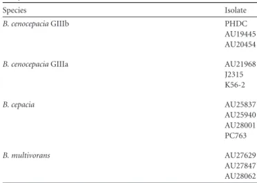
TABLE 1Burkholderia cepaciacomplex clinical isolates used in this study
Species Isolate
B. cenocepaciaGIIIb PHDC
AU19445 AU20454
B. cenocepaciaGIIIa AU21968
J2315 K56-2
B. cepacia AU25837
AU25940 AU28001 PC763
B. multivorans AU27629
AU27847 AU28062
Statistical analysis.All data are shown as means⫾standard errors. Data were analyzed using Prism software (GraphPad Software, Inc.). Nonparametric one-way analysis of variance (ANOVA; Kruskal Wallis) was used to compare multiple groups.Pvalues of⬍0.05 were considered statistically significant. All experiments were performed a minimum of three times.
RESULTS
Antimicrobial activity of SPLUNC1 against Bcc strains.The Bcc epidemic Edinburg-Toronto (ET)-12 strainB. cenocepaciaJ2315 is known to cause cepacia syndrome (42). Therefore, SPLUNC1’s antimicrobial effects were initially tested against this strain in a dose-dependent manner. SPLUNC1 was found to have antimi-crobial activity against J2315, with a 50% inhibitory concentration (IC50 ⫽ 0.28 M) similar to that observed for P. aeruginosa (IC50⫽0.12M) (Fig. 1A). We next tested SPLUNC1’s ability to affect a Gram-positive bacterium (S. aureus). Consistent with pre-vious reports that SPLUNC1 affects only Gram-negative bacteria (22,30),S. aureuswas insensitive to SPLUNC1. We also found that SPLUNC1 was more potent than tobramycin (IC50⫽0.33 M) to reduce growth of J2315, while the antibiotic polymyxin B, to which B. cenocepacia is resistant, had relatively little effect (Fig. 1B) (43). To test SPLUNC1’s ability to affect the growth of other Bcc clinical isolates, a physiological concentration of 0.4M
SPLUNC1, which is comparable to that found in nasal and saliva lavage fluids from healthy humans (21), was added along with Bcc clinical isolates at time zero, and bacterial growth was determined 24 h later. The data indicate that SPLUNC1 significantly affected the growth of many, but not all, of the Bcc clinical isolates that were tested (Fig. 1CtoE). Increasing concentrations of up to 4M SPLUNC1 were also tested against the Bcc clinical isolates that were not initially susceptible, and these isolates remained insensi-tive to SPLUNC1 (n⫽3) (data not shown). These data suggest that susceptibility to SPLUNC1 is strain dependent. SPLUNC1 has previously been shown to bind to bacterial lipopolysaccharide (LPS) (22,32). The LPS composition varies between the different Bcc strains, which may alter its susceptibility to SPLUNC1. To determine if the different LPS structures of Bcc strains play a role in their susceptibility to SPLUNC1, we used theB. cenocepacia K56-2 strain and its⌬hldEand⌬wbxELPS mutants. ThehldE gene codes for a heptokinase and is required for the assembly of the inner core oligosaccharide region of LPS, while thewbxEgene encodes a glycosyltransferase that mediates assembly of the O-an-tigen subunits (44). SPLUNC1 (0.4M) reduced the bacterial growth of wild-type K56-2 to a degree similar to that for J2315 (Fig. 1F). While the ⌬wbxE mutant was susceptible to 0.4M SPLUNC1, growth of the ⌬hldE mutant was unaffected by
FIG 1SPLUNC1 has antimicrobial activity againstB. cepaciacomplex clinical isolates. (A) SPLUNC1 was coincubated with 106CFU/mlB. cenocepaciaJ2315
(),P. aeruginosaPAO1 (), orS. aureusCDL (Œ) for 24 h, and growth was measured. The number of CFU per milliliter was determined, and inhibition was
calculated as follows: % inhibition⫽[(CFU/ml from vehicle⫺CFU/ml from SPLUNC1 present)/(CFU/ml from vehicle)]⫻100. (B) SPLUNC1 (䊐), tobramycin (Œ), and polymyxin B (o) were incubated with 106CFU/ml J2315 for 24 h, and growth was measured. (C to E) SPLUNC1 (0.4M) was incubated
for 24 h with 106CFU/ml ofB. cenocepacia(C),B. cepacia(D), andBurkholderia multivorans(E) isolates, and growth was measured. (F) SPLUNC1 (0.4M) was
incubated for 24 h with 106CFU/ml ofB. cenocepaciaK56-2 and its⌬wbxEand⌬hldELPS mutants, and growth was measured. Open bars, vehicle; closed bars,
SPLUNC1, suggesting that different regions of the LPS structure play a role in SPLUNC1 susceptibility.
SPLUNC1 has bacteriostatic activity againstB. cenocepacia
J2315.To better understand SPLUNC1’s effects on Bcc strains, we subsequently focused on its effects on the epidemic strain J2315 (42). As growth was still seen after 24 h, even with higher concen-trations of SPLUNC1 (Fig. 1A), we next determined whether SPLUNC1 also exerted bacteriostatic and/or bactericidal activity against J2315. We therefore incubated this strain with or without 0.4M SPLUNC1 for 2 h. Bacteria were then washed with 0.1% Triton X-100 to remove SPLUNC1 and grown for an additional 9 h. In the presence of SPLUNC1, bacterial growth was inhibited at 2 h. However, after removal of SPLUNC1, bacterial growth re-sumed, reaching levels similar to those of nontreated J2315 (Fig. 2). In addition, after 24 h, some J2315 bacteria were still present in the medium (Fig. 1B), suggesting that SPLUNC1 has bacterio-static rather than bactericidal activity.
SPLUNC1 has antibiofilm activity against Bcc strains. SPLUNC1 has previously been shown to exhibit antibiofilm activ-ity against Gram-negative bacteria (22, 30, 32). To determine whether SPLUNC1 exerts antibiofilm activity againstB. cenocepa-cia, increasing concentrations of SPLUNC1 were coincubated with 106CFU/ml J2315 for 24 h. Biofilm biomass was then mea-sured by crystal violet staining. Our data indicated that SPLUNC1 prevented J2315 biofilm formation, with an IC50of 0.10M (Fig.
3A). To determine if SPLUNC1 also affected preformed biofilms, J2315 was grown for 24 h to allow for biofilm formation, and SPLUNC1 was added over a range of concentrations to the pre-formed biofilms and incubated for 1 h and 24 h. We found that SPLUNC1 significantly reduced preformed J2315 biofilms after both 1 h and 24 h (Fig. 3B), therefore suggesting that SPLUNC1 can exert antibiofilm activity.
Bacterial attachment is the first step in biofilm formation; we therefore tested whether SPLUNC1 affected biofilm attachment. In addition to reducing biofilm formation, 0.4M SPLUNC1 also inhibited initial J2315 attachment for up to 3 h (Fig. 3C), suggest-ing an additional role for SPLUNC1 in biofilm inhibition.
Since SPLUNC1’s antimicrobial activities against the various Bcc clinical strains differed, its antibiofilm activities against these strains were also tested. At 0.4M, SPLUNC1 reduced some but
not all Bcc biofilm biomass (Fig. 3DtoF). Increasing concentra-tions of up to 4M SPLUNC1 were also tested against the Bcc clinical isolates that were not initially susceptible. However, these strains remained insensitive to SPLUNC1, suggesting that SPLUNC1’s antibiofilm activity is also strain specific (n⫽3) (data not shown). Additionally, while B. cenocepacia strains PHDC, AU20454, and AU21968 were resistant to SPLUNC1’s antimicro-bial activity (Fig. 1C), they were susceptible to SPLUNC1’s anti-biofilm activity (Fig. 3D). Since LPS plays a role in Bcc strain susceptibility to SPLUNC1’s antimicrobial activity (Fig. 1F), it may also be involved in susceptibility to SPLUNC1’s antibiofilm activity. While 0.4M SPLUNC1 reduced the K56-2 biofilm bio-mass to levels similar to those for J2315, its effects on the⌬hldE and⌬wbxELPS mutants varied (Fig. 3E). However, in contrast to the antimicrobial activity results, SPLUNC1 reduced⌬hldE bio-film biomass but did not affect⌬wbxEbiofilm biomass, suggesting that different regions of LPS are involved in Bcc strain suscepti-bility to SPLUNC1’s antibiofilm activity.
SPLUNC1 mutants reduce growth and biofilm formation of
B. cenocepacia J2315. Since SPLUNC1 was effective against J2315, we then sought to determine which domains of SPLUNC1 were responsible for its antimicrobial activity. SPLUNC1’s N-ter-minal S18 region is responsible for regulating ENaC (25), and we recently showed that deletion of the␣4 helix resulted in attenu-ated antimicrobial activity (32). However, the following addi-tional peptides and mutants of SPLUNC1 were generated to test whether the antimicrobial and antibiofilm activities were localized to a specific region of SPLUNC1: (i) the S18 peptide (residues G22 to A39), an N-terminal region containing only the ENaC inhibi-tory domain; (ii) the⌬44 mutant (residues T45 to V256), which lacks the SPLUNC1 N terminus, including the S18 region; (iii) the ⌬␣4 mutant, which lacks the␣4 helix (residues I76 to I105 and includes nonnative Gly-Ser-Gly-Ser linker to L75 to I106); (iv) a peptide corresponding to the␣4 helix (residues K77 to L101); (v) the⌬␣6 mutant, in which the␣6 helix (residues I242 to V256) is absent; and (vi) the␣6 helix peptide (residues I242 to V256) (Fig. 4A). The⌬␣4 and⌬␣6 mutants were chosen because (i)␣4 and ␣6 are two novel helixes that were present in our SPLUNC1 crystal structure and do not share homology with BPI (40) and (ii) our previous studies demonstrated that deletion of the␣4 helix re-duced SPLUNC1’s antimicrobial effects (32). SPLUNC1 mutants were coincubated with 106CFU/ml J2315 for 24 h, and bacterial growth and biofilm biomass were determined (Fig. 4BandC). The S18 peptide exhibited neither antimicrobial nor antibiofilm activ-ity against J2315. Consistent with this observation, the⌬44 mu-tant retained full antimicrobial activity (IC50⫽0.14 M) and antibiofilm activity (IC50⫽0.12M). The helix mutants varied in their effects against J2315. The ⌬␣4 mutant lost antimicrobial activity against J2315 and also had significantly diminished anti-biofilm activity (IC50⫽0.46M). However, the␣4 peptide pos-sessed antimicrobial activity against J2315 (IC50⫽0.36M) but did not have antibiofilm activity (Fig. 4BandC). The⌬␣6 mutant retained its antimicrobial activity (IC50⫽0.15M) but had sig-nificantly reduced antibiofilm activity (IC50⫽0.24M), while the ␣6 peptide had neither antimicrobial nor antibiofilm activity against J2315 (Fig. 4BandC).
Neutrophil elastase maintains SPLUNC1’s antibiofilm activ-ity.Chronic neutrophilia is a hallmark of CF lung disease and leads to elevated levels of NE in the lung lumen (45). SPLUNC1 is known to be a substrate for NE, and in some cases, NE may alter
FIG 2SPLUNC1 is bacteriostatic, not bactericidal.B. cenocepaciaJ2315 was incubated with or without 0.4M SPLUNC1 for 2 h (starting at⫺2 h). Bac-teria were then washed at 0 h with 0.1% Triton X-100 to remove SPLUNC1, grown for an additional 9 h, and measured by determining the OD600every 3 h.
Open bars, vehicle; closed bars, 0.4M SPLUNC1. *,P⬍0.05 compared to vehicle at⫺2 h; **,P⬍0.01 compared to vehicle at⫺2 h;⫹⫹,P⬍0.01 compared to 0.4M SPLUNC1 at⫺2 h (n⫽3).
SPLUNC1’s activity (25,31,46). To determine the effects of NE cleavage on SPLUNC1’s antimicrobial and antibiofilm activities, SPLUNC1 was exposed to 1M NE for defined periods, after which NE activity was inhibited by sivelestat as previously re-ported (47). SPLUNC1 was extensively cleaved by NE, as shown by SDS-PAGE followed by Coomassie blue staining (Fig. 5A). In-hibition of NE by sivelestat was then confirmed by measuring the ability of NE to cleave the fluorogenic substrate Suc-Ala-Ala-Ala-MCA (Fig. 5B). SDS-PAGE fully denatures SPLUNC1, allowing
individual fragments to be separated by size. However, under the nondenaturing conditions seen in the airways, NE cleavage of SPLUNC1 may not result in its dissociation, and SPLUNC1 may remain cohesive in the airway surface liquid (ASL) after cleavage. To determine if SPLUNC1 remained intact after NE exposure, we measured SPLUNC1’s molecular size in a physiological solution after NE exposure by multiangle static light scattering. Molecular mass was determined by measuring the intensity of scattered light against SPLUNC1. SPLUNC1’s initial (0 h) molecular mass was
FIG 3SPLUNC1 has antibiofilm activity against Bcc strains. (A) Dose-response curve for SPLUNC1 coincubated with 106CFU/mlB. cenocepaciaJ2315 for 24 h.
(B) Preformed (24 h) J2315 biofilms were incubated with increasing concentrations of SPLUNC1 for 1 h () or 24 h (Œ). (C) Attachment assay for J2315
coincubated with 0.4M SPLUNC1 for up to 3 h. (D to F) SPLUNC1 (0.4M) was coincubated for 24 h withB. cenocepacia(D),B. cepacia(E), andB. multivorans(F) Bcc clinical isolates. (G) SPLUNC1 (0.4M) was incubated for 24 h with 106CFU/ml ofB. cenocepaciaK56-2 and its⌬wbxEand⌬hldELPS
mutants. Bcc strains were stained with 1% crystal violet and measured by determining the OD590, and inhibition was calculated as follows: % inhibition⫽
[(CFU/ml from vehicle⫺CFU/ml from SPLUNC1 present)/(CFU/ml from vehicle)]⫻100. Open bars, vehicle; closed bars, 0.4M SPLUNC1. *,P⬍0.05; **,
23.5 kDa (Fig. 5C). After a 1-h exposure to NE, SPLUNC1’s mo-lecular mass was 22.3 kDa. There was a slight reduction in molec-ular mass within the initial 6 h after NE exposure, to 19.6 kDa. After 24 h of exposure to NE, SPLUNC1’s molecular mass further decreased, to 18.6 kDa. However, these sizes were still greater than those of the individual fragments detected by SDS-PAGE (⬃15 to 17 kDa) (Fig. 5A). To further examine changes in SPLUNC1’s structure after NE exposure, cleaved SPLUNC1 was analyzed by circular dichroism (CD) spectroscopy in the far-UV spectral re-gion (190 to 250 nm) to observe SPLUNC1’s secondary structures. SPLUNC1 initially had a secondary alpha-helical structure, as in-dicated by a positive signal at 194 nm and two small negative signals, at 208 and 222 nm (Fig. 5D). Despite being cleaved by NE, SPLUNC1 retained its secondary alpha-helical structure for up to 12 h, but it lost this structure after 24 h of incubation with NE, as shown by a random coiling effect, with a negative signal at 200 nm and an increasing signal at 210 nm.
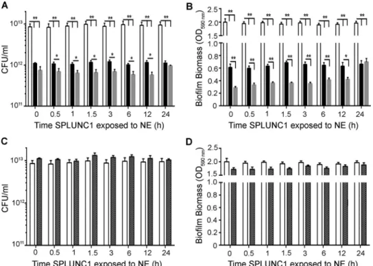
We next tested cleaved SPLUNC1, created by timed incuba-tions with NE, for antimicrobial and antibiofilm activities by coincubation with 106 CFU/ml J2315 for 24 h. NE-exposed SPLUNC1 had increased antimicrobial and antibiofilm activities against J2315 compared to those of SPLUNC1 alone (Fig. 6Aand
B). However, after 24 h, NE-exposed SPLUNC1 had levels of an-timicrobial activity similar to those of SPLUNC1 alone. Surpris-ingly, the effect of SPLUNC1 to disrupt biofilm formation was significantly potentiated by NE pretreatment for up to 12 h (n⫽3; P⬍0.01) (Fig. 6B). Importantly, these effects were not due to
active NE, since NE activity had been halted by sivelestat (Fig. 5B). As a control, we tested the effects of NE plus sivelestat against J2315 growth, and these compounds had no antimicrobial or an-tibiofilm activity (Fig. 6CandD).
DISCUSSION
The airways contain many antimicrobial agents, including pep-tides, such as cathelicidins and-defensins, and proteins, includ-ing SPLUNC1, as part of the first line of innate defense against pathogens (27,33). Previous reports showed that knockout of SPLUNC1 in mice led to increases in bacterial infections byP. aeruginosa,K. pneumoniae, andH. influenzae(26,30,31), suggest-ing that SPLUNC1 plays an important role in reducsuggest-ing bacterial infections. Furthermore, 0.4M SPLUNC1 reducedP. aeruginosa growth by 80%in vitro(48). While Bcc growth is not affected by cathelicidins or-defensins (49), our results show that 0.4M SPLUNC1, which is within the physiological range of SPLUNC1 in the ASL (0.4 to 1M), also reduces J2315 growth (Fig. 1Aand
B) (22). SPLUNC1 is thought to exert its antimicrobial activity againstP. aeruginosaby formation of pores in the bacterial cell wall, thus increasing cell wall permeability (22). SPLUNC1 shares structural homology with BPI and binds through hydrophobic interactions with the LPS of Gram-negative bacteria, such asK. pneumoniae andP. aeruginosa (50). Despite being structurally smaller than BPI, SPLUNC1 is thought to have similar mecha-nisms of interaction withP. aeruginosa(22). Although J2315 was
FIG 4The␣4 helix is required for SPLUNC1’s antimicrobial activity againstB. cenocepaciaJ2315. (A) Three-dimensional rendering of SPLUNC1 structure with the intrinsically disordered S18 region appended (labeled in red). Also indicated are⌬44 SPLUNC1, which lacks the S18 region (blue arrow); the␣4 region (labeled in purple), which is absent in the⌬␣4 mutant; and the␣6 region (labeled in green), which is absent in the⌬␣6 mutant. Increasing concentrations of SPLUNC1 (black closed circles), the⌬44 (blue closed squares),⌬␣4 (purple closed squares), and⌬␣6 (green closed triangles) SPLUNC1 mutants, and the␣4 (purple open squares),␣6 (green open triangles), and S18 (red closed triangles) peptides were coincubated with 106CFU/ml J2315 for 24 h and measured for
antimicrobial activity by CFU counts and calculation of % growth inhibition as previously described (B) and for antibiofilm activity by 1% crystal violet staining, OD590reading, and calculation of % biofilm biomass inhibition as previously described (C). **,P⬍0.001 for S18 peptide compared to SPLUNC1; ***,P⬍0.0001
for S18 peptide compared to SPLUNC1;⫹⫹,P⬍0.001 for⌬␣4 mutant compared to SPLUNC1;⫹⫹⫹,P⬍0.0001 for⌬␣4 mutant compared to SPLUNC1; #,P⬍0.01 for⌬␣6 mutant compared to SPLUNC1;⫻⫻,P⬍0.05 for␣4 peptide compared to SPLUNC1;⫻⫻⫻,P⬍0.001 for␣4 peptide compared to SPLUNC1; °,P⬍0.05 for␣6 peptide compared to SPLUNC1; °°°,P⬍0.001 for␣6 peptide compared to SPLUNC1 (n⫽3 for all panels).
susceptible to SPLUNC1, SPLUNC1’s antimicrobial activity var-ied among the different Bcc species (Fig. 1CtoE). This variation in susceptibility among the different Bcc species may be due to the unusual composition of the LPS structure, which differs among the Bcc species (51). Indeed, susceptibility to other antibiotics has been reported to vary among the Bcc members (11,52). Our re-sults demonstrate that changes to Bcc strains’ LPS structure alter their susceptibility to SPLUNC1 (Fig. 1Fand3E). As LPS plays an important role in bacterial sensitivity to antimicrobial agents, more studies will be needed to compare the LPS structures of these strains to determine their interaction with SPLUNC1.
Researchers have proposed that the ASL is bacteriostatic rather than bactericidal (53–55) and must act in concert with functional mucociliary clearance (MCC) to remove bacteria. That is, as bac-terial growth is impaired, MCC removes the bacteria in the air-ways, preventing bacterial colonization (56). SPLUNC1 has been shown to coatP. aeruginosato inhibit growth rather than killing bacteria (22, 32). Indeed, our results revealed that when SPLUNC1 was removed, J2315 growth was restored to levels sim-ilar to those of the controls (Fig. 2), confirming that SPLUNC1 also has bacteriostatic activity against Bcc strains.
In order to determine which domain of SPLUNC1 is required for its antimicrobial activity, SPLUNC1 mutants and peptides were tested. The S18 peptide exerted neither antimicrobial nor antibiofilm activity against J2315. The⌬44 mutant, which lacks the S18 region, had antimicrobial and antibiofilm activities com-parable to those of full-length SPLUNC1. Although the S18 region does not exert antimicrobial or antibiofilm activity against J2315, since this region is required to regulate ENaC (25), it still plays a role in mechanically clearing bacteria via the mucociliary escalator in vivo. Deletion of the␣4 but not␣6 helix resulted in a loss of
antimicrobial activity. Consistent with this observation, the␣4 peptide restored antimicrobial activity, suggesting that this region of SPLUNC1 is absolutely required for SPLUNC1’s antimicrobial activity. In addition, both the⌬␣4 and⌬␣6 mutants had reduced biofilm activity, but here it was less clear, since the␣4 deletion exerted a much stronger effect than the␣6 deletion. In addition, the␣4 and␣6 peptides alone did not exert antibiofilm activity. However, it is likely that these helixes both play roles in SPLUNC1’s antibiofilm activity while present in the SPLUNC1 protein (Fig. 4). While the⌬44 mutant retained antimicrobial activity (Fig. 4B), other SPLUNC1 fragments formed by NE activ-ity may further expose the domains for antimicrobial and anti-biofilm activities of SPLUNC1 for enhancement of the reduction of J2315 growth and biofilm formation.
Although biofilms in CF patients are rare, Bcc strains have been shown to form biofilmsin vitroand to form thick biofilms in sputa of CF patients (57–59). Biofilms increase the bacterium’s antibi-otic resistance. However, we found that 0.4M SPLUNC1 both prevents biofilm formation and reduces preformed J2315 biofilms (Fig. 3AandB). Surfactants change flagellar development, leading to altered bacterial attachment and altered biofilm formation (30,
60). SPLUNC1 has surfactant activity (21,32), which may play a role in antibiofilm activity. Indeed, our results have shown that SPLUNC1 reduces J2315 attachment (Fig. 3C), and we speculate that SPLUNC1’s surfactant activities may play a role in antibiofilm activity against Bcc strains. Additionally, SPLUNC1’s antibiofilm activity varied among the Bcc clinical isolates (Fig. 3DandE), as was seen with SPLUNC1’s antimicrobial activity (Fig. 1CtoE), which may be due to differences in the LPS or flagellar proteins of Bcc clinical isolates.
Persistent bacterial infection in CF lungs leads to airway
FIG 5SPLUNC1 does not dissociate and retains secondary structure after cleavage with NE. (A) Time course showing cleavage of 40M SPLUNC1 by 1M NE by SDS-PAGE with Coomassie blue staining. (B) Inhibition of 1M NE activity without () or with (䊐) 1M sivelestat and 10M substrate (Suc-Ala-Ala-Ala-MCA protein) (error bars are obscured by the symbols). A.U., arbitrary units. (C) Static light scattering of SPLUNC1 before and at timed intervals after exposure to NE and sivelestat. (D) Circular dichroism analysis of SPLUNC1 before and at timed intervals after exposure to NE and sivelestat. *,P⬍0.01; **,P⬍
inflammation, chronic neutrophilia, increased protease activ-ity, and subsequent lung damage (23, 37, 61). NE readily cleaves SPLUNC1. However, we previously showed that the S18 peptide, which is analogous to SPLUNC1’s ENaC inhibi-tory domain, remains as an intact and functional peptide capa-ble of regulating ENaC even after NE exposure (25). Con-versely, Jiang et al. reported that addition of NE to normal human tracheobronchial epithelia impaired their antimicro-bial activity againstMycoplasma pneumoniaeandH. influenzae (31). Here we found that NE-cleaved SPLUNC1 maintains anti-microbial/antibiofilm activity for up to 24 h against J2315 and that this activity is significantly enhanced compared to that of whole SPLUNC1 for a limited period (Fig. 6AandB). During the early stages of infection, both SPLUNC1 and NE expression levels in-crease (22, 23,37), which may serve to potentiate SPLUNC1’s antimicrobial activities, leading to a quicker resolution of the in-fection. However, in CF airways, SPLUNC1 is inactive due to the acidic environment, leading to a failure to regulate ENaC and to clear mucus, and chronic neutrophilia occurs, resulting in in-creased NE levels (29,37,40). We noted that the beneficial effects of NE on SPLUNC1 were eventually abolished (Fig. 6). Chroni-cally increased NE levels may therefore lead to an altered ASL milieu that contributes to SPLUNC1’s degradation and further impairment of SPLUNC1’s antimicrobial activities (31). Indeed, we previously reported that SPLUNC1 is differentially cleaved in CF versus normal sputum (25). Our data indicate that as NE ini-tially cleaves SPLUNC1, SPLUNC1 iniini-tially retains its secondary
alpha-helical structure (Fig. 5D), which may allow the protein to continue to exert antimicrobial activity against J2315 as well as releasing the S18 peptide, which can help to flush out the airways by inhibiting ENaC and increasing hydration and MCC.
In conclusion, Bcc strain resistance to many antibiotics poses a problem for immunocompromised individuals (62,63). For ex-ample, as Bcc strains colonize CF lungs in the later stages of the disease, patients exhibit a greater decline in pulmonary function and an increase in mortality (64). Our data have shown that SPLUNC1 affects J2315 by (i) bacteriostatic effects to reduce growth and (ii) antibiofilm activity to prevent and reduce biofilm formation via its␣4 and␣6 helixes. Further investigation into these helixes may provide novel therapies for treating Bcc infec-tions. While the impact of CF lung disease on SPLUNC1-Bcc in-teractions is not currently known, our data suggest that under-standing this phenomenon may have important implications for CF lung disease.
ACKNOWLEDGMENTS
We thank John LiPuma from the University of Michigan for sending the Bcc isolates, Miguel Valvano (Queens University Belfast) for the Bcc K56-2 strain and LPS mutants, and Colin Bingle (University of Sheffield, United Kingdom) for the wild-type SPLUNC1 construct.
This work was funded by NIH grant R01 HL108927 and by an INOVCF grant from the UK Cystic Fibrosis Trust.
Robert Tarran has equity in Spyryx Biosciences Inc. No other conflicts of interest exist.
FIG 6Cleaved wild-type SPLUNC1 exerts larger effects onB. cenocepaciaJ2315 growth and biofilm formation than those seen with whole SPLUNC1. Aliquots of SPLUNC1 were exposed to NE for timed intervals, and NE activity was then halted with sivelestat. NE-cleaved SPLUNC1 was then incubated for 24 h with 106
CFU/ml J2315. (A) CFU counts to show antimicrobial activity after incubation with whole versus cleaved SPLUNC1. (B) Inhibition of biofilm formation as measured by crystal violet staining followed by OD590readings. NE (1M) plus sivelestat (1M) alone had neither antimicrobial activity (C) nor antibiofilm
activity (D). White bars, vehicle; black bars, 0.4M SPLUNC1; gray bars, 0.4M NE-cleaved SPLUNC1; hatched bars, 1M NE plus 1M sivelestat (control). *,P⬍0.05; **,P⬍0.01 (n⫽5 for all panels).
FUNDING INFORMATION
This work, including the efforts of Jean Tyrrell, William G. Walton, Mat-thew R. Redinbo, and Robert Tarran, was funded by HHS | National Institutes of Health (NIH) (HL108927). This work, including the efforts of Saira Ahmad and Robert Tarran, was funded by Cystic Fibrosis Trust (SRC003).
REFERENCES
1.Mahenthiralingam E, Baldwin A, Vandamme P. 2002. Burkholderia cepaciacomplex infection in patients with cystic fibrosis. J Med Microbiol
51:533–538.http://dx.doi.org/10.1099/0022-1317-51-7-533.
2.Vanlaere E, Lipuma JJ, Baldwin A, Henry D, De Brandt E, Mahenthi-ralingam E, Speert D, Dowson C, Vandamme P. 2008.Burkholderia latenssp. nov.,Burkholderia diffusasp. nov.,Burkholderia arborissp. nov.,
Burkholderia seminalissp. nov. andBurkholderia metallicasp. nov., novel species within theBurkholderia cepaciacomplex. Int J Syst Evol Microbiol
58:1580 –1590.http://dx.doi.org/10.1099/ijs.0.65634-0.
3.Dedeckova K, Fila L, Skalicka V, Bartosova J, Kucerova T, Vavrova V, Zemkova D, Kalferstova L, Melter O, Cinek O, Drevinek P.2012. PCR detection ofBurkholderia cepaciacomplex as one of key factors to handle a long-term outbreak. J Cyst Fibros11:440 – 445.http://dx.doi.org/10 .1016/j.jcf.2012.04.005.
4.Saldias MS, Valvano MA.2009. Interactions ofBurkholderia cenocepacia
and otherBurkholderia cepaciacomplex bacteria with epithelial and phagocytic cells. Microbiology155:2809 –2817.http://dx.doi.org/10.1099 /mic.0.031344-0.
5.Loutet SA, Valvano MA.2010. A decade ofBurkholderia cenocepacia
virulence determinant research. Infect Immun78:4088 – 4100.http://dx .doi.org/10.1128/IAI.00212-10.
6.Amin R, Lam M, Dupuis A, Ratjen F.2011. The effect of early Pseu-domonas aeruginosatreatment on lung function in pediatric cystic fibrosis. Pediatr Pulmonol46:554 –558.http://dx.doi.org/10.1002/ppul.21417. 7.Coutinho CP, Dos Santos SC, Madeira A, Mira NP, Moreira AS,
Sa-Correia I.2011. Long-term colonization of the cystic fibrosis lung by
Burkholderia cepacia complex bacteria: epidemiology, clonal variation, and genome-wide expression alterations. Front Cell Infect Microbiol1:12.
http://dx.doi.org/10.3389/fcimb.2011.00012.
8.Mahenthiralingam E, Urban TA, Goldberg JB.2005. The multifarious, multirepliconBurkholderia cepaciacomplex. Nat Rev Microbiol3:144 – 156.http://dx.doi.org/10.1038/nrmicro1085.
9.Isles A, Maclusky I, Corey M, Gold R, Prober C, Fleming P, Levison H.
1984.Pseudomonas cepaciainfection in cystic fibrosis: an emerging prob-lem. J Pediatr104:206 –210. http://dx.doi.org/10.1016/S0022-3476 (84)80993-2.
10. Valvano MA, Keith KE, Cardona ST.2005. Survival and persistence of opportunisticBurkholderiaspecies in host cells. Curr Opin Microbiol
8:99 –105.http://dx.doi.org/10.1016/j.mib.2004.12.002.
11. Nzula S, Vandamme P, Govan JR.2002. Influence of taxonomic status on the in vitro antimicrobial susceptibility of theBurkholderia cepacia
complex. J Antimicrob Chemother50:265–269.http://dx.doi.org/10.1093 /jac/dkf137.
12. Sousa SA, Ramos CG, Almeida F, Meirinhos-Soares L, Wopperer J, Schwager S, Eberl L, Leitao JH.2008.Burkholderia cenocepaciaJ2315 acyl carrier protein: a potential target for antimicrobials’ development? Mi-crob Pathog45:331–336.http://dx.doi.org/10.1016/j.micpath.2008.08 .002.
13. Leitao JH, Sousa SA, Cunha MV, Salgado MJ, Melo-Cristino J, Barreto MC, Sa-Correia I.2008. Variation of the antimicrobial susceptibility profiles ofBurkholderia cepaciacomplex clonal isolates obtained from chronically infected cystic fibrosis patients: a five-year survey in the major Portuguese treatment center. Eur J Clin Microbiol Infect Dis27:1101– 1111.http://dx.doi.org/10.1007/s10096-008-0552-0.
14. Burns JL, Jonas M, Chi EY, Clark DK, Berger A, Griffith A. 1996. Invasion of respiratory epithelial cells byBurkholderia(Pseudomonas) ce-pacia. Infect Immun64:4054 – 4059.
15. Martin DW, Mohr CD.2000. Invasion and intracellular survival of Burk-holderia cepacia. Infect Immun68:24 –29.http://dx.doi.org/10.1128/IAI .68.1.24-29.2000.
16. Schwab U, Abdullah LH, Perlmutt OS, Albert D, Davis CW, Arnold RR, Yankaskas JR, Gilligan P, Neubauer H, Randell SH, Boucher RC.2014. Localization ofBurkholderia cepaciacomplex bacteria in cystic fibrosis
lungs and interactions withPseudomonas aeruginosain hypoxic mucus. Infect Immun82:4729 – 4745.http://dx.doi.org/10.1128/IAI.01876-14. 17. Forier K, Messiaen AS, Raemdonck K, Nelis H, De Smedt S, Demeester
J, Coenye T, Braeckmans K.2014. Probing the size limit for nanomedi-cine penetration intoBurkholderia multivoransandPseudomonas aerugi-nosabiofilms. J Control Release195:21–28.http://dx.doi.org/10.1016/j .jconrel.2014.07.061.
18. Conway BA, Venu V, Speert DP. 2002. Biofilm formation and acyl homoserine lactone production in theBurkholderia cepaciacomplex. J Bacteriol184:5678 –5685.http://dx.doi.org/10.1128/JB.184.20.5678-5685 .2002.
19. Dales L, Ferris W, Vandemheen K, Aaron SD. 2009. Combination antibiotic susceptibility of biofilm-grownBurkholderia cepaciaand Pseu-domonas aeruginosaisolated from patients with pulmonary exacerbations of cystic fibrosis. Eur J Clin Microbiol Infect Dis28:1275–1279.http://dx .doi.org/10.1007/s10096-009-0774-9.
20. Bingle CD, Bingle L.2000. Characterisation of the human plunc gene, a gene product with an upper airways and nasopharyngeal restricted expres-sion pattern. Biochim Biophys Acta1493:363–367.http://dx.doi.org/10 .1016/S0167-4781(00)00196-2.
21. Gakhar L, Bartlett JA, Penterman J, Mizrachi D, Singh PK, Mallampalli RK, Ramaswamy S, McCray PB, Jr.2010. PLUNC is a novel airway surfactant protein with anti-biofilm activity. PLoS One5:e9098.http://dx .doi.org/10.1371/journal.pone.0009098.
22. Sayeed S, Nistico L, St Croix C, Di YP.2013. Multifunctional role of human SPLUNC1 inPseudomonas aeruginosainfection. Infect Immun
81:285–291.http://dx.doi.org/10.1128/IAI.00500-12.
23. Bingle L, Barnes FA, Cross SS, Rassl D, Wallace WA, Campos MA, Bingle CD.2007. Differential epithelial expression of the putative innate immune molecule SPLUNC1 in cystic fibrosis. Respir Res8:79.http://dx .doi.org/10.1186/1465-9921-8-79.
24. Garcia-Caballero A, Rasmussen JE, Gaillard E, Watson MJ, Olsen JC, Donaldson SH, Stutts MJ, Tarran R.2009. SPLUNC1 regulates airway surface liquid volume by protecting ENaC from proteolytic cleavage. Proc Natl Acad Sci U S A106:11412–11417.http://dx.doi.org/10.1073/pnas .0903609106.
25. Hobbs CA, Blanchard MG, Alijevic O, Tan CD, Kellenberger S, Bencharit S, Cao R, Kesimer M, Walton WG, Henderson AG, Redinbo MR, Stutts MJ, Tarran R.2013. Identification of the SPLUNC1 ENaC-inhibitory domain yields novel strategies to treat sodium hyperabsorption in cystic fibrosis airway epithelial cultures. Am J Physiol Lung Cell Mol Physiol305:L990 –L1001.http://dx.doi.org/10.1152/ajplung.00103.2013. 26. Liu Y, Di ME, Chu HW, Liu X, Wang L, Wenzel S, Di YP. 2013. Increased susceptibility to pulmonaryPseudomonasinfection in Splunc1 knockout mice. J Immunol 191:4259 – 4268.http://dx.doi.org/10.4049 /jimmunol.1202340.
27. Di YP.2011. Functional roles of SPLUNC1 in the innate immune re-sponse against Gram-negative bacteria. Biochem Soc Trans39:1051– 1055.http://dx.doi.org/10.1042/BST0391051.
28. Britto CJ, Liu Q, Curran DR, Patham B, Dela Cruz CS, Cohn L.2013. Short palate, lung, and nasal epithelial clone-1 is a tightly regulated airway sensor in innate and adaptive immunity. Am J Respir Cell Mol Biol48:
717–724.http://dx.doi.org/10.1165/rcmb.2012-0072OC.
29. Tarran R, Redinbo MR.2014. Mammalian short palate lung and nasal epithelial clone 1 (SPLUNC1) in pH-dependent airway hydration. Int J Biochem Cell Biol52:130 –135.http://dx.doi.org/10.1016/j.biocel.2014.03 .002.
30. Liu Y, Bartlett JA, Di ME, Bomberger JM, Chan YR, Gakhar L, Mal-lampalli RK, McCray PB, Jr, Di YP.2013. SPLUNC1/BPIFA1 contrib-utes to pulmonary host defense againstKlebsiella pneumoniaerespiratory infection. Am J Pathol182:1519 –1531.http://dx.doi.org/10.1016/j.ajpath .2013.01.050.
31. Jiang D, Wenzel SE, Wu Q, Bowler RP, Schnell C, Chu HW.2013. Human neutrophil elastase degrades SPLUNC1 and impairs airway epi-thelial defense against bacteria. PLoS One8:e64689.http://dx.doi.org/10 .1371/journal.pone.0064689.
32. Walton WG, Ahmad S, Little MS, Kim CS, Tyrrell J, Lin Q, Di YP, Tarran R, Redinbo MR.2016. Structural features essential to the antimi-crobial functions of human SPLUNC1. Biochemistry55:2979 –2991.http: //dx.doi.org/10.1021/acs.biochem.6b00271.
34. Hiemstra PS, McCray PB, Jr, Bals R.2015. The innate immune function of airway epithelial cells in inflammatory lung disease. Eur Respir J45:
1150 –1162.http://dx.doi.org/10.1183/09031936.00141514.
35. Parker D, Prince A.2011. Innate immunity in the respiratory epithelium. Am J Respir Cell Mol Biol45:189 –201.http://dx.doi.org/10.1165/rcmb .2011-0011RT.
36. Elborn JS, Cordon SM, Parker D, Delamere FM, Shale DJ.1993. The host inflammatory response prior to death in patients with cystic fibrosis and chronicPseudomonas aeruginosainfection. Respir Med87:603– 607.
http://dx.doi.org/10.1016/S0954-6111(05)80263-X.
37. Voynow JA, Fischer BM, Zheng S.2008. Proteases and cystic fibrosis. Int J Biochem Cell Biol 40:1238 –1245. http://dx.doi.org/10.1016/j.biocel .2008.03.003.
38. Richman-Eisenstat J.1996. Cytokine soup: making sense of inflammation in cystic fibrosis. Pediatr Pulmonol 21:3–5. http://dx.doi.org/10.1002 /1099-0496(199601)21:1⬍3::AID-PPUL1950210103⬎3.0.CO;2-B. 39. Belaaouaj A.2002. Neutrophil elastase-mediated killing of bacteria:
les-sons from targeted mutagenesis. Microbes Infect4:1259 –1264.http://dx .doi.org/10.1016/S1286-4579(02)01654-4.
40. Garland AL, Walton WG, Coakley RD, Tan CD, Gilmore RC, Hobbs CA, Tripathy A, Clunes LA, Bencharit S, Stutts MJ, Betts L, Redinbo MR, Tarran R.2013. Molecular basis for pH-dependent mucosal dehy-dration in cystic fibrosis airways. Proc Natl Acad Sci U S A110:15973– 15978.http://dx.doi.org/10.1073/pnas.1311999110.
41. Margolis JJ, El-Etr S, Joubert LM, Moore E, Robison R, Rasley A, Spormann AM, Monack DM.2010. Contributions ofFrancisella tular-ensissubsp.novicidachitinases and Sec secretion system to biofilm forma-tion on chitin. Appl Environ Microbiol76:596 – 608.http://dx.doi.org/10 .1128/AEM.02037-09.
42. Vandamme P, Holmes B, Coenye T, Goris J, Mahenthiralingam E, LiPuma JJ, Govan JR.2003.Burkholderia cenocepaciasp. nov.—a new twist to an old story. Res Microbiol154:91–96.http://dx.doi.org/10.1016 /S0923-2508(03)00026-3.
43. Loutet SA, Valvano MA.2011. Extreme antimicrobial peptide and poly-myxin B resistance in the genusBurkholderia. Front Cell Infect Microbiol
1:6.http://dx.doi.org/10.3389/fcimb.2011.00006.
44. Loutet SA, Flannagan RS, Kooi C, Sokol PA, Valvano MA.2006. A complete lipopolysaccharide inner core oligosaccharide is required for resistance ofBurkholderia cenocepaciato antimicrobial peptides and bac-terial survival in vivo. J Bacteriol188:2073–2080.http://dx.doi.org/10 .1128/JB.188.6.2073-2080.2006.
45. Conese M, Copreni E, Di Gioia S, De Rinaldis P, Fumarulo R.2003. Neutrophil recruitment and airway epithelial cell involvement in chronic cystic fibrosis lung disease. J Cyst Fibros2:129 –135.http://dx.doi.org/10 .1016/S1569-1993(03)00063-8.
46. Gally F, Di YP, Smith SK, Minor MN, Liu Y, Bratton DL, Frasch SC, Michels NM, Case SR, Chu HW.2011. SPLUNC1 promotes lung innate defense againstMycoplasma pneumoniaeinfection in mice. Am J Pathol
178:2159 –2167.http://dx.doi.org/10.1016/j.ajpath.2011.01.026. 47. Kawabata K, Suzuki M, Sugitani M, Imaki K, Toda M, Miyamoto T.
1991. ONO-5046, a novel inhibitor of human neutrophil elastase. Biochem Biophys Res Commun177:814 – 820.http://dx.doi.org/10.1016 /0006-291X(91)91862-7.
48. Zhou HD, Li XL, Li GY, Zhou M, Liu HY, Yang YX, Deng T, Ma J, Sheng SR.2008. Effect of SPLUNC1 protein on thePseudomonas aerugi-nosaand Epstein-Barr virus. Mol Cell Biochem309:191–197.http://dx.doi .org/10.1007/s11010-007-9659-3.
49. Baird RM, Brown H, Smith AW, Watson ML.1999.Burkholderia cepacia
is resistant to the antimicrobial activity of airway epithelial cells.
Immu-nopharmacology 44:267–272. http://dx.doi.org/10.1016/S0162-3109 (99)00122-8.
50. Meszaros K, Parent JB, Gazzano-Santoro H, Little R, Horwitz A, Parsons T, Theofan G, Grinna L, Weickmann J, Elsbach P, Weiss J, Conlon PJ.1993. A recombinant amino terminal fragment of bactericid-al/permeability-increasing protein inhibits the induction of leukocyte re-sponses by LPS. J Leukoc Biol54:558 –563.
51. De Soyza A, Silipo A, Lanzetta R, Govan JR, Molinaro A.2008. Chem-ical and biologChem-ical features ofBurkholderia cepaciacomplex lipopolysac-charides. Innate Immun 14:127–144. http://dx.doi.org/10.1177 /1753425908093984.
52. Rose H, Baldwin A, Dowson CG, Mahenthiralingam E.2009. Biocide susceptibility of theBurkholderia cepaciacomplex. J Antimicrob Che-mother63:502–510.http://dx.doi.org/10.1093/jac/dkn540.
53. Schutte BC, McCray PB, Jr.2002. Beta-defensins in lung host defense. Annu Rev Physiol 64:709 –748. http://dx.doi.org/10.1146/annurev .physiol.64.081501.134340.
54. Schnapp D, Harris A.1998. Antibacterial peptides in bronchoalveolar lavage fluid. Am J Respir Cell Mol Biol19:352–356.http://dx.doi.org/10 .1165/ajrcmb.19.3.3384.
55. Konstan MW, Chen PW, Sherman JM, Thomassen MJ, Wood RE, Boat TF.1981. Human lung lysozyme: sources and properties. Am Rev Respir Dis123:120 –124.
56. Cole AM, Dewan P, Ganz T.1999. Innate antimicrobial activity of nasal secretions. Infect Immun67:3267–3275.
57. Kennedy S, Beaudoin T, Yau YC, Caraher E, Zlosnik JE, Speert DP, LiPuma JJ, Tullis E, Waters V.2016. Activity of tobramycin against cystic fibrosis isolates ofBurkholderia cepaciacomplex grown as biofilms. Anti-microb Agents Chemother60:348 –355.http://dx.doi.org/10.1128/AAC .02068-15.
58. Silva IN, Tavares AC, Ferreira AS, Moreira LM.2013. Stress conditions triggering mucoid morphotype variation inBurkholderiaspecies and ef-fect on virulence inGalleria mellonellaand biofilm formation in vitro. PLoS One8:e82522.http://dx.doi.org/10.1371/journal.pone.0082522. 59. Al-Bakri AG, Gilbert P, Allison DG.2005. Influence of gentamicin and
tobramycin on binary biofilm formation by co-cultures ofBurkholderia cepaciaandPseudomonas aeruginosa. J Basic Microbiol45:392–396.http: //dx.doi.org/10.1002/jobm.200510011.
60. Splendiani A, Livingston AG, Nicolella C.2006. Control of membrane-attached biofilms using surfactants. Biotechnol Bioeng94:15–23.http: //dx.doi.org/10.1002/bit.20752.
61. Jiang D, Persinger R, Wu Q, Gross A, Chu HW.2013.␣1-Antitrypsin promotes SPLUNC1-mediated lung defense againstPseudomonas aerugi-nosainfection in mice. Respir Res14:122.http://dx.doi.org/10.1186/1465 -9921-14-122.
62. Drevinek P, Mahenthiralingam E.2010.Burkholderia cenocepaciain cys-tic fibrosis: epidemiology and molecular mechanisms of virulence. Clin Microbiol Infect16:821– 830.http://dx.doi.org/10.1111/j.1469-0691.2010 .03237.x.
63. Saiman L, Siegel J, Cystic Fibrosis Foundation Consensus Conference on Infection Control Participants.2003. Infection control recommen-dations for patients with cystic fibrosis: microbiology, important patho-gens, and infection control practices to prevent patient-to-patient trans-mission. Am J Infect Control31:S1–S62.
64. Courtney JM, Dunbar KE, McDowell A, Moore JE, Warke TJ, Steven-son M, Elborn JS.2004. Clinical outcome ofBurkholderia cepacia com-plex infection in cystic fibrosis adults. J Cyst Fibros3:93–98.http://dx.doi .org/10.1016/j.jcf.2004.01.005.