BIOPHYSICAL MECHANISMS OF PROTEIN AGGREGATION
Sagar Khare
A dissertation submitted to the faculty of the University of North Carolina at Chapel Hill in partial fulfillment of the requirements for the degree of Doctor of Philosophy in the
Department of Biochemistry and Biophysics (School of Medicine).
Chapel Hill 2005
Approved By
ABSTRACT
SAGAR KHARE: Biophysical mechanisms of protein aggregation (Under the direction of Nikolay V. Dokholyan, PhD)
Protein aggregation related toxicity is implicated in a variety of neurodegenerative diseases including Alzheimer's, Huntington’s, prion and Amyotrophic Lateral Sclerosis (ALS). The proteins or peptides known to aggregate in disease are unrelated in their amino acid sequence and native structure but form structurally similar aggregates – amyloids. Studies outlined in this dissertation were aimed at uncovering the underlying biophysical mechanisms of amyloid formation, and lay the groundwork to develop rational strategies to combat neurodegenerative diseases.
To uncover the molecular determinants of SOD1 apo-monomer oligomerization, the rate-limiting step in aggregation, we have developed two complementary in silico approaches: (a) we have identified sequence fragments of SOD1 that have a high self-association propensity, and (b) we have performed molecular dynamics simulations of model SOD1 monomer and dimer folding and misfolding. In both cases, we have identified key residue-residue interactions in SOD1 responsible for maintaining fidelity to its native state. We have proposed that the disruption of one or more of these key interactions (“hot spots”) is implicated in non-native oligomerization.
DEDICATION To my parents,
ACKNOWLEDGEMENTS
I would like to first thank my advisor, Dr. Nikolay Dokholyan, whose enthusiasm, guidance, support, patience, insightful ideas, and friendship have allowed me to carry out this work. To Professor Michael Caplow, I owe a great deal of thanks for his sharp insights, designing some elegant experiments, and for patiently teaching me the rigors of lab work. I would also like to thank the other members of my committee, Drs. Sharon Campbell, Brian Kuhlman, and Max Berkowitz for their helpful suggestions and feedback. To Drs. Jan Hermans, Barry Lentz, Andrew Lee, Ashutosh Tripathy, Alexander Makhov, John Sheehan, Glenn Butterfoss, Hao Hu, Jainab Khatun, Lanette Fee and Brian Dominy, I would like to express my appreciation for their help and time.
TABLE OF CONTENTS Page
LIST OF TABLES ………... ix
LIST OF FIGURES ………. x
LIST OF ABBREVIATIONS ………..…. xii
Chapter 1. INTRODUCTION ………. 1
1.1. Aggregation and cytotoxicity in disease………..……… 2
1.2. Amyotrophic Lateral Sclerosis and mutant Cu, Zn superoxide dismutase ……..…... 2
1.3. Statement of the problem………. 3
1.4. Structural frameworks for the mechanism of polypeptide aggregation…….…...…... 7
1.5. Biophysical properties of mutant and wild type SOD1……….…… 13
1.6. Outline of the dissertation……….. 16
1.7. References……..……….... 17
2. AN IN VITRO MECHANISM FOR SOD1 AGGREGATION………...27
2.1. Abstract ……..………... 28
2.2. Introduction ………... 29
2.3. Methods ………. 30
2.4. Results ………... 33
2.6. References ………. 45
2.7. Supplementary material………... 54
3. ESTIMATION OF THE EFFECT OF MUTATION ON SOD1 STABILITY... 61
3.1. Abstract ………. 62
3.2. Introduction ………... 63
3.3. Methods………..……….... 65
3.4. Results & Discussion ….……….…………...68
3.5. Conclusion... 71
3.6. References ………. 72
3.7. Supplementary material ……..……….………. 80
4. SEQUENCE AND STRUCTURAL DETERMINANTS OF SOD1 AGGREGATION……….... 83
4.1. Abstract...………... 84
4.2. Introduction ………... 85
4.3. Methods ………. 87
4.4. Results ………... 91
4.5. Discussion ……….…… 97
4.6. References ………... 103
5. FOLDING AND MISFOLDING OF SOD1 MONOMER………... 116
5.1. Abstract...………. 117
5.2. Introduction ………. 118
5.3. Results ………..…... 121
5.4. Discussion ………..………. 124
5.6. Conclusion ………..…….... 130
5.7. References ………..………. 132
6. DYNAMICAL SIGNATURES OF FALS MUTATIONS……….……… 145
6.1. Abstract...……….…… 146
6.2. Introduction ………. 147
6.3. Methods……… 149
6.4. Results & Discussion.………... 153
6.5. References ………... 165
7. CONCLUSIONS ………... 178
7.1. Summary ………. 179
7.2. Implications and future directions ………..… 181
LIST OF TABLES
Table 2.1 Measurement of the Kd for SOD1 dimer dissociation using AUC …………... 49 Table 3.S.1 Free energies of the unfolded state ………..……. 80 Table 3.S.2 Conformational Free Energies of SOD1 mutants ………... 81 Table 5.1 Mutations in the identified kinetically important residues
also found in patients with FALS ………..………. 136 Table 5.2 Comparison of kinetically important residues ….………..………... 138 Table 6.1 Sum of correlations and anti-correlations within
LIST OF FIGURES
Figure 2.1 Size exclusion profiles of SOD1 at pH7.8 ….………... 50
Figure 2.2 The dimer dissociation rates of SOD1 measured by SPR …………..……….. 51
Figure 2.3 Kinetics of aggregation of SOD1……….. 52
Figure 2.4 A scheme for the aggregation of SOD1………..……….. 53
Figure 2.S.1 Size exclusion profiles of SOD1 and biotinylated SOD1 ………..………….. 58
Figure 2.S.2 Column calibration at pH3.5 ……… 59
Figure 2.S.3 Sequence alignment of SOD1 sequences …...………...…………... 60
Figure 3.1 Conformational free energies of 75 FALS point mutants ……… 74
Figure 3.2 Comparison of calculated free energies with experiments ………... 78
Figure 3.3 Superimposition of SOD1 structures ……… 79
Figure 4.1 Schematic and contact map of SOD1………... 108
Figure 4.2 Free energies of SOD1 regions obtained in MD simulations ….……… 109
Figure 4.3 Folding of SOD1 monomer and dimer ………110
Figure 4.4 Inter-monomer interactions during misfolding ……….. 112
Figure 4.5 Self-association and domain-swapping ……..……… 113
Figure 4.6 The correlation between the free energies of dimerization into anti-parallel β-strands obtained in MD simulations with physical-chemical properties of the fragment …………...……….. 114
Figure 5.1 SOD1 thermodynamic properties ……..……….… 140
Figure 5.2 Typical DMD trajectories near TF …….………..………... 141
Figure 5.3 Contact map and CHARMM generated energy map of the SOD1 monomer ………..….. 142
Figure 5.4 Two projections of SOD1 monomer ……….. 143
Figure 6.1 MD simulations of the SOD1 dimers and monomers ……..………..… 169
Figure 6.2 Covariance matrices of dimers ….…….………..………... 171
Figure 6.3 Essential dynamics analysis of dimers ……….….. 172
Figure 6.4 Motions of the β5-β6 loop ……….. 173
Figure 6.5 The network of interactions in SOD1 .……….….. 174
LIST OF ABBREVIATIONS
ALS Amyotrophic lateral sclerosis AUC Analytical ultracentrifugation CCS Copper chaperone for SOD DMD Discrete molecular dynamics EDTA Ethylene diamine tetraacetic acid FALS Familial amyotrophic lateral sclerosis MD Molecular dynamics
PDB Protein databank
RMSD Root mean square deviation
SALS Sporadic amyotrophic lateral sclerosis SEC Size exclusion chromatography SOD1 Cu, Zn superoxide dismutase SPC Simple point charge
SPR Surface plasmon resonance
CHAPTER 1
Protein aggregation-related toxicity is implicated in a plethora of neurodegenerative diseases including Alzheimer's, Parkinson's, prion, and motor neuron diseases1-5. The proteins found to aggregate in diseases are unrelated in their native structures and primary sequences, but form similar fibrils with characteristic cross-β sheet morphologies (amyloid6) when aggregated. The aggregates can be intra- or extra-cellular; however aggregation and the induced cytotoxicity are tissue-specific. A small number of the cases of the diseases, called familial cases, are linked to genetically inherited mutations in the respective aggregating proteins, in contrast with most sporadic cases.
1.1 Aggregation and cytotoxicity in disease.
The existence of a causative link between protein aggregation and cytotoxicity in neurodegeneration remains controversial. While aggregation is usually co-incident with cell death, it is not clear if aggregation is the cause of cytotoxicity or an effect – it may represent a protective response of the cell which depletes the pool of the cytotoxic soluble protein. Evidence has accumulated that soluble oligomeric species, rather than the insoluble fibrils, may cause cytotoxicity. For example, in various animal models7-11, it was observed that the triggering of cell-death mechanisms precedes the appearance of insoluble plaque12,13. The
cellular components, such as transcription factors and the proteasomal machinery18. Another mechanism of oligomers-induced cytotoxicity is the permeabilization of membranes, which disrupts ion-homeostasis and results in cell death19. While membrane permeabilization is known to occur by the spontaneous formation of oligomeric ion-channels in mixtures of disease-associated peptides and artificially reconstituted membrane bilayers in vitro20, evidence for ion-channels in vivo, in studies where ion homeostasis was found to be disrupted upon aggregation, remains controversial21,22. Finally, in some instances, cell death can be ascribed to mechanical disruption of the cell by the large quantities of insoluble material23. Thus, several scenarios linking aggregation and cytotoxicity have been proposed, and cytotoxicity may well be multi-factorial involving simultaneously more than one mechanism of cell death24. It is clear, however, that aggregation is an important event during neurodegeneration, and given the proposed direct involvement of oligomers in cytotoxicity, the mechanism of aggregation provides insights into the molecular basis of disease.
It has been shown that many proteins unrelated to any known disease can form disease-like ß-sheet amyloid aggregates in vitro, indicating that amyloid-formation is a common property of polypeptides chains25. Amyloid fibrils of different peptides share
common structural features: they bind to dyes such as Congo Red, and Thioflavin T, and display a characteristic X-ray diffraction pattern called the cross-β pattern25. An antibody raised against oligomers of one peptide cross-reacts with oligomers of several other proteins, suggesting that the oligomers, like the amyloid fibrils, may also share a common structural epitope26. A better understanding of the underlying biophysical principles of aggregation may
the knowledge of its in vitro aggregation mechanism, are under clinical trials for treating transthyretin aggregation-associated systemic amyloidosis27.
1.2 Amyotrophic Lateral Sclerosis and mutant Cu, Zn superoxide dismutase.
Amyotrophic lateral sclerosis (ALS) is a fatal neurodegenerative disease that targets the motor neurons in the spinal cord, brain stem, and cortex24,28. It is typically a disease of mid-life, with an average age-of-onset of 55±15, and is the most common motor neuron disease24. The symptoms start first with weakness in peripheral limbs and death generally occurs because of respiratory failure within 2-5 years of the appearance of the first symptoms. Most (90-95%) of the disease cases have no known cause and are called sporadic ALS (SALS), in the remaining (5-10%) cases there is a family history associated with the disease and these cases are called familial ALS (FALS). Of the familial cases, 20-25% of the cases are associated with inheritable mutations in a gene on chromosome 21 that codes for the ubiquitous cytoplasmic enzyme Cu, Zn superoxide dismutase (SOD1)29. More than 100
point mutations in SOD1 have been identified as linked to FALS30. The FALS mutations are scattered throughout the three-dimensional structure and primary sequence of the protein and do not show any obvious chemical patterns in their distribution.
ions are present in the inclusions or are involved in their formation in vivo (it is known, though, that SOD1 aggregates formed in vitro are not active32). It is also unknown if the SOD1 polypeptide is fragmented or covalently modified in the aggregates in vivo. As in the case of other aggregation-associated diseases, it is not clear of the final insoluble aggregates themselves are toxic or represent a dysfunction of the protective response of the cell where the toxic misfolded SOD1 protein cannot be degraded sufficiently by the proteasomal machinery. Soluble oligomerized species of SOD1, which may be toxicity-related, are found in the spinal cords of mice expressing mutant SOD1 well before disease onset or the appearance of the much larger microscopically visible fibrils or inclusions34. Nevertheless, it is well-accepted that mutant SOD1 proteins represent a toxic gain-of-function that facilitates misfolding and/or oligomerization in the disease24,29.
Mitochondrial dysfunction coupled with excitotoxicity has been postulated as a mechanism of tissue-specific toxicity in ALS35. A fraction of the SOD1 is transported to the mitochondria36, and mitochondrial abnormalities have been identified in transgenic mouse models of FALS expressing mutant SOD137. It was found that these abnormalities begin prior to the clinical and pathological onset of the disease, suggesting that mitochondrial dysfunction may be causally involved in pathogenesis38. Although the mechanisms by which mutant SOD1 damages mitochondria remain to be fully understood, mutant SOD1 is known to form aberrant aggregates and protein interactions in mitochondria39. It was found that misfolded mutant SOD1, but not wild type, was selectively associated with mitochondrial membranes in motor neurons40. Motor neurons are known to be particularly sensitive to the
Ca2+-related cascades (resulting in excitotoxicity) and thereby, tissue-specific neurodegeneration.
homeostasis of both metals. A possible dysfunction of metal homeostasis may lead to a higher proportion of misfolding- and aggregation-prone apo-state of the protein.
Thus, the picture that emerges from studies of FALS suggests that a toxic gain-of-function that is related to the misfolding and/or oligomerization of mutant SOD1 is causally implicated in the disease. The molecular mechanism of misfolding and aggregation is not well-understood.
1.3 Statement of the problem.
In view of the important biological role of protein misfolding and/or aggregation in neurodegenerative diseases in general, and mutant SOD1 misfolding and/or aggregation in FALS in particular, this dissertation attempts to answer the following questions: What is(are) the molecular mechanism(s) of SOD1 aggregation? What, if any, key sequence and structural elements of the SOD1 dimer determine the aggregation pathway of mutant SOD1? Why do more than 100 structurally and chemically diverse genotype changes (point mutations) in SOD1 all result in an identical aggregate phenotype?
1.4 Structural frameworks for the mechanism of polypeptide aggregation.
aggregating proteins, and conditions which promote aggregation are expected to destabilize the native-state. Such conditions include mutations in the protein 52-55, changes in the local cell environment that affect processing of the polypeptide chain during localization/transport into organelles, post-translational modifications, interactions with reactive metabolite species, and aberrant interactions with the proteolytic or chaperone machinery 56.
While it is long established that destabilization or (at least partial) unfolding is necessary for aggregation, the driving forces for formation of a specific aggregate topology, the cross-β amyloid fibril, are less well understood. There are three main proposed driving forces for amyloid formation from partially unfolded or misfolded states of polypeptides: Edge-strand hydrogen bonding. In the non-diseased cell, a large fraction of proteins folds to its native state, avoiding off-pathway processes that might lead to aggregation. It has been postulated that in the course of evolution, nature has selected sequences which not only fold into stable native states but also avoid non-native aggregation into amyloid fibrils. This is the so-called principle of negative design57, which offers structural clues into protein aggregation. Upon a systematic analysis of the database of protein structures, it was postulated that all β-sheet proteins can form edge-to-edge aggregates unless they employ some ‘blocking’ strategies to prevent aggregation. There are two such ‘blocking’ strategies: 1. Minimization of unsatisfied hydrogen bonds (H-bonds) 2. Inward-pointing charged residues that block aggregation58.
significantly larger than the average number of unsatisfied H-bonds in naturally occurring proteins (6%)59. Furthermore, the β-helix, which is a putative topology of amyloid fibrils, does not contain unsatisfied H-bond donors/acceptors. Thus, the formation of H-bonds may be the driving force for protein aggregation. In this scenario, formation of protein aggregates is governed by a competition between specific amino acid interactions within proteins, and non-specific hydrogen bond formation between the unprotected edge-strands of different proteins54,60.
The template hypothesis of aggregation61,62 states that preformed oligomers or aggregation nuclei provide edge-strands, and these interaction centers cause the conversion of a normally soluble protein into an aggregation-prone conformation. The docking of the aggregation-prone conformation to the pre-formed nucleus results in growth of the oligomer. Consistent with the template hypothesis, computational studies have shown that pre-formed or spontaneously-formed templates with hydrogen-bond donors/acceptors can cause the inter-conversion of α-helical peptides to β-strands that propagate the aggregate63.
Domain-swapping. In this scenario, aggregates are formed from partially unfolded protein chains by a mechanism called “runaway domain swapping”. Three-dimensional domain swapping is an event by which a monomer exchanges part of its chain with other identical monomers to form an oligomer in which each subunit has a similar structure to the monomer, albeit the subunit is now made of more than one chains. Domain swapping, initially proposed as a mechanism of functional regulation, has also been proposed to lead to misfolding and aggregation64-66. Although there is little direct evidence for domain swapping as a mechanism
example, a correlation between domain swapping propensity of the protein p13suc1 was found to be correlated with its rate of aggregation70. Eisenberg and coworkers have designed both domain-swapped dimers and high-order oligomers from the same three-helix bundle structural motif but with different topologies64,71,72. Furthermore, domain-swapped forms of both the human prion protein and the amyloidogenic human cystatin C67,73 have been crystallized.
Domain-swapping has been most clearly implicated in the aggregation of polyglutamine (polyQ)-containing model systems. The aggregation of polyQ-containing proteins is implicated in at least nine neurodegenerative diseases, and model systems in which polyQ stretches are inserted into small, well-characterized, non-disease-associated proteins have been used to understand the structural basis of aggregation. It has been found that at small repeat lengths of polyQ, the destabilization induced by their insertion can be overcome by domain-swapped dimer formation by the chimeric protein74. For longer repeat lengths, the presence of polyQ stretches can destabilize the protein sufficiently to form higher-order oligomers and fibrils. A striking example of this phenomenon was recently demonstrated in the model system ribonuclease A by Eisenberg and co-workers75. They had
previously found that ribonuclease A chains form domain-swapped dimers and trimers by exchanging identical structural elements on either side of a hinge region64,72. Upon a Q10
insertion in this hinge loop of ribonuclease A, polyQ self-association induces a “runaway” domain swap i.e. higher-order oligomerization and amyloid fibril formation.
Computational studies67-69 using simplified native-structure based Gō-models76,77
computational domain-swapping studies of the SH3 domain69,78, two types of topologies have been detected: “closed” domain-swapped dimers which are observed in X-ray crystal structures79, and more “open” oligomers which can be propagated to form fibrils by a “runaway domain swap”. Structural features of the computationally obtained fibrils agree with the X-ray diffraction pattern of amyloid fibrils obtained in experiments80.
Thus, in the domain-swapping framework, the strong bias towards the native-state of the protein leads to “closed” domain swapped dimers or trimers upon small destabilization. As the degree of destabilization increases, lower-order oligomers can no longer stabilize the protein chain in a native-like conformation. Instead, a “runaway” domain-swap occurs, resulting in the formation of amyloid fibrils, in which elements of native structure may still be retained.
Self-association of amyloidogenic fragments. In this scenario, unfolding may result in the exposure of amyloidogenic sequence fragments which, in turn, self-associate to induce oligomerization. This scenario is supported by the findings that several small (>5 residues long) sequence fragments of many aggregating proteins themselves form amyloid fibrils in vitro81. In some cases, these amyloidogenic sequence fragments have been found in the
aggregated state to be arranged as parallel β-strands in a sheet in which the amino acid sequence is in exact register82. Thus, it has been argued that unfolding makes the self-association of amyloidogenic “hotspots” possible, and the self-self-association of amyloidogenic fragments nucleates the aggregation of the entire chain.
fragments based on polypeptide sequence alone, and it has been found that in well folded globular protein sequences amyloidogenic fragments are surrounded by residues that have a very low aggregation propensity (“amyloid-breakers”)84. Analysis of protein structures also suggests that natural selection has led to amyloidogenic sequence fragments being protected in the native states of protein structures found in nature57,85. Therefore, the ability of amyloidogenic sequences to induce aggregation is modulated by the global stability and the structure of proteins. Protein aggregation propensity is then, the interplay between the stability of the native structure, which prevents protein aggregation, and the self-association of amyloidogenic sequence fragments from different polypeptide chains into in-register structures, which promotes protein aggregation. Consequently, mutations associated with familial forms of neurodegenerative diseases may promote aggregation by either destabilizing the native state and/or increasing the self association propensities of exposed sequence fragments under destabilizing conditions.
These different scenarios provide statistical mechanical frameworks to understand the molecular mechanism of polypeptide aggregation.
1.5 Biophysical properties of mutant and wild type SOD1
Stability. SOD1 is an unusually stable protein, both in vitro and in vivo. The FALS mutations that occur throughout the SOD1 polypeptide have been shown to decrease the half-life of the protein in vivo. Mouse wild-type SOD1, expressed in human kidney cells, had a half-life of 100 hours whereas mouse A4V and I113T had half-lives of 14 and 45 hours, respectively86. Human SOD1 overexpressed in COS1 cells was extremely stable with a half-life of 30 hours, whereas FALS mutants (I113T, G93C, G37R, G41D, G85R, and A4V) exhibited decreased half-lives from 20 hours for I113T to 7.5 hours for G85R and A4V87. A4T also exhibited a significantly reduced half-life of 18 hours when expressed in COS7 cells compared to human wild-type SOD1 which had a half-life of 25 hours88.
the most common mutant SOD1 found in FALS patients (A4V) unfolds at a guanidine-hydrochloride concentration 1M lower than the wild type enzyme.
The metal-content of SOD1 is a crucial determinant of its stability. It was shown that for five FALS mutants – A4V and C6F (dimer disrupting), G93A and G93C (decreasing backbone flexibility) and D90A (surface mutant) – the apo-state of the mutants is markedly destabilized compared to the wild-type while the holo-state is not significantly affected (except for the dimer disrupting mutations)33 suggesting that for some classes of mutants, the apo-state of the enzyme may be the species contributing to FALS linked pathology. Further support for the involvement of the apo-state in FALS was found in a weak correlation between the in vitro stabilities of 15 mutant apo-dimers and average survival times of patients with these mutations90. However, the stability of the apo-state may not be the “common denominator” responsible for FALS, as postulated by Lindeberg et al90 based on their folding studies on fifteen mutants. It was shown by Valentine and co-workers that other mutations in the metal-binding regions of SOD1, distinct from the ones considered by Lindeberg et al., have similar or, in some cases, higher stabilities compared to the wild type apo-dimer, but cause FALS nevertheless91. Therefore, although decreased stability of FALS
may be an important factor in disease, it is not the single underlying factor. These finding suggest that events downstream of unfolding affect the aggregation-propensity of mutants or the toxicity of oligomers, at least in some cases. A more complete understanding of the aggregation pathway, on the lines of the one developed in Chapter 2, is required to elucidate the common biophysical denominator of mutant SOD1 in causing FALS.
Monomer-dimer equilibria. SOD1 is a homodimer, with a dissociation constant, Kd ~ 10-11
monomer-dimer equilibrium (see Chapter 3), and as shown in Chapter 2, dimer dissociation is required for the aggregation of protein. A range of parameters affects the dimer dissociation properties of SOD1 in vitro93, most importantly, the metal content of the protein and the presence of an intact disulfide between Cys57 and Cys146. It is therefore possible that mutant SOD1 may affect the monomer-dimer equilibrium either by structurally destabilizing the disulfide bond or by diminishing the ability for the protein to bind metals. Crystal structures. Several crystal structures of mutant and wild type SOD1 have been solved, and in all known crystal structures mutant SOD1 adopts the same fold as wild type, the differences between them are limited to subtle changes in backbone and side-chain packing29. This observation is not surprising given that most mutants are known to be active, and are therefore expected to maintain the active-site geometry required for activity. However, subtle packing differences between the wild type and mutant, and, as we will see in Chapter 6, the dynamic propagation of these packing defects may be responsible for the enhanced aggregation propensities of the mutants. A comparison of the crystal structures shows that the A4V mutation causes significant shifts in side-chain positions in the vicinity of the mutation site causing defects in local packing94. In addition, a crystal structure of the
G37R mutant shows higher atomic displacement parameters (B-factors) for the side-chains compared to the wild type, but the backbone conformation is not significantly different, thus indicating greater molecular flexibility in some portions of the structure possibly explaining the observations that mutant SOD1s exhibit accelerated turn-over in vivo or increased susceptibility to proteolytic digestion95. Thus, one scenario for SOD1 aggregation evident
Protein dynamics. Mutations in the SOD1 polypeptide may destabilize the protein by altering the protein dynamics. It was found that the apo-state of the A4V FALS mutant had much higher rates of H/D exchange compared to the wild-type SOD191. The higher exchange rate of the mutant persisted even at low temperatures (4oC), indicating that the mutant has significantly higher flexibility. Using Protein NMR to probe motions on the ps-ns timescale, it was also found that holo-G93A shows a general increase in backbone mobility which is localized to two specific β-strands of the SOD1 fold important for maintaining fidelity to its native fold96 (Chapter 5). Moreover, as we will see in Chapter 6, the motion of the two β -strands may correspond to a transient opening of the β-barrel fold of SOD1, and is observed in molecular dynamics simulations of apo-SOD1 wild type mutants, suggesting a specific dynamic signature of the mutations on the ps-ns timescale. Mutant SOD1 was also found to be more prone to disulfide-reduction97. Since the native disulfide bond is well-buried in the structure of SOD1, the observation of increased reduction susceptibility implies a greater dynamic tendency of mutant SOD1 to open. The increased flexibility of mutant SOD1 is also reflected in the observation that mutant SOD1 shows significantly higher affinity to bind hydrophobic beads in vitro compared to wild type98. However, the details of the motions
implicated in misfolding and aggregation are not fully understood, especially in the μs-ms or higher timescales.
1.6 Outline of the Dissertation
1.6 References
1. Dobson, C. M. Protein misfolding, evolution and disease. Trends in Biochemical Sciences 24, 329-332 (1999).
2. Dobson, C. M. Protein folding and its links with human disease. From Protein Folding to New Enzymes 1-26 (2001).
3. Dobson, C. M. Experimental landscapes for protein folding and misfolding. Abstracts of Papers of the American Chemical Society 221, U392 (2001).
4. Dobson, C. M., Ellis, R. J. & Fersht, A. R. Protein misfolding and disease - Preface. Philosophical Transactions of the Royal Society of London Series B-Biological Sciences 356, 129-131 (2001).
5. Dobson, C. M. The structural basis of protein folding and its links with human disease. Philosophical Transactions of the Royal Society of London Series B-Biological Sciences 356, 133-145 (2001).
6. Sipe, J. D. & Cohen, A. S. Review: History of the amyloid fibril. Journal of Structural Biology 130, 88-98 (2000).
7. Hsia, A. Y., Masliah, E., Mcconlogue, L., Yu, G. Q., Tatsuno, G., Hu, K., Kholodenko, D., Malenka, R. C., Nicoll, R. A. & Mucke, L. Plaque-independent disruption of neural circuits in Alzheimer's disease mouse models. Proc. Natl. Acad. Sci. U. S. A 96, 3228-3233 (1999).
8. Mucke, L., Masliah, E., Yu, G. Q., Mallory, M., Rockenstein, E. M., Tatsuno, G., Hu, K., Kholodenko, D., Johnson-Wood, K. & Mcconlogue, L. High-level neuronal expression of abeta 1-42 in wild-type human amyloid protein precursor transgenic mice: synaptotoxicity without plaque formation. J. Neurosci. 20, 4050-4058 (2000). 9. Dodart, J. C., Bales, K. R., Gannon, K. S., Greene, S. J., DeMattos, R. B., Mathis, C.,
DeLong, C. A., Wu, S., Wu, X., Holtzman, D. M. & Paul, S. M. Immunization reverses memory deficits without reducing brain Abeta burden in Alzheimer's disease model. Nat. Neurosci. 5, 452-457 (2002).
10. Westerman, M. A., Cooper-Blacketer, D., Mariash, A., Kotilinek, L., Kawarabayashi, T., Younkin, L. H., Carlson, G. A., Younkin, S. G. & Ashe, K. H. The relationship between Abeta and memory in the Tg2576 mouse model of Alzheimer's disease. J. Neurosci. 22, 1858-1867 (2002).
potently inhibit hippocampal long-term potentiation in vivo. Nature 416, 535-539 (2002).
12. Yankner, B. A. Mechanisms of neuronal degeneration in Alzheimer's disease. Neuron 16, 921-932 (1996).
13. Selkoe, D. J. Neuroscience - Alzheimer's disease: Genotypes, phenotype, and treatments. Science 275, 630-631 (1997).
14. Yang, W., Dunlap, J. R., Andrews, R. B. & Wetzel, R. Aggregated polyglutamine peptides delivered to nuclei are toxic to mammalian cells. Human Molecular Genetics 11, 2905-2917 (2002).
15. Schaffar, G., Breuer, P., Boteva, R., Behrends, C., Tzvetkov, N., Strippel, N., Sakahira, H., Siegers, K., Hayer-Hartl, M. & Hartl, F. U. Cellular toxicity of polyglutamine expansion proteins: Mechanism of transcription factor deactivation. Molecular Cell 15, 95-105 (2004).
16. Sanchez, I., Mahlke, C. & Yuan, J. Y. Pivotal role of oligomerization in expanded polyglutamine neurodegenerative disorders. Nature 421, 373-379 (2003).
17. Muchowski, P. J., Schaffar, G., Sittler, A., Wanker, E. E., Hayer-Hartl, M. K. & Hartl, F. U. Hsp70 and Hsp40 chaperones can inhibit self-assembly of polyglutamine proteins into amyloid-like fibrils. Proceedings of the National Academy of Sciences of the United States of America 97, 7841-7846 (2000).
18. Chen, S., Berthelier, V., Yang, W. & Wetzel, R. Polyglutamine aggregation behavior in vitro supports a recruitment mechanism of cytotoxicity. Journal of Molecular Biology 311, 173-182 (2001).
19. Kroemer, G. Mitochondrial control of apoptosis: an introduction. Biochemical and Biophysical Research Communications 304, 433-435 (2003).
20. Kourie, J. I. & Henry, C. L. Ion channel formation and membrane-linked pathologies of misfolded hydrophobic proteins: The role of dangerous unchaperoned molecules. Clinical and Experimental Pharmacology and Physiology 29, 741-753 (2002).
21. Kourie, J. I., Culverson, A. L., Farrelly, P. V., Henry, C. L. & Laohachai, K. N. Heterogeneous amyloid-formed ion channels as a common cytotoxic mechanism - Implications for therapeutic strategies against amyloidosis. Cell Biochemistry and Biophysics 36, 191-207 (2002).
23. Kyriakides, T., Marquez, B., Panousopoulos, A., Kyriacou, E. & Kyriacou, K. Amyloid myopathy: evidence for mechanical injury to the sarcolemma. Clinical Neuropathology 21, 145-148 (2002).
24. Cleveland, D. W. & Rothstein, J. D. From Charcot to Lou Gehrig: Deciphering selective motor neuron death in ALS. Nature Reviews Neuroscience 2, 806-819 (2001).
25. Dobson, C. M. Protein-misfolding diseases: Getting out of shape. Nature 418, 729-730 (2002).
26. Kayed, R., Head, E., Thompson, J. L., McIntire, T. M., Milton, S. C., Cotman, C. W. & Glabe, C. G. Common structure of soluble amyloid oligomers implies common mechanism of pathogenesis. Science 300, 486-489 (2003).
27. Adamski-Werner, S. L., Palaninathan, S. K., Sacchettini, J. C. & Kelly, J. W. Diflunisal analogues stabilize the native state of transthyretin. Potent inhibition of amyloidogenesis. Journal of Medicinal Chemistry 47, 355-374 (2004).
28. Cleveland, D. W. From Charcot to SOD1: Mechanisms of selective motor neuron death in ALS. Neuron 24, 515-520 (1999).
29. Valentine, J. S., Doucette, P. A. & Potter, S. Z. Copper-zinc superoxide dismutase and amyotrophic lateral sclerosis. Annual Review of Biochemistry 74, 563-593 (2005).
30. Rosen, D. R., Siddique, T., Patterson, D., Figlewicz, D. A., Sapp, P., Hentati, A., Donaldson, D., Goto, J., Oregan, J. P., Deng, H. X., Rahmani, Z., Krizus, A.,
Mckennayasek, D., Cayabyab, A., Gaston, S. M., Berger, R., Tanzi, R. E., Halperin, J. J., Herzfeldt, B., Vandenbergh, R., Hung, W. Y., Bird, T., Deng, G., Mulder, D. W., Smyth, C., Laing, N. G., Soriano, E., PericakVance, M. A., Haines, J., Rouleau, G. A., Gusella, J. S., Horvitz, H. R. & Brown, R. H. Mutations in Cu/Zn Superoxide-Dismutase Gene Are Associated with Familial Amyotrophic-Lateral-Sclerosis. Nature 362, 59-62 (1993).
31. Bruijn, L. I., Miller, T. M. & Cleveland, D. W. Unraveling the mechanisms involved in motor neuron degeneration in ALS. Annual Review of Neuroscience 27, 723-749 (2004).
32. DiDonato, M., Craig, L., Huff, M. E., Thayer, M. M., Cardoso, R. M. F., Kassmann, C. J., Lo, T. P., Bruns, C. K., Powers, E. T., Kelly, J. W., Getzoff, E. D. & Tainer, J. A. ALS mutants of human superoxide dismutase form fibrous aggregates via
framework destabilization. Journal of Molecular Biology 332, 601-615 (2003). 33. Lindberg, M. J., Tibell, L. & Oliveberg, M. Common denominator of Cu/Zn
34. Johnston, J. A., Dalton, M. J., Gurney, M. E. & Kopito, R. R. Formation of high molecular weight complexes of mutant Cu,Zn-superoxide dismutase in a mouse model for familial amyotrophic lateral sclerosis. Proceedings of the National Academy of Sciences of the United States of America 97, 12571-12576 (2000).
35. Manfredi, G. & Xu, Z. S. Mitochondrial dysfunction and its role in motor neuron degeneration in ALS. Mitochondrion 5, 77-87 (2005).
36. Higgins, C. M. J., Jung, C. W., Ding, H. L. & Xu, Z. S. Mutant Cu, Zn superoxide dismutase that causes motoneuron degeneration is present in mitochondria in the CNS. Journal of Neuroscience 22, (2002).
37. Higgins, C. M. J., Jung, C. W. & Xu, Z. S. ALS-associated mutant SOD1G93A causes mitochondrial vacuolation by expansion of the intermembrane space and by involvement of SOD1 aggregation and peroxisomes. Bmc Neuroscience 4, (2003). 38. Higgins, C. M. J., Jung, C. W., Ding, H. L. & Xu, Z. S. Mutant Cu, Zn superoxide dismutase that causes motoneuron degeneration is present in mitochondria in the CNS. Journal of Neuroscience 22, (2002).
39. Higgins, C. M. J., Jung, C. W. & Xu, Z. S. ALS-associated mutant SOD1G93A causes mitochondrial vacuolation by expansion of the intermembrane space and by involvement of SOD1 aggregation and peroxisomes. Bmc Neuroscience 4, (2003). 40. Liu, J., Lillo, C., Jonsson, P. A., Velde, C. V., Ward, C. M., Miller, T. M.,
Subramaniam, J. R., Rothstein, J. D., Marklund, S., Andersen, P. M., Brannstrom, T., Gredal, O., Wong, P. C., Williams, D. S. & Cleveland, D. W. Toxicity of familial ALS-linked SOD1 mutants from selective recruitment to spinal mitochondria. Neuron 43, 5-17 (2004).
41. Xu, Z. S., Jung, C. W., Higgins, C., Levine, J. & Kong, J. M. Mitochondrial degeneration in amyotrophic lateral sclerosis. Journal of Bioenergetics and Biomembranes 36, 395-399 (2004).
42. Rodriguez, J. A., Valentine, O. S., Eggers, D. K., Roe, J. A., Tiwari, A., Brown, R. H. & Hayward, L. J. Familial amyotrophic lateral sclerosis-associated mutations
decrease the thermal stability of distinctly metallated species of human copper/zinc superoxide dismutase. J. Biol. Chem. 277, 15932-15937 (2002).
43. Wang, J., Xu, G. L., Gonzales, V., Coonfield, M., Fromholt, D., Copeland, N. G., Jenkins, N. A. & Borchelt, D. R. Fibrillar inclusions and motor neuron degeneration in transgenic mice expressing superoxide dismutase 1 with a disrupted copper-binding site. Neurobiology of Disease 10, 128-138 (2002).
45. Fandrich, M. & Dobson, C. M. Protein folding aspects of amyloid fibril formation. Amyloid-Journal of Protein Folding Disorders 8, 26 (2001).
46. Guijarro, J. I., Sunde, M., Jones, J. A., Campbell, I. D. & Dobson, C. M. Amyloid fibril formation by an SH3 domain. Proceedings of the National Academy of Sciences of the United States of America 95, 4224-4228 (1998).
47. Baldwin, M. A., James, T. L., Cohen, F. E. & Prusiner, S. B. The three-dimensional structure of prion protein: implications for prion disease. Biochemical Society Transactions 26, 481-486 (1998).
48. Baskakov, I. V., Legname, G., Baldwin, M. A., Prusiner, S. B. & Cohen, F. E. Pathway complexity of prion protein assembly into amyloid. J. Biol. Chem. 277, 21140-21148 (2002).
49. Cohen, F. E. & Prusiner, S. B. Pathologic conformations of prion proteins. Annual Review of Biochemistry 67, 793-+ (1998).
50. Petkova, A. T., Ishii, Y., Balbach, J. J., Antzutkin, O. N., Leapman, R. D., Delaglio, F. & Tycko, R. A structural model for Alzheimer's beta -amyloid fibrils based on experimental constraints from solid state NMR. PNAS 99, 16742-16747 (2002). 51. Ding, F., Borreguero, J. M., Buldyrev, S. V., Stanley, H. E. & Dokholyan, N. V. A
Mechanism for the alpha-helix to beta-hairpin transition. Proceedings of the National Academy of Sciences of the United States of America submitted (2002).
52. Bullock, A. N., Henckel, J., DeDecker, B. S., Johnson, C. M., Nikolova, P. V.,
Proctor, M. R., Lane, D. P. & Fersht, A. R. Thermodynamic stability of wild-type and mutant p53 core domain. Proceedings of the National Academy of Sciences of the United States of America 94, 14338-14342 (1997).
53. Canet, D., Last, A. M., Tito, P., Sunde, M., Spencer, A., Archer, D. B., Redfield, C., Robinson, C. V. & Dobson, C. M. Local cooperativity in the unfolding of an
amyloidogenic variant of human lysozyme. Nature Structural Biology 9, 308-315 (2002).
54. Chiti, F., Taddei, N., Bucciantini, M., White, P., Ramponi, G. & Dobson, C. M. Mutational analysis of the propensity for amyloid formation by a globular protein. Embo Journal 19, 1441-1449 (2000).
55. Villegas, V., Zurdo, J., Filimonov, V. V., Aviles, F. X., Dobson, C. M. & Serrano, L. Protein engineering as a strategy to avoid formation of amyloid fibrils. Protein Sci 9, 1700-1708 (2000).
57. Richardson, J. S. & Richardson, D. C. Natural beta-sheet proteins use negative design to avoid edge-to-edge aggregation. Proceedings of the National Academy of Sciences of the United States of America 99, 2754-2759 (2002).
58. Otzen, D. E., Kristensen, O. & Oliveberg, M. Designed protein tetramer zipped together with a hydrophobic Alzheimer homology: A structural clue to amyloid assembly. Proceedings of the National Academy of Sciences of the United States of America 97, 9907-9912 (2000).
59. Dima, R. I. & Thirumalai, D. Exploring the propensities of helices in PrPc to form beta sheet using NMR structures and sequence alignments. Biophys. J. 83, 1268-1280 (2002).
60. Chiti, F. Amyloid formation by a protein not associated with amyloid diseases. Italian Journal of Biochemistry 49, 4 (2000).
61. Harrison, P. M., Chan, H. S., Prusiner, S. B. & Cohen, F. E. Conformational propagation with prion-like characteristics in a simple model of protein folding. Protein Sci 10, 819-835 (2001).
62. Kelly, J. W. Towards an understanding of amyloidogenesis. Nature Structural Biology 9, 323-325 (2002).
63. Malolepsza, E., Boniecki, M., Kolinski, A. & Piela, L. Theoretical model of prion propagation: A misfolded protein induces misfolding. Proceedings of the National Academy of Sciences of the United States of America 102, 7835-7840 (2005).
64. Liu, Y. S., Gotte, G., Libonati, M. & Eisenberg, D. A domain-swapped RNase A dimer with implications for amyloid formation. Nature Structural Biology 8, 211-214 (2001).
65. Rousseau, F., Schymkowitz, J. W. H. & Itzhaki, L. S. The unfolding story of three-dimensional domain swapping. Structure 11, 243-251 (2003).
66. Liu, Y. & Eisenberg, D. 3D domain swapping: As domains continue to swap. Protein Sci 11, 1285-1299 (2002).
67. Ding, F., Dokholyan, N. V., Buldyrev, S. V., Stanley, H. E. & Shakhnovich, E. I. Molecular dynamic simulation of the SH3 domain aggregation suggests a generic amyloidogenesis mechanism. Journal of Molecular Biology 324, 851-857 (2002). 68. Yang, S. C., Cheung, M. S., Onuchic, J. N. & Levine, H. Molecular dynamics
simulations on domain swapping. Biophys. J. 86, 267A-268A (2004).
69. Yang, S. C., Cho, S. S., Levy, Y., Cheung, M. S., Levine, H., Wolynes, P. G. & Onuchic, J. N. Domain swapping is a consequence of minimal frustration.
70. Rousseau, F., Schymkowitz, J. W. H., Wilkinson, H. R. & Itzhaki, L. S. Three-dimensional domain swapping in p13suc1 occurs in the unfolded state and is controlled by conserved proline residues. Proceedings of the National Academy of Sciences of the United States of America 98, 5596-5601 (2001).
71. Liu, Y. S., Hart, P. J., Schlunegger, M. P. & Eisenberg, D. The crystal structure of a 3D domain-swapped dimer of RNase A at a 2.1-angstrom resolution. Proceedings of the National Academy of Sciences of the United States of America 95, 3437-3442 (1998).
72. Liu, Y. S., Gotte, G., Libonati, M. & Eisenberg, D. Structures of the two 3D domain-swapped RNase A trimers. Protein Sci 11, 371-380 (2002).
73. Janowski, R., Kozak, M., Jankowska, E., Grzonka, Z., Grubb, A., Abrahamson, M. & Jaskolski, M. Human cystatin C, an amyloidogenic protein, dimerizes through three-dimensional domain swapping. Nature Structural Biology 8, 316-320 (2001). 74. Chen, Y. W., Stott, K. & Perutz, M. F. Crystal structure of a dimeric chymotrypsin
inhibitor 2 mutant containing an inserted glutamine repeat. Proceedings of the
National Academy of Sciences of the United States of America 96, 1257-1261 (1999).
75. Sambashivan, S., Liu, Y. S., Sawaya, M. R., Gingery, M. & Eisenberg, D. Amyloid-like fibrils of ribonuclease A with three-dimensional domain-swapped and native-Amyloid-like structure. Nature 437, 266-269 (2005).
76. Go, N. & Abe, H. Noninteracting local-structure model of folding and unfolding transition in globular proteins. I. Formulation. Biopolymers 20, 991-1011 (1981). 77. Abe, H. & Go, N. Noninteracting local-structure model of folding and unfolding
transition in globular proteins. II. Application to two-dimensional lattice proteins. Biopolymers 20, 1013-1031 (1981).
78. Ding, F., Dokholyan, N. V., Buldyrev, S. V., Stanley, H. E. & Shakhnovich, E. I. Molecular dynamics simulation of the SH3 domain aggregation suggests a generic amyloidogenesis mechanism. Journal of Molecular Biology 324, 851-857 (2002). 79. Kishan, K. V. R., Scita, G., Wong, W. T., DiFiore, P. P. & Newcomer, M. E. The
SH3 domain of Eps8 exists as a novel intertwined dimer. Nature Structural Biology 4, 739-743 (1997).
80. Sunde, M., Serpell, L. C., Bartlam, M., Fraser, P. E., Pepys, M. B. & Blake, C. C. F. Common core structure of amyloid fibrils by synchrotron X-ray diffraction. Journal of Molecular Biology 273, 729-739 (1997).
82. Balbach, J. J., Petkova, A. T., Oyler, N. A., Antzutkin, O. N., Gordon, D. J., Meredith, S. C. & Tycko, R. Supramolecular structure in full-length Alzheimer's beta-amyloid fibrils: Evidence for a parallel beta-sheet organization from solid-state nuclear magnetic resonance. Biophys. J. 83, 1205-1216 (2002).
83. Chiti, F., Stefani, M., Taddei, N., Ramponi, G. & Dobson, C. M. Rationalization of the effects of mutations on peptide and protein aggregation rates. Nature 424, 805-808 (2003).
84. Fernandez-Escamilla, A. M., Rousseau, F., Schymkowitz, J. & Serrano, L. Prediction of sequence-dependent and mutational effects on the aggregation of peptides and proteins. Nature Biotechnology 22, 1302-1306 (2004).
85. Dobson, C. M. Protein folding and misfolding. Nature 426, 884-890 (2003). 86. Hoffman, E. K., Wilcox, H. M., Scott, R. W. & Siman, R. Proteasome inhibition
enhances the stability of mouse Cu/Zn superoxide dismutase with mutations linked to familial amyotrophic lateral sclerosis. Journal of the Neurological Sciences 139, 15-20 (1996).
87. Borchelt, D. R., Guarnieri, M., Wong, P. C., Lee, M. K., Slunt, H. S., Xu, Z. S., Sisodia, S. S., Price, D. L. & Cleveland, D. W. Superoxide-Dismutase-1 Subunits with Mutations Linked to Familial Amyotrophic-Lateral-Sclerosis do Not Affect Wild-Type Subunit Function. J. Biol. Chem. 270, 3234-3238 (1995).
88. Nakano, R., Inuzuka, T., Kikugawa, K., Takahashi, H., Sakimura, K., Fujii, J., Taniguchi, N. & Tsuji, S. Instability of mutant Cu/Zn superoxide dismutase (Ala4Thr) associated with familial amyotrophic lateral sclerosis. Neuroscience Letters 211, 129-131 (1996).
89. Forman, H. J. & Fridovich, I. On the stability of bovine superoxide dismutase. The effects of metals. J. Biol. Chem. 248, 2645-26499 (1973).
90. Lindberg, M. J., Bystrom, R., Boknas, N., Andersen, P. M. & Oliveberg, M. Systematically perturbed folding patterns of amyotrophic lateral sclerosis (ALS)-associated SOD1 mutants. Proceedings of the National Academy of Sciences of the United States of America 102, 9754-9759 (2005).
91. Rodriguez, J. A., Shaw, B. F., Durazo, A., Sohn, S. H., Doucette, P. A., Nersissian, A. M., Faull, K. F., Eggers, D. K., Tiwari, A., Hayward, L. J. & Valentine, J. S.
Destabilization of apoprotein is insufficient to explain Cu,Zn-superoxide dismutase-linked ALS pathogenesis. Proceedings of the National Academy of Sciences of the United States of America 102, 10516-10521 (2005).
92. Stroppolo, M. E., Malvezzi-Campeggi, F., Mei, G., Rosato, N. & Desideri, A. Role of the tertiary and quaternary structures in the stability of dimeric copper,zinc
93. Doucette, P. A., Whitson, L. J., Cao, X. H., Schirf, V., Demeler, B., Valentine, J. S., Hansen, J. C. & Hart, P. J. Dissociation of human copper-zinc superoxide dismutase dimers using chaotrope and reductant - Insights into the molecular basis for dimer stability. J. Biol. Chem. 279, 54558-54566 (2004).
94. Cardoso, R. M. F., Thayer, M. M., DiDonato, M., Lo, T. P., Bruns, C. K., Getzoff, E. D. & Tainer, J. A. Insights into Lou Gehrig's disease from the structure and instability of the A4V mutant of human Cu,Zn superoxide dismutase. Journal of Molecular Biology 324, 247-256 (2002).
95. Hart, P. J., Liu, H. B., Pellegrini, M., Nersissian, A. M., Gralla, E. B., Valentine, J. S. & Eisenberg, D. Subunit asymmetry in the three-dimensional structure of a human CuZnSOD mutant found in familial amyotrophic lateral sclerosis. Protein Sci 7, 545-555 (1998).
96. Shipp, E. L., Cantini, F., Bertini, I., Valentine, J. S. & Banci, L. Dynamic properties of the G93A mutant of copper-zinc superoxide dismutase as detected by NMR
spectroscopy: Implications for the pathology of familial amyotrophic lateral sclerosis. Biochemistry 42, 1890-1899 (2003).
97. Tiwari, A. & Hayward, L. J. Familial amyotrophic lateral sclerosis mutants of copper/zinc superoxide dismutase are susceptible to disulfide reduction. J. Biol. Chem. 278, 5984-5992 (2003).
CHAPTER 2
AN IN VITRO MECHANISM FOR SOD1 AGGREGATION
2.1 Abstract
Mutation-induced aggregation of the dimeric enzyme Cu, Zn superoxide dismutase (SOD1) has been implicated in the familial form of the disease ALS (FALS), but the mechanism of aggregation is not known. Here, we show that in vitro SOD1 aggregation is a multi-step reaction that minimally consists of dimer dissociation, metal loss from the monomers, and oligomerization of the apo-monomers:
-off m agg
on holo m apo
2
2
k k k
k k
D
←⎯⎯
⎯⎯→
M
←⎯⎯
⎯⎯→
+M
⎯⎯→
A
where Dholo, Mholo, Mapo and A are the holo-dimer, holo-monomer, apo-monomer and aggregate
2.2 Introduction
Amyotrophic Lateral Sclerosis (ALS) involves selective motor neuron death in the brain and the spinal cord (1-6) initiating a progressive paralysis in mid-life. Point mutations in the cytoplasmic homodimeric enzyme Cu, Zn superoxide dismutase (SOD1, Mw≈32kDa) were identified as the primary cause of approximately 20% cases of the familial form of ALS (FALS) (7, 8) in contrast with the sporadic form (SALS). Approximately 90 distinct FALS mutations are known (9). The toxic gain-of-function of the mutants is believed to be associated with either intracellular misfolding and aggregation or oxidative damage caused by mutant SOD1-catalyzed aberrant reactions, although the two scenarios may not be mutually exclusive (10, 11). The aggregation hypothesis is supported by the observations that in both mice and cell culture models, death of motor neurons is preceded by formation of cytoplasmic aggregates containing mutant SOD1 (12-15), that SOD1 knockout mice do not develop motor neuron disease (16), and that even Cu-depleted SOD1 mutants cause the disease in mice (17). Although aggregation of SOD1 has also been found in some cases of SALS (18-20), the sporadic disease is believed to have a different molecular basis (1). FALS and SOD1-linked SALS may belong to the general class of protein conformational disorders in which perturbation of protein folding leads to a relatively higher population of misfolded or partially folded (21) protein molecules which then aggregate into regular (22) structured fibrils. Toxicity may arise due to the saturation of the cellular chaperone machinery by the misfolded or aggregated SOD1 molecules (23).
is common to study SOD1 aggregation by perturbing the environmental conditions – by lowering the pH to 3.5 (24), by adding mild denaturants such as trifluoroethanol, or by heat-treatment (25) – such that disease-like aggregates can be detected on experimental time-scales. Aggregation-prone FALS mutations and/or loss of metals also decrease SOD1 stability (26, 27), indicating that exposure to non-physiological conditions, such as low pH, mimics the effect of mutation by similarly lowering the barrier for SOD1 aggregation. Although the rate of aggregation can be enhanced in vitro, the molecular mechanism of SOD1 aggregation is not well-understood.
We show here that the relatively rapid aggregation of SOD1 that results from perturbations in the environmental conditions occurs because these promote dimer dissociation and/or the loss of metals, and that the thus formed apo-monomers undergo multimeric assembly. We propose that this same reaction path holds for some or all FALS mutant forms of SOD1 and that increased aggregation results from perturbation of one or more of the steps in the pathway demonstrated here: dimer dissociation, metal loss from monomers, aggregation of apo-monomers.
2.3 Methods
Protein and buffers.NaCl, pH2.5, pH2.7, pH3.0. pH 10.8, pH 11.85, pH 12.5: 50mM phosphate buffer,150mM NaCl. All incubations were at 37 0C.
Biotinylation reaction.
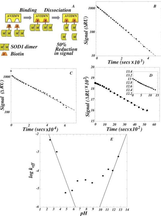
Biotinylated SOD1 was prepared at low stoichiometry (1 mol equivalents of biotinylating agent/SOD1 dimer) for Surface Plasmon Resonance (SPR) studies and high (10 fold excess of biotinylating agent/SOD1 dimer) stoichiometry for Size Exclusion Chromatography (SEC) studies as previously described (29). Biotinylated SOD1 was found to be fully active after biotinylation ((30); data not shown) and had identical chromatographic properties compared to the unmodified protein (Supplementary Material S1).
Analytical ultracentrifugation (AUC).
The Kd for SOD1 dimer dissociation was determined using a Beckmann-XLA ultracentrifuge at a speed of 16,000 rpm at 40 C or 250C using a Ti-50 rotor. Radial UV absorbance (258 nm) was monitored every 2h in cells with a path length of 12 mm. One blank buffer sample was used for each protein sample to obtain baselines. A total of 7 scans were obtained for each sample and the corresponding blank cell to reach equilibrium, followed by over-spinning at 45,000 rpm to obtain another estimate of baseline absorbance (typically 0.01 units). The UV-absorbance profiles were superimposed to ensure equilibrium, analyzed for residuals and the equilibrium profile(s) were fit to the Lambert equation to obtain estimates of Kd and apparent molecular weight Mw.
SEC was performed with a Pharmacia Akta chromatography system using an Amersham-Pharmacia SuperdexHR 10/30 column (Piscataway, NJ), with a 200-µl injection loop,total volume of 23.56 ml and a void volume estimate of 7.8 ml, working at 5°C. The column flow rate was 0.45 ml/min. Fractions (150-250 µl) were collected in glass tubes and these were analyzed immediately after completing the chromatogram. In reactions with low (nanomolar) SOD1 concentrations, the reaction mixture and the column buffer contained BSA at10 mg/l to prevent nonspecific binding of SOD1 to test tubesand to the column matrix. UV absorbance was monitored spectrophotometrically at 254nm.
Dot blotting.
Column fractions were analyzed by a dot-blot assay, with Avidin-AP, as previously described (29) and/or by immunostaining with anti-SOD1 antibody (Supplementary Material S2).
Surface Plasmon Resonance measurements.
biotin-SOD1 but received protein-free buffer. To obtain the pH dependence of the rate constant, the zero-order dissociation reaction was followed sequentially with increasing or decreasing pH, starting at pH 7.8.
2.4 Results
We show that the minimal sequence of steps constituting the aggregation pathway is:
-off m agg
on holo m apo
2
2
k k k
k k
D
←⎯⎯
⎯⎯→
M
←⎯⎯
⎯⎯→
+M
⎯⎯→
A
(1)where Dholo, Mholo, Mapo and A are the holo-dimer, holo-monomer, Zn-free monomer (31) and aggregate respectively. Next, we show that: (i) the Kd and koff for SOD1 dimer dissociation are enhanced under aggregation-promoting conditions; (ii) aggregation requires dissociation of the SOD1 dimer to monomers and a subsequent loss of metals. Based on the observed aggregation kinetics, we calculated the rate and equilibrium constants for each step in the reaction sequence.
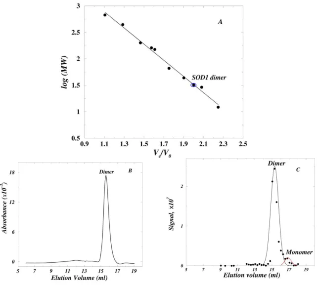
The Kd for SOD1 dimer dissociation is 6×10³-fold higher under aggregation-enhancing conditions than under near-physiological conditions.
≈16% monomer. In contrast, 30μM SOD1 was ≈100% dimer under near-physiological conditions (pH 7.8), where aggregation does not occur.
The Kd for SOD1 dimer dissociation at pH 7.8 could not be measured by AUC because the optical system used to detect the protein is limited to micromolar concentrations. However, nM SOD1 concentrations could be analyzed by using size exclusion chromatography (SEC) and a dot-blot assay. Based on the calibration of the Superdex HR 10/30 column (Fig. 1A), the SOD1 dimer and monomer are expected to elute at 15.5 and 17.0 ml respectively; SOD1 at 30μM in pH 7.8 buffer eluted as ≈100% dimer at 15.6 ml (Fig. 1B), in accord with AUC measurements (Table I). The Kd at pH 7.8 was estimated with 5nM SOD1 which gave column fractions at the limit of detection. The bulk of the protein eluted as a dimer at 15.3 ml with a small reproducible monomer peak at 16.8 ml, comprising ≈20% of the total protein (Fig. 1C). The dimer and monomer were expected to elute as distinct peaks because they were in slow equilibrium: the half-time for dimer dissociation was ≈6 h at pH 7.8 and 250C (see below), whereas the time required for chromatographic separation was ≈10 minutes at 50C. A Kd≈0.16 nM was calculated from a double-Gaussian least-square fit to the data, and is in reasonable agreement with the Kd≈1nM estimated from the unfolding studies of human SOD1 (32). Thus, the Kd≈1×10⁻⁶ M at pH 3.5 is larger than the Kd≈1.6×10⁻¹⁰ M at pH 7.8 by a factor of 6×10³. This corresponds to a large (≥5.2 kcal/mol) reduction in dimer stability, indicating that a higher fraction of SOD1 is monomeric under aggregation-enhancing conditions than under near-physiological conditions.
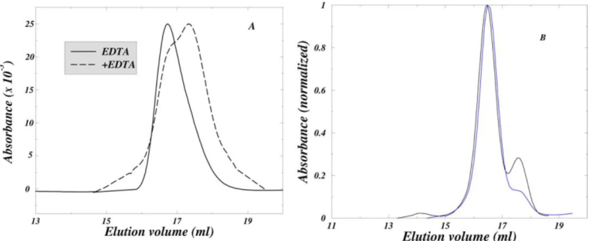
reversibility of dimer dissociation under aggregation-promoting conditions was directly demonstrated by SEC (Supplementary Material S4).
The koff for SOD1 dimer dissociation is 30-fold higher under aggregation-enhancing conditions than under near-physiological conditions.
Having determined that the Kd for dimer dissociation was enhanced at least 20,000-fold at pH 3.5, we used Surface Plasmon Resonance (SPR) to determine the kinetics of dimer dissociation. SOD1, biotinylated at low stoichiometry (≤1 biotin/SOD1 dimer), was immobilized on a strepavidin-coated gold surface. As shown in Fig. 2A, the biotinylated dimer has one subunit bound to strepavidin and the non-biotinylated subunit is progressively lost by flowing SOD1-free buffer through the flow-cell, leading to a SPR signal decrease of approximately 50%. The dissociation is made irreversible by a 2000 nl/minute flow of protein-free buffer through the 7 nl flow-cell. The rate constant for dissociation koff was determined by an exponential fit to the entire signal decay corresponding to a first-order reaction (Figs. 2B and 2C), or by a linear fit to the initial signal decay corresponding to a zero-order reaction (Fig. 2D). The dissociation at pH 3.5 had a half-time of ≈600 s (koff≈1.0×10⁻³ s-1; Fig. 2B) whereas the dissociation at pH 7.8 had a half time of ≈6 h (koff ≈ 3.1×10⁻⁵ s-1; Fig. 2C). Therefore, under conditions where aggregation was observed (pH 3.5), the rate of dimer dissociation is a 30-fold faster than under near-physiological conditions. This corresponds to an approximately 2.0 kcal/mol reduction in the energy barrier for dissociation.
The dramatic increase in koff at pH 3.5 suggests that insights into the forces stabilizing the SOD1 dimer could be obtained from the pH-dependence of the reaction. The koff increased sharply on the acidic and basic ends of the pH range 2.5-12.7 (Fig. 2D and 2E). In the simplest unified model to account for this effect, dimer dissociation is linked with the titration of carboxyl (Asp, Glu) and amine (Lys, Arg) groups forming a salt-linkage. At any given pH, the observed koff reflects the dissociation of three sub-populations of the SOD1 dimer, in which the salt-linkage is: (i) intact; (ii) disrupted because of the protonation of the carboxylic acid; and (iii) disrupted because of the de-protonation of the amine. The species from sub-populations (i), (ii), and (iii) have intrinsic dissociation rate constants kind, kacid, and kbasic respectively, which are weighted by their pH-dependent population size, so that the observed koff is:
[ ]
[ ] [ ]
obs b
off acid basic ind
a b
K H
k k k k
H K K H
+
+
⎧ ⎫ ⎧ ⎫
= ⎨ ⎬+ ⎨ ⎬
+ +
⎩ ⎭ ⎩ + ⎭+ (2)
where Ka and Kb are the equilibrium constants for the de-protonation of the carboxyl and amine groups involved. The first and the second term dominate at low and high pH respectively, and the rate is pH-independent at intermediate pH values. According to Eq.(2), in the low pH regime the titration-induced increase in log is predicted to have a slope of -1 when
obs off
k
a
pH >> pK . Similarly, a slope of 1 is predicted for the corresponding increase in the high pH regime when , and such increases were observed at both low and high pH (Fig. 2E). The apparent pKa and pKb values of <2.5 and >12.5, presumably correspond to
carboxyl (Asp; free pK=3.86) and amine (Arg; free pK=12.48) groups forming a highly stabilizing salt-linkage. The linkage of the protonation of the carboxylic acid and the de-protonation of the amine to the dimer dissociation leads to the abnormal apparent pK values. The 100-fold change in the observed koff in the intermediate pH range 4.8-10.8 is indicative of
b
other titrations, possibly of charged amino acid side-chains which may not form salt-linkages, so that they have a smaller effect on the rate kind.
Human SOD1 (hSOD1), which is highly homologous (82% sequence identity) to the bovine enzyme, exhibits a similar pH-dependence of dimer dissociation. The koff values were similar at pH 6.6 (Fig. 2D, inset) and at pH 7.8: 2.74×10⁻⁵ s-1 for hSOD1 (data not shown) and 3.13×10⁻⁵ s-1 for bovine SOD1 (Fig. 2B).
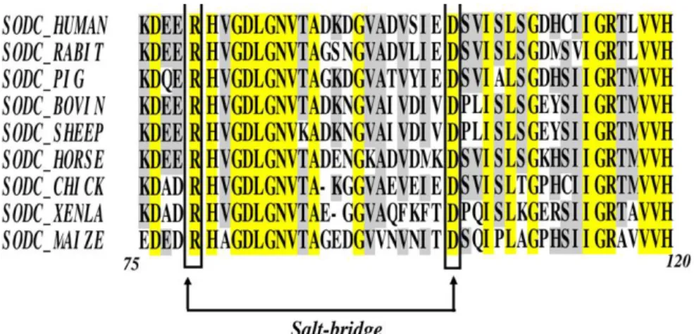
It is interesting that there are no salt-linkages on the dimer interface of either bovine or human SOD1 so that the enhanced dissociation rate is caused indirectly by the titration of a salt-linkage that is not in the proximity of the dimer interface. The Arg79-Asp101 interaction was identified as a candidate for such a linkage (Supplementary Material S5). This salt-linkage mechanism provides a unified framework that accounts for the steep pH-dependence of the reaction at low and high pH, and is consistent with the previous finding that charge-charge interactions in SOD1 contribute substantially to its stability and folding kinetics (21). An alternative mechanism in which ionization of surface exposed residue(s) dramatically influences dimer stability is less likely since it does not simultaneously predict the behavior at extremes of pH, and the modest pH-dependence of the rate at intermediate pH values.
Rapid oligomerization of SOD1 occurs under conditions where it is monomeric.
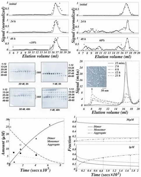
30μM SOD1 eluted predominantly as a dimer (elution volume 16.5 ml; Supplementary material Fig. S2A) on incubation for 40 minutes (Fig. 3A, top panel). 40 minutes was sufficient for the establishment of equilibrium since the dimer dissociation has a half-time of ≈600s at this pH (Fig. 2B). The elution profiles remained largely unaltered for 48 h (Fig. 3A, middle and bottom panels) and there was little aggregation since nearly identical peak heights and areas in the UV-absorbance profiles were obtained after incubation. This agreed with AUC results in which 30µM SOD1 gave nearly identical UV-absorbance signals and apparent Mw estimates (Table I). After 48 h, most of the protein remained dimeric or monomeric (Fig. 3A), and less than 10% was aggregated.
Relatively rapid aggregation occurred with 1μM SOD1 (Fig. 3B). After 40 minutes of incubation, the protein eluted in a broad peak centered at 17.2 ml corresponding to a 40:60 mixture of dimer and monomer, as expected from Kd≈1μM. The rate of aggregation was initially rapid and after 24 and 48 h approximately 60% of the protein was aggregated. The aggregation rate is expected to decrease markedly with time, and the failure to observe a significant increase in the fraction of aggregated protein at 48 h can be attributed to experimental error. These results show that at a given concentration, SOD1 aggregation is proportional to the fraction of monomer, rather than dimer, in solution.
Rapid oligomerization of SOD1 occurs under conditions where metals are lost irreversibly.
where metal loss was made irreversible by dialysis against metal free buffer. Dialysis at pH 3.6 results in a rapid loss of 95% of the Zn (31) and the unbound metals were expected to be removed rapidly since we had found that the loss of cupric acetate from the dialysis bag has a concentration-independent (range 100μM-10mM) half-time of 10 minutes under the conditions of the experiment (data not shown). A substantial amount of aggregation was expected if the loss of metals from SOD1 induces formation of a species that is aggregation-competent.
During incubation under dialysis conditions the amount of dimer rapidly decreased, the degree of dimer dissociation increased, and the protein was almost completely aggregated in 21 h (Fig. 3D). Electron micrographs of the aggregates obtained by negative staining, show that they have fibrillar morphology (Fig. 3D, inset). Despite the fact that the dimer concentration decreased monotonically, the monomer concentration initially increased and subsequently gradually decreased (Fig. 3E), indicating that under dialysis conditions, dimer dissociation is coupled to metal loss and aggregation of the apo-monomers in the reaction sequence:
-off m agg
on holo m apo
2 ( ) 2
k k k
dialysed
k k
D←⎯⎯⎯⎯→ M ←⎯⎯⎯⎯→+ Zn + M ⎯⎯→A (3)
where Dholo, Mholo, Mapo and A are the holo-dimer, holo-monomer, Zn-free monomer and aggregate respectively. In contrast, in AUC studies described above, the dominant species after incubation remained Dholo and Mholo and there was no detectable aggregation. The coincidence between the increase in Mapo and the increased rate of aggregation shows that the apo-monomer is the reactive species for this process. Moreover, since the apo-monomer accumulated before aggregation, it is concluded that the oligomerization of apo-monomers is the rate-limiting step in Eq.(3).
determined from SPR and kon≈103 M-1s-1 was calculated from Kd ≈1µM. The constants
≈1.2×10⁻⁴ s-1, ≈5.2×10⁴ M-1s-1, and kagg ≈0.1×10³ M-1s-1 were estimated from the observed kinetics of dialysis-induced aggregation (Supplementary material S3). The estimated value of ≈1.2×10⁻⁴ s-1 agrees well with the previously measured
m
k− km+
m
k− km−=4.2×10⁻⁴ s-1 (31) for the
loss of Zn from SOD1 at pH 3.6 (half-time of ≈27 minutes at 20oC). The differential master equations corresponding to Eq.(3) were solved numerically (Supplementary Material S3) and the above rate constant estimates were refined, fitting to the observed time-profile of dialysis-induced aggregation (Fig. 3E). The values that best account for the experimental results are: koff = 3×10⁻³ s-1, kon= 1.6×10³ M-1 s-1, km
−= 1×10⁻³ s-1,
m
k+= 9.8×10³ M-1 s-1 and kagg = 1×10³ M-1s-1. The rate constant values were found to be robust within an order of magnitude: the fit worsened dramatically on variations in rate constants greater than an order of magnitude. In accord with the above rate constants, which predict 12% aggregation in 30μM SOD1 after 24 h under non-dialysis conditions, we had found little aggregation (Fig. 3A). Similarly, 14% and 63% aggregation is predicted at 30 and 1μM in 48 h (Fig.3F), which is in good agreement with the observed values of 10% and 60% respectively.
2.5 Discussion
To create amyloidogenic conditions under which the aggregation of proteins can be readily induced, acidic conditions are routinely employed. For example, amyloidogenic intermediates of transthyretin (TTR), β2-microglobulin, prion and lysozyme have been