THE PROTECTIVE AND PATHOLOGIC ROLES OF TOLL-LIKE RECEPTORS IN ARTHRITOGENIC ALPHAVIRUS INFECTION
Lauren Michelle Neighbours
A dissertation submitted to the faculty of the University of North Carolina at Chapel Hill in partial fulfillment of the requirements for the degree of Doctor of Philosophy in the Department of Microbiology and Immunology.
Chapel Hill 2013
Approved by:
ii © 2013
iii ABSTRACT
LAUREN NEIGHBOURS: The Protective and Pathologic Roles of Toll-like Receptors in Arthritogenic Alphavirus Infection
(Under the direction of Mark T. Heise)
iv
To my parents, who always encouraged me to pursue my dreams
And provided me every opportunity to do so.
And to Stuart and Evan, who make every day a blessing,
Who remind me what is most important,
v
ACKNOWLEDGMENTS
I would like to thank all of the individuals who have aided in my professional development and accomplishments during my graduate experience. Firstly to my mentor, Mark Heise, I am so thankful for his continued support and encouragement. In addition to providing valuable advice and guidance during the course of my research, Mark has celebrated every professional success and motivated me through every tribulation that I have encountered throughout my graduate career. I am grateful for the freedom that I was allotted in the laboratory to manage my research project and develop as an independent scientist, but I am also thankful for the constructive criticisms that were necessary for me to reach my full scientific potential.
vi
to thank Bianca Trollinger, Charlie McGee, Adam Cockrell, Charissa Kam, Desi Matthews, Clancy Mullan, Clayton Morrison, and Doug Widman, who have each contributed to the success of our laboratory and have been wonderful colleagues.
vii
TABLE OF CONTENTS
LIST OF TABLES...x
LIST OF FIGURES...xi
LIST OF ABBREVIATIONS...xiv
CHAPTER ONE: INTRODUCTION...1
Overview of alphaviruses...1
Alphavirus taxonomy...1
Alphavirus emergence and transmission...3
Virion organization and life cycle...5
Human infection and animal models of disease...7
Pathogenesis of human arthritogenic alphavirus infection...7
Animal models of arthritogenic alphavirus-induced disease...9
Toll-like receptor signaling...12
Overview of toll-like receptors...12
Toll-like receptor signaling during viral infection...15
The role of toll-like receptors in autoimmunity and arthritis...17
Host response to arthritogenic alphavirus infection...19
Type I/II IFN responses to infection...18
viii
The role of pattern-recognition receptors during infection...22
Dissertation objectives...25
CHAPTER TWO: MYD88-DEPENDENT TLR7 SIGNALING MEDIATES PROTECTION FROM SEVERE ROSS RIVER VIRUS-INDUCED DISEASE IN MICE...33
2.1 Overview...33
2.2 Introduction...34
2.3 Materials and Methods...36
2.4 Results...43
2.5 Discussion...52
CHAPTER THREE: MYD88-DEPENDENT TLR7 SIGNALING PROTECTS MICE FROM SYSTEMIC COMPLEMENT- AND ANTIBODY-MEDIATED DISEASE FOLLOWING ROSS RIVER VIRUS INFECTION...66
3.1 Overview...66
3.2 Introduction...67
3.3 Materials and Methods...70
3.4 Results...74
3.5 Discussion...81
CHAPTER FOUR: TLR4 PROMOTES ROSS RIVER VIRUS-INDUCED DISEASE IN MICE...94
4.1 Overview...94
ix
4.3 Materials and Methods...98
4.4 Results...104
4.5 Discussion...110
CHAPTER FIVE: DISCUSSION...121
5.1 TLR7 mediates protection from severe RRV-induced disease...121
TLR7 deficiency promotes antibody- and complement-mediated enhancement of RRV-induced disease...121
Insights into mechanisms of TLR7-mediated protection...123
Future directions...126
5.2 TLR4 promotes RRV pathogenesis...128
TLR4 induces complement-mediated disease during RRV infection...128
Insights into mechanisms of TLR4-induced pathogenesis...131
Future directions...133
5.3 Conclusions...134
x
LIST OF TABLES
xi
LIST OF FIGURES
Figure 1.1: Alphavirus transmission cycles...29
Figure 1.2: Alphavirus genome...30
Figure 1.3: Mouse model of RRV infection...31
Figure 1.4: Overview of TLR signaling...32
Figure 2.1: TLR7 and Myd88 contribute to protection from RRV-induced disease in vivo...58
Figure 2.2: TLR7- and Myd88-deficientmice show enhanced tissue damage following RRV infection...59
Figure 2.3: Inflammatory cell recruitment is similar in WT and TLR7-/- mice following RRV infection...60
Figure 2.4: Type I IFN production in sera of TLR7-/- and Myd88-/- mice is similar to that in sera of WT mice early during infection...61
Figure 2.5: Mice lacking TLR7 or Myd88 show an inability to control viral titer at late times postinfection...62
Figure 2.6: TLR7-/- mice show reduced neutralizing antibody production following RRV infection...63
Figure 2.7: Antibody from TLR7-/- mice shows decreased virus-specific affinity and fails to protect from RRV-induced disease...64
Figure 2.8: TLR7-/- mice show reduced germinal center formation following RRV infection...65 Figure 3.1: Passive transfer of antisera from TLR7- and Myd88-deficient
xii
RRV infection...87 Figure 3.2: TLR7-/- mice show enhanced IgG and C3 deposition in
RRV-infected muscle tissue...88 Figure 3.3: Myd88- and TLR7-deficient mice show severe cardiac
muscle tissue damage following RRV infection...89 Figure 3.4: Myd88- and TLR7-deficient mice show IgG and C3
deposition in cardiac muscle tissue following RRV infection...90 Figure 3.5: Myd88-/- mice show IgG and C3 deposition in the kidneys
following RRV infection...91 Figure 3.6: TLR7xC1q DKO mice do not show enhanced disease
following RRV infection...92 Figure 3.7: TLR7 regulates antibody affinity responses to non-RNA
stimuli...93 Figure 4.1: TLR4 contributes to the development of RRV-induced
disease in mice...115 Figure 4.2: TLR4-deficient mice show reduced numbers of inflammatory
leukocyte and NK cell populations in RRV-infected quadriceps
at late times postinfection...116 Figure 4.3: Viral titers are similar between WT and TLR4-deficient
mice following RRV infection...117 Figure 4.4: TLR4-deficient mice show reduced complement deposition
and activation in RRV-infected muscle tissue...118 Figure 4.5: TLR4-deficient macrophages show reduced complement
xiii
Figure 5.1: Plasmacytoid DCs from WT and TLR7-/- mice produce
similar amounts of type I IFN following RRV infection...120 Figure 5.2: Expression of complement-associated inflammatory markers
is reduced in TLR4- and MBL-deficient macrophages following co-culture with RRV-infected but not RRV-DM-infected
myotubes...121 Figure 5.3: MBL treatment enhances C3 expression in WT macrophages
but not TLR4-deficient macrophages following co-culture with
RRV-infected myotubes...122 Figure 5.4: Macrophage cytotoxicity is not impacted by TLR4 signaling
xiv
LIST OF ABBREVIATIONS BFV Barmah Forest virus
CHIKV Chikungunya virus CNS Central nervous system CPE Cytopathic effect
DAMP Damage-associated molecular pattern DC Dendritic cell
dsRNA Double-stranded RNA
EEE Eastern equine encephalitis ER Endoplasmic reticulum HSP Heat-shock protein
IFN Interferon
IkB Inhibitor of NFκB IKK IκB kinase
IPS-1 Interferon promotor stimulator-1 IRF Interferon regulatory factor
ISRE Interferon-stimulated response element ISG Interferon-stimulated gene
xv
Myd88 Myeloid differentiation primary response gene 88 NF-κB Nuclear factor kappa B
NSP Nonstructural protein
PAMP Pathogen-associated molecular pattern pDC Plasmacytoid dendritic cell
PRR Pattern recognition receptor RdRp RNA-dependent RNA polymerase RLR Rig-I-like receptor
RRV Ross River virus SFV Semliki Forest virus SINV Sindbis virus
ssRNA Single-stranded RNA
TICAM-1 TIR-domain-containing adaptor molecule 1 TIRAP TIR-domain-containing adaptor protein TLR Toll-like receptor
TRAM TRIF-related adaptor molecule
TRIF TIR-containing-adaptor inducing interferon-b VEEV Venezuelan equine encephalitis virus
CHAPTER ONE: INTRODUCTION Overview of alphaviruses
Alphavirus taxonomy
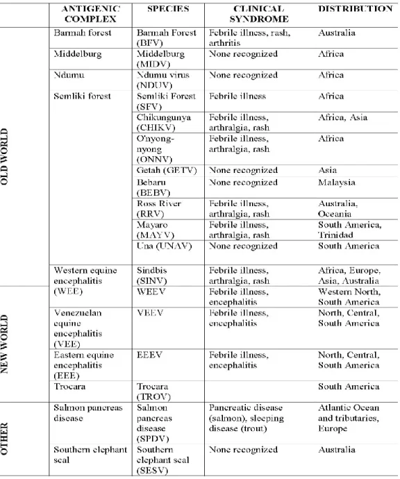
Alphaviruses are positive-sense single-stranded RNA (ssRNA) arboviruses and are members of the family Togaviridae (1). The Togaviridae family consists of two genera, Alphavirus and Rubivirus, and 29 of the 30 species of togaviruses are
alphaviruses, with Rubella virus being the only identified rubivirus (1). Alphaviruses and other arboviruses were originally grouped according to their antigenicity as determined using various serological and biochemical assays (2). Modern technological advances have expanded the classification of alphaviruses into distinct clades by comparing the genomic sequences of the viral species, which adds a complex genetic and evolutionary dimension to the taxonomy (3).
2
encephalitis (WEE) virus induce encephalitis (4, 5). Old World and New World alphaviruses are further segregated into seven distinct antigenic complexes or clades, including the Barmah Forest, Semliki Forest, Venezuelan equine encephalitis (VEE), EEE, WEE, Trocara, Middelburg, and Ndumu clades (1). An overview of alphavirus classification and epidemiology is included in Table 1, which focuses on Old World
Alphavirus taxonomy
Alphaviruses are positive-sense single-stranded RNA (ssRNA) arboviruses and are members of the family Togaviridae (1). The Togaviridae family consists of two genera, Alphavirus and Rubivirus, and 29 of the 30 species of togaviruses are
alphaviruses, with Rubella virus being the only identified rubivirus (1). Alphaviruses and other arboviruses were originally grouped according to their antigenicity as determined using various serological and biochemical assays (2). Modern technological advances have expanded the classification of alphaviruses into distinct clades by comparing the genomic sequences of the viral species, which adds a complex genetic and evolutionary dimension to the taxonomy (3).
Alphaviruses are categorized into two groups, Old World and New World, which distinguish the viruses based on their geographic distribution and epidemiological characteristics (3). The Old World alphaviruses, including Ross River virus (RRV), chikungunya virus (CHIKV), and Sindbis virus (SINV), are associated with debilitating arthralgia and myalgia, while New World alphaviruses such as Venezuelan equine encephalitis virus (VEEV), eastern equine encephalitis (EEE) virus, and western equine encephalitis (WEE) virus induce encephalitis (4, 5). Old World and New World
3
including the Barmah Forest, Semliki Forest, Venezuelan equine encephalitis (VEE), EEE, WEE, Trocara, Middelburg, and Ndumu clades (1). An overview of alphavirus classification and epidemiology is included in Table 1, which focuses on Old World arthritogenic alphaviruses for the purposes of this work and was adapted with permission from Weaver and colleagues (1).
Alphavirus emergence and transmission
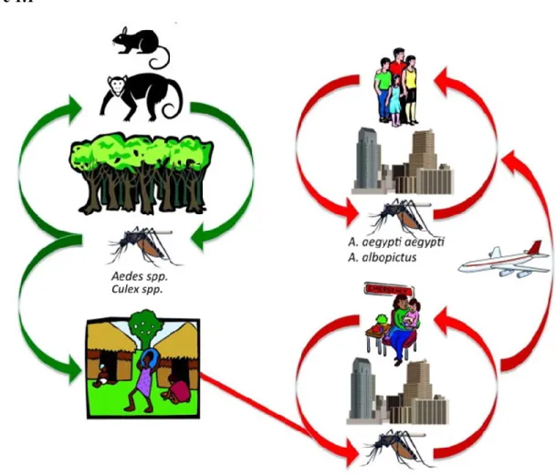
Alphaviruses are vector-borne viruses that cause a variety of diseases in humans, other vertebrates, and fish (1). Most alphaviruses are transmitted to their vertebrate hosts by mosquito vectors, and viral transmission is primarily maintained within an enzootic cycle via Culex and Aedes mosquito species that inhabit humid, marshy regions (1). As depicted in Figure 1.1, the enzootic transmission cycle of alphaviruses allows for
maintenance of viral infection in zoonotic reservoir hosts with endemic disease occurring in certain geographic areas (Figure 1.1, which was adapted with permission from Weaver and colleagues)(1). However, alphaviruses also cause sporadic epidemics that can lead to widespread morbidity and mortality in humans (Figure 1.1) (4). Most epidemics
involving New World alphaviruses, including VEE, EEE, and WEE viruses, have occurred in South, Central, and North America (5). Alternatively, Old World alphavirus epidemics have occurred in Africa, Asia, and Australia, with cases recently spreading to Europe and the United States after infected individuals traveled to endemic regions (6-8).
4
in Africa, where the virus is believed to have originated, and the virus caused multiple outbreaks through the 1980s in Africa and Southeast Asia (10). However, CHIKV outbreaks have re-emerged and expanded with widespread distribution since 2004, with thousands to millions of infections presenting with persistent debilitating arthraligia and a case fatality rate estimated at approximately 0.1% (1, 11). Additionally, the regions where CHIKV outbreaks tend to occur are also endemic for tropical diseases with similar symptomatic presentations such as dengue virus fever and malaria, and CHIKV
infections are thus believed to be grossly underreported (12). Epidemics of CHIKV-like disease, which were characterized as dengue outbreaks but were likely caused by CHIKV, have been traced back as far as the 19th century (13). Moreover, genotyping of viral isolates from the La Réunion outbreak of 2005-2007 identified a mutation in the CHIKV strain that allowed more efficient transmission by Aedes Albopictus mosquitoes and resulted in rapid dissemination of the virus (11).
Increasing cases of endemic and epidemic disease induced by other arthritogenic alphaviruses such as RRV and Barmah Forest virus (BFV) have also spurred recent concerns (14). RRV, which results in over 4,000 cases of disease in Australia every year, is considered the most substantial arboviral threat to public health in the Australasian region because of the long-lasting morbidities associated with infections (14, 15). RRV was first isolated in 1959 from mosquitoes found near the Australian Ross River, and it was the causative agent of a 1979 epidemic in the South Pacific that resulted in over 60,000 cases of disease (16). BFV results in approximately 1,000 human cases of disease in Australia each year, with symptoms typically less severe and shorter than those
5
concerned Australian public health authorities, and the growing number of arthritogenic alphavirus cases in the Australasian region results in a considerable economic burden as well (14). Moreover, models of climate change in alphavirus-endemic regions have predicted that alterations in environmental conditions, such as increasing rainfall and temperatures, may result in enhanced alphavirus activity and thus escalate the risk of human infections (14, 18).
Virus organization and life cycle
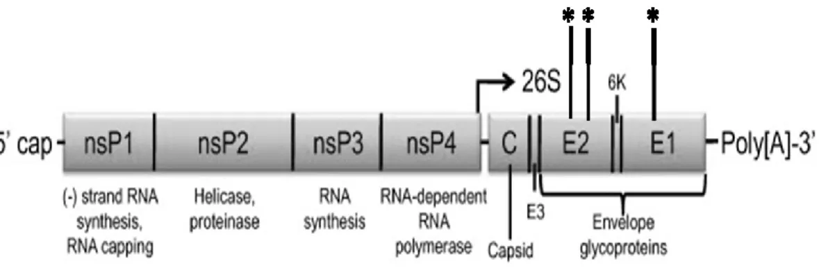
Alphaviruses are enveloped viruses of approximately 70 nm in diameter and contain an ssRNA genome of approximately 12 Kb in length (19-21). The virion contains a host-derived lipid membrane, which is embedded with heterodimeric trimer spikes formed by the highly glycosylated E1 and E2 viral glycoproteins (22). Within the membrane-bound virion is an icosahedral nucleocapsid, which is formed by the viral capsid proteins. The alphavirus genome encodes two open reading frames, from which the four nonstructural proteins (NSPs) are encoded from the genomic RNA and the five structural proteins are encoded from the 26S subgenomic strand (23). The genomic and subgenomic RNA species contain a 5’ methylguanosine cap and 3’ polyadenylated tail, and each strand is initially translated into polyproteins before being cleaved
posttranslationally by viral and host proteases. A simplified schematic of the alphavirus genome, which was adapted with permission from Weaver and colleagues, can be found in Figure 1.2 (1).
6
membrane have also been reported (23). Upon receptor-mediated endocytosis of the virion, viral uncoating occurs to allow for the release of alphavirus ssRNA into the cytoplasm. The initial translation of the viral RNA, which is infectious upon entry, results in the formation of the NSP polyprotein, NSP1234 and structural polyproteins (24). Cleavage of NSP1234 into NSP123 and NSP4 allow for the formation of the replication complex, which synthesizes the negative strand RNA, and complete cleavage of the nonstructural proteins allows for the complete stable replication complex to be formed and initiates positive full-length and subgenomic RNA synthesis (23). Cleavage of the structural polyprotein releases the capsid protein, which associates with the newly transcribed viral RNA and facilitates packaging of the RNA into nucleocapsid vesicles (25). A signaling sequence on the E3 protein facilitates the relocation of the remaining structural polyprotein to relocate and bud into the endoplasmic reticulum (ER) for further processing by host proteases, while 6K facilitates downstream processing of the E1 glycoprotein and aids in viral budding (23). E1 and E2 interact shortly after synthesis to form dimers, which are transported from the ER-Golgi complex to the host plasma membrane (26). The nucleocapsids formed in the cytoplasm can then bud into the host membrane containing E1-E2 spikes and release envelope-containing particles (22).
7
has numerous enzymatic activities and functions, performs multiple roles during replication and packaging but also mediates cellular cytopathic effects (CPE), induces host shut-off and antagonizes type I/II type I IFN induction during infection (30-33). Additionally, the use of NSP2 as an adjuvant in a CHIKV DNA vaccine enhanced protection of mice challenged with CHIKV when compared to mice immunized with the vaccine alone (34). Little is known about the functions of NSP3, which is required for RNA synthesis (22). However, NSP3 plays a role in neurovirulence and in the
suppression of stress granule formation, and it may also influence vector specificity (35-38). The viral RNA-dependent RNA polymerase, NSP4, is proposed to have multiple functions during RNA replication and was recently reported to modulate the host
translational machinery to enable more efficient replication during CHIKV infection (22, 39). Aside from their roles in virus entry and assembly, the structural proteins are also important mediators of viral pathogenesis. The precursor E2 protein is involved in RRV-induced inflammatory disease, while the glycans on the E1 and E2 glycoproteins
modulate type I IFN production and mediate pathogenesis during RRV infection [(27, 40), Gunn and Heise, unpublished].
1.1Human infection and animal models of disease
Pathogenesis of human arthritogenic alphavirus infection
8
develop a debilitating polyarthralgia and myalgia that can last for several months and in rare cases can induce chronic disease or mortality (41, 42). Chikungunya virus, which is named for the Makonde Tanzanian term for “that which bends up,” is considered the most threating arthritogenic alphavirus to global public health because of its capacity to induce catastrophic epidemics and persistent disease (43). Severe cases of arthritogenic disease tend to occur in individuals with chronic underlying conditions such as diabetes or impaired kidney function or in patients that have adverse reactions to therapeutic drug treatment (43). Chronic pain associated with arthritogenic alphavirus infection can significantly impact the quality of life of infected individuals and results in a substantial economic burden in affected regions because of prolonged care and decreased
productivity (14, 44, 45).
The incubation period of the virus is usually between 2-14 days prior to the onset of symptoms, with a viremia that lasts approximately 5-7 days (1, 15). The virus
replicates in skeletal muscle and joint tissues during the acute infection, although RNA and viral proteins were found in various tissues and synovial macrophages for several weeks to months following the resolution of viral replication (41, 46). The pathology induced by arthritogenic alphavirus infection is believed to be mediated by host
inflammatory responses induced by viral replication or viral products, although these are not considered autoimmune responses (15).
9
of the exposed neonates became infected with CHIKV (47). All of the infected neonates developed symptoms of infection shortly after birth, and more than half of the infected neonates developed severe neurological disease including encephalopathy and persistent neurodevelopmental disabilities. Transmission rates of CHIKV were highest in mothers that were infected very late during pregnancy, although infection may have contributed to early fetal deaths that were also observed.
There are currently no effective treatments or vaccines available for arthritogenic alphavirus infections. The current treatment regimen typically consists of treatment with non-steroidal anti-inflammatory drugs (NSAIDs) and/or steroids (41, 42). Anti-rheumatic drugs, including methotrexate, salazopyrine, leflunomide, and hydoxychloroquine, and tumor necrosis factor alpha (TNFα) inhibitors, have shown varying efficacy in patients with chronic disease following CHIKV infection (48). Additionally, long-term and combinatorial drug treatments for severe or persistent arthritogenic alphavirus infections can lead to adverse side effects such as gastrointestinal bleeding and bone deterioration in affected patients (15, 41, 42).
Animal models of arthritogenic alphavirus-induced disease
10
effects of SINV infection on neurological pathogenesis (50). More common models to study viral pathogenesis include subcutaneous infections into the footpads of C57BL/6 mice to study RRV- and CHIKV-induced inflammation and tissue damage (51-54).
The mouse model of RRV pathogenesis utilized by Morrison and colleagues was the model used in the studies described in this work (53). This model of alphavirus-induced arthritis/myositis involves a subcutaneous infection of 1000 PFU of RR64, an infectious clone of the T48 mouse-adapted strain of RRV, into the left-rear footpad of a twenty-four day old C57BL/6 mouse (53, 55). Over the course of infection,
RRV-infected mice show transient weight loss and develop disease characterized by hind limb weakness and altered gait (depicted in Figure 1.3, which is modified from Morrison and colleagues)(53). The clinical disease in these mice peaks in severity between 7-10 days post-infection, after which mice recover their hind limb function and are
indistinguishable from mock-infected animals by 30 days post-infection.
11
phase, wherein the inflammation resolves, and the muscle fibers repair. Notably, peak viral replication in the tissues occurs between 24-48 hours post-infection, suggesting that the disease correlates with inflammatory infiltration and not with viral replication (53). Additionally, numerous studies using the RRV mouse model have demonstrated the roles of innate immune components, including macrophages and complement, in mediating RRV-induced disease (51, 56-60).
Several treatment therapies and vaccine approaches are efficacious in animal models. Because macrophages are critical mediators of RRV-induced disease, some therapies focused on targeting this cellular population to ameliorate the disease (51, 56, 57). Treatment of RRV-infected mice with bindarit, an inhibitor of monocyte chemotactic protein synthesis, resulted in significantly reduced tissue inflammation and damage compared to untreated mice (61). Additionally, a recent study showed complete protection from CHIKV challenge in mice immunized with a recombinant adenoviral vaccine, while another report demonstrated that a synthetic DNA vaccine protected mice and non-human primates from CHIKV-induced disease and elicited protective
12 1.2Toll-like receptor signaling
Overview of toll-like receptors
Toll-like receptors (TLRs) are innate immune receptors that recognize conserved microbial patterns on the surface of pathogens such as viruses, fungi, parasites, and bacteria (64). TLRs are a class of pattern recognition receptors (PRRs), which are
classified as such because they recognize specific pathogen-associated molecular patterns (PAMPs) that enable the receptor to identify the microbe as foreign and initiate an
immune response. Other PRRs include the RIG-I-like receptors (RLRs) and Nod-like receptors (NLRs), and these PRRs signal exclusively from the cytosol. PRRs are
expressed on a variety of immune cells, including phagocytic macrophages and dendritic cells (DCs), which use these receptors as a first line of defense against invading
pathogens (65).
The basic structure of TLRs consists of a leucine-rich repeat extracellular domain, a membrane-spanning region and an intracellular region containing a Toll/interleukin-1 receptor (TIR) domain (66). The binding of the horseshoe-shaped extracellular domains of TLRs by their specific PAMP-containing ligands induces a conformational change in the receptors and the accompanying dimerization of their extracellular domains.
13
receptors in their monomeric forms, although multimeric and clustered forms of TLRs in their native transmembrane conformation are not available (66).
Upon recognition of a PAMP via receptor complex engagement, TLRs signal to their downstream adaptor molecules to activate a downstream signaling cascade that will lead to the appropriate immune response to the invading pathogen (64). The
TLR-induced signaling cascade leads to the activation of specific transcription factors, which can then induce the expression of various genes, such as pro-inflammatory cytokines, type I IFN, and IFN-stimulated genes (ISGs) (65). An overview of TLR signaling is depicted in Figure 1.4, which was adapted from Kawai and Akira with permission (69).
TLR signaling is modulated by the specificity of the TLR ligands and by the cellular localization of the receptors, which can affect ligand accessibility and
downstream signal transduction. Based on these specific criteria, TLRs can be segregated into two groups: group A consists of TLRs 1, 2, 4, 5, 6 and 11, while group B consists of TLRs 3, 7, 8 and 9 (64). Group A TLRs are primarily localized to the plasma membrane except for TLR4, which can also be localized to endosomal compartments. Group B TLRs are localized to intracellular vesicles such as endosomes, lysosomes,
endolysosomes, and the ER. With regards to PAMP sensing, Group A TLRs recognize a range of lipids and other microbial membrane components, while Group B TLRs
14
TLR4, which recognizes bacterial lipopolysaccharide (LPS), fungal mannans, and other microbial components, the chemotherapeutic drug, Taxol, and several endogenous molecules, is localized to both the cellular plasma membrane and to endocytic vesicles (64, 75-77). At the plasma membrane, TLR4, in complex with MD-2, signals through the adaptor molecule myeloid differentiation primary response gene 88 (Myd88), as well as the adaptor Mal/ TIR-domain-containing adaptor protein (TIRAP), within the Myd88-dependent pathway. However, TLR4 also signals through a Myd88-inMyd88-dependent pathway in endocytic vesicles, and this pathway recruits the adaptor molecules, TRIF-related adaptor molecule (TRAM) and TIR-containing-adaptor inducing interferon-b (TRIF)/TIR-domain-containing adaptor molecule 1 (TICAM-1). The Myd88-dependent TLR4 pathway leads to the activation of nuclear factor kappa B (NF-κB) through the phosphorylation and release of its inhibitory factor, IκB, by IκB kinases (IKKs) (78). The liberated NF-κB can then translocate into the nucleus and initiate pro-inflammatory cytokine gene expression. The TRAM/TRIF-dependent TLR4 pathway leads to NF-κB activation and interferon regulatory factor (IRF)3 serine phosphorylation, after which both transcription factors can transverse the nucleus to bind their respective promoters and commence gene transcription (64, 79). The activation of NF-κB and IRF3 signaling pathways lead to pro-inflammatory cytokine and type I IFN induction, respectively (65). TLR4 is expressed on a variety of immune cells, including macrophages, DCs, neutrophils, eosinophils, mast cells, B and T lymphocytes, and can be induced in a variety of non-professional tissues (75, 80-84).
15
signaling (64, 85). TLR7 recognizes viral ssRNA and synthetic ligands such as the imidazoquinoline derivatives, imiquimod and R848/resiquimod (86). Recognition of TLR7-specific PAMPs leads to the activation of Myd88-dependent signaling pathways, resulting in IRF7 and NF-κB activation. Serine phosphorylation of IRF7 leads to its translocation into the nucleus wherein it can bind to IFN-α and -β gene promoters and initiate the expression of Type I IFN genes, while NF-κB activation initiates pro-inflammatory cytokine induction (64, 79). Plasmacytoid DCs (pDCs) preferentially express high levels of TLR7 and secrete type I IFN, particularly IFN-α, following TLR7 activation (87). However, TLR7 signaling plays important and distinct roles in other immune cells such as myeloid DCs, eosinophils, macrophages, and B and T lymphocytes (75, 80, 82, 83).
Toll-like receptor signaling during viral infection
The role of toll-like receptors in antiviral immunity is well-documented and established in the literature (88). One of the most important roles of TLRs during viral infection is to induce type I IFNs and other pro-inflammatory cytokines to stimulate an antiviral response in the host. Stimulation of TRIF-dependent signaling cascades via TLR3 or TLR4 activation lead to IRF3 phosphorylation and early IFN-β production during viral infection (79). Myd88-dependent TLR signaling leads to type I IFN
16
Intracellular nucleic acid-sensing TLRs, including TLRs 3, 9 and 7, may recognize viral nucleic acids through multiple mechanisms during infection (65). The first mechanism through which intracellular TLRs may recognize viral nucleic acids is through direct recognition following endocytosis of the virion. Because of the highly acidic and enzymatic environment within endocytic compartments (such as lysosomes), the endocytosed viral particle may become damaged within the endosome, which could lead to the release of nucleic acid into the vesicle and result in TLR engagement. An alternative mechanism of intracellular TLR activation could be through the uptake of virally infected cells by phagocytes. Nucleic acid-containing viral capsids from apoptotic cells may be trafficked to lysosomal compartments in the phagocytes wherein the capsids can be degraded to release the viral RNA or DNA for TLR activation (65).
17
papillomavirus-induced genital warts by enhancing antigen presentation and promoting antigen-specific T helper type 1 (Th1) cell-mediated immune responses (97).
The mechanisms through which plasma membrane-expressed TLRs are activated during viral infection are not well defined. Evidence suggests that certain viral proteins, including viral envelope glycoproteins, can serve as TLR ligands during infection (88). TLR2 has been implicated to recognize a variety of viral proteins, and TLR4 has been implicated to recognize mouse mammary tumor virus (MMTV) and respiratory syncytial virus (RSV) envelope proteins (88, 98-102). MMTV-mediated activation of TLR4 leads to IL-10 production, which promotes an anti-inflammatory response during infection and prevents viral clearance (100). TLR4 signaling also plays protective and pathologic roles in a variety of viral infections, including KSHV, hepatitis C virus, influenza virus, hantaan virus, and Chandipura virus, although the mechanism of TLR4 recognition of these viruses has not been reported (103-108). TLR4-mediated cytokine induction mediates inflammatory cell infiltration and activation at the sites of infection, and TLR4 agonists have shown efficacy in promoting protective antibody responses to viral
vaccines (64, 96).
The role of toll-like receptors in autoimmunity and arthritis
In addition to mediating immune responses to microbial pathogens, TLRs
18
production of autoantibodies (109, 110). Moreover, the expression of multiple TLRs is elevated in rheumatoid arthritis patients (109).
Because of its ubiquitous expression in immune cells and non-professional tissues and its ability to recognize a wide range of ligands, TLR4 is implicated in the
development of multiple autoimmune conditions (75, 109). In addition to recognizing bacterial LPS and other PAMPs, TLR4 has been shown to recognize several endogenous molecules that can trigger immune responses in the presence or absence of infection (75). TLR4 recognizes several damage-associated molecular patterns (DAMPs) such as heat-shock proteins (HSPs), which are produced following exposure to various environmental stress conditions (111-113). Heparan sulfate proteoglycans, hyaluronic acid
oligosaccharides, fibrinogen, oxidized lipoproteins, and saturated fatty acids also induce TLR4 activation in mice and macrophages (114-118).
Besides its potential roles in diabetes, inflammatory bowel disease, multiple sclerosis, and cancer, multiple studies demonstrate that TLR4 signaling plays a
pathologic role in human and mouse models of arthritis (109, 119-127). The predominant mechanism of TLR4-mediated disease in arthritis and other autoimmune disorders is through its role in exacerbating local and systemic inflammation, which has led to the development of several TLR4 antagonists that are currently undergoing clinical trial evaluation (128). Additionally, TLR4 is stimulated by the myeloid inflammatory
19
enhanced in the synovial fluid of rheumatoid arthritis patients, suggesting that multiple TLRs may function in the development of arthritic diseases (109, 134).
1.3Host response to alphavirus infection
Type I/II IFN responses to infection
20
during SFV infection, and the induction of type I IFN is dependent on viral replication (140, 146). Furthermore, IFN-γ-deficient mice show increased susceptibility to SFV-induced mortality and succumb to infection between 7-10 days post-infection, suggesting that IFN-γ is necessary for stimulating protective adaptive immune responses to
alphavirus infection (147).
Because IRFs mediate type I IFN and inflammatory gene induction, IRF-dependent gene transcription is essential for protective antiviral responses during alphavirus infection. Both IRF3 and IRF7 are required for efficient type I IFN signaling during viral infection, with IRF3 responsible for initial type I IFN responses and IRF7 required for late IFN-α/β transcription (148). IFN induction via IRF3 and IRF7 activation pathways are required for protection against CHIV-induced mortality in mice, and mice deficient in both IRF3 and IRF7 succumb to fatal CHIKV infection despite robust IFN-γ responses (149, 150). IFN-α induction is mediated by IRF7 following neurovirulent SINV infection in microglial cultures (149, 151). Furthermore, protection from WEEV cytopathology is dependent on IRF3-mediated type I IFN responses in mice. (152). Other IRFs such as IRF-1 and IRF-2 may also be important in protection from alphavirus-induced disease because mice deficient in these IRFs showed increased susceptibility to VEEV replication and virus-induced pathology (143).
Alphavirus infection induces the expression of an array of antiviral genes, including interferon-stimulated genes (ISGs) (135, 142, 153-156). ISG15 is critical for controlling SINV and CHIKV infections, and virus-induced expression of ISG15 protects IFN-α/βR-deficient mice from lethal SINV infection (157-159) Furthermore, mice
virus-21
induced pathology during CHIKV infection compared to WT mice (160). Expression of the human MxA protein confers resistance to SFV replication in vitro, and bone marrow stromal antigen 2 (BST-2) reportedly functions to antagonize CHIKV infection by retaining progeny viral particles on the surface of the host cell (161, 162). Additionally, the ISG PARP inhibited the replication of multiple alphaviruses, including VEEV, SINV, and CHIKV, and expression of the IFN-induced 2’, 5’-Oligoadenylate Synthetase
(OAS)3 inhibited CHIKV replication in cell culture (163, 164). Moreover, data suggests that the zinc finger antiviral protein (ZAP), which attenuates SINV virulence in neonatal mice, synergizes with multiple ISGs to mediate alphavirus inhibition (156, 165).
Because of the important roles of IFN in controlling viral replication and
modulating adaptive immune responses during alphavirus infection, Old World and New World alphaviruses have developed mechanisms of IFN inhibition to allow for the establishment and dissemination of infection (31, 166). Old World alphaviruses,
22
Immunopathology associated with alphavirus infection
The immune-mediated pathology associated with alphavirus infection is well-characterized (15, 136, 170). Arthritogenic alphaviruses such as RRV and CHIKV induce severe myositis and arthritis in humans that correlate with host inflammatory responses and persist in the absence of active viral replication (15, 46). Gene profiling reveals that CHIKV-induced arthritis generates a similar gene expression signature to rheumatoid arthritis and collagen-induced arthritis, indicating that the host inflammatory pathways induced by these arthritides overlap (171). Additionally, the host complement pathway promotes RRV-induced morbidity and tissue damage in mice, although complement reportedly functions to limit neuropathology during encephalitic alphavirus infection (53, 59, 60, 172-174). Evidence also suggests that macrophages contribute to arthritogenic alphavirus pathogenesis (15). Depletion of macrophages or macrophage-dependent factors results in significantly reduced inflammation and tissue destruction following RRV infection, and mice treated with Bindarit, an inhibitor of monocyte chemotactic protein-1 (MCP-1), show significantly reduced disease following RRV or CHIKV
infection (51, 57, 61, 175, 176). Alphavirus-associated immunopathology is not limited to arthritogenic alphaviruses, however, because depletion of NK cells resulted in a delayed mean time to death following SFV infection, and NK cell-induced immunopathology is mediated by the granule-exocytosis and Fas cytotoxic pathways (147).
The role of pattern-recognition receptors during infection
23
responses to alphavirus infection (165, 177, 178). MDA-deficient macrophages show slightly impaired IFN-α production following SINV infection, and both RIG-I and MDA5 can mediate a synergistic effect with the ISG ZAP to induce antiviral activity against SINV in vitro (165, 179). However, because RIG-I-deficient mice are embryonic lethal or die shortly after birth, it is unclear whether the antiviral activity of RIG-I during alphavirus infection is biologically relevant in vivo (180). IPS-1, which is an adaptor for MDA5 and RIG-I, mediates IRF-dependent type I IFN and antiviral gene transcription during CHIKV infection (150, 181). Additionally, mice deficient in IPS-1 succumb to avirulent SINV infection, and CHIKV-infected IPS-1-deficient mice show increased viremia and increased foot swelling compared to WT mice (150, 182). Another cytoplasmic RNA sensor, the double-stranded RNA-binding protein kinase R (PKR), induces type I IFN and regulates IFN-α/β mRNA stability during SFV infection (183). Moreover, SINV infection induces translational shutoff via dependent and PKR-independent mechanism in vitro, suggesting a role for multiple PRRs in the host response to alphavirus infection (184).
There is also evidence that host c-type lectin receptors (CLRs), which recognize viral glycans, also regulate the host response to alphavirus infection. Alphavirus
glycoproteins, E1 and E2, are N-linked glycosylated, and the viral glycosylation patterns are dependent on the species within which the virus is propagated (1). Type I IFN
expression during alphavirus infection is modulated by differential glycosylation patterns between mosquito- and mammalian-derived viruses, and mutations in SINV N-linked glycans results in altered viral infectivity and virulence in mice (40, 185, 186).
24
when they express the CLRs DC-SIGN or L-SIGN, suggesting that DC-SIGN or L-SIGN may facilitate SINV entry into host cells (187). The complement-dependent activity of the soluble CLR, mannose binding lectin (MBL), promotes RRV-induced disease in mice and correlates with severe RRV-induced disease in humans (60). Furthermore, mice deficient in the dendritic cell immune receptor (DCIR), a CLR that negatively regulates the host inflammatory response, showed increased morbidity and tissue damage
following CHIKV infection, suggesting that DCIR plays a protective role in regulating the inflammatory response to CHIKV (188).
There is limited data regarding the role of TLRs in alphavirus infection. The TLR adaptor, Myd88, protects CHIKV-infected mice from significant viral dissemination during infection (141). Although Myd88-deficient mice do not show enhanced disease as indicated by foot swelling during CHIKV infection, TRIF-deficient mice show increased serum viremia and enhanced foot swelling compared to WT mice, suggesting that a TRIF-dependent, Myd88-independent TLR pathway may contribute to protection from severe CHIKV-induced disease (150). Treating cells with the TLR3 agonist, Poly (I:C), inhibits CHIKV replication through TLR3-dependent antiviral responses (189). Although TLR3- and Myd88-deficient mice show similar phenotypes to WT mice following
25 1.4 Dissertation objectives
Despite significant contributions to the understanding of the host response to alphavirus infection, many of the signaling pathways and mechanisms underlying
alphavirus pathogenesis remain poorly defined. As mentioned in the previous discussion, IRF-mediated IFN induction is required for protection from alphavirus-induced disease, and some studies have reported that IPS-1 contributes to IRF activation. However, it is unclear which PRRs are required for effective antiviral responses during alphavirus infection in vivo, and the mechanisms by which PRRs mediate inflammatory responses to alphavirus infection have not been reported. In particular, the role of TLRs during
alphavirus infection is largely undefined. Furthermore, the host pathways that contribute to immune-mediated pathology during arthritogenic alphavirus infection are poorly understood. Although macrophages and complement activation mediate RRV-induced disease, it is unclear whether other cell types or pathways contribute to arthritogenic alphavirus pathogenesis.
RRV-26
induced disease was dependent on TLR7, and we demonstrated that Myd88-dependent TLR7 deficiency resulted in a failure to control viral replication late during infection that correlated with a loss in protective virus-specific antibody responses. Moreover, we found that mice deficient in TLR7 or Myd88 showed antibody- and
complement-dependent disease in both skeletal and cardiac muscle tissue. Because of its demonstrated roles in viral infections and arthritic diseases, we also tested whether TLR4 contributed to arthritogenic alphavirus-induced disease in a mouse model of RRV arthritis/myositis and found that TLR4 promoted RRV-induced morbidity and tissue damage in a complement-dependent manner. Additionally, we demonstrated that complement-complement-dependent
macrophage activation during RRV infection was dependent on TLR4 expression. Taken together, these findings demonstrate that TLRs contribute to protection from and
enhancement of arthritogenic alphavirus infection. The aims addressed herein are:
Aim 1: To determine the cell types involved in mediating TLR-induced pathogenesis during RRV infection.
Aim 2: To determine whether TLRs and complement activate the same signaling pathways to induce pathogenesis during RRV infection.
27
29 Figure 1.1
Figure 1.1: Alphavirus transmission cycles
Enzootic and epidemic/endemic transmission cycles of alphaviruses.
30 Figure 1.2
Figure 1.2: Alphavirus genome
31 Figure 1.3
Figure 1.3: Mouse model of RRV infection
32 Figure 1.4
Figure 1.4: Overview of TLR signaling
†Lauren M. Neighbours, Kristin Long, Alan C. Whitmore, and Mark T. Heise. Departments of Microbiology & Immunology and Genetics, UNC-Chapel Hill.
First published in Journal of Virology, October 2012, Vol. 86, No. 19: p. 10675-85.
DOI:10.1128/JVI.00601-12. Reproduced with permission from American Society for Microbiology,
Copyright © 2012.
CHAPTER TWO:
MYD88-DEPENDENT TLR7 SIGNALING MEDIATES PROTECTION FROM SEVERE ROSS RIVER VIRUS-INDUCED DISEASE IN MICE†
2.1 Overview
Arthralgia-associated alphaviruses, including chikungunya virus (CHIKV) and Ross River virus (RRV), pose significant public health threats because they cause explosive outbreaks of debilitating arthralgia and myalgia in human populations. Although the host inflammatory response is known to contribute to the pathogenesis of alphavirus-induced arthritis and myositis, the role that toll-like receptors (TLRs), which are major regulators of host antiviral and inflammatory responses, play in the
34
compared to WT mice at early times post-infection, both Myd88 and TLR7 knockout mice exhibited higher viral loads than WT mice at late times post-infection. Furthermore, while high levels of RRV-specific antibody were produced in TLR7-deficient mice, this antibody had very little neutralizing activity and had lower affinity compared to WT antibody. Additionally, TLR7- and Myd88-deficient mice showed defects in germinal center activity, and the passive transfer of antisera from TLR7-deficient mice failed to protect WT mice from RRV-induced disease, suggesting that TLR7-dependent signaling is critical for the development of protective antibody responses against RRV.
2.2 Introduction
Mosquito-transmitted alphaviruses cause a variety of disease states in animals and humans, ranging from joint and muscle pain to severe neuropathology and encephalitis (4, 5, 45). Because of the wide distribution of their viral vectors and their ability to cause explosive outbreaks of debilitating arthralgia and myalgia, arthralgia-associated viruses such as chikungunya virus (CHIKV) and Ross River virus (RRV) are considered to be significant emerging disease threats (42, 190-192). Though rarely fatal, alphavirus-induced arthritis can be quite debilitating and can progress to chronic disease in a significant subset of individuals, thereby significantly affecting the patients’ quality of life and placing substantial burdens on health care systems (16).
Several studies have demonstrated that immune pathology contributes to
35
alphavirus infection have implicated inflammatory macrophages and complement in the development of alphavirus-induced disease (51, 53, 56, 57, 59, 175, 193). However, the signaling pathways underlying alphavirus pathogenesis remain poorly understood, and further characterization of the pathways contributing to alphavirus-induced arthritis and myositis may lead to the development of more effective antiviral therapies to treat affected individuals (192).
Toll-like receptors (TLRs) are pattern-recognition receptors that recognize conserved microbial patterns on pathogens, including bacteria, fungi and viruses (64). Following TLR engagement and stimulation by invading microbes, specific adaptor molecules interact with the activated TLRs and then initiate downstream signaling cascades to promote innate immune responses and target the associated infection (194). Although TLRs have been found to play a role in many virally-induced diseases, usually functioning in a protective capacity, and certain TLRs, including TLRs 1, 2, 3, 7, 8 and 9, and the TLR adaptor molecule myeloid differentiation primary response gene 88
(Myd88), have been found to be upregulated during alphavirus infection (144, 195, 196), the importance of TLRs in the pathogenesis of alphavirus-induced arthritis/myositis has not been elucidated.
To determine whether TLRs influence alphavirus pathogenesis, we evaluated the role of Myd88 in a mouse model of RRV-induced arthritis/myositis. Because Myd88 is essential for signaling by all TLRs except for TLR3 and partial TLR4 signaling, we chose first to focus our studies on Myd88 to determine whether RRV-induced disease is
36
mortality compared to RRV-infected wild-type C57BL/6J (WT) mice, and that TLR7-deficient animals exhibited an almost identical phenotype to the Myd88-TLR7-deficient mice during RRV infection. Moreover, mice deficient in TLR7 produced less neutralizing antibodies following RRV infection when compared with WT animals, suggesting that Myd88-dependent TLR7 signaling is required for the control of RRV replication and for protection from severe RRV-induced disease.
2.3 Materials and Methods
Virus stocks and cells. Viral stocks of the mouse-virulent T48 strain of Ross River Virus (RRV) were generatedfrom the full-length T48 cDNA clone (generously provided by Richard Kuhn,Purdue University) as previously described (55). Viral titrations were determined by plaque assay on Vero cells as described below.Fresh and confluent Vero cell monolayer, grown in DMEM/F12 (Gibco) with 10% Hyclone FBS, 1% non-essential amino acids, 1% Pen/Strep, 2.5% NaHCO3 (Gibco) and 1% L-glutamine, were used for
all plaque assay and PRNT assay experiments.
37
all in vivo studies. Mice were anesthetizedwith isoflurane (Halocarbon Laboratories)
prior to subcutaneous inoculation in the leftrear footpad with 103 PFU of virus in phosphate-bufferedsaline (PBS) diluent in a 10 µl volume.Mock-infected animals were inoculated with PBS diluent alone. Mice were weighed and monitoreddaily for clinical disease signs. Disease scoreswere determined by assessing grip strength, hind-limb weaknessand altered gait as described previously (53). Briefly, mice were scored on a scale from 0 to 5, wherein 0 indicated no disease signs, 1-2 indicated ruffled fur and mild hind limb weakness, 3-4 indicated moderate hind limb weakness and altered gait, 5 indicated severe hind limb weakness and dragging of hind limbs, and 6 indicated that the mice were moribund and thus unable to consume adequate amounts of food and water, thus meeting euthanasia criteria.
Viral titers. To determine viral titers in RRV-infected murine tissues, mice were anesthetized and sacrificed by exsanguination. Quadriceps tissues were excised and homogenized and serum was extracted prior to freezing at -80°C until viral titers were assessed. Viral tissues were thawed on ice and subsequently diluted in PBS diluent in 10-fold dilutions. After dilution, 200 µl of infected tissue samples were pipetted into each well of 70% confluent Vero cells in duplicate for each dilution. The samples were incubated on the cells for 1 hour at 37°C in a CO2 incubator and gently agitated every 15
38
counterstained with a 0.25% Crystal Violet solution in water, and viral plaques were counted.
Histological analysis. At 10 days post-infection, mice were sacrificed and perfused with 4% paraformaldehyde, pH 7.3. Excised tissues were embeddedin paraffin, and 5-µm sections were prepared. To determinethe extent of inflammation and tissue pathology, tissues werestained with hematoxylin and eosin (H & E) (53). Stained sectionswere blinded and scored for overall inflammatory cellinfiltration and tissue damage as described (53). Both scoring systems utilized a 10-point scale where a scoreof 0-3 represents no to mild inflammation or damage, 4-6 represents moderate inflammation or damage, and 7-10 represents severe inflammation or damage. For the analysis of
germinal center formation, spleen tissue from WT and TLR7-/- mice were stained with H & E, and stained sections were blinded and scored for the number of germinal centers (GCs) and the number of total follicles within the section. GCs were identified within each follicle by their characteristic staining pattern of a pale, circular region surrounded by a darker region containing the mantle and marginal zones.
39
totals were determined by trypan blue exclusion.Isolated cells were incubated with various antibodies, according to the associated staining panel, for 30 min to an hour at 4°C in fluorescence-activatedcell sorter staining buffer (1x Hanks balanced salt solution, 1% fetal bovine serum, 2% normal rabbit serum). Cells were washed, fixed overnightin 2% paraformaldehyde and analyzed on a Cyan cytometer (BectonDickinson) using Summit software. Isolated splenocytes from mock-infected animals were prepared similarly and used for single-color staining controls. The lymphocyte staining panel included the followingantibodies: NK1.1-phycoerythrin (PE) (eBioscience), anti-CD3-fluoresceinisothiocyanate (FITC) (eBioscience), anti-B220-PE Texas Red (PETR) (Invitrogen), LCA-PECy5 (eBioscience), F4/80-PECy7 (eBioscience), anti-CD4-pacific blue (PB) (Caltag Laboratories), anti-CD8-pacific orange (PO) (Invitrogen), anti-CD49b-allophycocyanin (APC) (eBioscience), and anti-GL7-Alexa88 (eBioscience). The monocyte staining panel included the following antibodies: anti-Ly6G-FITC (BD Pharmingen), SigLecF-PE (BD Pharmingen), CD11c-PETR (Invitrogen), anti-LCA-PECy5 (eBioscience), anti-F4/80-PECy7 (eBioscience), anti-CD11b-eF450 (eBioscience),anti-MHC class II-APC (eBioscience), and anti-B220-eF780 (eBioscience).
The gating strategies were as follows: For the inflammatory leukocytes, CD11c+, LCA+ viable cells were displayed on a histogram of Gr-1 plotted versus SigLecF.
40
high groups gated out, the remaining CD11c positive cells are displayed on a plot of MHC class II versus B220. The B220 high, MHC class II low pDCs are gated out and the remaining cells are displayed on a plot of MHC class II versus CD11b; the MHC class II low cells are enumerated as monocyte-derived DCs (also referred to as TIP-DCs in some publications), and MHC class II high, CD11b hi cells are classified as inflammatory DCs (or CD11b high DCs). For lymphocyte populations, viable lymphocytes (selected by forward/side scatter characteristics) are plotted on a histogram of CD3 versus
autofluorescence, and the CD3+ cells are further displayed on a plot of CD4 versus CD8. Lymphocytes that are both CD19+ and B220+ are counted as B cells and those that are both CD49b+ and NK1.1+ are counted as NK cells.
Type I IFN (IFN-α/β) bioassay. IFN-α/β levels in cell culture supernatants were measuredby an IFN bioassay as described previously (40, 185). Briefly, L929 mouse fibroblasts (ATCC CCL-1) were seeded into 96-well plates andgrown in αMEM media. Samples were diluted 1:5 in αMEM media, acidifiedto a pH of 2.0 for 24 hours and then neutralized to pH 7.4. Samples were then subjected to UV light for 15 minutes to
inactivate any remaining virus, followed by titration of the samples by two-fold serial dilutions across the seeded 96-well plate.Twenty-four hours later, encephalomyocarditis virus was addedto each well at an MOI of 5. At 18 to 24 hpi,
3-(4,5-dimethyl-2-thiazolyl)-2,5-diphenyl-2H-tetrazoliumbromide (MTT; Sigma) was added to the plate to assess the viability in eachwell. The MTT product produced by viable cells was
41
(Chemicon or R&D Systems) that was used to determinethe number of international units of IFN-α/β per milliliterof the unknown samples.
Plaque reduction neutralization test (PRNT) assay. Neutralizing antibody titers were determined by PRNT assay on Vero cell monolayers. Briefly, mice sera were heat-inactivated at 56°C for 30 minutes in a water bath and then placed on ice. Identical volumes (200 µl) of two-fold mice sera dilutions (1:2) and wild-type RRV (103 PFU virus/ml of PBS diluent) were mixed together and incubated at 37°C for 30 minutes in a water bath. After incubation, 200 µl of serum/virus sample mixture were pipetted into each well of 70% confluent Vero cells in duplicate for each dilution. The samples were incubated on the cells for 1 hour at 37°C in a CO2 incubator and gently agitated every 15
minutes to ensure a uniform distribution of the inoculum on the cell monolayer. After incubation, the cells were overlayed with an agar overlay solution consisting of 50% of 2.5% CMC (Sigma), 50% 2X Modified Eagles Media, 3% FBS, 1% Pen/Strep, 1% L-glutamine and 1% 1M HEPES buffer (Mediatech), and incubated for 48-72 hours at 37°C in a CO2 incubator. Cells were then fixed with 4% Paraformaldehyde for 24 hours,
counterstained with a 0.25% Crystal Violet solution in water, and viral plaques were counted. Neutralizing antibody titers are expressed as PRNT50, which is the reciprocal
dilution wherein 50% of the total number of viral plaques in the virus/PBS diluent samples was reduced.
Enzyme-linked immunosorbent assay (ELISA). RRV-specific antibody titers in sera of infected mice were determined by ELISA with inactivated RRV (1µg/ml) on
42
factor that gave an OD450 = 0.2 and is derived from nonlinear regression analysis of the
serial dilution curve. Detection of bound antibody is obtained using an HRP-conjugated goat anti-mouse secondary antibody (Abcam, Cambridge, MA) and ABTS substrate (Invitrogen).
Antibody Affinity ELISA. RRV-specific antibody titers in sera of WT and TLR7 -/-infected mice were first determined by ELISA as described above. Inactivated RRV (1µg/ml) was then plated on high-binding 96-well plates for 12-24 hours, and sera was diluted in antibody diluent (PBS containing 0.05% Tween-20 and 10% Sigma Block) and then plated at a known dilution across the plate to obtain maximum antibody-antigen binding and then incubated at 4ºC for 2 hours. Following the sera incubation, increasing concentrations of sodium thiocyanate (NaSCN) were added to the plate to serially dissociate the antibody-antigen complexes, followed by incubation with a conjugated secondary IgG antibody and substrate incubation as described in the ELISA protocol above. Values were reported as the percent maximum total IgG binding at each NaSCN concentration.
Passive serum transfer. Twenty-four day old WT and TLR7-/- mice were
43
Statistical analyses. Data were analyzed using Prism software (GraphPad Software, Inc.). Comparisons of one-variable data were performed using a two-tailed unpaired Student’s t test. Percent starting weight for WT, TLR7-/- and Myd88-/- mice was analyzed using a one-way ANOVA with multiple comparisons corrections (P<0.01 is considered significant). Clinical scores for WT, TLR7-/- and Myd88-/- mice were analyzed by Mann-Whitney analysis with Bonferroni’s correction (P<0.01 is considered significant). Bar graphs represent mean values ± standard deviation. Significant differences are
represented by comparison (*) with the following legend: *P≤0.05, **P≤0.01, ***P≤0.001, ****P≤0.0001, unless otherwise indicated.
2.4 Results
Myd88 mediates protection from RRV-induced disease.
44
C57BL/6J animals over the course of infection (Figure 2.1A and 2.1B). Consistent with previous studies, RRV-infected WT mice lost weight between days 6 and 10 post-infection and reached peak disease scores between day 9 and 10 post-post-infection (Figure 2.1A and 2.1B). Although Myd88-/- mice showed a similar disease progression, they lost more weight and exhibited more severe disease than WT animals over the course of RRV infection (Figure 2.1A and 2.1B). Furthermore, while RRV-induced disease is self-limited in WT animals, with the animals completely recovering from RRV-induced disease, 100% of Myd88-/- mice were moribund by day 10 post-infection and had to be euthanized. These results suggest that rather than contributing to disease pathogenesis, Myd88-dependent signaling plays an essential protective role during RRV infection.
TLR7-deficient mice exhibit an identical phenotype to Myd88-deficient animals.
45
2.1C). WT and TLR7-/- mice reached peak clinical disease scores between days 9 and 10 post-RRV infection (Figure 2.1D). However, mice deficient in TLR7 developed disease more rapidly than WT animals following RRV infection, and peak disease was more severe, with peak disease scores in TLR7-/- mice (5.7 ± 0.4) significantly higher than disease scores in WT animals (4.3 ± 0.2) (Figure 2.1D). Similar to Myd88-/- mice, TLR7 -/-mice became moribund by day 10 post-infection and had to be euthanized. Therefore, although we cannot rule out a role for other Myd88-dependent TLRs, such as TLR2 or IL-1/IL-18 receptor signaling in the pathogenesis of RRV infection, these results indicate that TLR7-dependent signaling through Myd88 plays a major protective role during RRV infection.
Tissue damage is enhanced in Myd88- and TLR7-deficient mice following RRV infection.
46
To quantify the extent of inflammation and damage present in the muscle tissues at 10 days post-infection, histological slides were blinded and scored using a blind scoring method that assigns scores based on the presence of inflammatory cell infiltrates and overall damage in the muscle tissue (Figure 2.2B and 2.2C). The results of the blind scoring analyses showed that while WT and TLR7-/- mice showed similar levels of inflammatory cell infiltration in skeletal muscle tissue following RRV infection, there was significantly more damage in the quadriceps tissue of TLR7-/- mice relative to WT mice (Figure 2.2B), and this was also observed following histological scoring of WT and Myd88-/- skeletal muscle tissues (Figure 2.2C). However, no differences were observed following histological analyses of non-target tissues such as the brain and spinal cord of WT, TLR7-/- and Myd88-/- mice at day 10 post-infection (data not shown), suggesting that the enhanced tissue damage observed in TLR7- and Myd88-deficient mice is specific to RRV-targeted skeletal muscle. These data suggest that TLR7 and Myd88 play a critical role in protection from severe skeletal muscle damage following RRV infection.
TLR7 deficiency does not affect macrophage recruitment but does affect T cell
recruitment into RRV-infected muscle tissue.
Previous studies have shown that inflammatory macrophages play a major role in promoting RRV-induced disease (51, 57, 175). Although the histological scoring
47
inflammatory macrophages, in RRV-infected quadriceps tissues as determined by flow cytometry (Figure 2.3A and 2.3C). Moreover, total numbers of LCA-positive cells isolated from RRV-infected muscle tissues were not different between WT and TLR7 -/-mice at both 5 and 7 days post-infection (data not shown). Analyses of infiltrating lymphocytes in the quadriceps muscle tissues revealed a slight decrease in the total numbers of lymphocyte cell populations, including B cells and CD4+ T cells, in TLR7 -/-mice when compared to WT -/-mice at 5 and 7 days post-RRV infection (Figure 2.3B and 2.3D). Additionally, CD3-positive and CD4-positive T cell populations were significantly decreased in TLR7-/- mice relative to WT mice at 7 days post-infection (Figure 2.3D). Therefore, although TLR7 deficiency did not have a general effect on inflammatory cell recruitment, it appears that TLR7 is required for the full recruitment of T cells into the inflamed muscle tissue during RRV infection.
Type I IFN production in sera of TLR7- and Myd88-deficient mice is similar to WT mice
at early times post-RRV infection.
48
(Figure 2.4B), suggesting that the early type I IFN response does not contribute to the enhanced disease phenotype observed in TLR7- and Myd88-deficient mice following RRV infection.
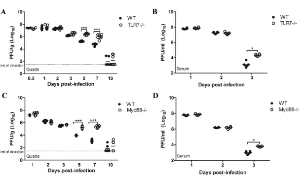
Viral titers are elevated in mice lacking TLR7 or Myd88 at late times post-infection.
To determine whether viral burden was altered in mice deficient in TLR7 or Myd88, we quantified the amount of virus present in the quadriceps and serum of WT, TLR7-/- and Myd88-/- mice at various times post-RRV infection. No significant
differences in viral titer were detected in the quadriceps muscles or serum of WT, TLR7 -/-and Myd88-/- mice at early times post-infection (Figure 2.5). WT and TLR7-/- mice did not show significant differences in viral titer in the quadriceps muscles by plaque assay at days 0.5, 1, 2 and 3 post-infection (Figure 2.5A). However, viral titers in the serum of TLR7-/- mice were significantly enhanced over WT mice at day 3 post-infection (Figure 5B), and analysis of viral burden in infected quadriceps tissues at later times
49
high levels of viral replication within joint and muscle tissues, with subsequent
overactive inflammatory responses in these tissues contributing to disease pathogenesis (51, 53, 58, 59), the inability of TLR7- or Myd88-deficient mice to control viral
replication at late times post-infection raised the possibility that the enhanced
disease/mortality in these mice might be due to enhanced viral replication in other tissues, such as the central nervous system (CNS). However, we observed no differences in viral load within the brain and spinal cord of WT and TLR7-/- mice at 1 and 2 days
post-infection, and viral titers in these tissues were below the limit of detection for both mouse strains by day 7 post-infection (data not shown). We also observed no differences in viral titer between WT and TLR7-/- mice in the kidney, liver, spleen and draining lymph node at 1 and 2 days post-infection (data not shown), suggesting that excess viral replication within the CNS or other non-target tissues of RRV infection does not explain the enhanced disease observed in TLR7-deficient mice.
TLR7-/- mice show reduced neutralizing antibody production and decreased antibody
affinity following RRV infection.
Because TLR7-/- mice showed enhanced viral titers when compared to WT mice at late times post-infection, we hypothesized that deficiency in TLR7 signaling may result in an ineffective antibody response that leads to an inability to control viral replication during RRV infection. To determine whether deficiency in TLR7 affected antibody production following RRV infection, RRV-specific antibody levels were quantified in the sera of WT and TLR7-/- mice by ELISA at 7 and 10 days post-infection (Figure 2.6A and 2.6B). TLR7-/- mice showed increased RRV-specific total IgG
50
2.6A and 2.6B), with TLR7-/- mice having significantly increased total RRV-specific IgG at day 7 post-infection (Figure 2.6A). Additionally, analysis of RRV-specific IgG
subtypes revealed that TLR7-/- mice showed significantly increased IgG1 and IgG2c antibody production at 7 days post-infection, as well as increased IgG2c antibody levels at day 10 post-infection, relative to WT mice (Figure 2.6A and 2.6B). These results suggest that RRV-specific antibody production is elevated in TLR7-/- mice following RRV infection. Furthermore, because IgG1 is indicative of T-helper 2 (Th2) immune responses and IgG2c is indicative of T-helper 1 (Th1) immunity, analysis of RRV-specific IgG subtypes reveals that TLR7-/- mice have a skewed Th1/Th2 antibody response following RRV infection relative to WT mice (Figure 2.6A and 2.6B) (214-216).
To determine whether the quality of RRV-specific antibody was altered in TLR7-deficient mice, we first tested the neutralization capacity of WT and TLR7-/- antisera from 7 and 10 days post-RRV infection by PRNT assay (Figure 2.5C). Antisera from TLR7-/- mice had significantly less neutralization activity when compare to WT antisera at both 7 and 10 days post-infection, suggesting that the quality of RRV-specific antibody produced by TLR7-deficient mice is defective and less neutralizing relative to WT
51
Lastly, we directly tested whether sera from TLR7-/- mice showed a reduced capacity to protect from RRV-induced disease in adoptive transfer studies. We passively transferred heat-inactivated sera obtained from WT or TLR7-/- mice at day 10 post-infection to naïve WT mice prior to RRV post-infection. As shown in Figure 2.7, the transfer of TLR7-deficient antisera failed to protect WT mice from RRV infection (Figure 2.7B and C). WT mice that received sera from RRV-infected TLR7-/- mice showed
significantly reduced weight gain and enhanced disease signs between days 5 and 10 post-infection compared to mice that received WT antisera, which were protected from RRV-induced disease (Figure 2.7B and C). Taken together, these results indicate that TLR7 is critically important in the establishment of an effective antibody response following RRV infection.
Myd88- and TLR7-deficiency alters germinal center development following RRV
infection.