The handle
http://hdl.handle.net/1887/20863
holds various files of this Leiden University
dissertation.
Author
: Maczuga, Piotr
Title
: Towards RNAi based therapy of liver diseases : diversity and complexity of shRNA
and miRNA processing and functions
Towards RNAi-based
therapy of liver diseases
Diversity and complexity
of shRNA and miRNA
processing and functions
Piotr Maczuga
of liver diseases
Diversity and complexity of shRNA
and miRNA processing and functions
ISBN: 978-94-6182-249-9
Layout & printing: Off Page, www.offpage.nl Cover: Piotr Maczuga & Off Page
Copyright © 2013, Piotr Maczuga. All rights reserved. No part of this thesis may be reproduced or transmitted in any form or by any means, without the prior permission in writing of the author.
A digital version of this thesis can be downloaded from Leiden University Repository: https://openaccess.leidenuniv.nl/handle/1887/9744
The printing of this thesis was financially supported by: uniQure Biopharma
Macrogen Leiden University
of liver diseases
Diversity and complexity of shRNA
and miRNA processing and functions
Proefschrift
ter verkrijging van
de graad van Doctor aan de Universiteit Leiden, op gezag van de Rector Magnificus Prof. Mr. C.J.J.M. Stolker,
volgens besluit van het College voor Promoties te verdedigen op dinsdag 7 mei 2013 klokke 10.00 uur
door
Piotr Maczuga
geboren te Le zajsk (Polen).
Promotor prof. dr. Sander J.H. van Deventer
Co-promotor dr. Pavlina Konstantinova
Overige leden
prof. dr. Theo van Berkel prof. dr. Pieter H. Reitsma dr. Hein W. Verspaget prof. dr. Bart van Hoek
Chapter 1 Introduction 9 Chapter 2 Apolipoprotein B knockdown by AAV-delivered
shRNA lowers plasma cholesterol in mice 39
Chapter 3 Optimization and comparison of knockdown efficacy between polymerase II expressed shRNA and artificial miRNA targeting luciferase
and Apolipoprotein B100 69
Chapter 4 Embedding siRNA sequences targeting Apolipoprotein B100 in shRNA and miRNA scaffolds results in differential processing
and in vivo efficacy 95
Chapter 5 Therapeutic expression of hairpins targeting Apolipoprotein B100 induces differential
changes in murine liver 125
Chapter 6 Regulated knockdown of Apolipoprotein B100 and Huntingtin by the GeneSwitch
mifiprestone-inducible system 175
Chapter 7 Discussion 197
Addendum Summary 221
Nederlandse samenvatting 223
Streszczenie 225
Curriculum Vitae 227
Publications 229
A1AD Alpha-1-antitrypsin deficiency
AAV Adeno-associated virus
Ago Argonaute protein
ALP Alkaline phosphatase
ALT Alanine aminotransferase
APC Antigen presenting cell
ApoB Apolipoprotein B
APOBEC2 Apolipoprotein B mRNA editing enzyme, catalytic polypeptide-like 2
AST Aspartate aminotransferase
bp basepairs
BGN Biglycan
CMV Cytomegalovirus
CTL Cytotoxic T lymphocyte
dsRNA double-stranded RNA
DGAT2 Diglyceride acyltransferase 2
F.IX Factor IX
FH Familial hypercholesterolemia
GS Gene Switch system
GFP Green fluorescent protein
hAAT human Alpha-1-antitrypsin
HBsAg Hepatitis B surface antigen
HBV Hepatitis B virus
HCC Hepatocellular carcinoma
HCV Hepatitis C virus
HMG-CoA 3-hydroxy-3-methyl-glutaryl-CoA reductase
IFN Interferon
INR Initiator sequence
ITR Inverted terminal repeat
JNK c-Jun N-terminal kinase
LDL-C Low density lipoprotein cholesterol
LDL-R Low density lipoprotein receptor
LDLRAP1 Low density lipoprotein receptor adaptor protein 1
lhRNA long-hairpin RNA
LNA Locked nucleic acid
LPL Lipoprotein lipase
MDR Multi drug resistance
MFP Mifiprestone
MHC Major histocompatibility complex
NHP Non-human primate
NGS Next generation sequencing
nt nucleotide
ORF Open reading frame
OVA Ovalbumin
pA polyadenylation signal
PAMP Pathogen-associated molecular pattern
PKR Protein kinase R
PCSK9 Proprotein convertase subtilisin/kexin type 9
p.i. post injection
pre-miRNA precursor micro RNA
pri-miRNA primary micro RNA
RISC RNA induced silencing complex
RNA Ribonucleic acid
RNAi RNA interference
shRNA short-hairpin RNA
siRNA small interfering RNA
SNALP Stable nucleic acid lipid particle
ssRNA single-stranded RNA
ss-siRNA single-stranded siRNA
TA Gene Switch transactivator
TATA TATA box
TLR Toll-like receptor
tRNA transfer RNA
TSS Transcription start site
TTS Transcription termination signal
VLDL-C Very low density lipoprotein cholesterol
Introduction
1
Introduction
Since its discovery RNA interference (RNAi) mechanism has become a valuable research tool for functional analysis of gene expression and a promising approach for treatment of many disorders including viral infections, hereditary disorders, and cancer. By introducing molecules that constitute substrates for the endogenous RNAi pathway, disease-related messenger RNA (mRNA) and thus production of related proteins can be inhibited. Liver plays a major role in the metabolism and has a number of functions in the body, including glycogen storage, decomposition of red blood cells, plasma protein synthesis, hormone production, and detoxification. Many liver diseases that currently cannot be adequately treated are good candidates for RNAi gene therapy. RNAi-based strategies have recently been explored for treating hepatocellular carcinoma (HCC), hepatitis B (HBV) and hepatitis C (HCV) virus, infections and genetic disorders such as familial hypercholesterolemia (FH) or alpha-1-antitrypsin deficiency (A1AD). The RNAi machinery can directly inhibit RNA, which plays important role in the etiology of these diseases i.e. target transcripts encoding for multidrug resistance proteins in HCC, viral genome of HBV, replication intermediates of HBV, HCV or mutated mRNAs which lead to production of amended proteins in FH and A1AD. Additionally, the RNAi machinery, in particular endogenous miRNAs can be exploited to regulate transgene expression. This approach was successfully applied to reduce immune response to Factor IX (F.IX) used for treatment of Hemophilia B by inhibiting F.IX expression in antigen presenting cells (APC) using hematopoietic cell-specific miRNA binding sites in 3’ untranslated region (3’UTR) of the transgene.
However, serious concerns have been raised about safety of the RNAi-based therapeutic approach when toxicity was described in rodents and non-human primates (NHP). This thesis addresses important questions about the long-term efficacy and safety of RNAi for treatment of liver diseases, which also involves aspects of processing
of RNAi molecules in vivo, transcriptome analysis and liver functioning after injection
with adeno-associated viruses (AAV) encoding RNAi-inducing molecules.
The natural RNAi pathway
RNAi is an evolutionary conserved mechanism for regulating gene expression. It plays an important role in defense against viruses but also in development and in normal functioning of the cell [1-3]. RNAi was initially discovered in plants where delivering DNA encoding the rate limiting enzyme in anthocyanin biosynthesis in
order to generate petunias (Petunia hybryda) with darker color, unexpectedly induced
1
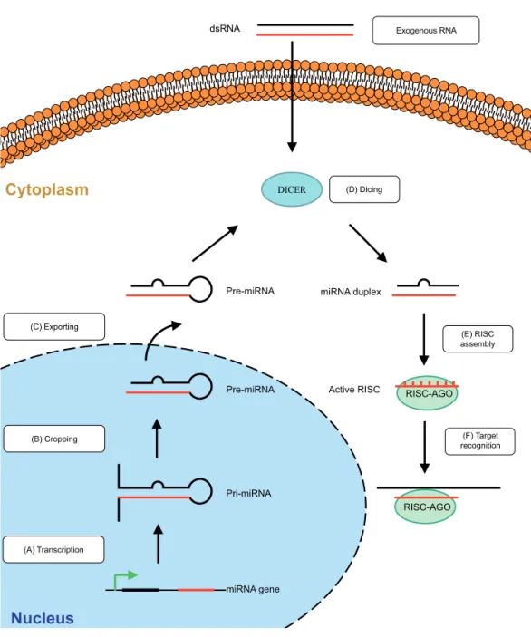
destruction or led to translational repression. The same mechanism was later found to be conserved in other species i.e. fungi Neurospora Crassa [6] and Caenorhabditis elgans worm [7]. In 1998 a breakthrough discovery was made by A. Fire and C. Mello which provided the explanation for these previously described phenomenons [8]. They identified molecules which were responsible for inducing target degradation of mRNA and showed that a double-stranded RNA (dsRNA) contamination in mRNA preparations rather than single stranded RNA was inhibiting a gene with complementary sequences [8]. Their discovery was a milestone which was just beginning of identification of novel mechanism of gene regulation. In 2006, A. Fire and C. Mello were awarded the Noble prize for their discovery of RNA interference-gene silencing by double-stranded RNA.The RNAi mechanism is initiated by dsRNA molecules (Fig. 1). The natural RNAi mechanism in mammalian cells functions by endogenous miRNA molecules, which are synthesized in cells as longer primary RNA transcripts (pri-miRNAs). Most miRNA are usually transcribed by RNA polymerase II; however RNA polymerase III (Pol III) transcribes some miRNAs, especially those with upstream Alu sequences and transfer RNAs (tRNAs) [9]. Pri-miRNAs are cleaved by the nuclear Microprocessor complex (Drosha-DGCR8 complex) to produce miRNA precursors (pre-miRNAs) of 70 nucleotides (nt) [10], which are then transported by Exportin 5 to the cytoplasm and processed by the RNAse III endonuclease family enzyme Dicer to produce a mature miRNA duplex of ~21-25 basepairs (bp) [11]. The guide strand of the miRNA enters a multiprotein
RNA-induced silencing complex (RISC) enabling it to bind to the 3′ untranslated region
(3’UTR) of its target mRNA. Although either strand of the duplex may potentially act as a functional miRNA, one strand called “guide strand” or “antisense strand” is preferentially incorporated into the RISC complex. The selection of guide strand is highly influenced by the thermodynamic stability of 5’ and 3’ ends of Dicer products [12]. Generally, the strand with the lower 5’ stability will be preferentially loaded into RISC, while the other strand is degraded. The active components of the RISC complex are endonucleases called Argonaute proteins (Ago) and in human cells the Ago family consists of four Argonaute proteins (Ago1-4) [13]. It is known that only RISC association with Ago2 has the capability to cleave target mRNA, whereas Ago1 association can mediate translational repression [12]. The process of loading Dicer
substrates on different mammalian Agos in vivo is not well established and it is known
that miRNA are not loaded into specific RISC-Ago complexes but can interact with any of them [12]. The RISC-Ago complex recognizes mRNA with the complementary target sequence and consequently induces cleavage or translational repression [14].
The specificity of miRNA target recognition is primarily determined by the
sequence from 2 to 8 nt from the 5′ end of the miRNA guide strand which is called the
1
1
therefore single miRNA is capable of regulating multiple cellular targets. Conversely, each gene can be controlled by several miRNAs [15].RNA entering RNAi pathway can also originate from an exogenous source, for example during a viral infection [17,18], which -when processed- can also generate mature miRNA. By targeting viral mRNA this can inhibit the expression of crucial viral proteins and prevent the spread of the infections. In fact, RNAi does work effectively as an antiviral agent in plants and mammalian cells [19-21].
RNAi therapeutics
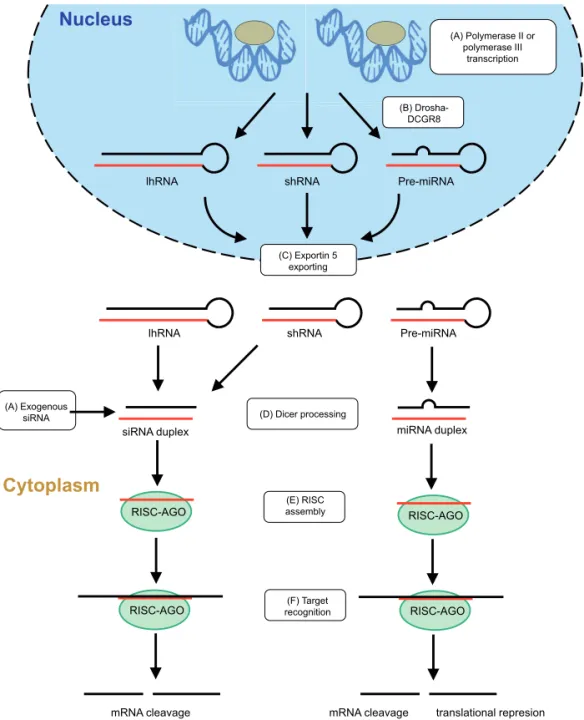
Identification of the RNAi pathway structural components and intermediates initiated studies on the application of RNAi as therapeutics. The general idea is that by introduction of molecules that constitute substrates for the endogenous pathway, specific inhibition of disease-related mRNA can be induced and as a consequence reduction of disease-related proteins can be achieved. Therapeutic RNAi can be induced in different ways (Fig. 2).
1
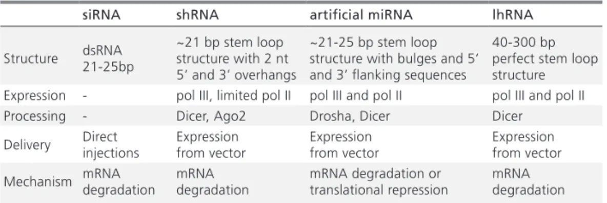
Table 1. Comparison of different approaches for inducing RNAi in cells
siRNA shRNA artificial miRNA lhRNA
Structure dsRNA 21-25bp ~21 bp stem loop structure with 2 nt 5’ and 3’ overhangs
~21-25 bp stem loop structure with bulges and 5’ and 3’ flanking sequences
40-300 bp perfect stem loop structure Expression - pol III, limited pol II pol III and pol II pol III and pol II
Processing - Dicer, Ago2 Drosha, Dicer Dicer
Delivery Direct injections Expression from vector Expression from vector Expression from vector
Mechanism mRNA degradation mRNA degradation mRNA degradation or translational repression mRNA degradation bulge structure in their stem that at least partially can explain differences in their functioning. siRNA, shRNA and lhRNA are perfectly complementary to their targets, which induce degradation rather than translational repression.
Another important difference is the mechanism of transcription. shRNAs are generally expressed from constitutive polymerase III (pol III) promoters and directly generate a mature stem-loop product, which is exported and processed by Dicer [32]. Although shRNA can be expressed from pol II promoters [12,34-35], this has not been very successful due to inefficient processing of the hairpins by the RNAi machinery and the lack of detailed knowledge of the transcription start site (TSS), transcription termination signal (TTS), and hairpin location. Alternatively, siRNA can be expressed as a long hairpin RNA (lhRNA) in which the stem can be extended up to 300 bp and can incorporate multiple ~21 bp siRNA sequences. lhRNAs can be expressed with pol III or pol II promoters and undergo processing by Dicer [26,28,36]. In contrast to shRNA, miRNAs transcripts are generally expressed from pol II promoters that allow for the use of tissue-specific or regulated expression systems. They have additional flanking sequences adjacent to hairpin structure therefore miRNA processing requires an additional step of excision from the longer pre-miRNA sequence by the Drosha-DGCR8 complex [10,37]. miRNA can be expressed in clusters allowing expression of different siRNA, which can be used for viral infections or cancer, where multiple sequences have to be targeted [38].
There are advantages and disadvantages inherent to the various ways of inducing RNAi in cells and the choice greatly depends of the desired effect. Characteristic features of different RNAi systems are summarized in Table 1.
1
1
can restore normal functioning of cells [41,42]. This approach is intensively investigated to prevent or treat metastasis of cancer cells. Inhibition of particular miRNA’s resulting in inhibition of its target mRNA is a promising treatment for viral infections [40].
Additionally by incorporation in the 3′ UTR region of transgenes, an intracellular RNAi
mechanism can be employed to regulate their expression in different cells [43,44]. In particular this approach is interesting for inhibition of transgene expression in non-target cells because cells which are not expressing the particular miRNA will not inhibit expression. Hence, genetic regulation by endogenous miRNA is a promising strategy of avoiding immune responses to transgene (i.e. F.IX for hemophilia B) that are seen as “foreign” by the immune system of the patient lacking F.IX expression [45].
RNAi therapeutics in the liver
Despite the progress in understanding causes of many diseases, current therapies involving pharmacological and biological drugs only alleviate disease symptoms.
Figure 3. Structure of (A) siRNA, (B) shRNA, (C) lhRNA and (D) miRNA targeting ApoB.
1
Inappropriate gene activity is being linked to a number of liver diseases and because RNAi controls gene expression at the level of RNA, a unique opportunity is beingoffered to stop the actual cause of diseases. Therefore, gene silencing in cells creates an entirely new horizon in the therapy of liver diseases allowing new treatment strategies to be developed. Proof of concept for the therapeutic efficacy of RNAi has already been convincingly demonstrated in a number of preclinical models of FH, hepatitis, cancer and others. The key of RNAi approach success is mainly dependent on careful target selection, the RNA inducer used and a convenient delivery system. In this paragraph, a short description of disease etiology, conventional treatments and possible RNAi targets is given.
Familial hypercholesterolemia (FH)
1
treatment often fails to reduce LDL-C levels. Therefore homozygous FH was among the first monogenic diseases targets for treatments with RNAi and conventional gene substitution therapies. Recently, a single intravenous injection of an AAV8 vector containing the mouse LDL-R gene to a humanized mouse model of FH, the LDL-R-/- APOBEC-/- mouse, was found to significantly reduce plasma cholesterol levels in chow-fed animals [54]. In addition to the LDL-R gene augmentation approach, the successful use of the VLDL-R for the complementation of mutated LDL-R function in homozygote FH has also been shown [55]. As alternative strategy, inhibition of Pcsk9 with siRNA has been shown to increase LDL-R levels in liver and in consequence reduce LDL-C levels [56]. However conventional gene therapy (resulting in LDL-R expression) and Pcsk9 inhibition is not suitable for FH patients that have ApoB
mutations. In those cases RNAi can be used to target ApoB. In 2004, Soutschek et al.
[57] showed silencing of ApoB in mice with systemically delivered modified siRNA,
which was associated with reduction in plasma cholesterol. Later, Zimmermann et al.
showed siRNA mediated ApoB silencing in human primates [58], however the effect was stable only for up to 11 days. Albeit ApoB reduction by siRNAs was successful in lowering LDL-C levels in plasma, the effect was transient and treatment caused liver steatosis [57,58]. In order to achieve stable reduction of ApoB and in consequence LDL-C without the need to re-administration, siRNA has to be embedded in shRNA or miRNA scaffolds, expressed from a pol II or pol III promoters and delivered to the target cells by using an appropriate vector system.
Alpha-1-antitrypsin deficiency (A1AD)
1
mice treated with rAAV vectors simultaneously expressing artificial miRNA targeting PiZ and codon optimized A1AT gene showed that serum PiZ level was stably reducedby an average of 80% [65]. In addition, decreased globular accumulation of misfolded Z-A1AT in hepatocytes and a reduction in inflammatory infiltrates in the liver were observed. Finally endogenous miRNAs were minimally affected by this treatment suggesting that miRNA mediated knockdown did not saturate the miRNA pathway.
Hepatocellular carcinoma (HCC)
HCC is the most common type of liver cancer. Most cases of HCC are secondary to viral infections or cirrhosis [66]. Prognosis and treatment depends on the stage of the disease, however ~70% of patients are diagnosed at intermediate and late stages when no curative treatment is available. These patients often develop multidrug resistance to chemotherapeutics due to overexpression of ABC family transporter proteins. Among them ABBC1 and ABBC2 are characterized for the liver and their implication in
chemotherapy resistance is suggested [67]. Borel et al. showed successful inhibition of
ABCC2 transporter in vivo by AAV delivered shRNA and artificial miRNA [68]. Despite
effectiveness mice exhibited serious side effects due to oversaturation of endogenous
RNAi machinery. Additionally, Borel et al. investigated panel of endogenous miRNA
whose expression pattern change in hepatocellular cancerogenesis [69]. These miRNAs not only can be used as marker for disease’s prognosis but also by overexpression of miRNA which are downregulated, normal functioning of the cell may be restored i.e. restoration of mir-122 expression in liver cancer correlates with suppression of both the malignant phenotype and metastatic properties [70]. Restoring endogenous miRNA pathway can also improve response to chemotherapy as many of MDR transporter transcripts have been identified as miRNA targets [69,71,72].
Hepatitis B virus (HBV) infections
HBV infection is still a major health problem. Even though effective vaccines have been available for the last 20 years, worldwide, over 350 million people are infected with this virus [73]. Chronic HBV infections may have serious consequences, including liver cirrhosis and HCC; prevention of this infection is a key to then prevent the HCC
[74]. HBV, which belongs to Hepadnaviruses, have small genome of 3,2 kb - partially
1
mutations. McCaffrey et al. showed 85% reduction in secreted HBsAg in mice plasma
when HBV genome plasmid was delivered with a plasmid encoding shRNA targeting the HBV sAg [76]. Importantly, viral inhibition occurred in absence of active viral replication suggesting that this strategy is independent from viral replication thus can be used for treatment of chronic HBV infections. An alternative to anti-HBV shRNA cassettes involves utilization of pri-miRNA scaffold sequences [77]. This is an appealing strategy as multiple miRNA can be expressed from single transcripts and this approach
has been already validated in vivo [78]. Another approach was used by Weinberg et
al., who assessed the efficacy of lhRNAs targeting the conserved hepatitis B protein X
(HBx) open reading frame of HBV [28]. After hydrodynamic injection in mice one of the lhRNA vectors decreased viral replication by 70–90% with no evidence of induction of an interferon response induction. RNAi therapy has a major disadvantage, being the inability to completely eliminate DNA genome, but it can complement conventional anti-HBV therapies by affecting multiple steps in the viral life cycle.
Hepatitis C virus (HCV) infections
Like HBV, HCV is one of the major causes of liver morbidity and also a leading cause of liver transplantation, with around 170 million people infected [79]. HCV
is small, enveloped member of family Flavividae [80]. Its genome consists of about
9,6kb single stranded positive sense RNA, which is translated into large multiprotein complex and translationaly processed into the structural components of the virus. HCV is particularly attractive target for RNAi, which in theory can target not only the replicative forms but also genomic RNA templates and eventually lead to viral clearance. However due to the high mutation rate only highly conserved regions of the virus can be targeted to prevent escaping from RNAi activity [81]. As with HBV, HCV has been the target of both synthetic and expressed activators of the RNAi pathway and effective inhibition of markers of virus replication has been described in
several studies. Yokota et al. used both synthetic siRNA and expressed shRNA effectors
against the 5′-NTR (5’ Non translated region) and approximately 80% suppression
1
mir-122 expression, which regulates HCV by binding directly to two adjacent sites close to the 5’ end of HCV RNA [89], Although the precise mechanism of regulationis not yet clear, inhibition of endogenous mir-122 function is a promising therapeutic approach. This can be accomplished by delivering single-stranded, modified RNA oligonucleotides complementary to miRNA guide strand, resulting in tight binding and efficient inhibition [90,91]. Santaris Pharma have developed drug- Miravirsen, which is a locked nucleic acid-based (LNA) antisense oligonucleotide to mir-122 that is delivered to the liver with intravenous injection. This molecule reduced HCV replication in a small-scale trial in chimpanzees without inducing liver toxicity [90].
Hemophilia B
Hemophilia is caused by hereditary genetic disorders that impair the body’s ability to establish blood coagulation. The defect in blood clotting in hemophilia B is caused by the lack of functional clotting Factor IX (F.IX) as a result of mutations in the gene encoding this protein. As an X-linked recessive chromosomal disorder, almost all people living with haemophilia are male, while females are generally asymptomatic carriers of the mutated allele [92]. It occurs in around 1 in about 30,000 male births. F.IX deficiency leads to an increased risk for bleeding in response to mild trauma or even spontaneously, such as in joints (haemarthrosis) or muscles. Patients are grouped according to circulating levels of F.IX, which correlate closely with clinical severity. Plasma concentrations of F.IX of 5–25%, 1–5%, and <1% of normal levels refer to mild, moderate and severe hemophilia respectively. Protein replacement is the current standard of care. Frequent intravenous administrations (two to three times a week) of recombinant F.IX are required to stop or prevent bleeding. A goal for the treatment of hemophilia B is the development of a gene transfer strategy that can maintain sustained production of clotting F.IX in the liver, where it is normally produced. At present, gene transfer mediated by viral vectors delivering F.IX expressing cassette shows the greatest promise for long-term correction of hemophilia B in the preclinical setting [93-96]. Improvement of levels to above 1% is sufficient to prevent most spontaneous and nearly all life-threatening bleeding episodes [95]. Unfortunately, initial evaluation of viral vectors expressing F.IX failed to achieve sustained F.IX expression in hemophilia B mice due to the recognition of F.IX as foreign antigen which resulted in anti-F.IX cellular immune response [97,98]. Further analysis suggested that this may be a result of off-target transgene expression in hematopoietic-lineage cells of the spleen which resulted in antigen presenting cells (APCs) maturation and activation of cytotoxic T lymphocytes (CTLs) [99,100]. As a consequence transduced hepatocytes were eliminated and expression diminished.
hematopoietic-1
specific microRNA, mir-142-3p in its 3’ UTR [45,101]. This allows expression in hepatocytes, while preventing F.IX production in hematopoietic cells- including APCs. By restricting transgene expression exclusively to the liver, while preventing F.IX production in hematopoietic cells including APCs, immunologic tolerance was induced. Importantly, analysis of miRNA revealed that the mir-142-3p–regulated vector did not affect the natural levels of expression of mir-142-3p or other miRNAs in either the liver or spleen. However these results are in opposite to F.IX gene transfer mediated by AAV vectors, which showed expression of clotting factor without inducing cellular response to transgene [96]. Interestingly expression of F.IX was limited by presence of anti-AAV immune response which probably eliminates transduced hepatocytes [102] while no anti-F.IX immune response was detected. Still this possibility has to be considered especially when strategies to overcome existing immune response to AAV will be applied (immune response to AAV is discussed in details in paragraph 7). Incorporation of mir-142-3p sites for transgenes expressed with AAV can be used as additional safety measure to prevent immune reaction and allowing long-term expression of transgenes.
RNAi toxicity
RNAi can interfere with normal functioning of cells and therefore safety is an important aspect of the implementation of RNAi based therapies. Non-specific effects resulting from the RNAi induction appear to have three separate mechanisms: innate immune response, off-target effects and oversaturation of the RNAi pathway. In addition, immune response to the delivery method can negatively contribute to the efficacy and safety of RNAi therapeutics. These side effects are often associated with severe toxicity that in some cases can be even fatal. In this chapter only RNAi toxicity and strategies to avoid them will be considered. Several toxicity issues involving response to the transgene or the RNAi inducer and the delivery vector will be described in paragraph 7. The first source of RNAi toxicity is related to activation of innate immune responses. It is now well established that RNAi therapeutics, in particular siRNAs
can be potent inducers of interferon (IFN) and inflammatory cytokines both in vivo
1
activate early response genes and genes encoding cytokines [107]. TLR activation by RNAi therapeutics leads in consequence to activation of immune response. Activationof the immune system by siRNAs is sequence-dependent as U and GU rich regions seem to be preferentially recognized by TLR [110]. Therefore, chemical modification of siRNA can prevent the recognition by the immune system [111]. It has been shown that modification of 2’ ribose with 2’-O-methyl, 2’fluoro-, 2’deoxy- or locked nucleic acid can reduce immune stimulation [110]. Although immunostimulatory side effects must be taken in account when inducing RNAi, for certain applications i.e. cancer or viral infections, activation of the immune system can be beneficial.
1
off target effects [115,116]. Modifications should include both strands of the siRNA: sense being loaded into RISC and antisense binding to off-targets. Similarly for shRNA and miRNA passenger strand’s interaction with endogenous transcripts can be minimized by expressing the so called “sponge” transcript containing complementary binding sites for the passenger strand. When the passenger strand is loaded into RISC, the complex will bind to sponge transcript instead of endogenous target [119].
A third toxicity aspect is oversaturation of the natural RNAi pathway. Introducing RNAi agents which make use the endogenous RNAi pathway and substitute intermediate products can create competition with the endogenous miRNA processing [68,120,121]. High expression levels of therapeutic shRNA or artificial miRNA can saturate the endogenous RNAi machinery and in consequence lead to deregulation of the complex miRNA network. As referred in paragraph 3, shRNAs are generally expressed from pol III promoters, which provide constitutive high expression levels necessary for potent knockdown of target mRNA. Although these features are valuable for research purposes, they can be disadvantages for therapeutic applications. It has been reported that pol III-expressed shRNA can lead to severe toxicity when high doses are used [68,121,122].
Grimm et al. observed liver failure and mortality in mice injected with different
1
regulatory proteins that are potentially immunogenic and inducer molecules that need to be safe for long-term applications.Adeno-associated virus (AAV) delivery
Among the different options of vehicles for delivery of genetic material to living organisms, viruses seem to be a vector of choice because of their natural ability to infect cells. The majority of vectors used in clinical trials are engineered human or animal viruses including adenoviruses, AAV, herpes simplex virus, lentiviruses and retroviruses [131]. Adeno-associated viruses with their unique properties have been shown to be a very promising gene-therapy vector for many genetic diseases [132].
AAV is member of the Parvoviridae family with a small single stranded genome
1
however some of the serotypes fail to meet the cross-reactivity requirement i.e. AAV6 can cross-react with AAV1 while AAV10 and AAV11 are not fully characterized [143]. Furthermore there are number of AAV variants for which serological data are not available. Each AAV serotype has diverse tissue tropism and therefore specific tissues can be targeted by using a particular AAV serotype. Another interesting approach is to generate hybrid viruses or create new serotypes in order to change tissue tropism or overcome pre-existing or acquired antibody response [144]. Tissue specificity is determined by the capsid composition and expression of AAV receptors, however apart from AAV2 all other serotypes’ receptors are not very well characterized.
The most common organ transduced by AAV following tail-vein injection in mice is liver. Biodistribution studies of different AAV serotypes vectors demonstrated superiority of some AAV serotypes in the context of liver targeting [145]. AAV serotypes that preferentially target mice hepatocytes after tail-vein injection are serotypes 1, 2, 5, 6, 7, and 8 [145]. Among them AAV7 has the strongest tropism to the liver, followed by AAV6. AAV1, 2, and 5 transduce also preferentially the liver, however with lower efficiency compared to AAV7 and AAV6. Additionally AAV8 and AAV9 demonstrate robust expression in the liver; however they also transduce other tissues more ubiquitously [145]. It is extremely important to understand that tissue specificity is species-specific and vectors which transducer murine liver do not necessary transduce human liver with the same efficiency.
Immune response to AAV capsid or the transgene
Side effects associated with gene therapy can originate from different sources and in the case of gene silencing; the outcome may be a combination of toxicity resulting from both
RNAi and the delivery vehicle. rAAV has been shown to stably express many genes in vivo
[146]. Surprisingly initial studies in humans led to identification of immune responses not to the transgene but to the AAV capsid [102]. A complete understanding of the interactions of rAAV with the immune system is pivotal for all gene therapy applications and both innate and adaptative immune responses should be taken into account.
1
after the presentation of AAV derived antigen by mature presenting cells in lymph Another important aspect is the cellular response to AAV [142], which is mountednodes to T cells. The manner in which the antigen is presented and the nature of the antigen itself determines the activation of antigen specific T cells. Antigen presentation can occur in two distinct manners: Antigens that are endogenously generated by the cellular machinery are presented on the Major histocompatibility complex (MHC) class I molecules which are present on all nucleated cells. Proteins exogenous to the cells are presented in context of MHC class II molecules exclusively on APC after phagocytosis or endocytosis of the pathogen [149]. During AAV infection, virus-derived antigens can be presented by APCs on both MHC class molecules. Opsonized AAVs can be phagocyted by APCs [150] and antigens will presented on MHC class II molecules. However it does not lead to CTL response as the iC3b and factor H influence APC chemokines and cytokies microenvironment. Additionally APCs are not natural targets of AAV which minimizes presentation of AAV derived antigens on MHC class I molecules. However, exogenous antigen can also be presented on MHC class I molecules via cross-presentation. Cross-presentation is the predominant mechanism by which AAV induces T cell immune response. Co-administration of an immunosuppressive regimen with AAV injection can be a good option to impede immune response to AAV [151,152]. It was shown that short-term immunosuppression did not lead to toxicity and does not interfere with AAV transduction of hepatocytes [152]. Application of immunosuppression prior injection may provide a solution for patients with pre-existing antibody response to AAV or for re-injecting patients with the same rAAV serotype as this strategy has already been validated in animal models [153,154].
Overview
Safe and long-term expression of siRNA is necessary for the treatment of patients with liver diseases that fail to respond to conventional treatment. This thesis addresses important questions about delivery of RNAi therapeutics to the liver, the efficacy of gene silencing and possible side effects. These aspects were considered in terms of constitutive and inducible expression of RNAi therapeutics in liver (Fig. 4). First,
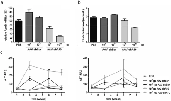
we show that ApoB can be inhibited in vivo using AAV vectors delivering shRNA
expressed from pol III promoters. By inhibiting ApoB, a stable reduction of total plasma cholesterol and LDL-C was achieved. To minimize possible toxic effects caused by shRNA overexpression pol II-driven expression of shRNA was optimized and compared
to artificial miRNA expression in vitro. As the latter appeared to be more potent, an
artificial miRNA construct targeting ApoB was developed, allowing expression from the weaker liver-specific LP1 promoter. Subsequently the long-term efficacy and safety
of shApoB and miApoB was assessed in vivo. Initially both shApoB and miApoB were
1
Gene
T
her
apy
for
liv
er
di
se
as
es
Regu
la
te
d
ex
pr
es
sion
Co
ns
titut
iv
e
sh
/miRNA
ex
pr
es
sion
Pol
II
sh
R
N
A
Pol
III
sh
R
N
A
Po
l I
I mi
R
N
A
m
iR
N
A-regu
lated
ex
pres
sion
G
en
eS
w
itch
regu
lated
mi
R
N
A
No
t ac
tiv
e,
no
ru
les
Dif
fer
ent
ial
pr
oce
ss
ing
As
ac
tiv
e
as
P
ol II
I
shRN
A
1 plas
mid
sy
stem
2 plas
mid
s
sy
stem
In vit
ro
In vivo
In vivo effica
cy
and
to
xic
ity Liv
er
mo
rp
ho
log
y
miRN
A
and
tr
anscr
ipt
ome
chan
1
of total cholesterol. Surprisingly, the effect diminished over the time over the course of 27 weeks and the loss was associated with virally delivered DNA loss. To addressthis issue we investigated liver function and morphology 8 weeks post injection with AAV encoding shApoB or miApoB. We showed differences in processing of shApoB and miApoB, which may have serious implications for RNAi therapy of liver diseases.
A second part of this thesis was to investigate the possibility of regulated expression using two separate approaches: inducible expression of artificial miRNA and transgene regulation using endogenous miRNA. We have shown expression of miRNA targeting ApoB using the GeneSwitch inducible system which allowed time-dependent expression of siApoB when the inducer was administered and no expression in its absence. A detailed analysis of the inducible promoter was performed in order to optimize induction rate and minimize leakiness. We also optimized an expression cassette for inducible expression of miRNA, combining two structural components of the GeneSwitch system. This is important safety measure to avoid possible side-effects resulting from miRNA overexpression.
1
Reference list
1. Pillai, RS (2005). MicroRNA function: multiple mechanisms for a tiny RNA? RNA; 11: 1753-1761.
2. Stark, A, Brennecke, J, Bushati, N, Russell, RB and Cohen, SM (2005). Animal MicroRNAs confer robustness to gene expression and have a significant impact on 3’UTR evolution. Cell; 123: 1133-1146.
3. Castanotto, D and Rossi, JJ (2009). The promises and pitfalls of RNA-interference-based therapeutics. Nature; 457: 426-433. 4. Ecker, JR and Davis, RW (1986). Inhibition of
gene expression in plant cells by expression of antisense RNA. Proc Natl Acad Sci U S A; 83: 5372-5376.
5. Napoli, C, Lemieux, C and Jorgensen, R (1990). Introduction of a chimeric chalcone synthase gene into petunia results in reversible co-suppression of homologous genes in trans. The Plant Cell Online; 2: 279-289.
6. Romano, N and Macino, G (1992). Quelling:
transient inactivation of gene expression in Neurospora crassa by transformation with homologous sequences. Mol Microbiol; 6: 3343-3353.
7. Guo, S and Kemphues, KJ (1995). Par-1, a gene required for establishing polarity in C. elegans embryos, encodes a putative Ser/Thr kinase that is asymmetrically distributed. Cell; 81: 611-620.
8. Fire, A, Xu, S, Montgomery, MK, Kostas, SA, Driver, SE and Mello, CC (1998). Potent and specific genetic interference by double-stranded RNA in Caenorhabditis elegans. Nature; 391: 806-811.
9. Borchert, GM, Lanier, W and Davidson, BL (2006). RNA polymerase III transcribes human microRNAs. Nat Struct Mol Biol; 13: 1097-1101. 10. Zeng, Y, Yi, R and Cullen, BR (2005). Recognition
and cleavage of primary microRNA precursors by the nuclear processing enzyme Drosha. EMBO J; 24: 138-148.
11. MacRae, IJ, Zhou, K and Doudna, JA (2007). Structural determinants of RNA recognition and cleavage by Dicer. Nat Struct Mol Biol; 14: 934-940.
12. Gu, S, Jin, L, Zhang, F, Huang, Y, Grimm, D, Rossi, JJ, et al. (2011). Thermodynamic stability of small hairpin RNAs highly influences the loading process of different mammalian Argonautes. Proc Natl Acad Sci U S A; 108: 9208-9213. 13. zuma-Mukai, A, Oguri, H, Mituyama, T, Qian, ZR,
Asai, K, Siomi, H, et al. (2008). Characterization
of endogenous human Argonautes and their miRNA partners in RNA silencing. Proc Natl Acad Sci U S A; 105: 7964-7969.
14. Gregory, RI, Chendrimada, TP, Cooch, N and Shiekhattar, R (2005). Human RISC couples microRNA biogenesis and posttranscriptional gene silencing. Cell; 123: 631-640.
15. Lewis, BP, Shih, Ih, Jones-Rhoades, MW, Bartel, DP and Burge, CB (2003). Prediction of Mammalian MicroRNA Targets. Cell; 115: 787-798.
16. Lewis, BP, Burge, CB and Bartel, DP (2005). Conserved seed pairing, often flanked by adenosines, indicates that thousands of human genes are microRNA targets. Cell; 120: 15-20. 17. Kini, HK and Walton, SP (2007). In vitro
binding of single-stranded RNA by human Dicer. FEBS Letters; 581: 5611-5616. 18. Aliyari, R and Ding, SW (2009). RNA-based
viral immunity initiated by the Dicer family of host immune receptors. Immunol Rev; 227: 176-188.
19. Tenllado, F and Díaz-Ruíz, JR (2001). Double-stranded RNA-mediated interference with plant virus infection. J Virol; 75: 12288-12297.
20. Kumar, A (2008). RNA interference: a multifaceted innate antiviral defense. Retrovirology; 5: 17.
21. Umbach, JL and Cullen, BR (2009). The role of RNAi and microRNAs in animal virus replication and antiviral immunity. Genes Dev; 23: 1151-1164.
22. Elbashir, SM, Harborth, J, Lendeckel, W, Yalcin, A, Weber, K and Tuschl, T (2001). Duplexes of 21-nucleotide RNAs mediate RNA interference in cultured mammalian cells. Nature; 411: 494-498.
23. Burnett, JC, Rossi, JJ and Tiemann, K (2011). Current progress of siRNA/shRNA therapeutics in clinical trials. Biotechnol J; 6: 1130-1146.
24. Rettig, GR and Behlke, MA (2012). Progress toward in vivo use of siRNAs-II. Mol Ther; 20: 483-512.
25. Paddison, PJ, Caudy, AA, Bernstein, E, Hannon, GJ and Conklin, DS (2002). Short hairpin RNAs (shRNAs) induce sequence-specific silencing in mammalian cells. Genes Dev; 16: 948-958.
1
Inhibition of human immunodeficiency virus type 1 by RNA interference using long-hairpin RNA. Gene Ther; 13: 1403-1413.27. Konstantinova, P, ter Brake, O, Haasnoot, J, de Haan, P and Berkhout, B (2007). Trans-inhibition of HIV-1 by a long hairpin RNA expressed within the viral genome. Retrovirology; 4: 15.
28. Weinberg, MS, Ely, A, Barichievy, S, Crowther, C, Mufamadi, S, Carmona, S, et al. (2007). Specific inhibition of HBV replication in vitro and in vivo with expressed long hairpin RNA. Mol Ther; 15: 534-541.
29. Boudreau, RL, Martins, I and Davidson, BL (2009). Artificial microRNAs as siRNA shuttles: improved safety as compared to shRNAs in vitro and in vivo. Mol Ther; 17: 169-175. 30. Couto, LB and High, KA (2010). Viral
vector-mediated RNA interference. Current Opinion in Pharmacology; 10: 534-542.
31. Chen, M, Du, Q, Zhang, HY, Wahlestedt, C and Liang, Z (2005). Vector-based siRNA delivery strategies for high-throughput screening of novel target genes. J RNAi Gene Silencing; 1: 5-11.
32. Siolas, D, Lerner, C, Burchard, J, Ge, W, Linsley, PS, Paddison, PJ, et al. (2005). Synthetic shRNAs as potent RNAi triggers. Nat Biotech; 23: 227-231.
33. Xia, H, Mao, Q, Paulson, HL and Davidson, BL (2002). siRNA-mediated gene silencing in vitro and in vivo. Nat Biotech; 20: 1006-1010. 34. Song, J, Pang, S, Lu, Y, Yokoyama, KK, Zheng,
JY and Chiu, R (2004). Gene silencing in androgen-responsive prostate cancer cells from the tissue-specific Prostate-specific antigen promoter. Cancer Research; 64: 7661-7663. 35. Giering, JC, Grimm, D, Storm, TA and Kay,
MA (2008). Expression of shRNA from a tissue-specific pol II promoter is an effective and safe RNAi therapeutic. Mol Ther; 16: 1630-1636.
36. Saayman, S, Arbuthnot, P and Weinberg, MS (2010). Deriving four functional anti-HIV siRNAs from a single Pol III-generated transcript comprising two adjacent long hairpin RNA precursors. Nucl Acids Res; 38: 6652-6663. 37. Han, J, Lee, Y, Yeom, KH, Kim, YK, Jin, H and
Kim, VN (2004). The Drosha-DGCR8 complex in primary microRNA processing. Genes Dev; 18: 3016-3027.
38. Zhou, H, Huang, C and Xia, XG (2008). A tightly regulated Pol III promoter for synthesis of miRNA genes in tandem. Biochim Biophys Acta; 11: 773-9.
39. Gregory, RI and Shiekhattar, R (2005). MicroRNA biogenesis and cancer. Cancer Res; 65: 3509-3512.
40. Sullivan, CS and Ganem, D (2005). MicroRNAs and viral infection. Mol Cell; 20: 3-7. 41. Coulouarn, C, Factor, VM, Andersen, JB,
Durkin, ME and Thorgeirsson, SS (2009). Loss of miR-122 expression in liver cancer correlates with suppression of the hepatic phenotype and gain of metastatic properties. Oncogene; 28: 3526-3536.
42. Valastyan, S, Reinhardt, F, Benaich, N, Calogrias, D, Szász, AM, Wang, ZC, et al. (2009). A pleiotropically acting microRNA, miR-31, inhibits breast cancer metastasis. Cell; 137: 1032-1046.
43. Karali, M, Manfredi, A, Puppo, A, Marrocco, E, Gargiulo, A, Allocca, M, et al. (2011). MicroRNA-Restricted Transgene Expression in the Retina. PLoS ONE; 6: e22166.
44. Brown, BD, Gentner, B, Cantore, A, Colleoni, S, Amendola, M, Zingale, A, et al. (2007). Endogenous microRNA can be broadly exploited to regulate transgene expression according to tissue, lineage and differentiation state. Nat Biotechnol; 25: 1457-1467. 45. Brown, BD, Cantore, A, Annoni, A, Sergi,
LS, Lombardo, A, Della, VP, et al. (2007). A microRNA-regulated lentiviral vector mediates stable correction of hemophilia B mice. Blood; 110: 4144-4152.
46. Sjouke, B, Kusters, D, Kastelein, J and Hovingh, G (2011). Familial Hypercholesterolemia:
Present and Future Management. Curr
Cardiol Rep; 13: 527-536.
47. van Aalst-Cohen, ES, Jansen, ACM, Tanck, MWT, Defesche, JC, Trip, MD, Lansberg, PJ, et al. (2006). Diagnosing familial hypercholesterolaemia: the relevance of genetic testing. Eur Heart J; 27: 2240-2246. 48. Myant, NB (1993). Familial defective
apolipoprotein B-100: a review, including
some comparisons with familial
hypercholesterolaemia. Atherosclerosis; 104: 1-18.
49. Tosi, I, Toledo-Leiva, P, Neuwirth, C, Naoumova, RP and Soutar, AK (2007). Genetic defects causing familial hypercholesterolaemia: Identification of deletions and duplications in the LDL-receptor gene and summary of all mutations found in patients attending the Hammersmith Hospital Lipid Clinic. Atherosclerosis; 194: 102-111.
1
of Monogenic Heterozygous Familial
Hypercholesterolemia: A HuGE Prevalence Review. Am J Epidemiol; 160: 407-420. 51. Cohen, MS (2004). Fetal and childhood onset
of adult cardiovascular diseases. Pediatr Clin North Am; 51: 1697-1719.
52. Rader, DJ, Cohen, J and Hobbs, HH (2003).
Monogenic hypercholesterolemia: new
insights in pathogenesis and treatment. J Clin Invest; 111: 1795-1803.
53. Brown, MS and Goldstein, JL (1986). A receptor-mediated pathway for cholesterol homeostasis. Science; 232: 34-47.
54. Al-Allaf, F, Coutelle, C, Waddington, S, David, A, Harbottle, R and Themis, M (2010). LDLR-Gene therapy for familial hypercholesterolaemia: problems, progress, and perspectives. Int Arch Med; 3: 36. 55. Chen, SJ, Rader, DJ, Tazelaar, J, Kawashiri, M,
Gao, G and Wilson, JM (2000). Prolonged correction of hyperlipidemia in mice with familial hypercholesterolemia using an adeno-associated viral vector expressing very-low-density lipoprotein receptor. Mol Ther; 2: 256-261.
56. Frank-Kamenetsky, M, Grefhorst, A,
Anderson, NN, Racie, TS, Bramlage, B, Akinc, A, et al. (2008). Therapeutic RNAi targeting PCSK9 acutely lowers plasma cholesterol in rodents and LDL cholesterol in nonhuman primates. Proc Natl Acad Sci U S A; 105: 11915-11920.
57. Soutschek, J, Akinc, A, Bramlage, B, Charisse, K, Constien, R, Donoghue, M, et al. (2004). Therapeutic silencing of an endogenous gene by systemic administration of modified siRNAs. Nature; 432: 173-178.
58. Zimmermann, TS, Lee, AC, Akinc, A, Bramlage, B, Bumcrot, D, Fedoruk, MN, et al. (2006). RNAi-mediated gene silencing in non-human primates. Nature; 441: 111-114. 59. Gettins, PGW (2002). Serpin Structure,
Mechanism, and Function. Chem Rev; 102: 4751-4804.
60. DeMeo, DL and Silverman, EK (2004). Alpha1-antitrypsin deficiency. 2: genetic aspects of alpha(1)-antitrypsin deficiency: phenotypes and genetic modifiers of emphysema risk. Thorax; 59: 259-264.
61. Schlade-Bartusiak, K and Cox, DW (1993). Alpha1-antitrypsin deficiency.
62. Lomas, DA and Mahadeva, R (2002). Alpha1-antitrypsin polymerization and the serpinopathies: pathobiology and prospects for therapy. J Clin Invest; 110: 1585-1590.
63. Perlmutter, DH (2002). Liver injury in alpha1-antitrypsin deficiency: an aggregated protein induces mitochondrial injury. J Clin Invest; 110: 1579-1583.
64. Li, C, Xiao, P, Gray, SJ, Weinberg, MS and Samulski, RJ (2011). Combination therapy utilizing shRNA knockdown and an optimized resistant transgene for rescue of diseases caused by misfolded proteins. Proc Natl Acad Sci U S A; 108: 14258-14263.
65. Mueller, C, Tang, Q, Gruntman, A, Blomenkamp, K, Teckman, J, Song, L, et al. (2012). Sustained miRNA-mediated knockdown of mutant AAT with simultaneous augmentation of wild-type AAT has minimal effect on global liver miRNA profiles. Mol Ther; 20: 590-600.
66. Wang, XW, Hussain, SP, Huo, TI, Wu, CG, Forgues, M, Hofseth, LJ, et al. (2002). Molecular pathogenesis of human hepatocellular carcinoma. Toxicology; 181;182: 43-47. 67. Bonin, S, Pascolo, L, Croce, LS, Stanta, G
and Tiribelli, C (2002). Gene expression of ABC proteins in hepatocellular carcinoma, perineoplastic tissue, and liver diseases. Mol Med; 8: 318-325.
68. Borel, F, van Logtenstein R., Koornneef, A, Maczuga, P, Ritsema, T, Petry, H, et al. (2011). In vivo knock-down of multidrug resistance transporters ABCC1 and ABCC2 by AAV-delivered shRNAs and by artificial miRNAs. J RNAi Gene Silencing; 7: 434-442.
69. Borel, F, Han, R, Visser, A, Petry, H, van Deventer, SJH, Jansen, PLM, et al. (2012). Adenosine triphosphate-binding cassette transporter genes up-regulation in untreated hepatocellular carcinoma is mediated by cellular microRNAs. Hepatology; 55: 821-832.
70. Coulouarn, C, Factor, VM, Andersen, JB, Durkin, ME and Thorgeirsson, SS (2009). Loss of miR-122 expression in liver cancer correlates with suppression of the hepatic phenotype and gain of metastatic properties. Oncogene; 28: 3526-3536.
71. Xia, L, Zhang, D, Du, R, Pan, Y, Zhao, L, Sun, S, et al. (2008). miR-15b and miR-16 modulate multidrug resistance by targeting BCL2 in human gastric cancer cells. Int J Cancer; 123: 372-379.
1
73. Ayoub, WS and Keeffe, EB (2011). Review article: current antiviral therapy of chronic hepatitis B. Aliment Pharmacol Ther; 34: 1145-1158.74. Williams, R (2006). Global challenges in liver disease. Hepatology; 44: 521-526.
75. Seeger, C and Mason, WS (2000). Hepatitis B Virus Biology. Microbiol Mol Biol Rev; 64: 51-68. 76. McCaffrey, AP, Nakai, H, Pandey, K, Huang, Z,
Salazar, FH, Xu, H, et al. (2003). Inhibition of hepatitis B virus in mice by RNA interference. Nat Biotech; 21: 639-644.
77. Gao, YF, Yu, L, Wei, W, Li, JB, Luo, QL and Shen, JL (2008). Inhibition of hepatitis B virus gene expression and replication by artificial microRNA. World J Gastroenterol; 14: 4684-4689.
78. Snyder, LL, Ahmed, I and Steel, LF (2009). RNA polymerase III can drive polycistronic expression of functional interfering RNAs designed to resemble microRNAs. Nucleic Acids Res; 37: e127.
79. Lee, LY, Tong, CYW, Wong, T and Wilkinson, M (2012). New therapies for chronic hepatitis C infection: a systematic review of evidence from clinical trials. Int J Clin Pract; 66: 342-355. 80. Chevaliez, S and Pawlotsky, JM (2006). HCV
genome and life cycle.
81. Pawlotsky, JM, Chevaliez, S and McHutchison, JG (2007). The hepatitis C virus life cycle as a target for new antiviral therapies. Gastroenterology; 132: 1979-1998. 82. Yokota, T, Sakamoto, N, Enomoto, N, Tanabe,
Y, Miyagishi, M, Maekawa, S, et al. (2003). Inhibition of intracellular hepatitis C virus replication by synthetic and vector-derived small interfering RNAs. EMBO Rep; 4: 602-608. 83. Sen, A, Steele, R, Ghosh, AK, Basu, A, Ray, R
and Ray, RB (2003). Inhibition of hepatitis C virus protein expression by RNA interference. Virus Res; 96: 27-35.
84. Krönke, J, Kittler, R, Buchholz, F, Windisch, MP, Pietschmann, T, Bartenschlager, R, et al. (2004). Alternative approaches for efficient inhibition of hepatitis C virus RNA replication by small interfering RNAs. J Virol; 78: 3436-3446. 85. Ashfaq, U, Yousaf, M, Aslam, M, Ejaz, R,
Jahan, S and Ullah, O (2011). siRNAs: Potential therapeutic agents against Hepatitis C Virus. Virol J; 8: 276.
86. Wilson, JA and Richardson, CD (2005). Hepatitis C virus replicons escape RNA interference induced by a short interfering RNA directed against the NS5b coding region. J Virol; 79: 7050-7058.
87. Akashi, H, Miyagishi, M, Yokota, T, Watanabe, T, Hino, T, Nishina, K, et al. (2005). Escape from the interferon response associated with RNA interference using vectors that encode long modified hairpin-RNA. Mol BioSyst; 1: 382-390. 88. Watanabe, T, Sudoh, M, Miyagishi, M, Akashi,
H, Arai, M, Inoue, K, et al. (2006). Intracellular-diced dsRNA has enhanced efficacy for silencing HCV RNA and overcomes variation in the viral genotype. Gene Ther; 13: 883-892. 89. Chang, J, Guo, JT, Jiang, D, Guo, H, Taylor,
JM and Block, TM (2008). Liver-specific microRNA miR-122 enhances the replication of hepatitis C virus in nonhepatic cells. J Virol; 82: 8215-8223.
90. Lanford, RE, Hildebrandt-Eriksen, ES, Petri, A, Persson, R, Lindow, M, Munk, ME, et al. (2010). Therapeutic silencing of microRNA-122 in primates with chronic hepatitis C virus infection. Science; 327: 198-201.
91. Haussecker, D and Kay, MA (2010). miR-122 continues to blaze the trail for microRNA therapeutics. Mol Ther; 18: 240-242. 92. Lee, CA, Kessler, CM, Varon, D, Martinowitz,
U, Heim, M and Lillicrap, D (1998). The molecular basis of haemophilia B. Haemophilia; 4: 350-357.
93. Manno, CS, Arruda, VR, Pierce, GF, Glader, B, Ragni, M, Rasko, J, et al. (2006). Successful transduction of liver in hemophilia by AAV-Factor IX and limitations imposed by the host immune response. Nat Med; 12: 342-347. 94. Margaritis, P (2010). Long-term expression of
canine FVIIa in hemophilic dogs. Thrombosis Research; 125, Supplement 1: S60-S62. 95. Hasbrouck, NC and High, KA (2008).
AAV-mediated gene transfer for the treatment of hemophilia B: problems and prospects. Gene Ther; 15: 870-875.
96. Harding, TC, Koprivnikar, KE, Tu, GH, Zayek, N, Lew, S, Subramanian, A, et al. (0 AD). Intravenous administration of an AAV-2 vector for the expression of factor IX in mice and a dog model of hemophilia B. Gene Ther; 11: 204-213.
97. Dai, Y, Schwarz, EM, Gu, D, Zhang, WW, Sarvetnick, N and Verma, IM (1995). Cellular and humoral immune responses to adenoviral vectors containing factor IX gene: tolerization of factor IX and vector antigens allows for long-term expression. Proc Natl Acad Sci U S A; 92: 1401-1405.
1
after vascular delivery of a lentiviral vector. Nat Biotech; 20: 53-57.
99. Mingozzi, F, Liu, YL, Dobrzynski, E, Kaufhold, A, Liu, JH, Wang, Y, et al. (2003). Induction of immune tolerance to coagulation factor IX antigen by in vivo hepatic gene transfer. J Clinical Investig; 111: 1347-1356.
100. Follenzi, A, Battaglia, M, Lombardo, A, Annoni, A, Roncarolo, MG and Naldini, L (2004). Targeting lentiviral vector expression to hepatocytes limits transgene-specific immune response and establishes long-term expression of human antihemophilic factor IX in mice. Blood; 103: 3700-3709.
101. Brown, BD, Venneri, MA, Zingale, A, Sergi, SL and Naldini, L (2006). Endogenous microRNA regulation suppresses transgene expression in hematopoietic lineages and enables stable gene transfer. Nat Med; 12: 585-591.
102. Nathwani, AC, Tuddenham, EGD,
Rangarajan, S, Rosales, C, McIntosh, J, Linch, DC, et al. (2011). Adenovirus-associated virus vector-mediated gene transfer in Hemophilia B. N Engl J Med; 365: 2357-2365.
103. Judge, AD, Sood, V, Shaw, JR, Fang, D, McClintock, K and MacLachlan, I (2005). Sequence-dependent stimulation of the mammalian innate immune response by synthetic siRNA. Nat Biotech; 23: 457-462. 104. Balachandran, S, Roberts, PC, Brown, LE,
Truong, H, Pattnaik, AK, Archer, DR, et al. (2000). Essential role for the dsRNA-dependent protein kinase PKR in innate immunity to viral infection. Immunity; 13: 129-141.
105. Bridge, AJ, Pebernard, S, Ducraux, A, Nicoulaz, AL and Iggo, R (2003). Induction of an interferon response by RNAi vectors in mammalian cells. Nat Genet; 34: 263-264. 106. Kleinman, ME, Yamada, K, Takeda, A,
Chandrasekaran, V, Nozaki, M, Baffi, JZ, et al. (2008). Sequence- and target-independent angiogenesis suppression by siRNA via TLR3. Nature; 452: 591-597.
107. Li, XL, Ezelle, HJ, Hsi, TY and Hassel, BA (2011). A central role for RNA in the induction and biological activities of type 1 interferons. WIREs RNA; 2: 58-78.
108. Alexopoulou, L, Holt, AC, Medzhitov, R and Flavell, RA (2001). Recognition of double-stranded RNA and activation of NF-[kappa]B by Toll-like receptor 3. Nature; 413: 732-738. 109. Diebold, SS (2008). Recognition of viral
single-stranded RNA by Toll-like receptors. Adv Drug Deliv Rev; 60: 813-823.
110. Honey, K (2004). TLR ligands from the natural world. Nat Rev Immunol; 4: 247.
111. Jackson, AL and Linsley, PS (2010). Recognizing and avoiding siRNA off-target effects for target identification and therapeutic application. Nat Rev Drug Discov; 9: 57-67.
112. Qiu, S, Adema, CM and Lane, T (2005). A computational study of off-target effects of RNA interference. Nucl Acids Res; 33: 1834-1847.
113. Brown, K and Samarsky, D (2006). RNAi off-targeting: Light at the end of the tunnel. J RNAi Gene Silencing; 2: 175-177.
114. Snøve, J and Holen, T (2004). Many commonly used siRNAs risk off-target activity. Biochem Biophys Res Commun; 319: 256-263. 115. Gredell, JA, Dittmer, MJ, Wu, M, Chan, C
and Walton, SP (2010). Recognition of siRNA asymmetry by TAR RNA binding protein. Biochemistry; 49: 3148-3155.
116. Wei, JX, Yang, J, Sun, JF, Jia, LT, Zhang, Y, Zhang, HZ, et al. (2009). Both strands of siRNA have potential to guide posttranscriptional gene silencing in mammalian cells. PLoS ONE; 4: e5382.
117. Wu, H, Ma, H, Ye, C, Ramirez, D, Chen, S, Montoya, J, et al. (2011). Improved siRNA/ shRNA Functionality by Mismatched Duplex. PLoS ONE; 6: e28580.
118. Fedorov, Y, Anderson, EM, Birmingham, A, Reynolds, A, Karpilow, J, Robinson, K, et al. (2006). Off-target effects by siRNA can induce toxic phenotype. RNA; 12: 1188-1196. 119. Ebert, MS and Sharp, PA (2010). Emerging
roles for natural microRNA sponges. Curr Biol; 20: R858-R861.
120. Bish, LT, Sleeper, MM, Reynolds, C, Gazzara, J, Withnall, E, Singletary, GE, et al. (2011). Cardiac gene transfer of short hairpin RNA directed against phospholamban effectively knocks down gene expression but causes cellular toxicity in canines. Hum Gene Ther; 22: 969-977.
121. Grimm, D, Streetz, KL, Jopling, CL, Storm, TA, Pandey, K, Davis, CR, et al. (2006). Fatality in mice due to oversaturation of cellular microRNA/short hairpin RNA pathways. Nature; 441: 537-541.
122. Ahn, M, Witting, SR, Ruiz, R, Saxena, R and Morral, N (2011). Constitutive expression of shRNA in vivo triggers build up of mature hairpin molecules. Hum Gene Ther; 22: 1483-1497. 123. McBride, JL, Boudreau, RL, Harper, SQ,
shRNA-1
mediated toxicity in the brain: Implications for the therapeutic development of RNAi. Proc Natl Acad Sci U S A; 105: 5868-5873. 124. Castanotto, D, Sakurai, K, Lingeman, R,Li, H, Shively, L, Aagaard, L, et al. (2007). Combinatorial delivery of small interfering RNAs reduces RNAi efficacy by selective incorporation into RISC. Nucleic Acids Res; 35: 5154-5164.
125. Liu, YP, Haasnoot, J, Ter, BO, Berkhout, B and Konstantinova, P (2008). Inhibition of HIV-1 by multiple siRNAs expressed from a single microRNA polycistron. Nucleic Acids Res; 36: 2811-2824.
126. Boudreau, RL, Monteys, AM and Davidson, BL (2008). Minimizing variables among hairpin-based RNAi vectors reveals the potency of shRNAs. RNA; 14: 1834-1844.
127. Kappel, S, Matthess, Y, Zimmer, B, Kaufmann, M and Strebhardt, K (2006). Tumor inhibition by genomically integrated inducible RNAi-cassettes. Nucleic Acids Res; 34: 4527-4536. 128. McJunkin, K, Mazurek, A, Premsrirut, PK,
Zuber, J, Dow, LE, Simon, J, et al. (2011). Reversible suppression of an essential gene in adult mice using transgenic RNA interference. Proc Natl Acad Sci U S A; 108: 7113-7118. 129. Premsrirut, P, Dow, L, Kim, S, Camiolo, M,
Malone, C, Miething, C, et al. (2011). A rapid and scalable system for studying gene function in mice using conditional RNA interference. Cell; 145: 145-158.
130. Rangasamy, D, Tremethick, DJ and Greaves, IK (2008). Gene knockdown by ecdysone-based inducible RNAi in stable mammalian cell lines. Nat Protocols; 3: 79-88.
131. Kay, MA, Glorioso, JC and Naldini, L (2001). Viral vectors for gene therapy: the art of turning infectious agents into vehicles of therapeutics. Nat Med; 7: 33-40.
132. Grieger, J and Samulski, R (2005). Adeno-associated virus as a gene therapy vector: vector development, production and clinical
applications. Gene Therapy and Gene
Delivery Systems; 99: 119-145
133. Goncalves, M (2005). Adeno-associated virus: from defective virus to effective vector. Virol J; 2: 43.
134. Surosky, RT, Urabe, M, Godwin, SG, McQuiston, SA, Kurtzman, GJ, Ozawa, K, et al. (1997). Adeno-associated virus Rep proteins target DNA sequences to a unique locus in the human genome. J Virol; 71: 7951-7959. 135. Kotin, RM, Siniscalco, M, Samulski, RJ, Zhu,
XD, Hunter, L, Laughlin, CA, et al. (1990).
Site-specific integration by adeno-associated virus. Proc Natl Acad Sci U S A; 87: 2211-2215. 136. Berns, KI and Linden, RM (1995). The cryptic
life style of adeno-associated virus. BioEssays; 17: 237-245.
137. Geoffroy, MC, Epstein, AL, Toublanc, E, Moullier, P and Salvetti, A (2004). Herpes simplex virus type 1 ICP0 protein mediates activation of adeno-associated virus type 2 rep gene expression from a latent integrated form. J Virol; 78: 10977-10986.
138. Geoffroy, MC and Salvetti, A (2005). Helper functions required for wild type and recombinant adeno-associated virus growth. Curr Gene Ther; 5: 265-271.
139. Penaud-Budloo, M, Le Guiner, C, Nowrouzi, A, Toromanoff, A, Chérel, Y, Chenuaud, P, et al. (2008). Adeno-associated virus vector genomes persist as episomal chromatin in primate muscle. J Virol; 82: 7875-7885. 140. Inagaki, K, Piao, C, Kotchey, NM, Wu, X and
Nakai, H (2008). Frequency and spectrum of genomic integration of recombinant adeno-associated virus serotype 8 vector in neonatal mouse liver. J Virol; 82: 9513-9524.
141. Coura, R and Nardi, N (2007). The state of the art of adeno-associated virus-based vectors in gene therapy. Virol J; 4: 99.
142. Vandenberghe, LH and Wilson, JM (2007). AAV as an immunogen. Curr Gene Ther; 7: 325-333. 143. Wu, Z, Asokan, A and Samulski, RJ (2006).
Adeno-associated virus serotypes: vector toolkit for human gene therapy. Mol Ther; 14: 316-327.
144. Wang, J, Faust, SM and Rabinowitz, JE (2011). The next step in gene delivery: Molecular engineering of adeno-associated virus serotypes. J Mol Cell Cardiol; 50: 793-802. 145. Zincarelli, C, Soltys, S, Rengo, G and
Rabinowitz, JE (2008). Analysis of AAV serotypes 1-9 mediated gene expression and tropism in mice after systemic injection. Mol Ther; 16: 1073-1080.
146. Mingozzi, F and High, KA (2011). Therapeutic in vivo gene transfer for genetic disease using AAV: progress and challenges. Nat Rev Genet; 12: 341-355.
147. Hensley, SE and Amalfitano, A (2007). Toll-like receptors impact on safety and efficacy of gene transfer vectors. Mol Ther; 15: 1417-1422. 148. Zaiss, AK, Cotter, MJ, White, LR, Clark, SA,
1
149. Robinson, JH and Delvig, AA (2002). Diversity in MHC class II antigen presentation. Immunology; 105: 252-262.
150. Rogers, GL, Martino, AT, Aslanidi, G, V, Jayandharan, GR, Srivastava, A and Herzog, RW (2011). Innate immune responses to AAV vectors. Frontiers in Microbiology; 2: 194 151. Haurigot, V, Mingozzi, F, Buchlis, G, Hui,
DJ, Chen, Y, Basner-Tschakarjan, E, et al. (2010). Safety of AAV factor IX peripheral transvenular gene delivery to muscle in hemophilia B dogs. Mol Ther; 18: 1318-1329. 152. Jiang, H, Couto, LB, Patarroyo-White, S,
Liu, T, Nagy, D, Vargas, JA, et al. (2006). Effects of transient immunosuppression on adenoassociated, virus-mediated,
liver-directed gene transfer in rhesus macaques and implications for human gene therapy. Blood; 108: 3321-3328.
153. Wang, Z, Kuhr, CS, Allen, JM, Blankinship, M, Gregorevic, P, Chamberlain, JS, et al. (2007). Sustained AAV-mediated dystrophin expression in a canine model of Duchenne muscular dystrophy with a brief course of immunosuppression. Mol Ther; 15: 1160-1166. 154. McIntosh, JH, Cochrane, M, Cobbold, S,
CHAPTER 2
Apolipoprotein B knockdown by AAV-delivered
shRNA lowers plasma cholesterol in mice
Annemart Koornneef
1, Piotr Maczuga
1,2, Richard van Logtenstein
1,
Florie Borel
1,3, Bas Blits
1, Tita Ritsema
1, Sander van Deventer
2,
Harald Petry
1and Pavlina Konstantinova
11Research & Development, Amsterdam Molecular Therapeutics, Amsterdam,
the Netherlands; 2Department of Gastroenterology and Hepatology,
Leiden University Medical Center, Leiden, the Netherlands; 3Department of
Gastroenterology and Hepatology, Academic Medical Center, Amsterdam, the Netherlands