Sensory experience-dependent synaptic modifications in the visual cortex
Koji Yashiro
A dissertation submitted to the faculty of the University of North Carolina at Chapel Hill in partial fulfillment of the requirements for the degree of Doctor of Philosophy
at the department of Cell and Molecular Physiology in the School of Medicine
Chapel Hill 2008
Approved by
ABSTRACT
Koji Yashiro
Sensory experience-dependent synaptic modifications in the visual cortex
Under the direction of Benjamin D. Philpot
ACKNOWLEDGEMENTS
I have greatly benefited from being a student in the research triangle area. My thesis work was supported by many faculty members not only in UNC but also in Duke and NIEHS. Interactions with excellent scientists in this collaborative environment greatly contributed to my development as a multidimensional thinker. Such an environment did not exist without a generous support by my mentor, Dr. Ben Philpot. Moreover, I would like to thank Ben for introducing me to neuroscience, being very supportive for my family life, and keeping me positive. I also would like to thank my friends in the Phipot’s lab, Adam, Jacquie, Jenny, Lindsey, Maile, Paul, Peter, Rebekah, Rohit, Roscoe, Rylan, Terreca, and Thorfinn. Special thanks to Adam for working on the Ube3A together with me and teaching me yin and yang of US political systems. I also want to thank Rebekah for her hard work on the NR2A/NR2B project.
interactions in his journal club that reads papers very critically with a lot of fun. Further, many thanks to faculty members in the physiology department and the neuroscience center for caring about my progress in life, science, and English, and the departmental coffee maker that facilitated my communication with them.
PREFACE
In any instance of our daily life, we receive constant sensory input through our ears, nose, tongue, skin, and eyes. These sensory inputs are processed by neurons in the brain, and some of these inputs alter neuronal circuits to form memories or to adapt the brain to the environment. It is widely believed that experience modifies neural circuits at specialized contacts between neurons, called synapses, because sensory experiences are known to alter the structures and properties of synapses. Thus, to understand mechanisms of memory formation or sensory adaptation, the question should be asked is, “How does sensory experience modify synapses?” My research seeks to answer this fundamental question and to solve how this process is distorted in a neurodevelopmental disorder. Specifically, I aim to understand 1) how experience modifies synapses in the visual cortex, and 2) how
disruption of the experience-dependent modifications results in a severe mental retardation known as Angelman syndrome.
found that their synapses are less plastic, explaining learning inabilities in this syndrome. These results have important implications for both mechanisms of sensory information encoding and cognitive disorders.
Chapter 1: Introduction to the roles of NMDA-type glutamate receptors (NMDARs) in synaptic plasticity and Angelman syndrome.
Chapter 2: The results of a study testing sensory experience-dependent modifications of NMDARs. Our results suggest that sensory experience modifies subunit composition of extrasynaptic NMDARs in the visual cortex of adult mice.
Chapter 3: Our initial study of the mouse model of Angelman syndrome is reported. Our results suggest that these mice are incapable of undergoing sensory experience-dependent synaptic development and that the lack of this development results from experience-guided loss of synaptic plasticity.
TABLE OF CONTENTS
Chapter 1: Introduction 1
1.1. Regulation of NMDA receptor subunit expression and its implications for 2 LTD, LTP, and metaplasticity
1.1.1. Abstract 2
1.1.2. Introduction 2
1.1.3. Characteristics of NR2A and NR2B subunits 5 1.1.4. Activity-dependent modulation of NR2A/NR2B ratio 10 1.1.5. Developmental and experience-dependent modification of the 17 NR2A/NR2B ratio in vivo
1.1.6. Is activity-dependent change in NR2A/NR2B ratio input 21 specific or global?
1.1.7. Roles of NR2A and NR2B in LTD and LTP 22
1.1.8. Interaction between NR2A/NR2B ratio and LTD/LTP in the 27
visual cortex
1.1.9. Roles of NR2A and NR2B in cortical functions in vivo 31
1.1.10. Conclusion 36
1.2. Angelman syndrome 40
Chapter 2: Visual deprivation modifies both presynaptic glutamate 44 release and the composition of perisynaptic/extrasynaptic
NMDA receptors in adult visual cortex.
2.1. Abstract 45
2.2. Introduction 46
2.3. Materials and Methods 48
Chapter 3: Sensory deprivation restores neocortical plasticity in a mouse 77 model of Angelman syndrome
3.1. Abstract 78
3.2. Main text 79
3.3. Methods 86
Chapter 4: Conclusions and future directions 100
4.1. Experience-dependent synaptic modifications in adults 102
4.1.1. Plasticity of adult brains 102
4.1.2 Reinstating synaptic plasticity in adult brains 105 4.2. Lack of sensory experience-dependent synaptic maturations in 108
Angelman syndrome
4.2.1. Roles of Ube3A in the experience-dependent synaptic development 108 4.2.2. Potential substrates for Ube3A involved in Angelman syndrome 114 4.2.3. Development of inhibitory connections in Ube3Am-/p+ mice 117
4.2.4. In vivo visual cortical plasticity 118
4.2.5. Elucidating mechanisms of neurodevelopmental disorders in the 120 primary sensory cortex
4.3. Closing remark 125
LIST OF TABLES AND FIGURES
Chapter 1
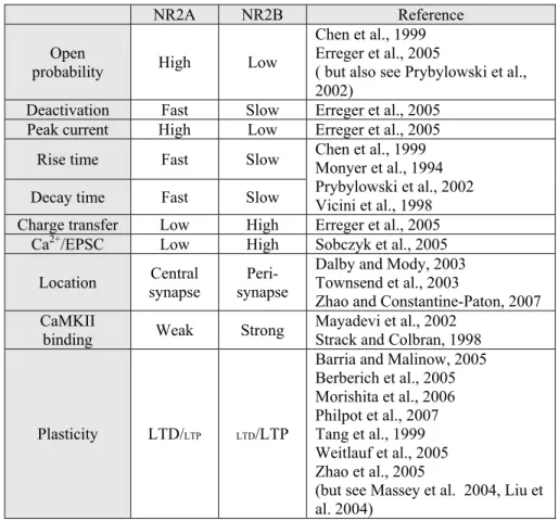
Table 1-1 Comparison of NR2A and NR2B-containing NMDARs 37 Figure 1-1 Hypothetical model of synaptic plasticity regulation by 38
NMDAR subunits
Figure 1-2 Activity-dependent regulation of NR2A and NR2B 39
Chapter 2
Figure 2-1 Visual deprivation in adult mice fails to modify NMDAR 69 EPSCs evoked by single pulses.
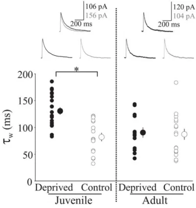
Figure 2-2 Visual deprivation in adult mice alters the temporal 70 summation of NMDAR EPSCs evoked by burst stimulation in the visual cortex
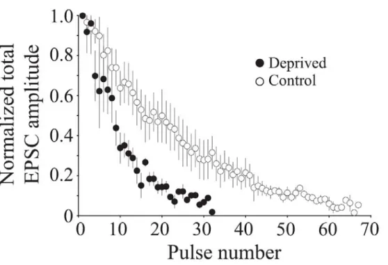
Figure 2-3 Visual deprivation increases the rate of synaptic depression 71 in adult mice
Figure 2-4 Visual deprivation increases the rate of neurotransmitter 72 release in adult mice
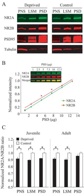
Figure 2-5 Visual deprivation in juvenile mice alters the NR2A/B ratio 73 in the postsynaptic density (PSD), but only in post-nuclear supernatant (PNS) and lysed synaptic membrane (LSM) fractions in adult mice
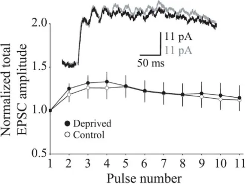
Figure 2-6 Minimal stimulation fails to reveal deprivation-induced 74 differences in the temporal summation of
NMDAR-mediated currents
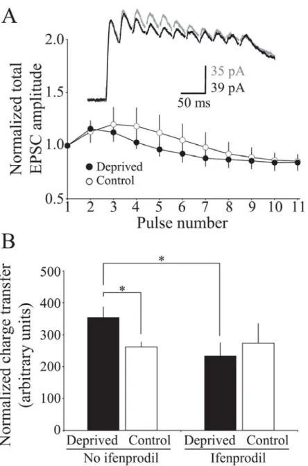
Figure 2-7 The enhanced temporal summation of NMDAR EPSCs in 75 deprived adult cortex can be blocked by acute administration of ifenprodil, an NR2B specific NMDAR antagonist
Chapter 3
Figure 3-1 Reduced functional maturation of neocortical synapses in 92
AS mice
Figure 3-2 Sensory experience augments excitatory synaptic 93 transmission in the neocortex of WT mice, but not AS mice Figure 3-3 Synaptic plasticity is impaired bidirectionally in the 94
neocortex of AS mice
Figure 3-4 Sensory deprivation restores neocortical plasticity in AS 95 mice
Supplemental figure 3-1 Immunohistochemical analysis of Ube3A expression in 96 the visual cortex from young (P24) WT, Ube3am-/p+, and
Ube3a KO mice
Supplemental figure 3-2 The absence of Ube3A causes severe deficits in 97 bidirectional synaptic plasticity in the visual cortex of
adult mice
Supplemental figure 3-3 Developmental loss of hippocampal LTP in AS mice 98 Supplemental table 3-1 Passive membrane properties of layer 2/3 pyramidal 99
neurons in which mEPSC’s are recorded
Chapter 4
LIST OF ABREVIATIONS
ACSF artificial cerebrospinal fluid AMPAR AMPA-type glutamate receptors
APV (2R)-amino-5-phosphonovaleric acid
AS Angelman syndrome
BDNF brain-derived neurotrophic factor CaMKII calcium/calmodulin kinase II CRE cAMP response element
CSPGs Chondroitin sulphate proteoglycans DIV days in vitro
DR dark-reared
ECM extracellular matrix
ER endoplasmic reticulum
fEPSP field excitatory postsynaptic potential FMRP fragile X mental retardation protein GABAARs GABAA receptors
GABAB3 β subunit of GABAA receptor
HEPES 4-(2-hydroxyethyl)-1-piperazineethanesulfonic acid
LSM lysed synaptosomal membrane
LTD long-term depression
LTP long-term potentiation
MD monocular deprivation MeCP2 methyl-CpG-binding protein mEPSC miniature excitatory post synaptic currents mGluR metabotropic glutamate receptor
MHCI major histocompatibility complex class I
Mib2 Mind bomb 2
MYCBP2 myc binding protein 2
NMDAR NMDA-type glutamate receptors NR normally-reared
NR1 NMDA-type glutamate receptor subunit 1 NR2A NMDA-type glutamate receptor subunit 2A NR2B NMDA-type glutamate receptor subunit 2B NR2C NMDA-type glutamate receptor subunit 2C NR2D NMDA-type glutamate receptor subunit 2C NR3A NMDA-type glutamate receptor subunit 3A NR3B NMDA-type glutamate receptor subunit 3B
P postnatal day
PBS phosphate-buffered saline
PirB Paired-immunoglobulin-like receptor B
PNS Post-nuclear supernatant
PSD postsynaptic density
Ras-GRF1 Ras-guanine nucleotide-releasing factor 1 RPM-1 regulator of presynaptic morphology
RTT Rett Syndrome
SAP102 synapse-associated protein 102
SDS-PAGE sodium dodecyl sulfate polyacrylamide gel electrophoresis
Ube3Am-/p+ maternally-deficient Ube3A heterozygous mouse (AS model mouse) Ube3Am+/p- paternally-deficient Ube3A heterozygous mouse
VEP visually evoked potential
Chapter 1
1.1. Regulation of NMDA receptor subunit expression and its implications for LTD, LTP, and metaplasticity
This was written as a review to be submitted for publication.
1.1.1. Abstract
NMDA-type glutamate receptors (NMDARs) mediate many forms of synaptic plasticity.
Two main regulatory subunits of NMDARs are NR2A and NR2B. In the neonatal
neocortex NR2B-containing NMDARs predominate, and sensory experience facilitates a
developmental switch of NMDARs from containing NR2B to predominantly containing
NR2A. In this review, I clarify roles of NR2 subunits in synaptic plasticity. While the
physiological importance of a shift in the ratio of NR2A and NR2B subunits is multifold, I
argue that a primary role of this shift is to control the threshold, rather than determining the
direction, for modifying synaptic strength. I also discuss recent studies that illuminate the
mechanisms regulating NR2 subunits, and I suggest that the NR2A/NR2B ratio is
regulated both locally at individual synapses and globally in a cell-wide manner. Finally, I
use the visual cortex as a model system to illustrate how activity-dependent modifications
in the NR2A/NR2B ratio may contribute to the development of cortical functions.
1.1.2. Introduction
NMDARs and AMPA-type glutamate receptors (AMPARs) are key mediators of
excitatory synaptic transmission in the brain. Both NMDARs and AMPARs are
glutamate-gated cation channels that convert a chemical signal (glutamate released from
though the receptors). Most NMDAR subtypes are unique in that their opening requires
both presynaptic transmitter release and strong postsynaptic membrane depolarization
(Mayer et al., 1984; Nowak et al., 1984). This coincidence detection arises because
NMDARs are typically blocked by Mg2+ at resting membrane potentials and can only be
activated when there is both receptor binding of glutamate and sufficient depolarization to
remove the Mg2+ block of the receptor (Mayer et al., 1984; Nowak et al., 1984). NMDARs
are permeable to Na+, K+, and Ca2+ ions, the latter of which acts as a second messenger to
modify synapses. These receptor properties insure input specificity of Ca2+-dependent
synaptic modifications by NMDARs.
Two paradigmatic examples of changes in synaptic strength are long-term depression
(LTD) and long-term potentiation (LTP), which are induced in a variety of brain regions
with diverse stimulation protocols (Malenka and Bear, 2004). Many forms of LTD and
LTP require NMDAR activation. Stimulations that induce NMDAR activation and
subsequent Ca2+ influx trigger a cascade of events to express LTD or LTP. Those events
include AMPAR removal from, or insertion into, postsynaptic membranes, respectively
(Malenka and Bear, 2004) and changes in spine morphology (Matsuzaki et al., 2004; Zhou
et al., 2004). The direction of the plasticity (weakening or strengthening) is controlled
largely by the kinetics and amount of Ca2+ influx through NMDARs. In the case of
frequency-dependent forms of synaptic plasticity, the magnitude and time course of Ca2+
entry is determined by the frequency of the conditioning stimulation given to axonal fibers.
For example, 100 Hz stimulation of axonal fibers for 1-3 seconds induces rapid and robust
0.5-5 Hz stimulation lasting for 5-30 minutes allows a smaller magnitude of Ca2+ entry
through NMDARs over a longer time course leading to LTD (Dudek and Bear, 1992). The
level of Ca2+ influx through NMDARs is determined in part by the level of postsynaptic
membrane depolarization, as this determines the extent that NMDARs are relieved from
Mg2+ block. Thus, even with low frequency stimulation, LTP can be induced if the
postsynaptic cells are held at depolarized membrane potentials (Kelso et al., 1986).
NMDARs are thought to consist of four subunits: two obligatory NR1 subunits and two
regulatory subunits that can be NR2A→D, or NR3A or B. The precise combination of
NMDAR subunits determines the functional properties of the NMDAR channels
(Cull-Candy and Leszkiewicz, 2004). Additional heterogeneity of NMDAR functions can arise
through alternative splicing. For example, NR1 and NR2D subunits undergo alternative
splicing to yield eight and two splice variants, respectively. Both the NMDAR subunit
composition (Chen et al., 2000; Liu et al., 2004b; Nase et al., 1999; Quinlan et al., 1999a;
Roberts and Ramoa, 1999) and the alternative splicing of NR1 subunits (Laurie and
Seeburg, 1994; Prybylowski and Wolfe, 2000) change during development. NR2A and
NR2B subunits, which predominate NMDARs in the forebrain, undergo a particularly
well-characterized developmental shift in the cortex. NR2B subunits are abundant in the
early postnatal brain, and NR2A levels increase progressively with development (Quinlan
et al., 1999a; Roberts and Ramoa, 1999; Sheng et al., 1994). Sensory deprivation retards
the NR2B→NR2A shift in NMDAR composition (Liu et al., 2004b; Nase et al., 1999;
Quinlan et al., 1999a; Roberts and Ramoa, 1999), suggesting this subunit change is guided
In this review, I will discuss three fundamental questions regarding the NR2A and NR2B
subunits. First, what are the molecular bases for the activity-dependent regulation of the
NR2A/NR2B ratio? Second, what are the roles of NR2A and NR2B in LTD and LTP?
Third, what is the functional consequence of the NR2A/NR2B ratio change in synaptic
plasticity in vitro and in vivo? This review is not meant to be comprehensive, but rather is
meant to highlight recent literature, to address controversies in the field, and to put forth
one viewpoint about the importance of the developmental changes in NMDAR subunit
composition. Readers are directed to other recent reviews for a more in-depth perspective
on the functions and regulation of NMDARs (Cull-Candy and Leszkiewicz, 2004; Kopp et
al., 2007; Lau and Zukin, 2007).
1.1.3. Characteristics of NR2A and NR2B subunits
Among the six regulatory subunits of NMDARs, NR2A and NR2B have been extensively
studied because they are broadly expressed in the brain, predominate in the postnatal
cortex, and are believed to play important roles in synaptic plasticity. NR2A and NR2B
subtypes of NMDARs are present as either di-heteromers (NR1/NR2A or NR1/NR2B) or
tri-heteromers (NR1/NR2A/NR2B). A recent biochemical study involving serial
immunoprecipitation from hippocampal lysates in young rats at P42 estimated that 60–
70% of NR2A and 70–85% of NR2B subunits were associated in NR1/NR2A or
NR1/NR2B di-heteromeric complexes, and one third of NMDARs are NR1/NR2A/NR2B
tri-heteromers (Al-Hallaq et al., 2007). It is likely that NMDARs in a single spine contain
currents by NR2B-specific antagonist, ifenprodil, are different among spines (Sobczyk et
al., 2005). NR2A and NR2B differ in channel kinetics, synaptic localization, and protein
binding partners, all of which are expected to influence the induction of synaptic plasticity
(Table 1-1), elaborated as follows:
Channel kinetics
At a macroscopic level, NR1/NR2A di-heteromeric channels exhibit faster rising and
decaying currents than NR1/NR2B di-heteromeric channels (Chen et al., 1999; Monyer et
al., 1994; Prybylowski et al., 2002; Vicini et al., 1998). NR1/NR2A/NR2B tri-heteromeric
channels reveal intermediate decay time courses (Vicini et al., 1998). The difference in the
decay kinetics arises from their single channel behaviors. That is, NR1/NR2A channels
have higher open probability and faster deactivation than NR1/NR2B channels (Chen et al.,
1999; Erreger et al., 2005) (but also see (Prybylowski et al., 2002)). Therefore, in response
to glutamate release, NR1/NR2A channels tend to open and close earlier than NR1/NR2B
channels, resulting in the faster rise and decay times observed macroscopically for
NR2A-containing NMDARs (Chen et al., 1999; Erreger et al., 2005). Although NR1/NR2B
channels may have lower peak currents, they carry about two-fold more charge than
NR1/NR2A channels (Erreger et al., 2005). This occurs because deactivation of
NR1/NR2B receptors is slow enough to compensate for their lower open probability
(Erreger et al., 2005). Moreover, Ca2+ imaging studies suggest that NR2B-containing
NMDARs carry more Ca2+ per unit of current than NR2A-containing NMDARs (Sobczyk
et al., 2005). Therefore, NR1/NR2B channels may carry a greater Ca2+ charge than
However, I stress that this viewpoint remains highly speculative, as this interpretation
awaits studies both that directly measure Ca2+ responses in isolated NR2A-only or
NR2B-only synapses and that more accurately assess and quantify the open probability statistics
of NR1/NR2B and NR1/NR2A receptors in mammalian neurons (see (Prybylowski et al.,
2002)).
Synaptic localization
NMDARs are found both at synaptic and extrasynaptic sites including the cell soma and
dendritic shaft. The understanding of these studies are, however, complicated by the
varied use of terminology to describe different cellular compartments. In this review, I
define the compartments of the neuronal surface as follows: the central part of postsynapse
= the area activated by spontaneous neurotransmitter release; the postsynapse = area
activated by action potential driven (evoked) neurotransmitter release; perisynapse =
region of the postsynaptic activated only by glutamate spillover from the synaptic cleft
arising from elevated levels of transmitter release driven by trains of action potentials or
arising from single action potentials in the presence of glutamate transporter inhibitors;
extrasynapse = area activated not by synaptic stimulations but by chemical stimulation
such as bath application of NMDA after blockage of synaptic NMDARs.
NR2A-containing NMDARs are thought to be concentrated in the central part of the
postsynapse in the adult brain. This view has arisen because, in rat dentate gyrus granule
cells, NMDAR-mediated miniature excitatory post synaptic currents (mEPSCs) reveal
Moreover, spontaneous synaptic events fail to activate NMDAR currents in the midbrain
of NR2A knockout mice, while evoked synaptic activity can drive NMDAR currents in the
absence of NR2A (Townsend et al., 2003; Zhao and Constantine-Paton, 2007). Thus,
miniature synaptic transmission primarily activates NR2A-containing NMDARs, whereas
action potential driven synaptic transmission engages both NR2A and NR2B-containing
NMDARs.
Subunit complements of NMDARs at extrasynaptic sites are controversial. Whereas some
studies suggest that NR2B-containing NMDARs are most prevalent at extrasynaptic sites
(Scimemi et al., 2004; Stocca and Vicini, 1998; Tovar and Westbrook, 1999), other studies
suggests that both NR2A- and NR2B-containing NMDARs exist extrasynaptically
(Mohrmann et al., 2000) and the ratio of the two subtypes is comparable to that of synaptic
NMDARs (Thomas et al., 2006). While the subunit composition of NMDARs at
extrasynaptic sites remains controversial, it is clear that synaptic and extrasynaptic
NMDARs couple to distinct intracellular signaling pathways (Ehlers, 2003; Hardingham et
al., 2002; Ivanov et al., 2006). However, because extrasynaptic NMDARs are activated
only by non-physiological stimulation such as bath application of NMDA or under
pathological conditions, and are unlikely to be activated under basal conditions (Herman
and Jahr, 2007), the subunit complement of extrasynaptic NMDARs may be irrelevant to
Protein interaction
To induce NMDAR-dependent LTD and LTP, downstream signaling pathways are tightly
coupled to NMDARs. Hence, proteins interacting with NMDAR subunits are important
determinates for the direction of synaptic plasticity. NR2A and NR2B interact with
different proteins intracellularly (reviewed in (Kennedy et al., 2005)). NR2B has many
unique or preferential binding partners. For example, NR2B interacts directly with
Ras-guanine nucleotide-releasing factor 1 (Ras-GRF1) (Krapivinsky et al., 2003), although
whether this interaction is occurring at synapses needs to be shown. NR2B is also
indirectly linked to synaptic Ras GTPase activating protein (RasGAP), presumably through
synapse-associated protein 102 (SAP102) (Kim et al., 1998). One of the most important
NMDAR binding partners is CaMKII, which has a well-documented role in the induction
of LTP (reviewed in (Lisman et al., 2002)). CaMKII binds with high affinity to NR2B
subunits (Leonard et al., 1999; Strack and Colbran, 1998; Strack et al., 2000), and, to a
much lesser extent, with NR2A subunits (Lisman et al., 2002). Ca2+ that enters through
NMDARs associates with a Ca2+ binding protein, calmodulin, and the Ca2+/calmodulin
complex interacts with and activates CaMKII. Activated CaMKII binds strongly to NR2B
(Strack and Colbran, 1998), allowing CaMKII to remain active even after dissociating
from Ca2+/calmodulin (Bayer et al., 2001). It has been shown that CaMKII activation and
its association to NR2B are required for LTP induction (Barria and Malinow, 2005).
NR2A also appears to have some unique associations with signaling molecules. A recent
study suggests that NR2A co-immunoprecipitates with neuronal nitric oxide synthase more
the association raises the interesting possibility that NO-mediated presynaptic forms of
LTP and LTD (Haghikia et al., 2007; Prast and Philippu, 2001; Zhang et al., 2006) may be
preferentially linked to NR2A-mediated signaling pathways.
Both NR2A and NR2B possess PDZ-binding motifs in their c-terminus. Through the
PDZ-binding motifs, they interact with membrane-associated guanylate kinase (MAGUK)
family of synaptic scaffolding proteins that in turn associate with important synaptic
signaling molecules and tether NMDARs to intracellular signaling pathways (Kennedy,
2000). The differential interaction of NR2A and NR2B subunits to MAGUKs is
controversial. It was once believed that, whereas NR2A preferentially bound to
postsynaptic density protein-95 (PSD-95), NR2B predominantly interacts with SAP102
(Sans et al., 2000; Townsend et al., 2003). Moreover, these interactions were thought to
control distinct synaptic localization of NR2A and NR2B (Townsend et al., 2003).
However, a recent biochemical study using a serial immunoprecipitation suggests that
MAGUK proteins such as PSD-95 and SAP102 interact with di-heteromeric NR1/NR2A
and NR1/NR2B receptors at comparable levels (Al-Hallaq et al., 2007). Thus, additional
studies are needed to clarify the association of NMDAR subunits with MAGUK family
members and what effects these associations may have on receptor localization and on
plasticity signaling pathways.
1.1.4. Activity-dependent modulation of NR2A/NR2B ratio
The NR2A/NR2B ratio is not fixed at synapses, rather, it changes with development and
al., 1999; Quinlan et al., 1999a; Roberts and Ramoa, 1999) as well as plasticity (Bellone
and Nicoll, 2007). This change may help to optimize the threshold for inducing synaptic
plasticity at different developmental points and/or under different sensory environments
(discussed later). An important question is how sensory experience and neuronal activity
regulate the ratio of the two subunits. Recent studies in cultured neurons have revealed
differential regulation of NR2A and NR2B subunits at various points in their synthesis,
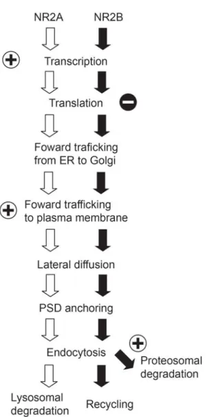
trafficking, and degradation. Here I describe mechanisms that control NR2A and NR2B at
each step and how these mechanisms can be regulated by neuronal activity (Fig.1-2).
Transcription and translation
Much of what we understand of the transcriptional and translational regulation of
NMDARs comes from studies of cultured neurons, and such studies reveal important
differences based on the maturity of neurons in culture. The early developmental increase
(days in vitro (DIV) 9 to 15) in the NR2A/NR2B ratio is largely due to an increase in
NR2A mRNA (Hoffmann et al., 2000), suggesting that the developmental shift is
controlled at a transcriptional level (but also see (Follesa and Ticku, 1996)). This increase
in NR2A levels can be suppressed by blockers of NMDARs
((2R)-amino-5-phosphonovaleric acid; APV), or voltage-gated calcium channels (Nifedipine). NR2B
mRNA levels in immature cultures are insensitive to APV treatment. These data indicate
that the early developmental increase in the NR2A/NR2B ratio is caused primarily by an
increase in NR2A levels driven by activity-dependent activation of NMDARs. These
observations also suggest that NR2A and NR2B transcripts are regulated by distinct Ca2+
subunits is currently unknown. However, analysis of transcriptional regulatory sequences
of NR2A revealed that a sequence between -1253 and -1180 in the up-stream region of
NR2A contains a cAMP response element (CRE)-like element and is necessary for the
developmental increase in NR2A (Desai et al., 2002a). Although it has not yet been tested
if this coding sequence is required for activity-dependent NR2A transcription, the possible
regulation of NR2A by CRE gives rise to the interesting possibility that the
activity-dependent developmental increases in NR2A may be mediated by the
NMDAR/PKA/CREB pathway.
In older cortical cultures (DIV22-30), responses of NR2A and NR2B to APV treatment is
quite different to that observed in younger cultures. One day of NMDAR blockade by
APV increases NR2B protein without affecting the level of NR2A protein, and this NR2B
increase is largely blocked by the translational inhibitors, cycloheximide and anisomycin
(Chen and Bear, 2006). Therefore, NMDAR activity in more mature neurons may
tonically suppress NR2B translation, and brief (one day) blockade of NMDARs may be
sufficient to relieve this suppression. Thus, neuronal activity appears to facilitate
transcription of NR2A in immature neurons, while activity and NMDAR activation
suppresses translation of NR2B in more mature neurons. Together, both these mechanisms
increase the NR2A/NR2B ratio in response to enhanced neural activity.
Forward trafficking
NMDAR subunits are assembled in the endoplasmic reticulum (ER) (Qiu et al., 2005),
and NR2B contain an ER export signal (HLFY) at the base of their c-terminus (Hawkins et
al., 2004). Because of the ER export signal on NR2 subunits, overexpression of NR2
subunits enhances the surface delivery of NMDARs by overcoming an ER retention signal
located in NR1 subunits (Scott et al., 2001; Standley et al., 2000). In this regard, the
increase in NR2B levels by NMDAR blockade seen in more mature neurons (Chen and
Bear, 2006) could also facilitate the surface delivery of NMDARs. Consistent with this
idea, chronic inactivation of NMDARs has been shown to increase synaptic NR1 (Crump
et al., 2001; Rao and Craig, 1997) and synaptic NR2B (Ehlers, 2003).
The activity-dependent mechanisms are distinct for regulating the synaptic delivery of
NR2A and NR2B (Barria and Malinow, 2002). Synaptic accumulation of
NR2A-containing NMDARs requires glutamate binding to NMDARs, whereas NR2B-NR2A-containing
NMDARs can accumulate at synapses regardless of synaptic activity level or ligand
binding (Barria and Malinow, 2002). These results provide further evidence that synaptic
activity preferentially drives NR2A-containing NMDARs to the synapse. Thus, neuronal
activity not only increases transcription of NR2A (Hoffmann et al., 2000) but also
facilitates synaptic delivery of NR2A-containing NMDARs over NR2B-containing
NMDARs. This differential regulation of NR2A and NR2B subunits ensures an
activity-dependent increase in the NR2A/NR2B ratio. The molecular mechanisms underlying this
Surface diffusion
Although it has been traditionally thought that NMDARs are relatively stable and
immobile at the cell surface, recent electrophysiological and imaging studies suggest that
NMDARs are highly mobile. By taking advantage of the irreversible open channel blocker,
MK-801, Tovar and Westbrook have shown in immature cultured hippocampal neurons
that NMDAR-mediated EPSC can quickly recover following MK-801 block (Tovar and
Westbrook, 2002). This recovery was shown to be due to lateral diffusion, by which 65%
of synaptic NMDARs exchange in less than 7 minutes. NMDAR lateral mobility has also
been observed by single molecular tracking (Groc et al., 2004, 2006, 2007). These studies
reveal that NMDARs are highly mobile both at synaptic and extrasynaptic membranes.
Importantly, the surface mobility of NMDARs appears to change with development in a
subunit composition specific manner (Groc et al., 2006). For example, NR2A-containing
NMDARs are less mobile than NR2B-containing NMDARs, and the synaptic residency
time of NR2B-containing NMDARs decreases over development. This decrease in
synaptic dwell time of NR2B-containing NMDARs is mediated by Reelin, an extracellular
matrix protein (Groc et al., 2007). Interestingly, the lateral mobility of NMDARs is
insensitive to acute changes in neuronal activity levels, and this is unlike AMPARs, whose
diffusion is bidirectionally controlled by neuronal activity (Groc et al., 2004). It is
unknown, however, if chronic activity manipulations for several days affect NMDAR
surface mobility and thereby contributes in a homeostatic fashion to differential synaptic
accumulations of NR2 subunits. Lastly, these studies were performed primarily in
Therefore, surface mobility of these receptors need to be examined in more intact
preparations.
Endocytosis
The ability of NMDARs to undergo endocytosis decreases with age (Roche et al., 2001).
This developmental change is likely a consequence of the fact that NR2B subunits
experience more robust endocytosis than NR2A subunits (Lavezzari et al., 2004), and the
proportion of NR2B subunits decreases with age. How are the distinct endocytic
mechanisms of NR2A and NR2B controlled? Both NR2A and NR2B contain a PDZ
binding motif (ESDV) at the c-terminus (Lin et al., 2004; Prybylowski et al., 2005), and
this helps to tether the subunits to the postsynaptic density through binding to MAGUK
proteins, such as PSD-95 and SAP102. Interestingly, PDZ binding is required for synaptic
localization of NR2B, but not NR2A (Lin et al., 2004; Prybylowski et al., 2005). The
c-terminus of both NR2A and NR2B contain the endocytic signals LL (Lavezzari et al., 2004)
and YEKL (Roche et al., 2001), respectively, which bind the AP2 clathrin adaptor protein
to initiate clathrin-dependent endocytosis. Additional regulation of NR2B endocytosis is
endowed through phosphorylation of tyrosine 1472 in YEKL by the Src-family kinase Fyn
(Prybylowski et al., 2005). This phosphorylation protects YEKL from AP2 binding, and
consequently limits NR2B subtypes from clathrin-dependent endocytosis. Thus,
localization of NR2B-containing NMDARs to the postsynaptic density is controlled by
Fyn and an as yet unidentified phosphatase which removes the phosphate from YEKL.
Such phosphorylation-dependent regulation of NR2A endocytosis has not yet been
NR2 subunits can also dictate the fate of endocytosed NMDARs. Both NR2A and NR2B
contain proximal motifs that direct NMDARs to the late enodosome/lysosome, where the
receptors are degraded. However, NR2B subunits possess an additional proximal motif,
which can help drive the receptors along a recycling pathway (Scott et al., 2004). As a
consequence, endocytosed NR2B-containing NMDARs are preferentially recycled back to
the plasma membrane surface, whereas NR2A di-heteromeric NMDARs are more likely to
be degraded when endocytosed.
Whether activity-dependent endocytosis of NMDARs (Morishita et al., 2005) is regulated
in a subunit-specific manner has not been examined. Given the recent finding of the rapid
increase in the NR2A/NR2B ratio upon LTP induction in hippocampus (Bellone and
Nicoll, 2007), one may speculate that LTP-inducing stimulation preferentially induces
endocytosis of NR2B-containing NMDARs and/or insertion of NR2A-containing
NMDARs. It is possible, however, that the change is mediated by posttranslational
modifications.
Degradation
By changing neuronal activity levels, NR2A and NR2B levels can be bidirectionally
regulated (Ehlers, 2003). This activity-dependent regulation can be prevented by
proteasome inhibitors (Ehlers, 2003), indicating that degradation pathways are important
for the activity-dependent regulation of NMDAR subunit levels. A recent study showed
Mib2 directly interacts with NR2B and ubiquitinates it when tyrosine 1472 in YEKL is
phosphorylated by Fyn. Because neuronal activity facilitates tyrosine phosphorylation of
NR2B and Mib2 binding (Jurd et al., 2007), Mib2 may play an important role in the
activity-dependent regulation of NR2 subunits. In an apparent paradox, phosphorylation at
tyrosine 1472 both increases the proteasomal degradation of NR2B but limits NR2B
endocytosis (Prybylowski et al., 2005). Thus, it will be interesting to discern how synaptic
localization and Mib2-mediated degradation of tyrosine 1472 phosphorylated-NR2B are
balanced.
In conclusion, NR2A and NR2B undergo differential activity-dependent regulation at
various points of the subunit turnover. Therefore, multiple layers of regulation contribute
to the experience-dependent modifications of the synaptic NR2A/NR2B ratio.
1.1.5. Developmental and experience-dependent modification of the NR2A/NR2B ratio in vivo
Developmental regulation
In many parts of the CNS, including the brain stem, hippocampus, and neocortex, the
NR2A/NR2B ratio increases during early postnatal development (Barth and Malenka, 2001;
Chen et al., 2000; Hestrin, 1992; Liu et al., 2004b; Nase et al., 1999; Quinlan et al., 1999a;
Roberts and Ramoa, 1999; Yoshimura et al., 2003). This change can occur both at the
mRNA (Liu et al., 2004b; Nase et al., 1999) and protein levels (Chen et al., 2000; Quinlan
NR2A-containing NMDARs, rather than a decrease in NR2B subunits, is believed to be the
primary factor contributing to the observation that the decay of NMDAR-mediated
currents becomes faster with development (Carmignoto and Vicini, 1992; Quinlan et al.,
1999a; Yoshimura et al., 2003). However, an increase in the NR2A/NR2B ratio may not
be the sole factor that regulates NMDAR decay kinetics. For example, changes in
NMDAR phosphorylation and the expression of NR1 splice variants also regulate
NMDAR current kinetics (Lieberman and Mody, 1994; Rumbaugh et al., 2000; Tong et al.,
1995). In some regions, the largest developmental decline in NMDAR-mediated current
duration can actually precede the most profound increase in the NR2A/NR2B ratio (Barth
and Malenka, 2001). Moreover, a mild but significant developmental decrease in
NMDAR-mediated current decay time is still observed in NR2A knockout mice (Lu et al.,
2001). Nonetheless, studies in NR2A knockout mice demonstrate that the upregulation of
NR2A underlies the largest developmental changes in NMDAR current duration (Fagiolini
et al., 2003).
Experience-dependent regulation
In some parts of the neocortex, including the primary visual cortex, the elevation of the
NR2A/NR2B ratio is dependent upon the level of neuronal activity (Liu et al., 2004b; Nase
et al., 1999; Quinlan et al., 1999a; Roberts and Ramoa, 1999). Sensory deprivation, such
as dark-rearing, reduces the developmental shift in the NR2A/NR2B ratio (Carmignoto and
Vicini, 1992; Philpot et al., 2001a; Quinlan et al., 1999a). This visual
experience-dependent control in the NR2A/B ratio is not restricted to young animals. I and others
ratio in the visual cortex (He et al., 2006; Yashiro et al., 2005). The present evidence
suggests, however, that the ratio change may be restricted to perisynaptic sites in adults,
unlike the experience-dependent modifications in NR2A/NR2B that can occur at synapses
in young rodents (Yashiro et al., 2005). This suggests that NR2A/B protein expression
level can be modified throughout development by visual experience, but the ability to
control synaptic NMDARs is restricted to young animals.
How is the NR2A/NR2B ratio controlled by visual experience? There are currently
conflicting reports on how visual experience regulates NR2A and NR2B at the mRNA
level. For example, one comprehensive microarray analysis of mouse visual cortex
revealed that both NR2A and NR2B mRNA are elevated in rodents reared in complete
darkness until P27 compared to those in age-matched light-reared animals (Tropea et al.,
2006). In contrast, single cell RT-PCR analysis of mRNA isolated from neurons in layer 4
of rat visual cortex showed that dark-rearing until P20 significantly retards the
developmental increase in NR2A mRNA (Nase et al., 1999). These differences may be a
subtle consequence of the ages studied or the techniques employed.
There is general agreement that visual experience increases the NR2A/NR2B ratio at the
protein level, although there is some disagreement as to whether this change is due to an
increase in NR2A and/or decrease in NR2B. Dark-rearing of rats until 6 weeks of age does
not change NR2B protein levels in synatoneurosome fractions, although this manipulation
significantly reduces NR2A protein levels starting at 3 weeks of age compared to
raises NR2A protein levels within one hour (Quinlan et al., 1999b). Reciprocally, 5 weeks
of dark-rearing reduces NR2A protein levels without affecting NR2B protein levels in cats
(Chen et al., 2000). Moreover, an immunohistochemical analysis reports that the NR2A
protein reductions occur in all layers of the visual cortex in dark-reared rats (Tongiorgi et
al., 2003). Collectively, these results indicate that the NR2A is the target of sensory
experience-dependent regulation, but NR2B is not. A recent study suggests, however, that
visual experience may regulate both NR2A and NR2B levels, although whether visual
deprivation enhances NR2B or retards NR2A expression is regulated tightly depending on
the age of onset of the visual deprivation (Chen and Bear, 2006). Therefore, both NR2A
and NR2B protein levels can be targets of sensory experience-dependent regulation, and
the two proteins are inversely regulated by experience.
I suggest that the visual experience-dependent increase in the NR2A/NR2B ratio is
controlled by several cellular processes. The developmental NR2A increase is likely
driven by activity-dependent facilitation in transcription (Hoffmann et al., 2000). The
NR2B level may be chronically attenuated by activity-dependent suppression of its
translation (Chen and Bear, 2006). Moreover, neuronal activity may limit NR2B levels by
facilitating its degradation by Mib2-mediated ubiquitin proteasome system (Jurd et al.,
2007). In the absence of visual experience, NR2A mRNA synthesis is slowed, the
suppression of NR2B translation is relieved, and NR2B degradation is attenuated, all
processes that result in a net decrease in the NR2A/NR2B ratio. Detailed biochemical
studies are required to dissect the visual experience-induced transcriptional and
1.1.6. Are activity-dependent changes in the NR2A/NR2B ratio input-specific or global?
I have described that the NR2A/NR2B ratio changes in response to neuronal activity both
in vitro and in vivo. An important question is whether the NR2A/NR2B ratio is controlled at the level of individual synapses or in a global, cell-wide manner. This question is
important, because it predicts whether the inactivation of a subset of synapses could result
in a global reduction in the NR2A/NR2B ratio. In contrast, a global (cell-wide) change in
the synaptic levels of NR2A/NR2B would alter the properties of synaptic plasticity
throughout a neuron, and such changes obviously have different consequences on dynamic
modifications of synapses across a neuronal network.
Most studies that have addressed the regulation of NR2A/NR2B have employed global
manipulations of synaptic function, such as chronic treatment with APV or dark-rearing.
Because these slow alterations of the NR2A/NR2B ratio are, at least in part, controlled by
transcription and translation of NR2 subunits, these slow changes are likely to be achieved
by global control of the NR2A/NR2B ratio on the neuronal surface. There is evidence,
however, that the NR2A/NR2B ratio may also be controlled in a local (input-specific)
manner. For example, the NR2A/B ratio is different between intercortical and intracortical
synapses in L5 pyramidal neurons in the cortex (Kumar and Huguenard, 2003). Moreover,
the synaptic distribution of NR2B subunits in the adult mouse hippocampus is
asymmetrical between the apical and basal dendrites of single neurons (Kawakami et al.,
2003). Such observations indicate that the NR2A/NR2B ratio must have a level of
the NR2A/NR2B ratio increases immediately after the application of LTP-inducing
stimulation in hippocampus in an input-specific manner (Bellone and Nicoll, 2007). Such
rapid changes are unlikely to be regulated at the transcriptional or translational levels.
Therefore, I hypothesize that the NR2A/NR2B ratio is regulated both at synaptic and
cellular levels. Global regulation of the NR2A/NR2B levels might regulate the threshold
of synaptic plasticity across the cell to direct the acquisition of stimulus-selective response
properties (Philpot et al., 1999, 2007), while a rapid, input-specific regulation of
NR2A/NR2B levels might limit runaway potentiation on individual synapses.
1.1.7. Roles of NR2A and NR2B in LTD and LTP
LTP
Given that NR2B-containing NMDARs reveal longer currents, carry more Ca2+ per unit of
current, and interact preferentially with CaMKII compared to NR2A-containing NMDARs,
it is tempting to speculate that NR2B subtypes are more likely to favor the induction of
LTP compared to NR2A subtypes (although see (Liu et al., 2004a)). A number of lines of
evidence support this contention, some of which are highlighted here. (1) Ifenprodil
completely blocks LTP induced by a pairing protocol in immature hippocampal slice
cultures, suggesting a critical requirement of NR2B subtypes for the induction of LTP
(Barria and Malinow, 2005). (2) Overexpression of NR2A, and a presumptive replacement
of NR2B subtypes with NR2A subtypes, attenuates the induction of LTP induced by a
pairing protocol (Barria and Malinow, 2005). (3) In the anterior cingulate cortex, an
theta-burst stimulation (Zhao et al., 2005). (4) In thalamocortical synapses in the barrel
cortex of postnatal day (P) 3-5 mice, ifenprodil blocks LTP induced by a pairing protocol
(Lu et al., 2001). (5) Genetic lesion of NR2A fails to abolish hippocampal LTP,
suggesting that NR1/NR2B di-heteromic receptors are sufficient to induce hippocampal
LTP (Berberich et al., 2005; Weitlauf et al., 2005). (6) Transgenic overexpression of
NR2B enhances hippocampal LTP (Tang et al., 1999). (7) Transient overexpression of
NR2B c-terminus, which blocks the NR2B and CaMKII interaction, attenuates
hippocampal LTP (Zhou et al., 2007). Thus, in various regions of the brain,
NR2B-containing NMDARs help to promote the induction of LTP induced by a variety of
stimulation protocols.
Contrary to these findings, it has been reported that NR2A-containing, but not
NR2B-containing, NMDARs mediate LTP (Liu et al., 2004a). Support for this hypothesis comes
from experiments utilizing a new pharmacological tool, NVP-AAM077, which was
believed to specifically block NR2A-containing NMDARs. In these experiments,
NVP-AAM077 was shown to block hippocampal LTP (Liu et al., 2004a). Similar observations
were made using this antagonist in the adult perirhinal cortex (Massey et al., 2004).
However, the specificity of NVP-AAM077 has been questioned. While NVP-AAM077
was reported to be 100 times more selective for NR1/NR2A channels than NR1/NR2B
channels using a cell line exogenously expressing human NMDARs (Liu et al., 2004a),
this antagonist is only 6-12 fold more effective for NR1/NR2A channels than NR1/NR2B
channels in rodents (Feng et al., 2004; Neyton and Paoletti, 2006). The low selectivity of
than 20% of the NMDAR current in hippocampal slices of NR2A-null mice even at 50 nM
concentration (Berberich et al., 2005). At this concentration less than 80% of
NMDAR-mediated currents are obstructed in a cell line expressing rodent NR1/NR2A channels.
These findings suggest that specificity of NVP-AAM077 is not sufficient to determine the
role of NR2A in synaptic plasticity.
While I suggest that the induction of LTP is more likely to be favored with NR2B subtypes
than NR2A subtypes, I caution that the roles of these receptors must be carefully
considered within a developmental and regional context. Moreover, NMDARs are not the
sole determinants for inducing plasticity. For example, both signaling molecules and
inhibitory inputs are important contributors to the induction of plasticity (Choi et al., 2002;
Steele and Mauk, 1999), and these factors clearly change across development and region
(Chattopadhyaya et al., 2004; Jiang et al., 2005; Morales et al., 2002 ; Yasuda et al., 2003).
LTD
Which subunits mediate LTD? Unlike the previous demonstration of complete block of
hippocampal LTD by ifenprodil (Liu et al., 2004a), studies in three independent
laboratories consistently found that hippocampal LTD is insensitive to NR2B blockade by
ifenprodil (Morishita et al., 2006). Another study even suggests that ifenprodil enhances
the induction of LTD in the hippocampus (Hendricson et al., 2002). These studies
demonstrate that the induction of LTD does not require activation of NR2B-containing
NMDARs. A caveat in these pharmacological studies are in the complex nature of
concentrations of glutamate, it may actually potentiate NMDAR currents at low glutamate
concentrations (Kew et al., 1996). Thus, ifenprodil might affect synapses differently
depending on the glutamate concentration in the synaptic cleft. Such complexities might
partially underlie observations that NR2B can be involved in synaptic weakening in some
conditions but not others. For example, in the adult perirhinal cortex, the
subunit-dependence of LTD relies on the state of the synapse (Massey et al., 2004); ifenprodil
blocks LTD that has been induced at a basal state, but the antagonist fails to block LTD
induced after synaptic potentiation (depotentiation).
Studies in mice lacking NR2A have attempted to illuminate a possible role for NR2A in
LTD. In the visual cortex of NR2A knockout mice, the standard 1 Hz stimulation protocol
(900 pulses), which induces LTD in wildtype mice, gives rise to LTP. On the other hand,
0.5 Hz stimulation (900 pulses) induces LTD in NR2A knockout mice comparable to
wildtype mice (Philpot et al., 2007). Therefore, one hypothesis is that the threshold for
inducing LTP is lowered in NR2A knockout mice, as activation of NR2B-containing
di-heteromeric NMDARs allows greater Ca2+ entry than possible through NR2A-containing
NMDARs. Such a threshold change by deleting NR2A may not be universal in the brain,
because 1 Hz stimulation induces LTD in NR2A knockout mice in the midbrain (Zhao and
Constantine-Paton, 2007). Thus, the consequences of deleting NR2A may vary depending
on age or brain region. Future studies that take advantage of either conditional NR2A
deletion and/or more specific NR2A antagonists are needed to clarify the role of NR2A in
Taken together, it seems that NR2A-containing NMDARs favor the induction of LTD by
limiting Ca2+ entry through NMDARs. Future studies examining possible interactions
between NR2A and LTD-inducing signaling components such as the PP1/PP2B pathway
may strengthen this hypothesis. Also, as mentioned previously, further work will be
necessary to determine the differences between NR2A and NR2B subtypes in open
probability, as the calcium signaling through these two receptor subtypes hinges critically
on how these receptors behave endogenously in mammalian neurons.
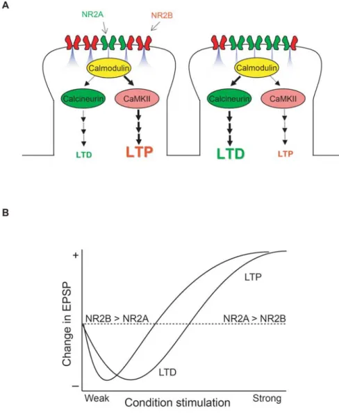
NR2A/NR2B ratio controls LTD and LTP
The NR2A/NR2B ratio changes during development (Quinlan et al., 1999a) and likely
differs dramatically among synaptic sites on dendritic trees (Kumar and Huguenard, 2003;
Sobczyk et al., 2005). How do these differences affect LTD and LTP? Given the
contributions of NR2 subunits on LTD and LTP mentioned above, it has been
hypothesized that the LTD/LTP induction threshold is determined by the ratio of
NR2A/NR2B expressed on dendritic spine surfaces (Fig.1-1). That is, if the ratio of
NR2A/NR2B is elevated, stronger stimulation (e.g. a higher stimulus frequency) would be
required to induce LTP compared to when the ratio of NR2A/NR2B is low. This
hypothesis is based on two observations; a higher NR2A/NR2B ratio limits both Ca2+ entry
through NMDARs (although see (Erreger et al., 2005)) and the accessibility of CaMKII at
the synapse. Therefore, with a high NR2A/NR2B ratio a stronger postsynaptic response is
needed to elevate Ca2+ and activate CaMKII to a level sufficient to induce LTP, whereas
weaker postsynaptic responses might suit activation of calcineurin and activate an LTD
would lower the LTP induction threshold, making it more likely that a modest response
can elevate Ca2+ and activate CaMKII to a level sufficient to induce LTP. In light of this
hypothesis, it is interesting to consider how experience-dependent changes in the
NR2A/NR2B ratio alter the LTD/LTP induction threshold (discussed below).
1.1.8. Interaction between NR2A/NR2B ratio and LTD/LTP in the visual cortex As I discussed above, the NR2A/NR2B ratio increases in many parts of the brain and this
increase is regulated by sensory experience in many regions, including the visual cortex.
Like the NR2A/NR2B ratio, properties of LTD and LTP change during development in an
experience-dependent manner (Bear, 2003). Here I discuss if the described changes in
LTD/LTP are mediated, in part, by the modification of the NR2A/NR2B ratio. I focus my
discussion onto two excitatory synaptic connections within the visual cortex, the thalamus
to layer 4 (thalamocortical) and the layer 4 to 2/3 (intracortical) connections, because these
pathways are well-studied and yet very distinct. Interestingly, although the NR2A/NR2B
ratio similarly increases in both of the excitatory connections, those synapses reveal
distinctive developmental and experience-dependent regulations of LTD and LTP.
Thalamocortical synapses
In the thalamocortical synapses of somatosensory and visual cortices, the magnitude of
both LTD and LTP diminish in the second to fourth postnatal week in rodents (Dudek and
Friedlander, 1996; Feldman et al., 1999; Jiang et al., 2007). Since the timing of the
developmental increase in the NR2A/NR2B ratio coincides with the loss of LTD and LTP
(Carmignoto and Vicini, 1992; Jiang et al., 2007; Quinlan et al., 1999b), it has been
hypothesized that the increase in the NR2A/NR2B ratio may limit the expression of
plasticity at thalamocortical synapses. This hypothesis has been challenged by a finding
that the developmental reduction in the thalamocortical LTP is shown to be conserved in
the somatosensory cortex of the NR2A knock-out mice (Lu et al., 2001). This suggests
that the developmental increase in the NR2A/NR2B ratio does not mediate the
developmental loss of thalamocortical LTP in the somatosensory cortex. Whether the
developmental loss of thalamocortical LTP is also conserved in visual cortex has not yet
been tested, but the current evidence indicates that the developmental loss of
thalamocortical LTP is not dependent solely on the NR2A/NR2B switch. For example,
while dark-rearing delays both the onset of the critical period for ocular dominance
plasticity (Mower, 1991b) and the developmental upregulation of the NR2A/NR2B ratio at
layer 4 (Carmignoto and Vicini, 1992; Quinlan et al., 1999a), this manipulation does not
delay the developmental loss of thalamocortical LTP in the visual cortex (Jiang et al.,
2007).
These data argue that the NR2A/NR2B ratio is not the sole determinant for the ability to
induce LTD and LTP at thalamocortical plasticity. However, it is still conceivable that the
relative levels of NR2A/NR2B might adjust the threshold for inducing plasticity within a
developmental time point. For example, it is possible that an increase in the NR2A/NR2B
ratio may increase the threshold for inducing LTP at thalamocortical synapses, but whether
or not LTP can be induced at all is likely a consequence of other factors including
et al., 2007), or downstream signaling molecules (Yasuda et al., 2003). Future studies that
manipulate NR2A and NR2B levels in vivo are needed to test these possibilities.
Intracortical synapses
Although there is general agreement that there is a developmental loss of thalamocortical
plasticity, it is less clear whether plasticity persists at the L4-L2/3 synapse, the first
intracortical relay. There are reports that both LTD (Kirkwood et al., 1997) and LTP
(Yoshimura et al., 2003) at the L4-L2/3 synapses diminish with development, but other
studies find that LTD (Jiang et al., 2007) and LTP (Frankland et al., 2001; Jiang et al.,
2007; Kirkwood et al., 1997) persist into adulthood at this synapse. These seemingly
contradictory findings make it impossible to determine whether observed changes in the
NR2A/NR2B ratio (Yoshimura et al., 2003) are associated with the ability to induce LTD
and LTP at this intracortical synapse. Because receptive field plasticity remains intact in
the superficial layers into adulthood (Daw et al., 1992), suggesting that LTD and LTP also
remain intact, it appears as though the developmental increase in NR2A/NR2B does not
eliminate the induction of plasticity at the L4-L2/3 synapse.
While the NR2A/NR2B ratio does not gate the absolute ability to induce LTD and/or LTP,
changes in NR2A/NR2B appear to affect the threshold for the frequency-dependent
induction of LTD/LTP. Visual experience/deprivation alters the frequency-response
relationship of LTD/LTP in the layer 4-2/3 synapses (Kirkwood et al., 1996; Philpot et al.,
2003), such that dark-rearing narrows the window of stimulus frequencies that induce LTD
Namely, previous sensory experience modifies the properties of synaptic plasticity in the
visual cortex, an effect known as “metaplasticity” (Bear, 2003). Because dark-rearing
reduces the NR2A/NR2B ratio, there is a striking correlation between the threshold for
modifying synaptic strength and the relative ratio of NR2A/NR2B. Thus, it has been
hypothesized that sensory experience slides the threshold for inducing LTP/LTD through
regulation of the NR2A/NR2B ratio (Philpot et al., 1999), with a low NR2A/NR2B ratio
favoring the induction of LTP (by lowering the plasticity threshold).
To test the hypothesis that the NR2A/NR2B ratio regulates the threshold of inducing
synaptic plasticity, a process termed metaplasticity, we have taken advantage of mice that
lack NR2A. The idea of this study was to lock the NR2A/NR2B in place, as this should
prevent experience-dependent modifications in the LTD/LTP threshold if this were
normally a consequence of changing the NR2A/NR2B ratio. we first demonstrated that the
visual experience-dependent shortening of NMDAR-current decay is absent in NR2A
knockout mice (Philpot et al., 2007), indicating that the shortening of NMDAR currents is
indeed due to an increase in the NR2A/NR2B ratio. Importantly, dark-rearing, which
normally lowers the threshold for inducing LTP in wildtype mice, failed to alter the
threshold for frequency-dependent plasticity in mice lacking NR2A. Moreover, the
threshold stimulus frequency for inducing LTP is greatly lowered in NR2A knockout mice,
such that 1Hz stimulation, which induces LTD in the wildtype mice, is sufficient to give
rise to LTP. This is consistent with the idea that a low NR2A/NR2B ratio favors the
induction of LTP. Currently the mechanism by which the NR2A/NR2B ratio alters the
NMDARs and, in its absence, NR2B lowers the plasticity threshold by promoting a greater
degree of Ca2+ entry into the postsynaptic neuron. These results clearly suggest that, at
least in developing rodents, NR2A is required for metaplasticity in the visual cortex and
strongly indicate that visual experience-dependent change in the NR2A/B ratio controls
metaplasticity by directly changing biophysical properties of postsynaptic NMDARs.
1.1.9. Roles of NR2A and NR2B in cortical functions in vivo
I have described the roles of NR2A and NR2B in synaptic plasticity in vitro, but how do
changes in these NMDAR types contribute to development of sensory systems? Here I
review the roles of the NR2 subunits in the development of two well-studied visual
functions: orientation selectivity and ocular dominance plasticity.
Orientation selectivity
Most neurons in the primary visual cortex respond vigorously to light-dark bars or edges
presented to animals at a particular range of orientations. Some degree of orientation
selectivity is innate in cortical neurons and the selectivity becomes more fine-tuned with
development. Visual experience is necessary for the proper development of orientation
selectivity, and fewer cells exhibit orientation selectivity in the absence of prior visual
experience (Fagiolini et al., 2003; White et al., 2001). Moreover, if the visual environment
is largely restricted to one orientation (by rearing in a striped cylinder), animals develop
orientation selectivity biased toward the orientation of the stripes (Sengpiel et al., 1999).
These results indicate that cortical neurons change their connectivity to respond more to
What is happening at synapses during the acquisition of enhanced stimulus selectivity?
One may hypothesize that patterned visual stimulation potentiates synapses of neurons,
which gain preferential responses to repeatedly experienced orientations by an LTP-like
mechanism. On the other hand, the same stimulation may depress synapses responding to
the non-favored orientation, by an LTD-like mechanism. The acquisition of stimulus
selective properties such as orientation selectivity are thought to require
experience-dependent modifications in the properties of synaptic plasticity (metaplasticity)
(Bienenstock et al., 1982). Thus, orientation selectivity is less likely to occur, and more
likely to be broad when it does occur, in the absence of metaplasticity. Given that the
NR2A/NR2B ratio controls visual cortex metaplasticity, one would predict that orientation
selectivity would be severely retarded if the NR2A/NR2B ratio were fixed (hence
preventing metaplasticity). Consistent with this hypothesis, the proportion of orientation
selective neurons is severely diminished in the visual cortex of NR2A knockout mice
(Fagiolini et al., 2003). It is tempting to speculate that, under normal conditions, a visual
experience-dependent increase in the NR2A/NR2B ratio mediates the establishment of
orientation selectivity by widening the window for LTD induction and strengthening
orientation-specific synaptic connections through an LTP-like mechanism.
Ocular dominance plasticity
One well-studied in vivo paradigm of synaptic plasticity is the ocular dominance shift
observed in the primary visual cortex. Classical studies demonstrate that closure of one
gain of responsiveness to the intact (non-deprived) eye (Wiesel and Hubel, 1963). Recent
studies, which investigate visually-evoked potential recordings in awake mice, reveal that
monocular deprivation induces depression of the closed eye response in the first 2 days
after MD followed by potentiation of the open eye response occurring in 5 days (Frenkel
and Bear, 2004). Is the initial depression and subsequent potentiation induced by LTD and
LTP-like mechanisms? It has been shown that monocular deprivation induces
dephosphorylation of AMPARs seen in LTD (Heynen et al., 2003). Moreover, in the
visual cortical slices prepared from monocularly deprived rats, LTD was suppressed,
suggesting that synaptic depression induced by monocular deprivation occludes LTD
(Heynen et al., 2003). Thus, the closed eye depression seems likely to involve a LTD-like
mechanism. Because the ocular dominance shift can not be reliably induced in mice
carrying a knock-in mutation in CaMKII at threonine 286, autophosphorylation of which is
required of LTP induction (Taha et al., 2002), its expression mechanism shares common
molecular pathways with LTP. It has not been investigated, however, if open eye
potentiation is absent in the CaMKII mutant mice.
Ocular dominance plasticity is most dramatic during a brief period of postnatal life, termed
the critical period. In mice, this period lasts roughly from 3 to 5 weeks of age.
Interestingly, at the onset of the critical period, NR2A protein levels markedly increase and
NMDAR decay kinetics decrease steeply (Chen et al., 2000; Erisir and Harris, 2003;
Roberts and Ramoa, 1999). This indicates that, at least in layers 2/3 of cortex, an increase
in the NR2A/NR2B ratio does not terminate the critical period and may even help to
delays the developmental increase in the NR2A/NR2B ratio also delays the initiation of the
critical period (Mower, 1991a). Moreover, genetic deletion of NR2A suppresses ocular
dominance plasticity without changing the timing or duration of the critical period
(Fagiolini et al., 2003). Together, these findings suggest that experience-dependent
increases in the NR2A/NR2B ratio may be required to enable certain forms of critical
period plasticity. This hypothesis is attractive because it predicts that in order to have full
expression of critical period plasticity, the NR2A/NR2B ratio needs to reach a high enough
level to help promote weakening (LTD?) of deprived eye inputs.
It was previously thought that the increase in the NR2A/NR2B ratio regulates the end of
the critical period (Carmignoto and Vicini, 1992; Fox and Zahs, 1994). But, as mentioned
above, studies in NR2A knockout mice revealed that the developmental increase in NR2A
is not essential for the end of the critical period either in the somatosensory (Lu et al., 2001)
or visual (Fagiolini et al., 2003) cortices. However, it is still premature to conclude that
changes in NR2 subunits do not contribute to the termination of the critical period in all
pathways. Indeed, a loss of NR2B immunoreactivity at layer 4 tightly coincides with the
end of the critical period in both somatosensory (Liu et al., 2004b) and visual (Erisir and
Harris, 2003) cortices. Therefore, although the end of the critical period is not regulated by
changes in NR2A, it may instead be controlled by the decrease in NR2B at layer 4
synapses (independent of changes in NR2A). Because NR2B knockout mice die shortly
after birth (Kutsuwada et al., 1995), future studies taking advantage of conditional NR2B
deletion are needed to test the hypothesis that a sharp reduction in NR2B at
Interestingly, recent findings suggest that the change in the NR2A/NR2B ratio could also
be involved in the dynamic regulation of the ocular dominance shift. Although monocular
deprivation initially causes deprivation of the deprived eye, the subsequent potentiation of
the open eye inputs at 5 days after monocular deprivation may be a result of a reduction in
the NR2A/NR2B ratio (Chen and Bear, 2006). This reduction in NR2A/NR2B may lower
the LTP induction threshold and allow weak ipsilateral inputs to induce LTP. This
hypothesis would explain why the potentiation of open eye responses is slow to emerge
(Frenkel and Bear, 2004). Moreover, the above observations are consistent with a global
regulation of NR2A/NR2B levels, as the reduced response at one set of inputs following
deprivation eventually leads to a change in the LTP threshold at a second set of synapses
corresponding to the open eye.
In conclusion, the experience-dependent increase in the NR2A/NR2B ratio is likely to
regulate the threshold for inducing plasticity. As such, the NR2A/NR2B switch is
important both for the acquisition of stimulus-selective properties such as orientation
selectivity and for the full expression of ocular dominance plasticity. Moreover, a change
in the NR2A/NR2B ratio might underlie the naturally occurring metaplasticity observed in
visual cortex, with an initial deprivation-induced depression and a delayed potentiation of
the open eye response. Clever uses of genetic manipulation of NR2 subunits combined
with chronic in vivo measurements will clarify the roles of NR2 subunits in in vivo