The Journal of Infectious Diseases
M A J O R A R T I C L E
HIV-1-RNA Decay and Dolutegravir Concentrations in
Semen of Patients Starting a First Antiretroviral Regimen
Arkaitz Imaz,1Javier Martinez-Picado,3,4,5Jordi Niubó,2Angela D. M. Kashuba,6Elena Ferrer,1Dan Ouchi,5Craig Sykes,6Nerea Rozas,1Laura Acerete,1 Jordi Curto,1Antonia Vila,1and Daniel Podzamczer11
HIV and STD Unit, Department of Infectious Diseases,2Department of Microbiology, IDIBELL-Hospital Universitari de Bellvitge, L’Hospitalet de Llobregat,3
Institució Catalana de Recerca i Estudis Avançats, Barcelona,4
University of Vic-Central University of Catalonia, Vic, and5
AIDS Research Institute IrsiCaixa, Institut d’Investigació en Ciències de la Salut Germans Trias i Pujol, Universitat Autònoma de Barcelona, Badalona, Spain; and6
UNC Center for AIDS Research, UNC Eshelman School of Pharmacy, University of North Carolina at Chapel Hill
(See the editorial commentary by Coombs and Krieger on pages 1473–4.)
Background. The objective of this study was to quantify human immunodeficiency virus (HIV) type 1 RNA decay and dolute-gravir (DTG) concentrations in the semen of HIV-infected patients receiving DTG-basedfirst-line therapy.
Methods. This was a prospective, single-arm, open-label study including 15 HIV-1–infected, antiretroviral therapy–naive men starting once-daily treatment with DTG (50 mg) plus abacavir-lamivudine (600/300 mg). HIV-1 RNA was measured in seminal plas-ma (SP) and blood plasplas-ma (BP) at baseline, on days 3, 7, and 14, and at weeks 4, 12, and 24. The HIV-1 RNA decay rate was assessed using nonlinear mixed-effects models. Total and free DTG concentrations were quantified 24 hours after the dose at weeks 4 and 24 by means of a validated liquid chromatography–tandem mass spectrometry method.
Results. Viral decay was faster in BP than in SP in thefirst decay phase (half-life, 4.5 vs 8.6 days;P= .001) with no statistically significant differences in the second phase. HIV-1 RNA suppression (<40 copies/mL) was reached earlier in SP (4 vs 12 weeks; P= .008) due to lower baseline HIV-1 RNA levels. The median total DTG 24 hours after the dose in SP was 119.1 ng/mL (range, 27.2–377 ng/mL), which represents 7.8% of BP exposure. The median DTG free-fraction in SP was 48% of the total drug. Seminal protein-unbound DTG concentrations exceeded the in vitro 50% inhibitory concentration (0.21 ng/mL) by a median of 214-fold.
Conclusions. DTG concentrations in SP are sufficient to contribute to rapid seminal HIV-1 RNA suppression.
Keywords. HIV-1; antiretroviral therapy; semen; male genital tract, HIV reservoirs; dolutegravir.
Sexual transmission is the most common route of human immunodeficiency virus (HIV) acquisition in most regions of the world [1], and semen of HIV-infected men is a major vector for new HIV infections. Treatment of HIV-infected patients with currently available combined antiretroviral (ARV) therapy (ART) reduces the risk of HIV acquisition by their sexual partners [2,3]. HIV transmission during unprotected sexual intercourse is associated with the presence of HIV in genital
fluids, and the efficacy of ART in preventing new infection is based on the ability to reduce HIV viral load in these fluids [4]. In addition, the male genital tract is a separate reservoir for HIV [5] and may contribute to HIV shedding in seminal
fluid, even in patients receiving ART [6,7]. Thus, the ability of ARV drugs to penetrate into the male genital tract is a key factor for achieving HIV suppression in seminalfluid and pre-venting sexual transmission of the virus [8].
Dolutegravir (DTG) is a new integrase inhibitor (INI) with high antiviral potency and a high genetic barrier to resistance [9]. The pharmacokinetic profile of this drug allows once-daily administration in INI-naive patients. In large phase III-a ran-domized clinical trials, DTG (50 mg once daily) in combination with 2 nucleos(t)ide reverse-transcriptase inhibitors (NRTIs) has shown noninferiority compared with raltegravir and superiority to efavirenz or ritonavir-boosted darunavir asfirst-line therapy in treatment-naive HIV-1–infected patients [10–12].
However, DTG is highly bound to plasma proteins (>99%, mainly albumin andαacid glycoprotein) [13] and is a substrate for the efflux transporter P-glycoprotein and breast cancer resis-tance protein (BCRP) [14], which might limit its penetration into viral reservoirs, such as the genital tract. A recent study in healthy volunteers showed that DTG penetration in seminal
fluid was <7% of DTG exposure in blood plasma (BP), and the median seminal concentration at the end of the dosing in-terval (C24h) was lower than the in vitro protein-adjusted (PA)
90% inhibitory concentration (IC90) for wild-type HIV-1 [15].
There is no information regarding DTG concentrations in the semen of HIV-1–infected patients or the antiviral activity of a DTG-based ARV combination in this compartment. The aim of the current study was to compare viral decay kinetics and DTG concentrations (total drug and unbound fraction) in the
Received 10 May 2016; accepted 14 July 2016; published online 30 August 2016. Presented in part: 15th European AIDS Conference, Barcelona, Spain, 21–24 October 2015. Correspondence: A. Imaz, HIV and STD Unit, Department of Infectious Diseases, Hospital Universitari de Bellvitge, Feixa Llarga S/N, L’Hospitalet de Llobregat, 08907 Barcelona, Spain ([email protected]).
The Journal of Infectious Diseases®
2016;214:1512–9
seminal plasma (SP) and BP in a group of treatment-naive HIV-1–infected patients starting DTG plus abacavir (ABC) and lam-ivudine (3TC) once daily.
METHODS
Study Design and Population
A prospective, single-arm, open-label, 24-week pilot study including 15 HIV-1–infected patients was conducted at the HIV outpatient clinic of the Bellvitge University Hospital in Barcelona, Spain, between February and September 2014. Eligible participants were male adults (≥18 years old) with chronic HIV-1 infection who had not been previously exposed to ART and had a screening plasma viral load (HIV-1 RNA) >5000 copies/mL, and negative status for the HLA-B*5701 allele. The exclusion criteria were evidence of primary viral resistance to the study drugs, concomitant sexually transmitted infection, hepatitis B, hepatitis C that might need treatment during the study, moderate or severe hepatic impairment, estimated glomerularfiltration rate <60 mL/min, active oppor-tunistic infections, or active malignant conditions. All partici-pants received ART with DTG (50 mg) together with the NRTI combination of ABC (600 mg( plus 3TC (300 mg) in a
fixed-dose tablet (Kivexa; ViiV Healthcare), all taken once daily. The study protocol was approved by the ethics review com-mittee of the Bellvitge University Hospital, in accordance with the principles of the 2008 Declaration of Helsinki and the Spanish regulatory authorities. Written informed consent was obtained from all participants before any study procedures were performed. This study was registered at the EU Clinical Trials Registry (EudraCT 2013-003243-36).
Procedures and Assessments
Study visits were scheduled at baseline, on days 3, 7, and 14, and at weeks 4, 12, and 24. At each study visit, paired blood and semen samples were obtained through peripheral venous puncture and self-masturbation, respectively, to assess HIV-1 RNA. At weeks 4 and 24, an additional set of blood and semen samples were col-lected 3 days apart for the analysis of DTG concentrations. These additional samples were planned in the study protocol to ensure that the quantity of seminalfluid would be enough to analyze both HIV-1 RNA and drug concentrations. Participants were ad-vised to take ABC/3TC and DTG every day at same time in the morning. After confirmation that the last doses of ABC/3TC and DTG were taken correctly and on time, blood and semen samples were obtained at the end of the dosing interval (23–25 hours after the last dose and before the next). Participants were recommended to abstain from sexual activity for at least 72 hours before each semen sample was obtained.
In addition, CD4+lymphocyte count, hematology, and chemis-try (liver function, renal function, electrolytes and lipids) tests were performed at baseline and at weeks 4, 12, and 24. Clinical events and ART-related adverse events were recorded at each study visit.
Laboratory Methods
Sexually transmitted infections were ruled out in all participants before ART was started. Urinary leukocytes were measured by
flow cytometry (negative at <6/µL),Chlamydia trachomatisand Neisseria gonorrhoeaewere tested by polymerase chain reaction in urine samples (Xpert CT/NG; Cepheid), and syphilis was screened for by serologic testing in plasma.
Semen samples (collected at home or in a private room at hospital) were transported in sterile sample collection contain-ers. Both blood and semen samples were processed within 2 hours after collection. Specimens were centrifuged to obtain BP and SP. HIV-1 RNA levels in BP and SP were measured using a real-time polymerase chain reaction assay (Abbott RealTime HIV-1) with a quantification limit of 40 copies/mL. The BP and SP samples used to measure DTG concentration were transferred to microvials and frozen at −80°C until analysis.
DTG concentrations were measured at the University of North Carolina Center for AIDS Research Clinical Pharmacol-ogy and Analytical Chemistry Core, using a validated liquid chromatography–tandem mass spectrometry (LC-MS/MS) method, as described elsewhere [15]. The calibration range was 20–20 000 ng/mL for BP and 1–1000 ng/mL for SP. All calibrators and quality control samples were within 15% of the nominal value for both within-day and between-day runs. Within-day and between-day analytical precision was <15%. Recovery of DTG and its internal standard with this method was approximately 100%. Protein-unbound DTG concentra-tions were analyzed in plasma and semen from 13 subjects using a modification of a method developed by Weller et al [16], with rapid equilibrium dialysis (Thermo Scientific) and LC/MS/MS analysis. To quantify both bound and unbound DTG concentrations, the extraction was slightly modified to allow for a calibration range of 0.25–5000 ng/mL. The removal of thefinal dilution step before analysis with an increased injec-tion volume allowed for the new lower limit of quantification.
Statistical Methods
Nonparametric statistical tests were used in the between- and within-group comparisons (Mann–WhitneyUand signed-rank tests, respectively) and to determine associations between variables (Spearman test). A log rank test and Cox regression model were used to assess the time to achieve HIV-1 RNA below the limit of detection. The ratio between log10HIV
RNA variances in BP and SP was evaluated using a paired ver-sion of the Levene test. Mean longitudinal changes in log10HIV
for model selection. All analyses were carried out using the R (version 3.1.2) statistical package [19].
RESULTS
Of 16 patients screened, 15 who were eligible according to the study entry criteria were enrolled and completed the study. The participants’characteristics are summarized in Table1. The me-dian age (range) was 35 (22–60) years, the median CD4+T-cell count at baseline was 504 (60–782) cells/µL, and the median BP and SP HIV-1 RNA levels were 5.03 (4.02–5.76) and 3.91 (2.97– 4.82) log10copies/mL, respectively. There were no differences in
median BP or SP HIV-1 RNA levels according to B or non-B HIV-1 subtype (data not shown).
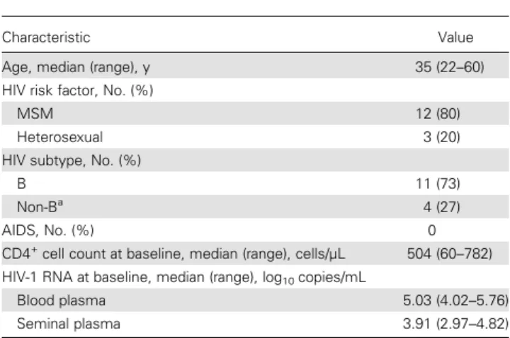
A significant correlation between HIV-1 RNA levels in BP and SP was observed at baseline (Spearmanρ= 0.575;P= .03). The HIV-1 RNA decrease at each study time point is shown in Figure1
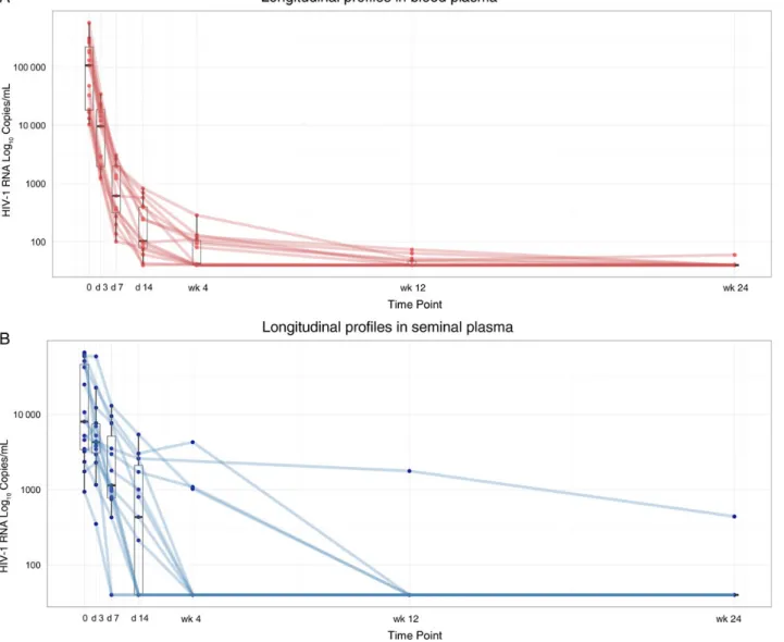
and Table2. The median HIV-1 RNA decrease from baseline was significantly smaller in SP than in BP samples at all time points. Furthermore, the Levene test showed higher heterogeneity of var-iance in SP than in BP samples (data not shown). Thesefindings are consistent with differing decay dynamics in the 2 compart-ments. To further evaluate the dynamics of viral decay after ART initiation based on HIV-1 RNA decreases in BP and SP sam-ples, the model that bestfit our data was 2-phase decay kinetics followed by a plateau (Figure 2 and Supplementary Table). Viral decay was significantly faster in BP than in SP in thefirst decay phase (half-life, 4.5 vs 8.6 days;P= .001). Although decay in the second phase was also faster in BP, the difference was not statistically significant (half-life, 54 vs 156 days;P= .43).
The median time to HIV-1 RNA <40 copies/mL was signifi -cantly shorter in SP than in BP samples (4 vs 12 weeks; P= .008). However, in a Cox regression model adjusted by HIV-1 RNA higher or lower than 5 log at baseline, no differenc-es were found between the 2 compartments (P= .88). HIV-1 RNA suppression at week 4 occurred in SP in 11 individuals
and in BP in 7. BP and SP viral loads at baseline did not differ significantly between patients with seminal HIV-1 RNA persis-tence or suppression at week 4 (4.39 vs 4.25 and 3.96 vs 3.66 log10copies/mL, respectively;P= .24 andP= .79).
Two patients had detectable HIV-1 RNA in BP at week 24 (60 and 41 copies/mL, respectively), whereas HIV-1 RNA in SP of these patients had already fallen to <40 copies/mL at weeks 2 and 4, respectively. Both patients achieved BP HIV-1 RNA <40 copies/mL 8 weeks later. One other patient had detectable seminal HIV-1 RNA (441 copies/mL) at week 24, whereas the BP viral load had been <40 copies/mL since week 2. This pa-tient, who remained on the same ART regimen, consented to provide a new semen sample 19 weeks after completion of the study, when the HIV-1 RNA level was <40 copies/mL.
A significant correlation was observed between DTG concen-trations in the paired BP and SP samples (SpearmanP< .001). The median (range) total DTG concentrations in BP at the end of the dosing interval (C24h) at weeks 4 and 24 were 1200 (124–
2290) and 1420 (353–3100) ng/mL (Table3), which exceed 18.7-and 22.2-fold, respectively, the in vitro PA-IC90for wild-type
HIV-1 (64 ng/mL). The median total DTG concentrations in SP at weeks 4 and 24 were 92.2 (15.7–331) and 129 (38.8–423) ng/mL, respectively (Table3). Overall, the median SP-to-BP ratio was 7.83% (3.65%–20.54%). The total DTG C24h was
below the in vitro PA-IC90in 5 patients at week 4 and also at
week 24 in 2. Those with the lowest seminal DTG C24h also
had the lowest BP DTG C24h.
Unbound DTG concentrations in paired BP and SP samples could be analyzed in 13 of the 15 participants (including those with the lowest total DTG concentrations). The median (range) unbound DTG fractions in BP and SP were 0.45% (0.39%– 0.56%) and 48% (34.2%–59.7%) of total drug concentration, respectively (Table 3). Extrapolating these percentages of DTG free concentrations to the total concentrations measured at weeks 4 and 24, the estimated median (range) unbound DTG concentrations in BP were 5.17 (3.11–10.01) and 6.23 (3.02–12.42) ng/mL, respectively. The estimated unbound DTG concentrations in SP were 34 (5.4–158.8) and 56 (13.4– 203) ng/mL, which exceed 170- and 280-fold the in vitro unbound 50% inhibitory concentration (IC50) for wild type
HIV-1 (0.21 ng/mL). Thus, the estimated average free DTG C24hin SP (45.05 ng/mL) was 214-fold higher than the in
vitro unbound IC50, and the patient with the lowest total
DTG C24hobserved in SP would have free seminal DTG C24h
26-fold the in vitro unbound IC50(Table3).
The patients with detectable HIV-1 RNA in BP or SP samples at week 24 had total DTG concentrations above the in vitro PA-IC90in both compartments. Interestingly, the patient with
de-tectable seminal HIV-1 RNA at week 24 had the highest seminal DTG concentrations at both 4 and 24 weeks. No significant cor-relations were found between DTG concentrations at day 28 and time to HIV-1 RNA <40 copies/mL (SpearmanP= .84).
Table 1. Patient Characteristics (n = 15)
Characteristic Value
Age, median (range), y 35 (22–60)
HIV risk factor, No. (%)
MSM 12 (80)
Heterosexual 3 (20)
HIV subtype, No. (%)
B 11 (73)
Non-Ba
4 (27)
AIDS, No. (%) 0
CD4+
cell count at baseline, median (range), cells/µL 504 (60–782) HIV-1 RNA at baseline, median (range), log10copies/mL
Blood plasma 5.03 (4.02–5.76)
Seminal plasma 3.91 (2.97–4.82)
Abbreviations: HIV, human immunodeficiency virus; MSM, men who have sex with men.
a
Figure 1. Decline in human immunodeficiency virus (HIV) type 1 RNA decline through week 24; times points denote days (d) and weeks (wk) after baseline.A, Blood plasma.
B, Seminal plasma.
Table 2. HIV-1 RNA Decrease in BP and SP
Time Point
HIV-1 RNA, Median (Range) Log10
Copies/mL
HIV-1 RNA Below Limit of Quantification, No.
of Samples/Total No.a
HIV-1 RNA Decrease From Baseline, Median (Range) Log10Copies/mL
PValueb
BP SP BP SP BP SP
Baseline 5.03 (4.02–5.76) 3.91 (2.97–4.82) 0/15 0/15
Day 3 3.99 (3.09–4.54) 3.63 (2.55–4.77) 0/15 0/15 −1.05 (−0.76 to−1.48) −0.23 (+0.18 to−1.18) .001 Day 7 2.79 (2.01–3.49) 3.06 (1.59–4.12) 0/15 2/15 −2.01 (−1.46 to−2.46) −0.74 (−0.19 to−1.92) .001 Day 14 2.02 (1.59–2.92) 2.64 (1.59–3.73) 1/15 6/15 −2.51 (−2.04 to−3.47) −1.41 (−0.48 to−2.44) .001 Week 4 1.62 (1.59–2.46) 1.59 (1.59–3.63) 7/15 11/15 −3.05 (−2.42 to−3.75) −1.95 (−0.68 to−3.21) .002 Week 12 1.59 (1.49–1.87) 1.59 (1.59–3.25) 10/15 14/15 −3.36 (−2.42 to−4.08) −2.13 (−1.38 to−3.21) .001 Week 24 1.59 (1.59–1.78) 1.59 (1.59–2.64) 13/15 14/15 −3.44 (−2.42 to−3.98) −2.18 (−1.38 to−3.21) .001
Abbreviations: BP, blood plasma; HIV, human immunodeficiency virus; SP, seminal plasma.
a
The limit of detection for the quantitative real-time HIV-1 polymerase chain reaction RNA assay is 1.59 log10copies/mL. b
DISCUSSION
HIV-1 RNA suppression in seminalfluid might be limited by ARV penetration into the male genital tract and compartmen-talized infection in this HIV reservoir. The antiviral activity of a
DTG-based ARV regimen in the genital tract of HIV-1–infected male patients has not been assessed to date. Furthermore, determi-nation of HIV-1 decay kinetics in seminalfluid might help to bet-ter understand the role of DTG-based regimens for preventing
Figure 2. Human immunodeficiency virus (HIV) type 1 RNA decay dynamics in blood plasma (BP) and seminal plasma (SP).
Table 3. DTG Concentrations in BP and SPa
Patient
DTG in BP, ng/mL
DTG Free Fraction in BP, %
DTG in SP, ng/mL
DTG Free Fraction in SP, %
SP/BP Ratio, %
Week 4 Week 24 Mean Week 4 Week 24 Mean Week 4 Week 24 Mean
1 2290 924 1607 0.441 171 67.2 119.1 52.8 7.47 7.27 7.37
2 124 353 238.5 NA 15.7 38.8 27.25 34.7 12.66 10.99 11.83
3 1710 2230 1970 0.557 137 152 144.5 50.8 8.02 6.81 7.41
4 1330 2000 1665 0.457 141 207 174 42.6 10.60 10.35 10.47
5 1460 1750 1605 0.412 92.6 164 128.3 34.2 6.342 9.37 7.86
6 1340 3100 2220 NA 92.5 264 178.25 NA 6.90 8.51 7.71
7 1200 1480 1340 NA 92.2 129 110.6 NA 7.68 8.71 8.19
8 1390 2450 1920 0.415 331 423 377 48.0 23.81 17.26 20.54
9 864 1420 1142 0.439 23.9 64.5 44.2 42.4 2.77 4.54 3.65
10 1100 829 964.5 0.470 160 182 171 51.9 14.5 21.95 18.25
11 1050 1990 1520 0.447 88.5 233 160.75 48.6 8.43 11.71 10.07
12 838 1290 1064 0.455 32.1 111 71.55 51.5 3.83 8.60 6.22
13 677 627 652 0.481 33 47 40 59.7 4.87 7.49 6.18
14 805 1010 907.5 0.387 60.9 72.9 66.9 38.6 7.56 7.22 7.39
15 1200 1060 1130 0.459 70.2 104 87.1 47.1 5.85 9.81 7.83
Median 1200 1420 1340 0.455 92.2 129 119.1 48 7.56 8.71 7.83
Abbreviations: BP, blood plasma; DTG, dolutegravir; NA, not available; SP, seminal plasma.
a
HIV-1 transmission. In the current study, we found that HIV-1 RNA decay dynamics in HIV-1–infected patients starting DTG plus ABC/3TC asfirst-line ART differ in the 2 compartments, being faster in BP than in SP, although statistically significant differences were observed only in thefirst decay phase.
We also found that the DTG concentration in SP was 7.8% that seen in BP, similar to thefindings reported in healthy vol-unteers [15], with the DTG C24hlower than the in vitro PA-IC90
in some patients. Nevertheless, all except 1 of the 15 patients studied achieved rapid HIV-1 RNA suppression in SP, with an even shorter median time to suppression than in BP.
In addition to other physicochemical properties, such as mo-lecular weight or lipophilicity, ARV penetration into the genital tract is thought to be determined mainly by BP protein binding [20]. Thus, the low penetration of DTG into the male genital tract may be explained mainly by the drug’s high BP protein binding (>99%). However, total DTG seminal exposure was higher than the free drug concentration in BP, suggesting that drug uptake transporters in addition to passive diffusion of unbound drug may be implicated in DTG penetration in the male genital tract [21].
Because of lower drug binding protein concentrations in SP than in BP [20,22], protein binding of certain ARV drugs has been found to be lower in SP [23]. Unbound DTG C24h was
measured in 13 of 15 participants. The median free DTG frac-tion in BP was 0.45% of the total drug, concordant with previ-ously published data [13], whereas the median free DTG fraction in SP was 48% of the total drug. Therefore, we could estimate that the seminal unbound DTG C24hwas a median
of 214-fold higher than the in vitro unbound IC50(0.21 ng/mL).
The subject with the lowest observed total DTG C24hin SP had an
unbound SP concentration 26-fold above the in vitro unbound IC50. Of note, 4 of the 5 patients with the lowest total DTG
con-centrations experienced rapid suppression of seminal HIV-1 RNA, even before that of BP.
Although ABC and 3TC achieve high seminal concentrations [20,24,25] and the activity of this NRTI backbone must also be taken also into account, the high free DTG concentrations in SP supports the contribution of DTG to HIV-1 suppression in this compartment. The only patient with detectable HIV-1 RNA at week 24 had the highest seminal DTG concentrations and the highest DTG SP/BP ratio. Resistance-associated mutations to INIs or NRTI were not observed with baseline BP genotypic resistance testing. We attempted to perform this testing on a stored baseline seminal remnant sample, but it was not techni-cally possible. Further testing showed that this patient achieved HIV-1 RNA suppression after the study period. In this case, compartmentalized HIV replication in the genital tract might have been the obstacle preventing earlier HIV suppression rath-er than insufficient ARV levels in this reservoir.
Consistent with thesefindings, no significant correlation was observed between DTG concentrations and the time to achieve
HIV-1 RNA below the detection limit in each compartment. In-terestingly, a good correlation between BP DTG concentration and HIV-1 RNA decline after 10 days of DTG monotherapy has been observed in a previous phase IIa trial [26]. This differing pharmacodynamic behavior highlights the multiple factors that can influence HIV suppression in seminalfluid, including drug penetration, compartmentalized and autonomous HIV replica-tion, and the possible interindividual variability related to these aspects.
A significant correlation between HIV-1 RNA in BP and SP was observed at baseline. Median HIV-1 RNA in SP at baseline was 1 log lower compared to median HIV-1 RNA in BP that is concordant with other previously published studies [4]. At week 24, all but 2 patients had HIV-1 RNA <40 copies/mL in BP al-though it is worthy of note that these 2 patients had a favorable HIV-1 RNA decrease (41 and 60 copies/mL). Only 1 patient did not have a favorable virologic response in SP, with the HIV-1 RNA level remaining at 441 copies/mL while undetectable in BP; thatfinding is concordant with compartmentalized HIV-1 RNA dynamics in the genital tract, as has also been previously described [5–7].
Significantly smaller decreases in HIV-1 RNA in SP com-pared with BP were observed at each time point, as well as greater heterogeneity in HIV-1 RNA decline over time. This heterogeneity is also concordant with the compartmentalized dynamics of HIV infection in the genital tract and the role of different organs within the male genital tract as the source of virions and/or infected cells for semen, such as the epididymis, seminal vesicles, prostate, and urethra [27].
We observed a faster HIV-1 RNA decay in BP than in SP, although statistically significant differences were only observed in thefirst decay phase. Although the absence of significant differences in the second decay phase could be explained by the limited sample size and the low HIV-1 RNA values in this second phase, a similar pattern of initially slower HIV-1 RNA decay in seminalfluid than in plasma, but no difference in the second-phase decay rate has been also observed in patients receiving efavirenz-based ART [28].
during the first 6 months after starting ART [31]. The early seminal HIV-1 suppression achieved by DTG-based regimens, and perhaps other INIs, could reduce the time needed for pre-exposure prophylaxis in serodiscordant couples with a high risk of HIV transmission.
Our study has some limitations. As in other studies assessing pharmacokinetics in these viral reservoirs, our sample size is small, which makes the results more susceptible to influence by interindividual and intraindividual variability. Nevertheless, we performed 2 separate determinations of seminal and BP DTG concentrations in each patient, and our results are similar to those observed in healthy volunteers. On the other hand, we only measured HIV-1 RNA in SP; therefore, the role of ongoing HIV-1 RNA production from long-lived infected cells in semen was not considered in our model.
In summary, this study showed that despite an initially slower HIV-1 RNA decline in SP than in BP samples, rapid HIV-1 RNA suppression in seminalfluid is achieved in most patients startingfirst-line ART with DTG plus ABC/3TC. Thesefindings could be of interest for reducing the HIV transmission risk after ART initiation. Although the seminal DTG concentration is only 7.8% of the concentration in BP, it is sufficient to contrib-ute to suppress HIV-1 replication in this compartment.
Supplementary Data
Supplementary materialsare available athttp://jid.oxfordjournals.org. Consisting of data provided by the author to benefit the reader, the posted materials are not copyedited and are the sole responsibility of the author, so questions or comments should be addressed to the author.
Notes
Acknowledgments. We are grateful to all the patients who participated in this study. We thank Antonio Navarro for assistance in samples process-ing and Celine Cavallo for English language support.
Author contributions. A. I. and D. P. designed the study; A. I., E. F., and D. P. recruited participants; A. I., E. F., and A. V. conducted the study visits; J. N. performed the microbiologic procedures; A. D. M. K. and C. S. per-formed LC-MS/MS to measure Dolutegravir concentrations in plasma and seminalfluid; N. R. and L. A. assisted in data collection and study co-ordination; D. O. and J. C. performed the statistical analysis; A. I., J. M.-P., and D. P. analyzed and interpreted the results; A. I. drafted the manuscript and J. M.-P., A. D. M. K., and D. P. reviewed it. All authors revised the manuscript for important intellectual content and contributed to thefinal version.
Disclaimer. ViiV Healthcare was given the opportunity to review a pre-liminary version of this manuscript for factual accuracy. The authors are solely responsible for the study design and the final content and interpretation.
Financial support. This work was provided by ViiV Healthcare. The study was also partially supported by the RD12/0017/0013 project as part of the Plan Nacional R + D + I and co-financed by Instituto de Salud Carlos III- Subdirección General de Evaluación and Fondo Europeo de Desarrollo Regional (FEDER), the University of North Carolina Center for AIDS Research (grant P30 AI050410 to A. D. M. K. and C. S.), Collaboratory of AIDS Researchers for Eradication (grant U19 AI096113 to A. D. M. K. and C. S.), and the National Institutes of Health (grant R01 AI111891 to A. D. M. K. and C. S.).
Potential conflicts of interest. A. I. has receivedfinancial compensation for lectures, consultancies, and educational activities or funds for research from Bristol-Myers Squibb (BMS), Gilead Sciences, Janssen-Cilag, Merck
Sharp & Dohme, and ViiV Healthcare. J. M.-P. has receivedfinancial compensation for consultancies, lectures, and educational activities and research support from Merck Sharp & Dohme, BMS, ViiV Healthcare, and Gilead. J. N. has receivedfinancial compensation for lectures and re-search from Abbott Molecular. A. D. M. K. has receivedfinancial compen-sation for a consultancy from Merck and grant support from Gilead, GlaxoSmithKline, and Merck. D. P. has received research grants and/or honoraria for advisories and/or conferences from Boehringer Ingelheim, GlaxoSmithKline, ViiV Healthcare, Pfizer, BMS, Abbott, Gilead, Janssen, and Merck. All other authors report no potential conflicts. All authors have submitted the ICMJE Form for Disclosure of Potential Conflicts of Interest. Conflicts that the editors consider relevant to the content of the manuscript have been disclosed.
References
1. UN Joint Programme on HIV/AIDS (UNAIDS). The gap report,2014.http:// www.refworld.org/docid/53f1e1604.html. Accessed 8 April 2015.
2. Donnell D, Baeten JM, Kiarie J, et al.; Partners in Prevention HSV/HIV Transmis-sion Study Team. Heterosexual HIV-1 transmisTransmis-sion after initiation of antiretrovi-ral therapy: a prospective cohort analysis. Lancet2010; 375:2092–8.
3. Cohen MS, Chen YQ, McCauley M, et al.; HPTN 052 Study Team. Prevention of HIV-1 infection with early antiretroviral therapy. N Engl J Med2011; 365:493–505.
4. Baeten JM, Kahle E, Lingappa JR, et al.; Partners in Prevention HSV/HIV Transmission Study Team. Genital HIV-1 RNA predicts risk of heterosexual HIV-1 transmission. Sci Transl Med2011; 3:77ra29.
5. Anderson JA, Ping LH, Dibben O, et al. Center for HIV/AIDS vaccine immunol-ogy. HIV-1 populations in semen arise through multiple mechanisms. PLoS Pathog2010; 6:e1001053.
6. Zhang H, Dornadula G, Beumont M, et al. Human immunodeficiency virus type 1 in the semen of men receiving highly active antiretroviral therapy. N Engl J Med 1998; 339:1803–9.
7. Lambert-Niclot S, Tubiana R, Beaudoux C, et al. Detection of HIV-1 RNA in seminal plasma samples from treated patients with undetectable HIV-1 RNA in blood plasma on a 2002–2011 survey. AIDS2012; 26:971–5.
8. Taylor S, Davies S. Antiretroviral drug concentrations in the male and female genital tract: implications for the sexual transmission of HIV. Curr Opin HIV AIDS2010; 5:335–43.
9. Kobayashi M, Yoshinaga T, Seki T, et al. In Vitro antiretroviral properties of S/GSK1349572, a next-generation HIV integrase inhibitor. Antimicrob Agents Chemother2011; 55:813–21.
10. RaffiF, Rachlis A, Stellbrink HJ, et al.; SPRING-2 Study Group. Once-daily dolutegravir versus raltegravir in antiretroviral-naive adults with HIV-1 infection: 48 week results from the randomised, double-blind, non-inferiority SPRING-2 study. Lancet2013; 381:735–43.
11. Walmsley SL, Antela A, Clumeck N, et al.; SINGLE Investigators. Dolutegravir plus abacavir-lamivudine for the treatment of HIV-1 infection. N Engl J Med 2013; 369:1807–18.
12. Clotet B, Feinberg J, van Lunzen J, et al.; ING114915 Study Team. Once-daily dolutegravir versus darunavir plus ritonavir in antiretroviral-naive adults with HIV-1 infection (FLAMINGO): 48 week results from the randomised open-label phase 3b study. Lancet2014; 383:2222–31.
13. Cottrell ML, Hadzic T, Kashuba AD. Clinical pharmacokinetic, pharmacodynamic and drug-interaction profile of the integrase inhibitor dolutegravir. Clin Pharma-cokinet2013; 52:981–94.
14. Reese MJ, Savina PM, Generaux GT, et al. In vitro investigations into the roles of drug transporters and metabolizing enzymes in the disposition and drug interac-tions of dolutegravir, a HIV integrase inhibitor. Drug Metab Dispos2013; 41:353–61.
15. Greener BN, Patterson KB, Prince HM, et al. Dolutegravir pharmacokinetics in the genital tract and colorectum of HIV-negative men after single and multiple dosing. J Acquir Immune Defic Syndr2013; 64:39–44.
16. Weller S, Borland J, Chen S, et al. Pharmacokinetics of dolutegravir in HIV-sero-negative subjects with severe renal impairment. Eur J Clin Pharmacol2014; 70:29–35.
17. Perelson AS, Neumann AU, Markowitz M, Leonard JM, Ho DD. HIV-1 dynamics in vivo: virion clearance rate, infected cell life-span, and viral generation time. Science1996; 271:1582–6.
18. Wu H, Ding AA. Population HIV1 dynamics in vivo: applicable models and in-ferential tools for virological data from AIDS clinical trials. Biometrics1999; 55: 410–8.
19. Gentleman R, Ihaka R, Bates D. The R project for statistical computing.2009.
20. Else LJ, Taylor S, Back DJ, Khoo SH. Pharmacokinetics of antiretroviral drugs in anatomical sanctuary sites: the male and female genital tract. Antivir Ther2011; 16:1149–67.
21. Klein DM, Wright SH, Cherrington NJ. Xenobiotic transporter expression along the male genital tract. Reprod Toxicol2014; 47:1–8.
22. Lizana J, Blad E. Immunonephelometry of specific proteins in human seminal plasma. Clin Chem1983; 29:618–23.
23. Brown KC, Patterson KB, Jennings SH, et al. Single and multiple dose pharmaco-kinetics of darunavir plus ritonavir and etravirine in semen and rectal tissue of HIV-negative men. J Acquir Immune Defic Syndr2012; 61:138–44.
24. Pereira AS, Kashuba AD, Fiscus SA, et al. Nucleoside analogues achieve high con-centrations in seminal plasma: relationship between drug concentration and virus burden. J Infect Dis1999; 180:2039–43.
25. van Praag RM, van Heeswijk RP, Jurriaans S, Lange JM, Hoetelmans RM, Prins JM. Penetration of the nucleoside analogue abacavir into the genital tract of men infected with human immunodeficiency virus type 1. Clin Infect Dis2001; 33:e91–2. 26. Min S, Sloan L, DeJesus E, et al. Antiviral activity, safety, and pharmacokinetics/
pharmacodynamics of dolutegravir as 10-day monotherapy in HIV-1-infected adults. AIDS2011; 25:1737–45.
27. Houzet L, Matusali G, Dejucq-Rainsford N. Origins of HIV-infected leukocytes and virions in semen. J Infect Dis2014; 210(suppl 3):S622–30.
28. Graham SM, Holte SE, Dragavon JA, et al. HIV-1 RNA may decline more slowly in semen than in blood following initiation of efavirenz-based antiretroviral therapy. PLoS One2012; 7:e43086.
29. Lennox JL, DeJesus E, Lazzarin A, et al.; STARTMRK investigators. Safety and efficacy of raltegravir-based versus efavirenz-based combination therapy in treat-ment-naïve patients with HIV-1 infection: a multicentre, double-blind rando-mised controlled trial. Lancet2009; 374:796–806.
30. DeJesus E, Rockstroh JK, Henry K, et al.; GS-236-0103 Study Team. Co-formulated elvitegravir, cobicistat, emtricitabine, and tenofovir disoproxil fumarate versus ritonavir-boosted atazanavir plus co-formulated emtricitabine and tenofovir disoproxil fumarate for initial treatment of HIV-1 infection: a randomised, double-blind, phase 3, non-inferiority trial. Lancet2012; 379:2429–38.