THE EFFECT OF ANTERIOR CRUCIATE LIGAMENT INJURY AND RECONSTRUCTION ON LOWER EXTREMITY BIOMECHANICS,
COORDINATION, AND VARIABILITY
Benjamin McMillen Goerger
A dissertation submitted to the faculty of the University of North Carolina at Chapel Hill in partial fulfillment of the requirements for the degree of Doctor of Philosophy in the Department of Interdisciplinary Human Movement Science (School of Medicine)
Chapel Hill 2012
Approved by:
© 2012
ABSTRACT
BENJAMIN MCMILLEN GOERGER: The Effect of Anterior Cruciate Ligament Injury and Reconstruction on Lower Extremity Biomechanics, Coordination, and Variability
(Under the direction of Darin A. Padua)
Individuals that suffer an ACL injury, and undergo reconstructive surgery are at an increased risk for the development of osteoarthritis and a secondary ACL injury.
ACKNOWLEDGEMENTS
This project would not have been possible without the help and guidance of several people, first being my committee of Drs. Darin Padua, Anthony Beutler, Troy Blackburn, Stephen Marshall, and John Wilckens. I am grateful for their time and effort to make this a successful project, and consider myself very lucky to have had the opportunity to learn from them. I would like to specifically acknowledge Dr. Marshall, Dr. Beutler, and Dr. Padua for their encouragement to pursue this project and their time and effort in
conducting the JUMP ACL study, without which this project would never have been possible. Last, I am very thankful to Dr. Padua. If it were not for his mentorship that began so many summers ago in Fishkill, NY, I would not have the ability or the opportunities that I have today.
I would also like to acknowledge others who have helped make this project a success along the way. A big thank you goes to Sue Wolf for helping me with everything from IRB forms and data, to shipping equipment and reimbursements. Thank you also to Barnett Frank, Rebecca Begalle, and Molly Grasso for their assistance with data
collection. They all braved a different adversity (i.e. extreme weather, disastrous travel, moving heavy equipment) to help with this project, and I am thankful.
TABLE OF CONTENTS
Page
LIST OF TABLES………...x
LIST OF FIGURES………...xii
CHAPTER ONE...1
1.1 Research Questions...5
1.2 Research Hypotheses...6
1.3 Operational Definitions...7
1.4 Assumptions & Limitations...9
1.5 Delimitations...10
1.6 Independent Variables...10
1.7 Dependent Variables...10
1.8 Significance...11
CHAPTER TWO...12
2.1 ACL Injury as a Risk Factor for Subsequent ACL Injury and Knee OA...12
2.2 Factors Contributing to the Loading of the ACL...16
2.3 Observed Mechanism of Non-Contact ACL Injury...23
2.4 Prospective Risk Factors for ACL Injury...26
2.7 Coordination and Variability Methodology Review...47
2.8 Summary of Literature Review...57
CHAPTER THREE...58
3.1 Experimental Design...58
3.2 Participants...59
3.3 Instrumentation...59
3.4 Procedures...61
3.5 Data Capture, Processing and Reduction...64
3.6 Statistical Analysis...69
CHAPTER FOUR...72
4.1 Introduction...72
4.2 Overview of Study and Participant Demographics...72
4.3 Results...73
4.3.1 Results Research Question 1...73
4.3.2 Results Research Question 2...78
4.3.3 Results Research Question 3...79
4.3.4 Results Research Question 4...80
CHAPTER FIVE...82
5.1 Introduction...82
5.2 Preparatory Phase Kinematics...82
5.3 Research Question 4...85
APPENDIX A. SUBJECT QUESTIONAIRE...164
LIST OF TABLES
Table Page
1. List of kinematic dependent variables...89
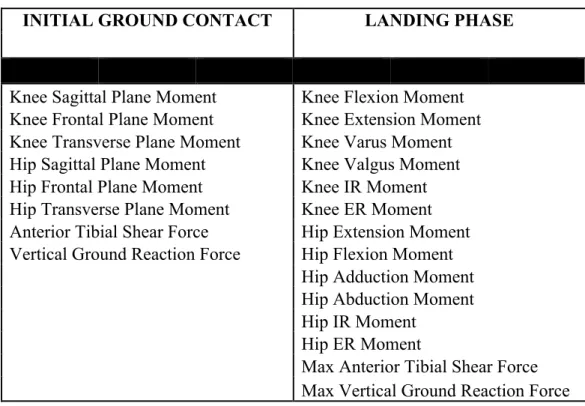
2. List of kinetic dependent variables...90
3. List of joint coordination and variability dependent variables...91
4.Participant demographics and anthropometrics for Research Question #1...92
5. Group chronological descriptive statistics for Research Question #1 ...93
6.Descriptive statistics for Research Question #1...94
7. Descriptive statistics for knee and hip kinematics at Initial Ground Contact for Research Question #1...95
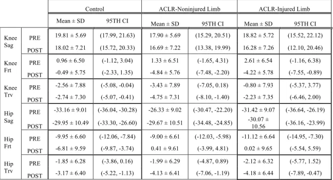
8. Descriptive statistics for knee and hip moments and kinetics at Initial Ground Contact for Research Question #1...96
9. Descriptive statistics for peak knee and hip kinematics at Landing Phase for Research Question #1...97
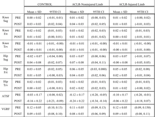
10. Descriptive statistics for knee moments and kinetics at Landing Phase for Research Question #1...98
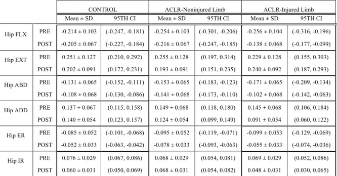
11. Descriptive statistics for hip moments at Landing Phase for Research Question #1...99
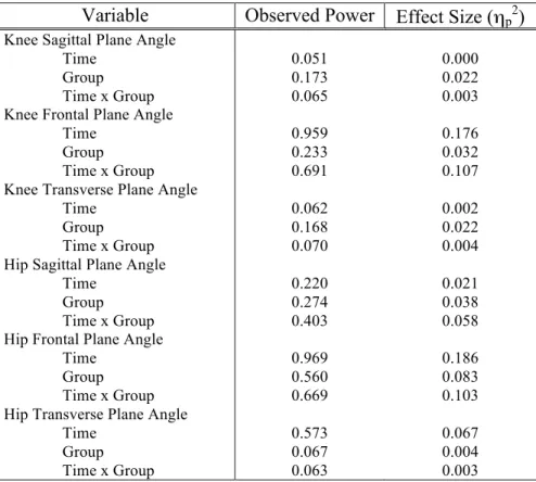
12. Observed power and effect size for analyses of knee and hip kinematics an Initial Ground Contact for Research Question #1...100
13. Observed power and effect size for analyses of knee and hip moments and kinetics at Initial Ground Contact for Research Question #1...101
15. Observed power and effect size for analyses of hip kinematics
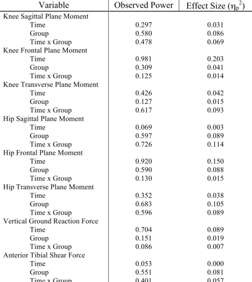
during Landing Phase for Research Question #1...103 16. Observed power and effect size for analyses of knee moments and
kinetics during Landing Phase for Research Question #1...104 17. Observed power and effect size for analyses of hip moments during
Landing Phase for Research Question #1...105
18.Participant demographics and anthropometrics
for Research Question #2...106 19. Group chronological descriptive statistics
for Research Question #2 ...107 20.Descriptive statistics for Research Question #2...108 21. Average coupling angle for Research Question #2...109 22. Observed power and effect size for analyses of mean coupling
angle during Landing Phase for Research Question #2...110 23.Participant demographics and anthropometrics
for Research Question #3...111 24.Descriptive statistics for Research Question #3...112 25. Asymmetry of kinematics, moments and kinetics for groups at
Initial Ground Contact for Research Question #3...113 26. Asymmetry of peak kinematics during Landing Phase for
Research Question #3...114 27. Asymmetry of peak moment and kinetic variables during Landing Phase
for Research Question #3...115 28.Participant demographics and anthropometrics
for Research Question #4...116 29.Descriptive statistics for Research Question #4...117 30. Asymmetry of mean coupling angles during Landing Phase for
LIST OF FIGURES
Figure Page
1. Double leg jump landing task...120 2. Visual representation of the calculation of coupling angles determined
using vector coding...121 3. Ensemble average plot of sagittal plane knee angle at Baseline and
Follow-Up during the Landing Phase for the ACLR-Injured Limb Group...122 4. Ensemble average plot of sagittal plane knee angle at Baseline and
Follow-Up during the Landing Phase for the ACLR-Noninjured Limb Group...123 5. Ensemble average plot of sagittal plane knee angle at Baseline and
Follow-Up during the Landing Phase for the Control Group...124 6. Ensemble average plot of frontal plane knee angle at Baseline and
Follow-Up during the Landing Phase for the ACLR-Injured Limb Group...125 7. Ensemble average plot of frontal plane knee angle at Baseline and
Follow-Up during the Landing Phase for the ACLR-Noninjured Limb Group...126 8. Ensemble average plot of frontal plane knee angle at Baseline and
Follow-Up during the Landing Phase for the Control Group...127 9. Ensemble average plot of transverse plane knee angle at Baseline and
Follow-Up during the Landing Phase for the ACLR-Injured Limb Group...128 10. Ensemble average plot of transverse plane knee angle at Baseline and
Follow-Up during the Landing Phase for the ACLR-Noninjured Limb Group...129 11. Ensemble average plot of transverse plane knee angle at Baseline and
Follow-Up during the Landing Phase for the Control Group...130 12. Ensemble average plot of sagittal plane hip angle at Baseline and
Follow-Up during the Landing Phase for the ACLR-Injured Limb Group...131 13. Ensemble average plot of sagittal plane hip angle at Baseline and
Follow-Up during the Landing Phase for the ACLR-Noninjured Limb Group...132 14. Ensemble average plot of sagittal plane hip angle at Baseline and
15. Ensemble average plot of frontal plane hip angle at Baseline and
Follow-Up during the Landing Phase for the ACLR-Injured Limb Group...134 16. Ensemble average plot of frontal plane hip angle at Baseline and
Follow-Up during the Landing Phase for the ACLR-Noninjured Limb Group...135 17. Ensemble average plot of frontal plane hip angle at Baseline and
Follow-Up during the Landing Phase for the Control Group...136 18. Ensemble average plot of transverse plane hip angle at Baseline and
Follow-Up during the Landing Phase for the ACLR-Injured Limb Group...137 19. Ensemble average plot of transverse plane hip angle at Baseline and
Follow-Up during the Landing Phase for the ACLR-Noninjured Limb Group...138 20. Ensemble average plot of transverse plane hip angle at Baseline and
Follow-Up during the Landing Phase for the Control Group...139 21. Ensemble average plot of sagittal plane knee moment at Baseline and
Follow-Up during the Landing Phase for the ACLR-Injured Limb Group...140 22. Ensemble average plot of sagittal plane knee moment at Baseline and
Follow-Up during the Landing Phase for the ACLR-Noninjured Limb Group...141 23. Ensemble average plot of sagittal plane knee moment at Baseline and
Follow-Up during the Landing Phase for the Control Group...142 24. Ensemble average plot of frontal plane knee moment at Baseline and
Follow-Up during the Landing Phase for the ACLR-Injured Limb Group...143 25. Ensemble average plot of frontal plane knee moment at Baseline and
Follow-Up during the Landing Phase for the ACLR-Noninjured Limb Group...144 26. Ensemble average plot of frontal plane knee moment at Baseline and
Follow-Up during the Landing Phase for the Control Group...145 27. Ensemble average plot of transverse plane knee moment at Baseline and
Follow-Up during the Landing Phase for the ACLR-Injured Limb Group...146 28. Ensemble average plot of transverse plane knee moment at Baseline and
30. Ensemble average plot of sagittal plane hip moment at Baseline and
Follow-Up during the Landing Phase for the ACLR-Injured Limb Group...149 31. Ensemble average plot of sagittal plane hip moment at Baseline and
Follow-Up during the Landing Phase for the ACLR-Noninjured Limb Group...150 32. Ensemble average plot of sagittal plane hip moment at Baseline and
Follow-Up during the Landing Phase for the Control Group...151 33. Ensemble average plot of frontal plane hip moment at Baseline and
Follow-Up during the Landing Phase for the ACLR-Injured Limb Group...152 34. Ensemble average plot of frontal plane hip moment at Baseline and
Follow-Up during the Landing Phase for the ACLR-Noninjured Limb Group...153 35. Ensemble average plot of frontal plane hip moment at Baseline and
Follow-Up during the Landing Phase for the Control Group...154 36. Ensemble average plot of transverse plane hip moment at Baseline and
Follow-Up during the Landing Phase for the ACLR-Injured Limb Group...155 37. Ensemble average plot of transverse plane hip moment at Baseline and
Follow-Up during the Landing Phase for the ACLR-Noninjured Limb Group...156 38. Ensemble average plot of transverse plane hip moment at Baseline and
Follow-Up during the Landing Phase for the Control Group...157 39. Ensemble average plot of vertical ground reaction force at Baseline and
Follow-Up during the Landing Phase for the ACLR-Injured Limb Group...158 40. Ensemble average plot of vertical ground reaction force at Baseline and
Follow-Up during the Landing Phase for the ACLR-Noninjured Limb Group...159 41. Ensemble average plot of vertical ground reaction force at Baseline and
Follow-Up during the Landing Phase for the Control Group...160 42. Ensemble average plot of anterior tibial shear force at Baseline and
Follow-Up during the Landing Phase for the ACLR-Injured Limb Group...161 43. Ensemble average plot of anterior tibial shear force at Baseline and
Follow-Up during the Landing Phase for the ACLR-Noninjured Limb Group...162 44. Ensemble average plot of anterior tibial shear force at Baseline and
CHAPTER ONE
INTRODUCTION
Injury to the anterior cruciate ligament (ACL) can be a devastating injury that requires a significant amount of time and effort to allow for recovery and return to one’s respective level of activity or sport. Anterior cruciate ligament reconstruction (ACLR) is often performed to restore mechanical stability to the knee and facilitate return to
participation in physical activity and sport. The true number of ACL injuries that occur each year in the United States is unknown, but recent projections have estimated a 67.8% increase in the number of ACLRs performed over a ten year period.1 For the most part, ACLR does meet the goals of restoring mechanical stability allowing persons to continue participation in physical activity.2,3
Returning to participation in a sport or high demand physical activity is a short-term goal. Reconstructive surgery and rehabilitation are often conducted with the goal of returning function to the injured limb. The marker for adequate function is returning the limb to how it functioned before injury, but this may be a flawed benchmark as this may reestablish factors that predisposed the person to injury in the first place. These
a second ACL injury, regardless of the initial leg injured.5-7 Those with a previous ACL injury reportedly have a four fold5 to ten fold 7 increased risk for injury compared to those with no previous ACL injury. The risk for reinjury is independent of the type of graft used for ACLR, and nonspecific to the previously injured knee.8 In fact, returning to high demand sports or activities, the goal of ACLR, is associated with risk for injury of the contralateral uninjured ACL.8 Faude et al6 postulated factors that predispose those with ACL injury to subsequent injury may be person specific and not knee specific. Whether the increased risk for reinjury is the product of ACL injury and reconstruction, or residual biomechanical risk factors that caused the initial injury is unknown.
Significant research has been devoted to understanding the factors that cause initial injury to the ACL. Cadaveric studies have demonstrated that the ACL is loaded when an anterior force is applied to the proximal tibia.9 Additional factors such as frontal plane loading10,11, transverse plane loading11, weight bearing12,13, and a combination of these factors can also increase the ACL strain.14-17 Going a step further, analyses of actual ACL injuries captured on video, it appears that a majority of these injuries occur during a landing from a jump18-20 and during a planting, cutting maneuver.20 The position at the time of injury has also been described as landing with less plantar flexion21, with the foot placed outside the knee creating a valgus alignment while the knee is extended20, and having greater hip flexion.21 Descriptions of ACL loading and the mechanism of injury provide limited information, as they describe how the ACL can be injured, but they do not necessarily provide information as to what places a person at greater risk for suffering injury. In addition, these descriptions are relevant to describing a first injury, not
There is, however, limited information on the prospective risk factors for non-contact ACL injury. Hewett et al22 demonstrated in a prospective analysis of adolescent female athletes that those that went on to suffer a non-contact ACL injury displayed greater knee valgus angle, lower maximum peak knee flexion angle, greater external knee valgus moment, greater peak vertical ground reaction force, and greater external hip flexion moment.22 While the information is limited in scope, because of the population studied and a relatively low number of injuries, it does identify biomechanical risk factors that may be important for non-contact ACL injury. One finding of particular use for
understanding risk factors for subsequent ACL injury, is that the athletes that went on to suffer injury displayed bilateral differences in external knee valgus moment.22
Other bilateral differences in lower extremity biomechanics have been identified in persons after ACLR.23-31 The presence of asymmetries in movement and loading are thought to be the result of injury to the ACL, but because they have also been identified as a prospective risk factor for initial injury, it may indicate the risk factors for initial injury are not being adequately addressed in rehabilitation. Further evidence of this problem was provided by Paterno et al23, when they observed that asymmetrical loading of the lower extremities during a double leg jump landing, particularly bilateral
magnitude of asymmetry for those with ACLR is any greater than those observed in healthy noninjured persons. Only this type of information can provide an indication of risk factors that are not being adequately addressed during reconstruction and
rehabilitation.
As informative as biomechanical analyses are, they only provide a limited view on the changes in movement after ACL injury and ACLR. Recent work has been done to characterize the coordination and variability of movement within the lower extremity in those with ACLR.32-35 Coordination and variability have been researched under the notion that quantifying these measures may provide an assessment of an individual’s risk for injury. Persons with ACLR, when compared to those without injury, have
demonstrated increased variability33,34, as well as altered coordination between the shank and thigh.32,35 There is, however, a lack of prospective data to indicate if these differences are inherent to the individual and were present prior to injury, or if they are the result from ACL injury and reconstruction. Again, research that incorporates repeated measures taken prior to ACL injury and after subsequent ACLR is needed to confirm the role that this type of information has in understanding risk for the initial and subsequent ACL injuries.
on the current information that is available for this population. The same is true for measures of lower extremity coordination and variability, which is thought to influence the risk for injury, but has not been used prospectively to identify risk factors for injury. The only way to determine if the above postulation is correct is to conduct research that incorporates repeated assessment that includes measures prior to the initial ACL injury and after the subsequent ACLR. In addition, bilateral differences in lower extremity biomechanics, coordination, and variability have been documented in those with ACLR, but there is little evidence to determine if these differences are greater than those with no previous ACL injury. Therefore, the purpose of this study is to determine if lower
extremity biomechanics and coordination obtained prior to ACL injury change following ACL injury and subsequent ACLR. A second purpose is to determine if between limb differences in lower extremity biomechanics, coordination, and variability are different as compared to individuals with no history of ACL injury.
1.1Research Questions
Research Question 1: Are lower extremity biomechanics during a double leg jump landing changed following ACL injury and subsequent ACLR?
Research Question 1a: Are lower extremity biomechanics of the injured limb changed following ACL injury and subsequent ACLR?
Research Question 2: Is lower extremity joint coordination during a double leg jump landing changed following ACL injury and subsequent ACLR?
Research Question 2a: Is lower extremity joint coordination of the injured limb changed following ACL injury and subsequent ACLR?
Research Question 2b: Is lower extremity joint coordination of the noninjured limb changed following ACL injury and subsequent ACLR?
Research Question 3: Is the magnitude of between limb differences in lower extremity biomechanics during a double leg jump landing different for persons with ACLR as compared to those with no history of ACL injury at Follow-Up?
Research Question 4: Is the magnitude of between limb differences in lower extremity joint coordination and variability during a double leg jump landing different for persons with ACLR as compared to those with no history of ACL injury at Follow-Up?
1.2Research Hypotheses
Research Hypothesis 1b: There will be no change in lower extremity biomechanics of the noninjured limb during a double leg jump landing from Baseline to Follow-Up for both the ACLR and Control groups.
Research Hypothesis 2a: There will be no change in lower extremity joint coordination of the injured limb during a double leg jump landing from Baseline to Follow-Up for both the ACLR and Control groups.
Research Hypothesis 2b: There will be no change in lower extremity joint coordination of the noninjured limb during a double leg jump landing from Baseline to Follow-Up for both the ACLR and Control groups.
Research Hypothesis 3: The ACLR Group will demonstrate greater differences in between limb lower extremity biomechanics at Follow-Up as compared to the Control Group.
Research Hypothesis 4: The ACLR Group will demonstrate greater differences in between limb lower extremity joint coordination and variability at Follow-Up as compared to the Control Group.
1.3Operational Definitions
ACL Injury: Disruption of the anterior cruciate ligament injury as confirmed by examination of medical records.
ACLR: Any surgery performed to replace the injured anterior cruciate ligament for the purpose of restoring stability to the knee.
Injured Limb: For the ACLR Group, the limb in which an ACL injury occurred. For the Control Group, the limb that was tested during initial testing for the JUMP ACL study.
Noninjured Limb: For the ACLR Group, the limb in which an ACL injury did not occur. For the Control Group, the limb that was not tested during initial testing for the JUMP ACL study.
Joint Coordination: The relationship in angular position between two joints, quantified by determining the average coupling angle across the phase of interest during the double leg jump landing task as measured using vector coding.
Joint Coordination Variability: The between trials variability of joint coordination during the double leg jump landing task as measured using vector coding.
Baseline: Testing that occurred during the JUMP ACL study, and resulted in
collection of biomechanical data on the participant’s dominant leg at the time of their enrollment in their respective service academy.
Follow-Up: Testing that occurred in the Spring of 2011 in which lower extremity biomechanical data were collected on those identified for the purposes of this study that were enrolled in the JUMP ACL study.
Initial Ground Contact: The first time point at which the vertical ground reaction force exceeds 10 N during a double leg jump landing.
Landing Phase: A time period during a double leg jump landing defined as the point from Initial Ground Contact to the time point at which the maximum knee flexion angle is reached.
Anterior Knee Laxity: The anterior displacement of the tibia, measured in millimeters, under a 30lb force using a knee arthrometer.
1.4Assumptions & Limitations
• All participants will perform tasks with their best effort.
• The type of graft or reconstructive procedure performed for the Injured Group
will not be considered for this study.
• Concomitant injury associated with ACL injury will not be considered for this
study.
• The amount of time since ACLR will not be considered for this study.
• Rehabilitation programs after ACLR are similar across service academies and
time for those in the Injured Group.
• The results of this study may be limited to those that are young and physically
active.
1.5Delimitations
• All participants were enrolled in the JUMP ACL study and had complete
biomechanical data collected at baseline testing.
• All participants were still enrolled in their respective service academy and the
JUMP ACL study.
• All participants were participating in physical activity.
• All participants were members of the 2007 or 2008 JUMP ACL cohort.
1.6Independent Variables
Time:
• Baseline: Testing performed for the JUMP ACL study in the Summers of 2007
and 2008 upon each participant’s entry in to the respective service academy.
• Follow-Up: Testing conducted in the Spring of 2011.
Group:
• ACLR Group: Participants that had suffered one ACL injury and undergone
subsequent ACLR since Baseline, had no prior history of ACL injury at Baseline, and had complete biomechanical data from Baseline.
• Control Group: Participants that have no history of ACL injury, and had complete
biomechanical data from Baseline.
1.7Dependent Variables
Kinetics: Sagittal, frontal, and transverse plane moments of the knee and hip; anterior tibial shear force; vertical ground reaction force
Joint Coordination and Variability: Knee sagittal plane - Hip sagittal plane; Knee frontal plane – Hip frontal plane; Knee transverse plane – Hip transverse plane; Knee frontal plane – Hip transverse plane; Knee transverse plane – Hip frontal plane
1.8Significance
CHAPTER TWO
REVIEW OF THE LITERATURE
The purpose of this study is to examine the effect of ACL injury and ACLR on a sample of young, physically active persons. This review of the available literature will indicate what additional information this study will provide to the current literature regarding the biomechanics and coordination of those with ACLR.
2.1 ACL Injury as a Risk Factor for Subsequent ACL Injury and Knee OA
returning to sport activity. In addition, suffering an initial ACL injury by a contact mechanism of injury was a significant predictor for reinjury of the reconstructed limb.8 This is despite no difference in median graft diameter, or any difference in the presence of articular cartilage or meniscal damage at the time of reconstruction.8 Graft type does not seem to influence the risk of reinjury either, as time to injury was not different
between those that had a BPTB or HS autograft used for the initial reconstruction.8 Injury to the healthy or uninjured ACL was predicted by a return to sports or activities that were classified as strenuous or moderate in demand after initial injury and reconstruction.8 The lack of any difference in risk for subsequent ACL injury between limbs may be the result of these factors being person specific rather than limb specific, as was suggested by Faude et al6 after they noted that when treating limbs as individual cases, the risk for reinjury was no longer associated with that of previous ACL injury.
and/or articular cartilage injury.2,37 Graft type used and the amount of anterior-posterior laxity that remains after reconstructive surgery does not determine the severity of knee OA in those with ACLR.37,38,41 Salmon et al43 did note, however, poorer radiographic findings for those with a loss of knee extension or greater laxity during a lachman test.43 There are contrasting findings for mensical involvement.2,39 Jarvela et al39 found that the presence of accompanying injuries, which included meniscal injuries, to an ACL injury only increased the number of subsequent surgical procedures performed on the knee, and did not affect radiologic findings of articular cartilage degradation. Similar findings were noted by Lebel et al2, indicating no difference in the rate of OA for those with meniscal involvement. They did find however, that delaying ACLR with the presence of a meniscal injury for more than one year was associated with an increased incidence of degenerative changes at the medial knee.2 The contradictory findings give some indication that degenerative changes are influenced by the integrity of the
meniscus, but they may also be influenced by other extraneous factors, such as how a person moves and loads the knee after ACLR.
For long term outcomes, ACLR may still be better than conservative treatment though, as Mihelic et al3 found that severity of knee OA was greater for those who received conservative treatment relative to those who had surgical reconstruction. Therefore, an interaction between providing mechanical stability and properly using and loading the knee may be best to manage the sequela of reinjury and joint degeneration following initial ACL injury and ACLR.
Summary of ACL Injury as a Risk Factor for Subsequent ACL Injury and Knee OA
To summarize the findings of the risk for subsequent injury, those with previous ACL injury are at a greater risk for subsequent ACL injury, the risk for reinjury is not knee dependent, nor graft dependent. Risk factors for subsequent injury are return to sport within 12 months for reinjury of the reconstructed knee, and return to participating in more demanding sports or activities. These findings suggest that it may be important to examine bilateral lower extremity biomechanics within those with ACLR and compared to those with no history of ACL injury, as risk factors may be person dependent and not limb dependent. Also, these findings suggest that risk factors for ACL injury are not being corrected with surgical intervention or rehabilitation. Therefore, understanding the lower extremity biomechanics before and after initial ACL injury and reconstruction may help identify factors that increase the risk for subsequent injury.
2.2 Factors Contributing to the Loading of the ACL
Injury to the ACL, as with any tissue, occurs when the loads placed on it exceed the mechanical properties of the tissue. Excessive loading of the ACL, therefore, can increase the risk of injury. Alignment of the tibia relative to the femur, and forces, whether
internally generated by soft tissue or externally applied by interaction with the
environment, can influence the loading of the ACL during dynamic activity. A review of known factors that load the ACL may help identify factors that are important for
understanding why those with ACLR are at an increased risk for subsequent ACL injury, this is with the assumption that ACLR is performed with a proper technique to replicate the in situ behavior of the native ACL. The purpose of this section of the literature review is to identify factors that increase loading placed on the ACL, are associated with non-contact ACL injury, and are predictive of non-non-contact ACL injury.
ACL Loading
Butler et al9 demonstrated that the ACL is the primary restraint to anterior tibial displacement. This contribution is present regardless of knee flexion angle, as it provided approximately 87% of the restraint at 30° of knee flexion, and approximately 85% at 90° of knee flexion.9 In addition, the ACL is able to withstand a significant amount of loading before rupture, approximately 2,100 Newtons of force in young cadaveric specimens.44 An anterior direct force placed on the tibia is able to load the ACL at all angles of knee flexion.15 The extent of loading though, is sensitive to the angle of knee flexion.45 The ACL undergoes greater strain and loading at lower degrees of knee flexion,
demonstrated using cadaveric models, but Beynnon et al46 were able to measure strain of the anteromedial bundle of the ACL in vivo. Strain was significantly greater during a Lachman test at 30° of knee flexion as compared to an anterior drawer at 90° of knee flexion.46 These findings indicate that the amount of knee flexion can affect the strain of the ACL or the load placed on it; with lower knee flexion angles being more likely to increase loading of the ACL. It can be inferred that the combination of an anterior directed force and lower knee flexion angle might place greater load on the ACL and increase the likelihood of injury during dynamic tasks.
torques that are applied to the tibia increase strain on the ACL, particularly at low knee flexion angles as the more recent studies have demonstrated.
There is a limited amount of research that has examined the effect of isolated axial torque on loading of the ACL. Miyasaka et al11 demonstrated that the application of an internal rotational torque increased loading of the ACL at knee flexion angles between 0° and 45° of knee flexion. Loading of the ACL with the application of an external
rotational torque was relatively small though.11 These findings indicate that internal rotation of the tibia relative to the femur increase loading of the ACL. Again, this loading appears to be limited to relatively low knee flexion angles, when the knee is closer to an extended position.
loading combination for high ACL forces. They also found that anterior tibial force with a valgus moment also produced high ACL forces, as well as the application of valgus moment to a knee already loaded with internal rotation torque also significantly increased ACL strain.15 Kanamori et al16 had complimentary results, in which a simulated pivot shift, valgus torque with internal rotational torque, produced greater anterior tibial translation near full knee extension as compared to the application of internal rotational torque alone. Internal rotational torque and valgus moment appear to primarily load the anteromedial bundle of the ACL as compared to the posterolateral bundle, although elevated forces are still present in both.47 Durselen et al17 though, found that combined varus and internal rotation torque strained the ACL between 20° and 40° of knee flexion. There was no significant effect for the combination of valgus and external rotational torque.17 These findings seem to indicate that axial loading of the knee may have more of an effect on ACL loading than frontal plane loading. This is what Markolf et al15
concluded, citing that the risk of ACL injury from the addition of valgus moment in combination with internal rotational torque was no different than the risk of injury from internal rotational torque alone.
Simulated muscle forces acting at the knee can have a significant effect on the loading of the ACL.17,46,48-50 Beynnon and colleagues46 were able to measure ACL strain in vivo during isometric quadriceps contractions. They found that an isometric quadriceps contraction significantly increased strain on the ACL at 30° of knee flexion when
potential to injure the ACL under circumstances that simulate a non-contact ACL injury. In addition, Li et al50 observed that a simulated isolated quadriceps muscle force
produced tibial internal rotation at lower levels of knee flexion (0°-30°) in a cadaveric model. Hamstrings muscle force, acting as the antagonistic muscle to the quadriceps muscle force, is able to counteract loading of the ACL.49,50 Draganich and Vahey49 first demonstrated this in 1990 using a cadaveric model. They found that simulated a
hamstrings muscle force at lower knee flexion angles was able to significantly reduce the strain placed on the ACL by an isolated quadriceps muscle force.49 Li et al50 found complimentary findings nine years later, noting the addition of a hamstrings muscle force reduced the in situ forces of the ACL at 15°, 30°, and 60° of knee flexion.
In the frontal plane, Fleming et al13 that frontal plane loading of the knee with a varus and valgus torque was significantly higher when weight bearing was simulated as
compared to a non-weight bearing condition. The authors noted, however, that loading of the ACL did not differ with the application of a varus or valgus torque in weight bearing, concluding that the ACL is not a primary restraint to frontal plane loading in weight bearing.13 The differences were likely the result of weight bearing alone.13 When
Withrow et al52 simulated a jump landing with impulsive loading, they found the addition of a valgus moment increased in vitro ACL strain. These results may be more informative as they are representative of a more dynamic condition that may more accurately simulate in vivo loading of the ACL.
relative increase was so small that it was not meaningful given the rather large increase in anterior tibial translation.54 This is an important note as the cadaveric model used was placed under large muscle loads that would be expected with a pivot landing, and as seen with the non-weight bearing models, muscle forces can affect loading of the ACL.
Summary of ACL Loading
The ACL appears to be most sensitive to loading from an anteriorly directed force on the tibia. Although an anterior force applied throughout the range of knee flexion loads the ACL, loading is greatest when the knee is in a relatively extended position. These conditions are not sensitive to weight bearing, indicating they are consistent factors for loading of the ACL. Externally applied valgus torque and internal rotational torque load the ACL in a non-weight bearing condition. These trends are not as consistent when weight bearing is included in the cadaveric model, and in fact, for axial loading it reverses with external rotational torque loading the ACL in weight bearing. Regardless, the key factors for ACL loading appear to be an anteriorly directed force that can be produced by the quadriceps, with the knee in a relatively extended position, with external moments applied in the frontal and/or transverse plane. Therefore, it would be inferred that a person who demonstrated these lower extremity patterns while performing dynamic tasks would place greater load on their ACL and have a greater risk for injury.
informative. Inclusion of such factors may improve our understanding of risk for ACL injury.
2.3 Observed Mechanism of Non-Contact ACL Injury
Another mean by which to assess what movement patterns load the ACL and place a person at greater risk for injury, is to examine the kinematics of individuals while suffering a non-contact ACL injury. The most common mechanism of injuries observed involve landing from a jump18-20 or a plant-cut maneuver.20 At the ankle, those that have injured their ACL have been described as landing with less plantar flexion at initial contact with the ground and maintaining this position during the time of injury as compared to selected controls.21 In addition, Olsen et al20 described those injuring their ACL as having the foot firmly fixed to the floor and placed outside of their knee during a plant-cut or foot firmly fixed to the floor and externally rotated during one-legged
landing in female handball players.
contact.21 Of those who suffer ACL injury, Hewett et al55 found that females had more knee valgus than males who injured their ACL, and went through more knee valgus as compared to females who did not injure their ACL. This is similar to the findings of Krosshaugh et al19, who noted that females went through more knee valgus as compared to males even though there were no differences at the time of initial contact with the ground.
Description of the position of the hip and thigh during ACL injury is limited. Those suffering ACL injury have been described as having grater hip flexion as compared to a sample of controls but demonstrating no difference in hip abduction.21 More information regarding the description of hip motion is provided by Krosshaug et al19, as in their description of ACL injury they noted that females displayed a “valgus collapse” mechanism more often than males. What they describe as “valgus collapse” consists of hip internal rotation, knee valgus, and external rotation of the tibia.19
Summary of Observed Mechanism of Non-Contact ACL Injury
Of the limited number of non-contact ACL injuries that have been captured on videotape and analyzed, an attempt, though limited, at describing lower extremity movement patterns associated with the injury can be developed. Injuries appear to occur during tasks that involve landing from a jump and planting the foot and cutting or
changing direction. Unfortunately the majority of research related to the identification of movement patterns associated with ACL injury has been directed at identifying
disregard the fact that both males and females injure their ACL by non-contact means. A complete picture of ACL injury, therefore, may not be developed.
At the ankle, those who injure their ACL land with less plantar flexion of the ankle, and with the ankle placed outside of the knee with the shank rotated. The knee tends to be in an extended position, though the amount of knee flexion may be no different than those who do not suffer an ACL injury. The amount of frontal plane motion at the knee also appears to be important, and may have some gender implications in those who injure their ACL. This emphasis has been placed on positioning of the hip, but it appears that the amount of hip flexion and internal rotation may be important factors for the non-contact mechanism of ACL injury. Again, what is missing from this body of literature is a full description of hip kinematics that are associated with ACL injury. Not only may factors related to the hip influence risk for initial ACL injury but may also add to the understanding of why those with ACLR have an increased risk for subsequent ACL injury.
These findings agree relatively well with factors identified in vivo and in vitro to load the ACL. However, obtaining accurate quantitative data of lower extremity kinematics from videotape data is hampered by technical limitations that may skew the findings because of the inability to confirm that motions occurred within the plane of view. The time of ACL injury is also not known with these videotape descriptions. This is
particularly important as Meyer et al56 recently noted that the motions observed
torsion and tibiofemoral compression.56 This evidence indicates that descriptions of ACL injury from videotape analysis may not actually describe factors that load the ACL but may describe the kinematics that result from tibiofemoral compression following ACL injury.
2.4 Prospective Risk Factors for ACL Injury
Describing the loading of the ACL and mechanisms by which non-contact ACL injury occur may not be helpful in understanding what places those with ACLR at greater risk for subsequent injury and knee OA. These factors describe the mechanism, by which the ACL is injured, but they have not been demonstrated in those with ACLR and they do not describe risk factors for injury. Identification of risk factors can begin by analyzing how those with ACLR differ from a matched healthy population and a longitudinal analysis of how ACL injury and ACLR change biomechanics.
may have influenced some results as kinetic measures were not normalized to body mass or height, and the control group consisted of pooling the data from both limbs of those that did not suffer injury which likely reduced the within subjects variance and made the statistical analysis of between group comparisons more powerful.
One factor that is often over looked in this study however, is the presence of between limb differences in those that went on to suffer an ACL injury and the lack of such a difference in the healthy controls. Specifically, the injured group had a significant bilateral difference in external knee valgus moment prior to ACL injury.22 This is of important note because bilateral differences are commonly observed in persons after ACLR, and this finding provides some evidence that such differences may not result solely from ACLR, but may be present before initial injury.
Summary of Prospective Risk Factors for ACL Injury
These findings indicate that predictors for initial non-contact ACL injury are similar to the predictors of a subsequent non-contact ACL injury after ACLR. The findings do not agree completely, which may be the result of assessing different populations, but it appears that the biomechanics of the hip and biomechanics of the knee in the frontal and sagittal planes influence the risk for non-contact ACL injury. In addition, there is
preliminary evidence that bilateral asymmetries related to these factors may also be important for understanding the risk for ACL injury as they have been found in separate studies to be present before initial ACL injury and before a second ACL injury. It is unknown though if biomechanics change after ACL injury and ACLR. Therefore, it is not possible to determine if what makes a person more susceptible to a second ACL injury after ACLR is a product of the loss of the native ACL and surgical technique, or is the result of underlying neuromuscular factors that are not being adequately addressed with rehabilitation and conditioning.
2.5 Biomechanical Characteristics of ACLR
use their reconstructed limb differently, which may be what places them at a greater risk of subsequent injury or abnormal joint loading that predisposes them to knee OA.
Kinematic Characteristics of ACLR
As would be expected, an ACLR knee displays kinematic differences when compared to its contralateral healthy knee and healthy individuals with no history of ACL injury. During gait, persons with ACLR demonstrate differences in sagittal plane kinematics as compared to noninjured persons.61 Webster et al61 examined gait patterns in those with ACLR and compared those who had a bone patellar tendon bone (BPTB) autograft, a 4-strand hamstring tendon (HS) autograft, and healthy individuals with no lower extremity abnormalities. As a group, those with ACLR demonstrated similar sagittal plane hip and ankle kinematics as the noninjured group.61 Those with a BPTB autograft, however, displayed less peak knee flexion during midstance when compared to the healthy controls. This difference in knee flexion was not present in the ACLR members with a HS autograft.61 The kinematic patterns observed in the ACLR knee, though, may be dependent on the nature of the movement performed. During a relatively low demand double leg squat, Salem et al24 noted only differences in the amount of ankle dorsiflexion between the reconstructed and healthy leg. When performing a double leg weighted squat, persons with ACLR had less dorsiflexion in the reconstructed limb, but displayed similar sagittal plane motion at the hip and knee.24 This analysis was limited to the sagittal plane only though.
These findings are consistent with other studies that have examined kinematics of ACLR knees during single-leg landing.27,60 Orishimo et al27 assessed between limb differences in persons with ACLR during takeoff and landing from a single-leg hop test. When the participants performed the single-leg hop with the ACLR knee, they displayed significantly less sagittal plane motion at the hip, knee and ankle during takeoff and landing.27 Not only did the participants move differently when landing, but also used less sagittal plane motion to perform the hop. All the participants were classified clinically as having a normal single-leg hop test, where the distance they could hop with the ACLR leg was within 85% of the noninjured leg.27 This may indicate that whether an individual can perform a task with their ACLR knee isn’t important, but it is how they perform it. Graft type was not considered by Orishimo et al27, but Webster et al60 found that between limb differences in knee flexion during single leg hop tests was present for those with BPTB autograft but not those with a HS autograft. The differences in knee flexion of the BPTB group were consistent for both a horizontal single-leg hop test and vertical
horizontal single-leg hop test.60
In contrast to these findings, Vairo et al28 found that when persons landed on their ACLR leg they landed in a more flexed position and achieved more sagittal plane motion as compared to the noninjured leg and a group of healthy matched controls. When
ground contact, and greater hip flexion, knee flexion, and ankle dorsiflexion at the moment of peak vGRF.28
Ortiz et al59 found that females with a longer time period, an average of
approximately 7 years, after ACLR had no between limb differences in peak hip and knee kinematics during a single-leg drop jump and single-leg hop task. The lack of differences were consistent in all three planes of motion.59 In addition, the peak hip and knee
kineamtics of those with ACLR were similar to healthy noninjured individuals. Nyland et al58 also examined single-leg landing adaptations in a group of individuals with ACLR that were farther removed from the time of their injury, 2-11 years. They found that when individuals performed a repetitive single-leg counter movement jump, there was no difference in how persons moved their leg when performing the task on the reconstructed and noninjured leg.58 They found, in particular, that the peak flexion angles of the hip, knee, and ankle, as well as the mean knee flexion velocity at each joint were not different between legs.58 Gender did not appear to influence how the reconstructed limb was used either.58 While in contradiction to the previous studies, these studies appear to indicate that sagittal plane kinematics during single-leg movements are not consistent across groups of individuals with ACLR. In addition, the differences are not present in individuals who are farther removed from the time of injury and ACLR.58,59
limbs as between limb kinematics may not be present in the sagittal plane during double leg tasks.24
In a comparison of differences in sagittal plane biomechanics between those with ACLR and healthy noninjured matched controls, Decker et al29 found that those with ACLR landed with the hip and ankle more extended initially, but went through greater sagittal plane motion and velocity at the ankle during landing. Interestingly, participants in both group were matched for how softly they landed, and even with this factor
accounted for, those with ACLR tended to land in a more extended position.29 Again, analysis was limited to sagittal plane motion.
Summary of Kinematic Characteristics of ACLR
The literature is not conclusive, but it appears that persons with ACLR display bilateral differences in kinematics. They are also kinematicallly different from persons who have never suffered an ACL injury. The literature demonstrates a consistent use of averaged values at specific time points. This approach limits the interpretation of movement differences as dynamic motion is reduced to a single number.
The lack of extensive research outside of the sagittal plane is a major limitation of the current research devoted to the kinematics after ACLR. Other planes of motion need to be assessed, particularly since recent evidence indicates that frontal plane motion of the knee is predictive of secondary injury in persons with ACLR.23 Paterno et al23 tested and prospectively followed a cohort of athletes who had completed rehabilitation and
during the landing phase of a double leg drop vertical jump as compared to those with ACLR who did not suffer an additional ACL injury. To better understand the effect of ACLR on risk factors for additional ACL injury and knee osteoarthritis, motion in the frontal and transverse plane at the knee and hip should be analyzed.
Kinetic Characteristics of ACLR
Not only do those with ACLR display differences in how they, both bilaterally and as compared to noninjured persons, but they also display some differences in the kinetics experienced during dynamic tasks. The kinetic differences between noninjured and persons with ACLR are more evident, though with more demanding tasks.57
Bush-Joseph et al57 compared sagittal plane moments of the knee across low and high demand tasks between a group with ACLR and a group of noninjured participants. The authors noted that the kinetic profiles of the two groups were very low demand tasks such as walking and stair-climbing. The ALCR group demonstrated a slight decrease in
external knee flexion moment during gait, when the effect of walking speed was introduced as a covariate.57 These findings are somewhat similar to those of Webster et al61 who noted minimal differences in sagittal plane kinetics when comparing ACLRs based on graft type to healthy noninjured persons; only those with a HS autograft showed reduced external knee extension moment during the terminal stance of gait. The more demanding tasks of jogging and jog-cut demonstrated more distinguishable group differences, as those with ACLR had significantly less peak external knee flexion
plane, they warrant the use of more demanding tasks to characterize the kinetic differences of those with ACLR.
Other authors have also noted differences in the kinetic profiles of those with ACLR. During single-leg movements there appears to be trend towards individuals trying to protect the reconstructed knee by minimizing the forces acting on the limb, reducing the internal moments produced at the knee, and increasing the internal moments produced at the hip and ankle.27,28,31,58-60 Ernst et al31 found that the reconstructed limb of those with ACLR had less internal knee extension moment as compared to the noninjured limb during a single-leg vertical jump. This was due to less internal extension moment of the reconstructed limb and not greater internal extension moment in the noninjured limb, as the noninjured limb did not differ in the magnitude of internal knee extension moment as compared to a healthy noninjured group.31 In addition, when the authors summated the extension moment of the entire reconstructed limb, it was significantly less than that of both the contralateral limb and the noninjured healthy group.31 These differences in internal knee extension moment were also found by Ortiz et al59, who noted lower values in the reconstructed knees as compared to the noninjured limb during a single-leg vertical hop task. These findings weren’t consistent within the sample across tasks, because there were no bilateral differences between limbs during a single-leg drop jump for internal knee extension moment.59
the ACLR group. These findings are in conflict with those of Orishimo et al27 who found that there was no bilateral difference in peak vGRF during single-leg landing or takeoff. However, they also found no bilateral difference for internal extension moment at either the hip, knee, or ankle during landing.27 During takeoff, however, the noninjured knee did produce greater internal knee extension moment as compared to the reconstructed knee.27 Another interesting finding was the lack of a significant difference in anterior tibial shear force experienced by the reconstructed knee, as this force is believed to represent loading of the ACL.27
Graft type used for reconstructed was not directly compared in these studies, but the findings of Webster et al60 provide some evidence that the kinetic profiles of persons with ACLR may be in some respects influenced by graft type. They found that when
comparing persons with BPTB autograft to those with HS autograft, the BPTB group had bilateral differences in external knee flexion moments and peak vGRF, with higher peak vGRF and lower external knee flexiom moment occurring when landing on the
knee extension moment in the reconstructed limb to complete the squat.24 These bilateral differences were present despite no difference in peak vGRF.24
During a double-leg drop vertical jump, Paterno et al30 observed similar bilateral differences in young female athletes with ACLR as was noted during single-leg tasks. There was significantly more peak vGRF with the noninjured limb during landing, which was also greater when compared to a group of healthy participants that had never suffered an ACL injury.30 The bilateral differences in peak vGRF were also present during the takeoff phase of the task.30 The authors took into account the rate at which the vGRF was applied to the body, and noted that the noninjured limb had a greater loading rate as compared to the reconstructed limb and healthy noninjured controls.30 These findings suggest that this sample of female athletes with ACLR unevenly loaded their lower extremities when landing from a jump; placing greater force on the noninjured leg and loading it at a greater rate than the reconstructed leg.30 In this case the compensation was to place greater load on the noninjured leg, as the vGRFs of the reconstructed leg were no different than those of the healthy noninjured controls.
With the presence of some inconsistencies in the literature there appears to be a trend in the research available indicating that kinetic bilateral differences exist in those with ACLR. The major deficit of the research thus far, is its limitation to analyzing sagittal plane kinetics and not exploring kinetic differences in the frontal or transverse plane. This deficit in the research is highlighted by the recent evidence provided by Paterno et al23 that transverse plane kinetics at the hip are predictive of secondary injury in those with ACLR. Those who went on to suffer a second ACL injury after initial ACLR and return to pre-injury physical activity/sport level had less hip external rotation moment in the noninjured leg during landing.23 Bilateral differences in internal knee extension moment were also predictive of subsequent injury, with the reconstructed limb producing
significantly greater moment than the uninvolved being associated with injury.23 The differences in internal hip external rotation moment was the variable that was most predictive of secondary injury.23 This provides evidence for exploring bilateral difference of kinetics at the hip and knee, as well as all planes of motion to better understand why those with ACLR may be at increased risk for subsequent injury.
Summary of Kinetic Characteristics of ACLR
Summary of Biomechanical Characteristics of ACLR
A lack in understanding of the biomechanical profile of those with ACLR before ACL injury and reconstruction makes it difficult to determine if the differences
previously mentioned are inherent to the individual and important to understanding their risk for future injury, or simply a residual effect of the reconstruction itself that has not adequately been addressed during rehabilitation. The ability to compare individuals pre-injury and post-pre-injury would provide better understanding of the deficits not being adequately addressed in rehabilitation, which may be placing those with ACLR at greater risk for subsequent injury.
2.6 Coordination and Variability Characteristics of ACLR
The limits of using traditional biomechanical measures to describe the movements of those with ACLR have been overcome by the use of some of techniques from Dynamical Systems Theory. These techniques include measures of coordination and non-linear measures that can describe the characteristics of an entire time-series rather than discrete point estimates.62
After ACL injury, bilateral differences in sagittal plane variability occur in those that do not undergo ACLR and remain ACL deficient (ACLD). 63,65,66 This difference in variability is consistent in the presence of small perturbations such as changes in walking speed65 and rather novel constraints such as walking backwards.66 Stergiou et al65 first characterized the bilateral differences of the dynamic stability of sagittal plane motion of the thigh and shank of persons with ACLD. The authors used the LyE of the knee
flexion-extension time series to characterize the local stability, defined as the sensitivity of the knee to small perturbations, of both knees during walking at different speeds.65 The ACLD knee demonstrated a larger LyE value, indicating less local stability, as compared to the contralateral noninjured knee. The difference was consistent regardless of walking speed.65 The authors concluded based on their results that the ACLD knee is less
sensitive to perturbations. This indicates that the ACLD knee is more locally unstable, and that the lack of differences across walking speed may indicate that those with ACLD alter their movement patterns to maintain what local stability they do have.65 One of the interesting factors of the sample of subjects used for this study was the length of time between their injury and the date of testing. The sample mean for the time between injury and testing was 33.5 months, indicating that the bilateral differences were resistant to time. This is contrary to what is observed in traditional biomechanical measures, where studies using ACLR tended to note less bilateral differences in those who were farther removed from the time of their injury and reconstruction.
variability in the knee flexion-extension time series during gait across different walking speeds, ApEn.63 The sample of participants had significantly different amounts of variability between knees; the ACLD knee demonstrated more regular, less variable movement in the sagittal plane as indicated by a lower ApEn value. Again, there was no limb by speed interaction, although ApEn values did increase as walking speed
increased.63 The lower ApEn values of the ACLD knee were interpreted as more regular, predictable motion in the sagittal plane and a decreased ability to adjust to unpredictable perturbations during walking.
The evidence of these studies indicates that under a very familiar task such as walking, the ACLD knee demonstrates differences in variability in the sagittal plane as compared to their bilateral noninjured knee, and those with no history of ACL injury. Zampeli et al66 though found that these patterns were consistent when persons with ACLD performed a relatively novel task, backwards walking. Again, they used the LyE value of stride-to-stride variability in the knee flexion-extension time series to perform a within subjects comparison of those persons with ACLD and between subjects
comparison of those with no history of ACL injury.66 Unlike the previous studies, this sample of persons with ACLD was tested at a time close to the date of their injury, a mean of 8.1 months. Their findings were in agreement with the previous works; the ACLD knee exhibited a higher value for LyE as compared to the noninjured contralateral knee, but a lower value than the healthy noninjured control group.66 The ACLD was again shown to be less variable and less complex in the knee flexion-extension time series as compared to those with no ACL injury, but to be more variable and complex than the contralateral noninjured knee. As may be assumed, the differences in the contralateral limb of the ACLD group may represent an attempt by this population to maintain some level of symmetry between the two limbs during walking.66
had never suffered an ACL injury. There was no difference in clinical measures or activity levels between those with BPTB and HS autograft. In addition, there was no difference in stride-to-stride variability between the two graft types. However, those with ACLR, regardless of graft type, demonstrated significantly greater variability, indicated by a larger ApEn value, as compared to the healthy noninjured control group.33 The authors explained their findings as ACLD results in a decrease in variability, but
reconstructive surgery may provide individuals with the comfort to add accessory motion to their movement patterns.33
Moraiti and colleagues essentially repeated their previous study in 2010, but
examined stride-to-stride variability using LyE value.34 Again, they compared those with BPTB autograft, HS autograft, and a healthy noninjured control group. This study, however, also included bilateral comparisons for those with ACLR. They confirmed the lack of difference in stride-to-stride variability of the knee flexion-extension time series between the two types of grafts. They also confirmed that those with ACLR, regardless of graft type, had significantly greater variability, as indicated by a larger LyE value.
Besides confirming their previous findings, they also noted that the contralateral noninjured knee of the ACLR group was significantly more variable than the
describe the motion after ACLR by using relative phase dynamics to assess the
coordination of shank and thigh movement while walking and jogging. This technique provides information about the degree to which two segments are in-phase (move in the same direction) or out-of-phase (move in the opposite direction). Shank-thigh and foot-shank coordination in the sagittal plane was assessed during walking and jogging at a self-selected speed. The mean absolute relative phase (MARP) value was calculated to provide a quantitative comparison. Participants with ACLR were tested on average 3.4 years after the date of surgery, and were compared to a group of gender and age matched control participants. Significant differences in the MARP values for the foot-shank and shank-thigh were present during walking. During the stance phase, the foot-shank motion was more in-phase for the ACLR group, while shank-thigh motion in the sagittal plane was more out-of-phase when compared to that of the healthy noninjured matched
controls. Running only produced differences in the coordination between the foot-shank, with the ACLR group demonstrating greater in-phase motion during the stance phase of running. These findings, in addition to the previously mentioned, indicate that not only do those with ACLR have different amounts of variability in the sagittal plane time series, but they also coordinate the movement differently between the foot and shank, and shank and thigh.
was no difference between the coordination of the healthy control group, and the
noninjured limb of the ACLR group. Additional analyses were performed to calculate the standard deviation of the relative phase, which is equivalent to the deviation phase (DP), to determine the variability or stability of movement coordination of each limb. Again, the reconstructed limb of the ACLR group had a significantly higher DP value as compared to the healthy controls, and was also higher than the noninjured limb of the ACLR group.35 This suggests that those with ACLR have different movement
coordination, and the coordination is less stable than those with no ACL injury.
Summary of Coordination and Variability Characteristics of ACLR
The research related to movement coordination and variability of the ACLD population indicates that they have bilateral differences in the variability of the knee flexion-extension time series during gait, and that they differ from those that have never suffered an ACL injury as well. In general, the ACLD knee has less complexity in the organization of movement in the sagittal plane. This may cause them to be less able to adapt to unexpected perturbations or changes that occur in the internal or external environment during walking. These differences are present during both relatively long and short periods of time after injury, suggesting that these differences are resistant to change over time, though no longitudinal studies have been performed. These differences also appear to be present despite ACLR.
of the coordination, as demonstrated by van Uden et al35. These techniques may also be more appropriate to used in more dynamic tasks, other than gait.
One limitation of the available literature is the lack of analyses assessing coordination outside of the sagittal plane. Kurz et al32 addressed this as a limitation to their study, indicating that the associated noise in marker motion associated with the other planes prevented them from performing these analyses in multiple planes. Still these planes are worthy of assessing as they may help identify additional information about the movement patterns of persons with ACLR. However, these analyses have a major advantage over traditional biomechanical measures because they provide more detailed information about motion and its sensitivity to changing conditions. A more thorough interpretation can then be generated about the differences in how those with ACLR perform a task, rather than inferring differences from a point estimate.
The previous studies that have examined variability associated with ACLD and ACLR are limited in the amount of information they provide with regards to movement by the analyses used. As the majority of these studies have quantified lower extremity variability with the measures of ApEn and LyE, they are only able to describe the magnitude of the variability associated with these measures. While variability is