Development of Peptidase-Resistant Peptide Substrates for
Measurement of Protein Kinase B and Bcr-Abl Kinase Activity
Angela Proctor
A dissertation submitted to the faculty of the University of North Carolina at Chapel Hill in partial fulfillment of the requirements for the degree of Doctor of Philosophy in the
Department of Chemistry.
Chapel Hill 2012
ii
iii
ABSTRACT
ANGELA PROCTOR: Development of Peptidase-Resistant Peptide Substrates for Measurement of Protein Kinase B and Bcr-Abl Kinase Activity
(Under the direction of Nancy L. Allbritton)
Synthetic peptides are widely used by the biomedical research community as kinase substrates for purified kinases, in cell lysates, and sometimes in intact single cells. Peptides are relatively straightforward to construct, they have a long shelf-life, and they are easily derivatized with labels to facilitate detection. However, despite the benefits of peptides, they suffer from susceptibility to degradation by intracellular peptidases. Peptidolysis is often extremely rapid, yielding peptide fragmentation within minutes of introduction into a cell lysate or single cell. While protease inhibitors can be used to slow degradation, they do not entirely eliminate protease activity and they are difficult to use with intact cells. In order to render peptides more suitable as substrates in in vivo settings, proteolytic degradation needs to be dramatically slowed or halted.
The work described in this dissertation develops an iterative strategy to screen rationally designed peptides for their suitability as kinase substrates in cell lysates and single cells. Peptide bonds susceptible to peptidolysis were identified and stabilized by replacement of native residues with non-native residues, which are poor substrates for peptidases.
Modified peptides were screened for substrate suitability as well as for resistance to
iv
multiple cellular functions, including stress response and programmed cell death, and is upregulated in many cancers, including pancreatic, breast, and prostate tumors. Bcr-Abl is the primary driver of chronic myelogenous leukemia. This dissertation outlines the
v
This work is dedicated to my nephews,
Rigel Austin and Antonin Sirius.
May you always be curious about the universe around you.
And to those I lost along the way,
my uncle Larry R. Dipoma and my grandmother
vi
Acknowledgements
There are so many individuals that I wish to thank for their assistance and
encouragement throughout this process. This was not a task to be undertaken alone and I am grateful to those who accompanied me on my journey. I would like to thank my advisor, Dr. Nancy Allbritton, for her guidance throughout my graduate career. I appreciate the time and dedication you took to see me to the end. I also wish to thank past and present members of the Allbritton Lab, and specifically the CE team, for making each and every day in the lab enjoyable. Without you, I could not have thrived and accomplished what I have. Dr. Chris Sims, for your patience and assistance whenever something went wrong. Dr. Sumith Kottegoda, you welcomed me into the lab from the first day and you taught me the ropes of CE and how to be a good lab citizen. I appreciate your help and your constant smile, and I still miss having you around. Dr. Rahul Dhopeshwarkar, Dr. Wei Xu, Dr. Michelle Kovarik, Dr. David Detwiler, Jazz Dickinson, Pavak Shah, and Nicholas Dobes, thanks for everyday laughs, fun, and scientific discussions. It would not have been the same without you.
vii
a part of your life. I also appreciate my fellow students who started and took this journey with me, particularly Jordan Stobaugh for your friendship and Derek Wolfe for your sense of humor and your assistance with my computer programming for my single cell system.
Dr. Shan Yang, you are the reason that I made it this far. I miss you every day and cannot wait to see you again. China is such a very long way from North Carolina and I am not quite sure how I finished this off without you here. To my original pod, Abby Turner, Colleen Phillips, and Ryan Phillips: thank you. I have never laughed so much as when I was with you, nor have I ever eaten so much ice cream. It was with you three that I realized there is nothing so bad that a little ice cream cannot fix, nor is there anything too small that is not worth celebrating with a trip to Ben and Jerry’s. I cannot thank you enough. Colleen, your friendship outside the lab has been something I cherish, as you help to bring me into the real world when I need it most. Laura Blue and Emily Oblath, thank you for being there with me every step of the way. From the ritualistic study sessions in our first semester to the regular lunches just to catch up, I have enjoyed your friendship. Thank you for listening to my complaints and triumphs over the years, I am grateful to have shared this journey with you both.
Dr. Mari Diaz, I cannot fully explain just how grateful I am to you. You joined my path when I thought I could go no further, and you showed me the way through while giving me the skills I needed to continue on. Your assistance and expertise has seen me through some of my darkest times. Thank you for listening.
viii
because of you that I chose to go to graduate school. You gave me the confidence that I could do this and you gave me the tools I needed to succeed. I appreciate your continued friendship and I will never forget that one conversation we had the last day of summer semester. That moment changed my life forever. To Dr. Stacy Palen in the Physics Department at Weber State University: you are amazing. You have been a wonderful example for me and I appreciate your advice and your friendship. You have given me an entirely new perspective to see from and I thank you for being my friend and mentor. Thank you for your support!! And to the others back home who are cheering from afar: Dr. Sue Harley, Dr. John Armstrong, Dr. Adam Johnston, Heather Jorgensen, Pam Tryon, and Dañelle Sundell, thanks for the friendship!
ix
Table of Contents
List of Tables ... xvii
List of Figures... xviii
List of Abbreviations and Symbols ... xxii
Chapter 1: Introduction...1
1.1 Kinase Pathways and Implications in Cancer...1
1.1.1 Kinases and Kinase Functions ...1
1.1.2 Protein Kinase B ...3
1.1.3 Abelson Tyrosine Kinase...5
1.2 Use of Peptides in Biomedical Research ...6
1.2.1 Peptides as Substrates ...7
1.2.1.1 Techniques for Measuring Peptide Phosphorylation...7
1.2.1.2 Peptide Substrates in Cell Lysates...9
1.2.1.3 Peptide Substrates in Intact Cells ...9
1.2.2 Peptide Susceptibility to Degradation ...10
1.2.3 Strategies to Stabilize Peptides Against Degradation...11
1.2.3.1 Cyclization...11
1.2.3.2 PEGylation...12
x
1.2.3.4 Non-Native Amino Acids ...13
1.3 Capillary Electrophoresis...14
1.4 Chemical Cytometry...17
1.5 Scope of the Dissertation ...18
1.6 Figures ...20
1.7 References...26
Chapter 2: Design, Construction and Validation of a Single-Cell CE-LIF Instrument...35
2.1 Introduction...35
2.1.1 Capillary Electrophoresis for Single-Cell Analysis...35
2.1.2 Single-Cell CE Instrument Designs...36
2.1.3 Instrument Design Rationale ...41
2.2 Experimental Design ...42
2.2.1 Instrument Parts ...42
2.2.2 Instrument Validation ...44
2.2.2.1 Chemicals and Materials...44
2.2.2.2 Optimum Electrophoretic Voltage...45
2.2.2.3 Limit of Detection...45
2.2.2.4 Cell Lysis ...46
2.2.2.5 Buffer Flow System...46
2.3 Results and Discussion ...47
2.3.1 Instrument Description ...47
2.3.2 Instrument Construction ...47
xi
2.3.2.2 Emission Pathway...48
2.3.2.3 Laser-Based Cell Lysis ...50
2.3.2.4 Temperature Controlled Buffer Flow System ...51
2.3.2.5 Capillary Electrophoresis...52
2.3.2.6 Software and Hardware Interfacing...53
2.3.3 Instrument Validation ...55
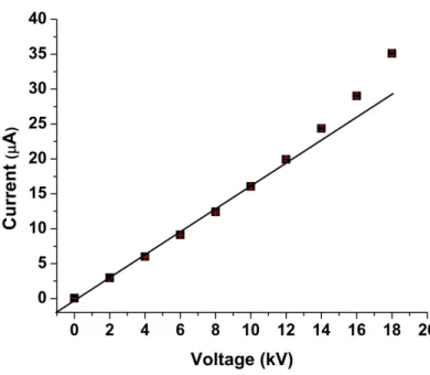
2.3.3.1 CE Optimal Electrophoretic Voltage...55
2.3.3.2 CE Limit of Detection ...57
2.4 Conclusions...58
2.5 Figures ...59
2.6 References...66
Chapter 3: Metabolism of Peptide Reporters in Cell Lysates and Single Cells...71
3.1 Introduction...71
3.1.1 Synthetic Peptides as Kinase Substrates and Inhibitors ...71
3.1.2 Peptides as Reporters in Cell Lysates and Single Cells...72
3.1.3 Limitations of Peptide Reporters ...73
3.1.4 Reporter Peptide Design and Evaluation...74
3.2 Experimental Design ...75
3.2.1 Chemicals ...75
3.2.2 Peptide Synthesis and Preparation...75
3.2.2.1 Synthesis of Full-Length Peptides Amidated on the C-Terminus ...75
xii
3.2.2.3 Synthesis of Peptide Fragment Standards...76
3.2.3 Cell Culture...77
3.2.4 Measurement of Peptide Degradation in a Cell Lysate ...77
3.2.5 in vitro Kinase Assay...78
3.2.6 Measurement of Kinetic Parameters...79
3.2.7 Capillary Electrophoresis...79
3.2.8 Single Cell Capillary Electrophoresis...80
3.3 Results and Discussion ...81
3.3.1 Selection of the Starting Peptide...81
3.3.2 Characterization of Peptide QW-III-67B Degradation in Cytosolic Lysates...82
3.3.3 Characterization of Peptides Following Lysine Replacement ...83
3.3.4 Identification of the Peptide Fragments...85
3.3.5 Characterization of Peptides with Proline Replacement ...85
3.3.6 Characterization of Peptides with Phenylalanine Replacement ...87
3.3.7 Design and Characterization of an (N-methyl)phenyl- alanine-substituted Lead Peptide ...88
3.3.8 Characterization of the Lead Peptide QW-V-48B in a Cytosolic Lysate ...89
3.3.9 Characterization of Peptides in Single HeLa Cells...90
3.4 Conclusions...93
3.5 Figures and Tables...94
xiii
Chapter 4: Development of a Peptidase-Resistant Substrate for
Single-Cell Measurement of Protein Kinase B Activation...110
4.1 Introduction...110
4.1.1 Protein Kinase B Activation and Function ...110
4.1.2 Methods for Measuring PKB...111
4.1.3 Peptides as Kinase Substrates...112
4.1.4 Iterative Strategy for Design of Peptidase-Resistant Kinase Substrates...113
4.2 Experimental Design ...113
4.2.1 Chemicals ...113
4.2.2 Peptide Synthesis and Preparation...114
4.2.2.1 Synthesis of Full-Length Peptides Amidated on the C-Terminus ...114
4.2.2.2 Alternative Coupling for Difficult Amino Acids...115
4.2.2.3 Synthesis of Peptide Fragment Standards...115
4.2.3 Cell Culture...116
4.2.4 Measurement of Peptide Degradation in a Cell Lysate ...116
4.2.5 in vitro Kinase Assay...117
4.2.6 Measurement of Kinetic Parameters...118
4.2.7 Capillary Electrophoresis...118
4.2.8 Single-Cell Capillary Electrophoresis ...119
4.3 Results and Discussion ...120
4.3.1 Selection of the Starting Peptide and Screening Lysates...120
xiv
4.3.3 Characterization of Peptides Following Replacement of
the Amino-Terminal Alanine...122
4.3.4 Characterization of Peptides Following Arginine Replacement ...123
4.3.5 Characterization of Peptides Following the Replacement of the Carboxy-Terminal Alanine...124
4.3.6 Characterization of Peptides Following Phenylalanine or Alanine Replacement ...125
4.3.7 Characterization of Peptides Following Truncation of the Substrate...125
4.3.8 Characterization of Lead Peptide VI-B in Cytosolic Lysates ...126
4.3.9 Characterization of Peptide Degradation in Single LNCaP Cells ...127
4.3.10 Phosphorylation of Peptide VI-B in Single LNCaP Cells ...129
4.3.11 Inhibition of PKB Activity in Single Cells...129
4.4 Conclusions...130
4.5 Figures and Tables...132
4.6 References...144
Chapter 5: Measurement of PKB Activity in Single Pancreatic Cancer Cells...150
5.1 Introduction...150
5.1.1 Pancreatic Ductal Adenocarcinoma...150
5.1.2 Protein Kinase B and PDA ...151
5.1.3 Xenografts of Human Cancer ...152
5.1.4 Single-cell Analysis of PKB Activity in Pancreatic Cancer Cells...153
xv
5.2 Materials and Methods ...153
5.2.1 Chemicals ...153
5.2.2 Peptide Synthesis and Preparation...154
5.2.3 Cell Culture...154
5.2.3.1 Immortalized Cell Lines ...154
5.2.3.2 Xenograft Cells...155
5.2.4 Single Cell Analysis of PKB Activity ...155
5.2.5 Single-Cell Capillary Electrophoresis ...156
5.3 Results and Discussion ...157
5.3.1 Selection of the Substrate Peptide and Cell Lines ...157
5.3.2 Characterization of Peptide Degradation in Single Tissue-Cultured Cells ...158
5.3.4 Inhibition of PKB Activity in Single Tissue-Cultured Cells ...164
5.3.5 Characterization of VI-B in Pancreatic Tumor Xenografts...165
5.4 Conclusions...166
5.5 Figures ...167
5.6 References...173
Chapter 6: Further Stabilization of a Bcr-Abl Kinase Substrate Reporter ...176
6.1 Introduction...176
6.2 Experimental Design ...176
6.2.1 Chemicals ...176
6.2.2 Peptide Synthesis and Preparation...177
xvi
6.2.2.2 Alternative Coupling for Difficult Amino Acids...178
6.2.2.3 Synthesis of Peptide Fragment Standards...178
6.2.3 Cell Culture...178
6.2.4 Measurement of Peptide Degradation in a Cell Lysate ...178
6.2.5 in vitro Kinase Assay...179
6.2.6 Capillary Electrophoresis...180
6.3 Results and Discussion ...181
6.3.1 Characterization of Peptides Following Replacement of the N-Terminal Alanine...181
6.3.2 Characterization of Peptides Following Replacement of the C-Terminal Alanine ...182
6.3.3 Characterization of VIII-B in a Baf/BCR-ABL Cytosolic Lysate ...183
6.3.4 Characterization of Peptide Following Truncation of the Substrate...184
6.4 Conclusions...185
6.5 Figures and Tables...186
6.6 References...195
Appendix A: Single-Cell CE-LIF Instrument Custom Parts ...196
Appendix B: Single-Cell CE-LIF Instrument Software Programming ...208
Appendix C: Peptide Sequences...214
References...220
xvii
List of Tables
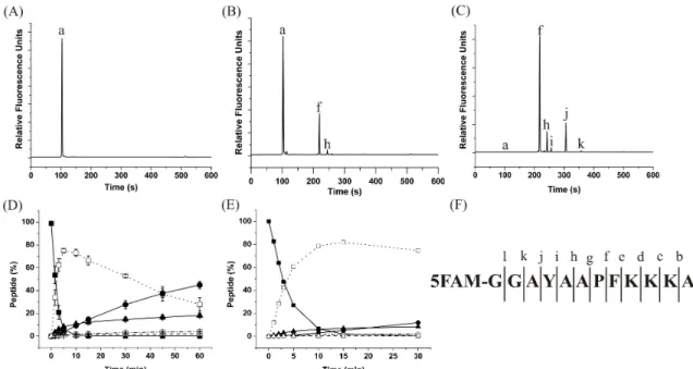
Table 3.1:Properties of the modified peptides derived from the
starting peptide (QW-III-67B)...103
Table 3.2: Percentage of each fragment formed following a 5 min incubation of the peptides in a single cell...104
Table 4.1: Properties of the modified peptides derived from starting peptide I ...143
Table 6.1: Properties of the modified peptides...194
Table C1: The non-standard abbreviations used in the peptide sequences ...215
Table C2: Peptides intended as Abl kinase substrates (Part 1 of 2) ...216
Table C3: Peptides intended as Abl kinase substrates (Part 2 of 2) ...217
Table C4: Peptides intended as PKB substrates (Part 1 of 2)...218
xviii
List of Figures
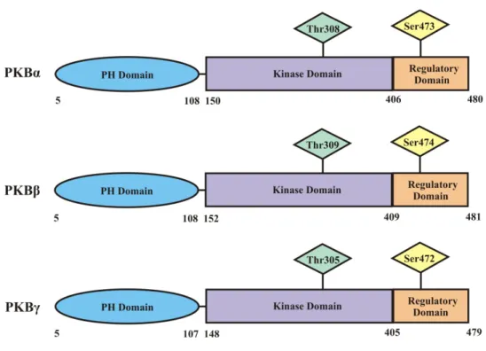
Figure 1.1: The domains of the three isoforms of PKB...20
Figure 1.2: A simplified schematic of PKB activation...21
Figure 1.3:The domains of Abl and Bcr-Abl...22
Figure 1.4: Formation of the Philadelphia chromosome ...23
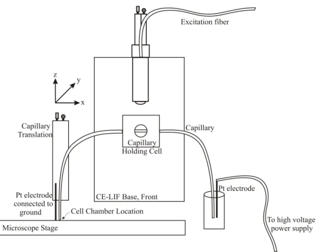
Figure 1.5: Diagram of a generic CE-LIF instrument ...24
Figure 1.6: Movement of analytes through a capillary during CE ...25
Figure 2.1: The single cell CE-LIF instrument described in this chapter...59
Figure 2.2: The CE portion of the CE-LIF instrument ...60
Figure 2.3: Schematic of the excitation pathway for the CE-LIF system ...61
Figure 2.4: Schematic of the emission pathway for the CE-LIF system ...62
Figure 2.5:Schematic of the capillary electrophoresis portion of the CE-LIF system...63
Figure 2.6:An Ohm’s plot generated for a 100 mM tris and 100 mM tricine, pH 8.1 buffer on the single cell CE-LIF instrument...64
Figure 2.7: Limit of detection calculations for the CE-LIF system...65
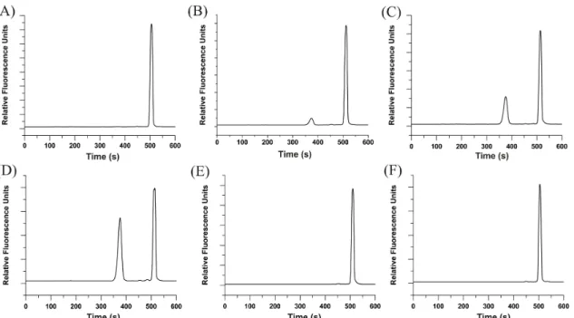
Figure 3.1: in vitro phosphorylation of the starting peptide (QW-III-67B) by recombinant Abl kinase ...94
Figure 3.2: Degradation profile of the starting peptide (QW-III-67B) in a cell lysate ...95
Figure 3.3:The native residues and the non-native residues that were inserted into the peptide to stabilize the peptide against hydrolysis...96
xix
Figure 3.5: Baf/BCR-ABL cytosolic lysate degradation (A) and
in vitro phosphorylation (B) of modified peptides ...98
Figure 3.6: Electropherograms of peptide QW-IV-85B incubated
in a Baf/BCR-ABL cytosolic lysate ...99
Figure 3.7: Electropherograms of QW-V-48B incubated with
recombinant Abl kinase ...100
Figure 3.8: Degradation profile of the lead peptide (QW-V-48B) in
a Baf/BCR-ABL cell lysate ...101
Figure 3.9:Degradation of peptides in single HeLa cells ...102 Figure 4.1: in vitro phosphorylation of starting peptide I over time ...132 Figure 4.2: Degradation of the starting peptide I in cytosolic lysates ...133 Figure 4.3: Electropherograms of the starting peptide I when
incubated in HeLa cytosolic lysate ...134
Figure 4.4: Electropherograms of the starting peptide I when
incubated in LNCaP cytosolic lysate...135
Figure 4.5: Schematic of rational peptide substrate design ...136 Figure 4.6: Line bond structures of native and non-native amino
acids utilized in peptide construction ...137
Figure 4.7: Electropherograms of the lead peptide VI-B when
incubated in HeLa cytosolic lysate ...138
Figure 4.8: Degradation of the lead peptide VI-B in cytosolic lysates...139 Figure 4.9: Electropherograms of the lead peptide VI-B when
incubated in LNCaP cytosolic lysate...140
Figure 4.10: Electropherograms of single LNCaP cells 90 s after microinjection of the starting peptide I (A) or lead
peptide VI-B (B) ...141
Figure 4.11: Electropherograms of single LNCaP cells 5 min after microinjecting the lead peptide QW-VII-48F without
(A) or with pre-treatment with wortmannin (B) ...142
xx
Figure 5.1: Select electropherograms from HPAF-II (A), CFPAC-1
(B), and PANC-1 (C) cells with no inhibitor treatment...167
Figure 5.2: The rate of degradation of intact peptide VI-B as a
function of initial substrate concentration ...168
Figure 5.3: Percent phosphorylation in HPAF-II (open squares), CFPAC-1 (open triangles), and PANC-1 (open circles)
cells with and without pre-treatment with wortmannin...169
Figure 5.4: The rate of phosphorylation of intact peptide VI-B as
a function of initial substrate concentration...170
Figure 5.5: Select electropherograms from HPAF-II (A), CFPAC-1
(B), and PANC-1 (C) cells pre-incubated with wortmannin ...171
Figure 5.6: Electropherograms from incubation of peptide 48F in human pancreatic cancer xenograft cells for 90 s
(A and B) and 5 min (C) ...172
Figure 6.1: The non-native amino acids utilized in the construction
of alternative peptides...186
Figure 6.2: Select electropherograms of peptide VII-A (A), VII-B (B), and VII-C (C) incubated for 60 min in a
Baf/BCR-ABL cytosolic lysate ...187
Figure 6.3: Baf/BCR-ABL cytosolic lysate degradation (A) and
in vitro phosphorylation (B) of the series VII peptides ...188
Figure 6.4: Select electropherograms of peptide VIII-A (A), VIII-B (B), and VIII-C (C) incubated for 60 min in a
Baf/BCR-ABL cytosolic lysate ...189
Figure 6.5: Baf/BCR-ABL cytosolic lysate degradation (A) and
in vitro phosphorylation (B) of the series VIII peptides...190
Figure 6.6: Degradation profile of peptide VIII-B in a Baf/BCR-ABL
cell lysate ...191
Figure 6.7: Degradation profile of peptide IX-A in a Baf/BCR-ABL
cell lysate ...192
Figure 6.8: Baf/BCR-ABL cytosolic lysate degradation (A) and
in vitro phosphorylation (B) of peptides...193
xxi
Figure A1: Blueprint for the CE-LIF microscope stage adapter ...197
Figure A2: Blueprint for the CE-LIF base, bottom ...198
Figure A3: Blueprint for the CE-LIF base, front...199
Figure A4: Blueprint for the CE-LIF base, top...200
Figure A5: Blueprint for the CE-LIF detector housing ...201
Figure A6: Blueprint for the Collection lens holder...202
Figure A7: Blueprint for the PMT housing ...203
Figure A8: Blueprint for the PMT filter drawer ...204
Figure A9: Blueprint for the PMT shutter plate ...205
Figure A10: Blueprint for the PMT shutter ...206
Figure A11: The modifications needed on the two (2) plate adapters...207
Figure B1: The front panel of the LabVIEW program created for the single cell CE-LIF system...209
Figure B2: The first portion of the block diagram of the LabVIEW program created for the single cell CE-LIF system...210
Figure B3: The event structure utilized in the block diagram of the LabVIEW program created for the single cell CE-LIF system ...211
Figure B4: The code contained within the CE case structure which is executed once when the user presses the green “Start CE” button on the front panel...212
xxii
List of Abbreviations and Symbols
A alanine
Abl Abelson tyrosine kinase
AGC Protein Kinase A, G, or C sub-family A/D analog to digital
Akt Protein kinase B
Ala alanine
amol attomole
Arg arginine
Arg(Me2) N-ω,ω-dimethyl-L-arginine (symmetrical)
ATP adenosine triphosphate
B band-broadening due to longitudinal diffusion β-Ala beta-alanine
β-Arg beta-arginine
bcr breakpoint cluster region Bcr-Abl Bcr-Abl fusion protein BNC Bayonet Neill-Concelman BSA bovine serum albumin oC degrees Celcius
c-Abl chromosomal Abl kinase CaCl2 calcium chloride
CE capillary electrophoresis
xxiii
CH2Cl2 dichloromethane
cm centimeter
CML chronic myelogenous leukemia CMOS complementary metal oxide silicon CO2 carbon dioxide
C-terminus carboxy terminus
CyTOF cytometry by time-of-flight
2D two-dimensional
d diameter
Da Daltons
D/A digital to analog
D-Ala D-alanine
DAQ data acquisition
DIC diisopropylcarbodiimide DIPEA N,N-diisopropylethylamine
DMEM Dulbecco’s Modified Eagle Medium DMF dimethylformamide
DNA deoxyribonucleic acid DPh D-phenylalanine
DTT dithiothreitol
E glutamic acid
xxiv
EDTA ethylene diamine tetraacetic acid e.g. for example
EO electroosmotic
eq equivalents
F phenylalanine
FAM carboxyfluorescein FBS fetal bovine serum
Fmoc 9-fluorenylmethoxycarbonyl FRET Förster resonance energy transfer
G glycine
g acceleration due to gravity
H theoretical plate height
h hours
h height
HCl hydrochloric acid
HCTU 2-(6-chloro-1H-benzotriazole-1-yl)-1,1,3,3-tetramethylaminium hexafluorophosphate
He/Ne Helium/Neon laser
HEPES 4-(2-Hydroxyethyl)piperazine-1-ethanesulfonic acid HF hydrofluoric acid
H2O water
HOBt N-hydroxybenzotriazole
xxv
HPLC-MS high performance liquid chromatography coupled with mass spectrometric detection
hPa hectopascals
HV high voltage
Hz Hertz
I isoleucine
ID inner diameter
IMAP immobilized metal ion affinity-based fluorescence polarization IMDM Iscove’s Modified Dulbecco’s Medium
K lysine
KCl potassium chloride kDa kiloDalton
kHz kilohertz
KH2PO4 potassium phosphate
kcat turnover number
KM Michaelis-Menten constant
kV kilovolt
L length
LIF laser-induced fluorescence LOD limit of detection
µ electrophoretic mobility
µA microamps
xxvi
µep electrophoretic mobility
µg microgram
µJ microJoules
µL microliter
µM micromolar
µm micrometers
M molar
M mass
m meter
MAP mitogen activated protein MeAla N-methyl alanine
MeArg N-methyl arginine
MEM Minimal Essential Medium MePh N-methyl phenylalanine MePhe N-methyl phenylalanine
mg milligram
MgCl2 magnesium chloride
min minutes
mL milliliter
MLCK myosin light chain kinase mM millimolar
mm millimeters
xxvii
MOPS 3-(N-morpholino)propane sulfonic acid MPh N-methylphenylalanine
MePh N-methylphenylalanine mRNA messenger ribonucleic acid MS mass spectrometry
ms millisecond
MSNT 1-(mesitylene-2-sulfonyl)-3-nitro-1,2,4-triazole mTOR mammalian target of rapamycin
mTORC2 mammalian target of rapamycin complex 2
mW milliwatt
η viscosity
NA numerical aperture NaCl sodium chloride NaH2PO4 sodium phosphate Nal β-(2-Napthyl)-L-alanine
NaOH sodium hydroxide
Nd:YAG neodymium-doped yttrium aluminum garnet
nL nanoliter
nM nanomolar
nm nanometer
xxviii
O ornithine
OD outer diameter
π pi
32P radioactive isotope of phosphorus
P proline
PBS phosphate buffered saline
PDA pancreatic ductal adenocarcinoma PDK-1 phosphoinositide-dependent kinase-1 PDMS poly(dimethyl)siloxane
PEG poly-ethylene glycol
PH pleckstrin homology domain Phe phenylalanine
Phe(F5) pentafluorophenylalanine
PHLPP PH-domain leucine-rich repeat protein phosphatase PI3-K phosphoinositide 3-kinase
PIP2 phosphatidylinositol 4,5-bisphosphate PIP3 phosphatidylinositol 3,4,5-trisphosphate PKB protein kinase B
pg picogram
pL picoliter
pM picomolar
pm picometer
xxix
Pro proline
psi pounds per square inch
PTEN phosphatase and tensin homolog
PyBrop bromo-tris-pyrrolidino phosphonium hexafluorophosphate
q particle charge
ρ density
R arginine
R resistance
r Stoke’s radius
RFU relative fluorescence units RNA ribonucleic acid
RPMI Roswell Park Memorial Institute RTK receptor tyrosine kinase
s seconds
Sarc sarcosine, N-methylglycine SDS sodium dodecyl sulfate
Ser serine
Ser473 serine residue in position 473 SH2 Src homology 2 domain SH3 Src homology 3 domain S/N signal to noise
xxx
Src sarcoma kinase
STI-571 Imatinib mesylate, Gleevec
T threonine
t time
t1/2 half-life
t50% P time to 50% phosphorylation TFA trifluoroacetic acid
Thr threonine
Thr308 threonine residue in position 308 TIS triisopropyl silane
TK tyrosine kinase Tris-HCl trizma hydrochoride Tyr(3-NO2) 3-nitrotyrosine
u linear velocity
UV ultraviolet
V volts
V partial specific volume
v velocity
W tryptophan
X times
Y tyrosine
CHAPTER 1
INTRODUCTION
1.1 Kinase Pathways and Implications in Cancer
1.1.1 Kinases and Kinase Functions
Nearly all proteins undergo post-translational modification after synthesis. These modifications can be irreversible, such as proteolysis of peptide bonds in the degradation and reprocessing of substrates, or reversible, such as acylation or glycosylation used to modify protein properties.1 One common form of reversible modification is phosphorylation by a kinase. A protein kinase catalyzes the transfer of the terminal phosphoryl group from adenosine triphosphate (ATP) to a serine, threonine, or tyrosine residue on a substrate. This reversible modification is often used as a switching mechanism to activate and deactivate proteins. Phosphorylation of a target protein alters its properties by addition of the
2
many of the signal transduction pathways leading to growth, metabolism, transcription differentiation, behavior, and apoptosis.1, 2 Kinases themselves are often regulated by phosphorylation and dephosphorylation.
There are over 500 known kinases in the human genome which have been grouped into eight different families based on their structure and function.2 For example, the tyrosine kinase (TK) family of the human genome contains all of the receptor tyrosine kinases (RTKs) responsible for signal transduction from the extracellular environment to the cytoplasm of cells.3 RTKs on the surface of cell membranes bind to ligands, resulting in propagation of signal transduction within the cell. Included in tasks accomplished by RTKs are roles in regulation of cell proliferation or differentiation, vital processes for organism growth and function.3 The TK family also contains multiple non-receptor tyrosine kinases (NRTKs), critical members of signaling cascades within a cell. Among other tasks, the NRTKs are involved in glucose uptake and metabolism, as well as in regulation of the immune system. Another family in the human kinome is the AGC family (related to protein kinases A, G, and C), which contains approximately 60 kinases responsible for a variety of functions including, but not limited to, proliferation, metabolism, protein synthesis, cell shape, and motility.4 As kinases are involved in modulation of multiple signal transduction pathways, dysregulation of TK, AGC or other family kinase activity is implicated in many diseases, including multiple cancers.2-5 It is for this reason that kinases are prime targets for therapeutic
3
1.1.2 Protein Kinase B
Protein kinase B (PKB, also known as Akt) has been implicated in many cellular functions such as insulin signaling, glucose metabolism, transcription, proliferation, stress response, and apoptosis.7 PKB was first described in 1977 by Staal et al. in their work with a murine leukemia virus found to be linked to lymphoma formation.8 It was not until 1991 that the genes encoding PKB were found, when three independent research groups published results regarding PKB.9-11 Interest in PKB has steadily increased as it has been implicated in assisting cellular survival during stressful events, such as prevention of apoptosis in cells programmed for cell death.12 Increased PKB activity has been found to enhance tumor progression and is present in multiple cancers, including pancreatic, breast and prostate tumors.13, 14 Many reasons exist for high PKB activity and any of these can lead to tumor survival and progression: i) PKB can be constitutively activated;15 ii) normal PKB can be over-expressed;12, 16 or iii) regulatory feedback control of PKB can be impaired.8, 17 While increased PKB activity may not be directly responsible for producing tumors, there is
abundant evidence that increased PKB activity does enable tumors to survive and proliferate, making PKB an attractive candidate for targeted therapy.13, 18, 19
PKB is a serine/threonine kinase and a member of the AGC family of kinases.2 There are three isoforms of PKB, termed PKBα, PKBβ, and PKBγ, each responsible for different functions within cells. All three isoforms are approximately 80% homologous and share similarities in three highly conserved functional domains (Figure 1.1). These domains are: i) a pleckstrin homology (PH) domain found on the N-terminus which is a region of
4
phosphorylated; and iii) a C-terminal regulatory domain containing one of the phosphorylation sites for PKB regulation.7, 14, 15, 17, 20
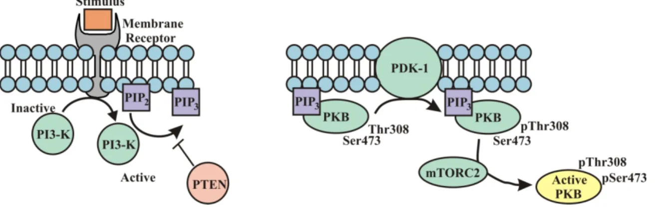
PKB activity is controlled by phosphorylation on two residues, Thr308, located in the kinase domain, and Ser473, found in the regulatory domain. Phosphorylation of both
5
1.1.3 Abelson Tyrosine Kinase
Abelson tyrosine kinase (Abl) is a member of the TK family of kinases as a non-receptor kinase and is normally encoded by a gene found on chromosome 9. Abl can be found in many locations within the cell and is known to be involved in many cellular
processes such as cell migration, DNA damage responses, and even regulation of growth and survival.22 Abl contains the same three core domains as other Src-family kinases—an SH3, SH2 and kinase domain (Figure 1.3).22, 23 Normal chromosomal Abl (c-Abl) contains a region upstream of the SH3 domain that is myristoylated and is responsible for regulation of the kinase. c-Abl is tightly regulated both temporally and spatially by auto-inhibition involving this myristate tail, the SH2-SH3 connector and the SH2-kinase domain linker.22-24
In the early 1960s, it was discovered that many chronic myelogenous leukemia (CML) patients had a shortened version of chromosome 22 (termed the Philadelphia
6
that competes with ATP for the binding pocket in the catalytic domain of Bcr-Abl29, 30 and prevents Bcr-Abl from phosphorylating its substrates. Treatment with Gleevec has proven useful for many CML patients, notably those still in the chronic phase of the disease.31 The success of Gleevec has led to the development of 2nd and 3rd generation inhibitors of Bcr-Abl as well as investigation and development of alternative drug therapies aimed specifically at targeting aberrant kinase activity.
As successful as Gleevec treatment has been for treatment of chronic phase CML, it has not worked for all patients, especially those in advanced or blast-stage CML.32-34 There is also a sub-population of CML patients that do not have the Philadelphia chromosome, so drugs targeting Bcr-Abl are useless as these patients do not express Bcr-Abl.26 Direct measurement of Bcr-Abl kinase activity would allow for quantification of Bcr-Abl activity and investigation of the likely response and sensitivity to Gleevec or to its 2nd and 3rd generation relatives. Chapters 3 and 6 of this dissertation discuss development of a peptide substrate reporter for Bcr-Abl kinase activity.
1.2 Use of Peptides in Biomedical Research
7
activity in vitro, in cell lysates, or in vivo. In addition, peptides can be constructed to contain only the select kinase’s consensus sequence, the short region of a handful of amino acids around the phosphoryl-accepting residue recognized by a specific kinase or kinase family. Sometimes, these short peptide regions can mimic a full-length substrate protein and large libraries of consensus sequences have been determined for many kinases and the preferred sequences are readily available for nearly all known kinases.20, 36-38 For these reasons many peptides are available commercially for use as substrates to measure kinase activity.
1.2.1 Peptides as Substrates
1.2.1.1 Techniques for Measuring Peptide Phosphorylation
Peptides are frequently utilized by commercial companies and research laboratories to quantify kinase activity of purified kinases. These formats are relatively inexpensive ways to quickly assess kinase activity and specificity. Select methods frequently utilized to measure phosphorylation of a peptide substrate by a kinase are discussed below. Transfer of a radioactive phosphoryl group from [32P]-γ-ATP to the substrate can be quantitatively
determined by capturing the radioactive substrates on a negatively charged phosphocellulose membrane and measuring radioactivity.35, 39 An alternative radioactive technique is a
homogenous scintillation proximity assay (SPA) that measures incorporation of the radioactive phosphoryl group on a substrate. When the radiolabeled substrate binds to the SPA bead, the emitted radiation activates the scintillant, which produces light that can be measured with a scintillation counter.40, 41 However, due to the cost of disposal of
8
(FRET).42 For example, a substrate constructed with a donor and acceptor fluorophore emits no fluorescence when the target amino acid is not phosphorylated as the conformation of the substrate separates the two fluorophores from one another. A conformational change is induced upon phosphorylation, usually when a phosphoryl-recognition sequence binds to the phosphoryl-group, arranging the fluorophores in close proximity to one another. When the proper wavelength of light illuminates the substrate, FRET occurs as the donor fluorophore emits in the acceptor fluorophore excitation region. The emission of the acceptor
fluorophore is measured over time and utilized to quantify phosphorylation. Additional techniques to measure phosphorylation include immobilization of biotin-labeled substrate peptides on a streptavidin-coated surface, where phosphorylation is detected via
radioactivity, colorimetric changes, or fluorescence of a labeled antiphosphoserine, -threonine, or -tyrosine antibody.43-45 Enzyme activity is inferred based on the amount of phosphorylated substrate relative to non-phosphorylated substrate. An alternative technique to monitor substrate phosphorylation measures fluorescence polarization in the presence of an appropriate antiphospho antibody or a metal nanoparticle.46, 47 Fluorescence polarization is high when the phosphorylated substrate is bound by the relatively large antibody or nanoparticle because the bulky addition prevents free rotation of the molecule, resulting in highly polarized emitted light. This fluorescence polarization change upon substrate phosphorylation permits quantification of enzyme activity. Additionally, capillary
electrophoresis can be used to monitor substrate phosphorylation over time, as the addition of the double negatively-charged phosphoryl group results in altered electrophoretic mobility between the substrate and its phosphorylated counterpart, allowing for separation and
9
but most often utilizes fluorescence detection of fluorescently-labeled substrates because of the exquisite sensitivity offered by laser-induced fluorescence detection. These techniques, as well as others not mentioned, can all be used to measure kinase activity of purified
kinases, for example, to determine substrate suitability or as validation of new technology to measure enzyme activity.50, 52, 53
1.2.1.2 Peptide Substrates in Cell Lysates
In addition to their usefulness in monitoring activity of purified kinases, peptides are also utilized in vitro to monitor kinase activity in a cytosolic lysate. Cytosolic lysates provide an environment in which many of the cofactors and regulatory elements are present, though minimally regulated, allowing for a simulation of kinase behavior in these
environments. Cytosolic lysates are also attractive because they are less expensive to use than intact cells for many reasons. Lysates are generated via a freeze-thaw cycle or by adding a lysis buffer, neither of which require specialized or costly materials. Many different measurement techniques can be utilized to accommodate assays performed in a cytosolic lysate, eliminating the need for purchase or construction of specialized detection instruments. Cytosolic lysates are easy to prepare in large volumes and can be modified to match nearly any detection strategy. Lysates are easily amenable to addition of co-factors or inhibitors to monitor enzyme reaction to changes and the substrate can be directly mixed with the lysate in vitro. A search of the literature yields multiple examples of peptides utilized as substrates of kinases in cell lysates.54-64
1.2.1.3 Peptide Substrates in Intact Cells
10
within an intact cell. Intracellular compartments exist within intact cells, which keeps cell components isolated from other components and localized to certain areas. When a lysate is generated, these subcellular compartments are ruptured, eliminating this separation and localization. Additionally, intracellular compartments also facilitate formation of local regions of high concentration of a specific analyte, also eliminated when the lysate is generated and diluted into a homogenous mixture.
However, despite the cost advantages of lysates over single cells, lysates cannot recapitulate the native environment of an intact cell and lysates eliminate the ability to detect heterogeneity within a cell population. To gain an understanding into protein behavior within a population of cells, peptides are also being increasingly used in single cells as substrates to measure protein activity at the single cell level, such as that of kinases, acyl transferases, and proteases.49, 51, 65-67 In these examples, peptide substrates were introduced into tissue-cultured cell lines via endocytosis,66 electroporation,65 or microinjection49, 51, 67 and measurements were made utilizing laser-induced fluorescence detection after capillary electrophoresis on custom-designed systems mounted on a microscope stage. These
substrate loading and measurement strategies are also amenable for use with primary cells, an exciting new frontier for studying protein activity in a population of diseased and healthy cells.
1.2.2 Peptide Susceptibility to Degradation
Despite all of the advantages listed above and the widespread use of peptides as substrates both in vitro and in vivo, peptides suffer from a susceptibility to degradation by intracellular proteases and peptidases. Cleavage of peptide bonds by endo- and
11
cells, responsible for breaking proteins and peptides into their constituent amino acids.68 While not a problem for use with purified kinases, this is a major concern when peptides are utilized in cell lysates or intact cells because of the prevalence of peptidases. Peptidolysis can be extremely rapid, yielding peptide fragmentation within minutes of introduction into a lysate or single cell.51, 69, 70 This is true for peptide substrates and inhibitors and low
bioavailability due to peptide hydrolysis is a major disadvantage for peptide-based therapeutics.71-75
While protease inhibitors can be used to slow degradation, several drawbacks exist to using these inhibitors. Protease inhibitors do not entirely eliminate protease activity, they are often poorly soluble in aqueous solutions, rendering them available only in organic solvents not compatible with enzymes. Most inhibitors cannot be used with intact cells since they are not membrane permeable. Once a peptide is cleaved, it is generally no longer an effective substrate for the desired kinase as it does not possess all of the elements required for kinase recognition. In order for peptides to become more useful in biomedical research, proteolytic degradation needs to be dramatically slowed or eliminated. Strategies for achieving
proteolytic stability are discussed in the following sections.
1.2.3 Strategies to Stabilize Peptides Against Degradation
1.2.3.1 Cyclization
Two strategies that exist for cyclizing peptides are head-to-tail cyclization or
12
rings used and in the methods utilized for cyclization, offering a range of useful strategies for prolonging peptide lifetime. However, cyclization methods are very challenging
synthetically, requiring excessive amounts of time and resources for a very low yield of substrate. While cyclization is a valid strategy for stabilization of peptides, it does not lend itself to rapid synthesis and screening of possible substrates because of its synthetic
challenges, expense, and reduction of substrate efficacy. 1.2.3.2 PEGylation
Addition of a poly-ethylene glyocol moiety (PEGylation) has also been demonstrated to reduce proteolytic activity on peptides.80-82 PEGylation is relatively straightforward to accomplish and is easily incorporated into regular SPPS methods. PEG groups of various sizes (with molecular weights ranging from 1 – 30 kDa) can be added to peptides, thus altering their properties depending on the PEG group added. Lower molecular weight PEGylated substrate peptides tend to have better substrate activity than higher molecular weight substrates, yet also demonstrate a diminished resistance to degradation when compared to their higher-molecular weight counterparts.81 While PEGylation of peptides allows them to resist degradation, problems exist due to the size and polydispersity of PEG groups. The molecular weights of most PEG groups are dramatically larger than the substrate peptides, rendering the mass difference between substrate and its phosphorylated counterpart much less and making electrophoretic separations difficult to achieve.
13
1.2.3.3 Terminal Modifications
Modifications to either or both the C- and N-terminus have also been shown to impart some stability to peptides.80, 83, 84 These modifications are varied and include lipid
conjugations, acetylation of the N-terminus and amidation of the C-terminus. All of these modifications are easily accomplished via standard SPPS techniques. Lipid conjugation of peptides serves the added benefit of making the reporter more lipophilic, allowing it to become more cell permeable while also increasing loading efficiency into cells. Terminal modifications provide added protection against carboxy- and amino-peptidases, but do not stabilize the peptide against endo-peptidase degradation. For this reason, terminal
modifications are often used in combination with one or more alternative stabilization strategies.
1.2.3.4 Non-Native Amino Acids
A strategy frequently utilized in peptide substrate and inhibitor design for promotion of peptide stability is utilization of non-native amino acids within the peptide. These can include D-amino acids, where the stereochemistry of the α-carbon is mirror image to the
natural L-amino acids;85 N-methylated amino acids, where a methyl group is added to the
backbone nitrogen;86β- or γ-amino acids, where the backbone nitrogen is on the β- or γ -carbon as opposed to the α-carbon;87 or a modification of any sort to the side chain.35 Many non-native amino acids are available and ready for synthesis using standard SPPS techniques, making their incorporation into a peptide straightforward. There are multiple examples of use of peptides constructed with non-native amino acids in the literature.35, 51, 77, 85, 86, 88, 89 An added benefit is that these peptides can be degradation resistant while simultaneously
14
in peptide synthesis, this is the strategy utilized to stabilize reporters chosen for the work described in this dissertation. A more thorough discussion of how this was accomplished is presented in Chapters 3 through 6 of this work.
1.3 Capillary Electrophoresis
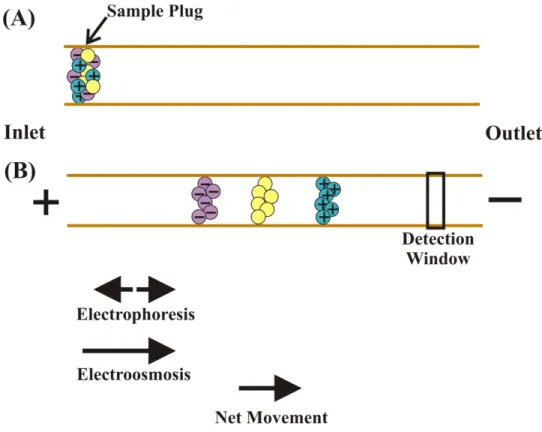
Capillary electrophoresis (CE) was first described in the early 1980s by Jorgenson and Lukacs.90 CE is a separation method utilizing small inner-diameter fused silica capillaries (on the order of 50 µm) filled with and immersed in a current-carrying buffer solution. Sample is introduced at the inlet and a high voltage (typically up to 30 kV) is applied across the capillary. A detector near the capillary outlet monitors the species as they move toward the column exit (Figure 1.5). The velocity and direction of electrophoretic migration is a property of the size, shape, and charge of the species. Overall migration is determined by a combination of the electrophoretic mobility of the analytes and the electroosmotic flow of the buffer. For a rigid sphere, electrophoretic mobility can be
approximated as shown in equations 1.1 and 1.2 and velocity of a species in CE is defined in equation 1.3.90, 91
Equation 1.1:
µ = electrophoretic mobility
q = particle charge η = buffer viscosity
15
Equation 1.2:
M = mass
r = Stoke’s radius
V = partial specific volume
Equation 1.3:
v = velocity
µep= electrophoretic mobility
µeo = electroosmotic mobility
E = applied electric field
16
capillary that bulk migration of all buffer contents occurs in one direction, as the double layer ions migrating in the electric field pull their associated components with them. This results in a nearly flat fluid profile as all capillary components migrate to one end of the capillary.90, 92 This flow, termed electroosmotic flow (EO flow) can be quite significant and it is utilized in CE to sweep all species toward the detector (Figure 1.6). Electrophoresis separates the analytes into discrete bands based on electrophoretic mobility and EO flow sweeps all bands toward the detector, allowing for detection of all species, including those of opposite charges as well as neutral species.
In this dissertation, CE was used for the separation of phosphorylated and non-phosphorylated peptides as well as peptide fragments. Phosphorylation of a peptide
sequence yields an addition of a relatively small amount of mass and two negative charges, altering the electrophoretic mobility of the peptide and causing it to migrate differently than its non-phosphorylated counterpart. A comparison of the integrated peak areas under the peaks corresponding to non-phosphorylated substrate and its phosphorylated counterpart can be used to measure phosphorylation over time and allow for an inference of kinase activity.49, 67, 93, 94 Also, when a peptide undergoes proteolysis, the differences in electrophoretic
mobility of each fragment can be exploited, allowing for separation of all possible fragments. The high resolving power of CE permits separation and quantification of peptide fragments differing by a single amino acid, with the area under each fragment peak correlating to the concentration of the peptide fragment in solution.51 Coupling of CE with ultrasensitive laser-induced fluorescence detection (LIF) allows for work at extremely low peptide
17
small sample volumes (less than 10 nL) and ease of automation, all amenable to work with single cells. Chapter 2 provides a more thorough description of the custom made CE system utilized in this work.
1.4 Chemical Cytometry
Classic cytometry refers to instrumental methods utilized in the characterization of individual cells, such as image and flow cytometry.97 These techniques are often
nondestructive and can process large numbers of cells in relatively short periods of time to provide a wealth of information about cell populations. Both image and flow cytometry rely on fluorescence detection and can simultaneously monitor multiple parameters. However, spectral overlap of fluorescence markers is an important limitation of classic cytometry techniques, as it limits the number of parameters that can be monitored simultaneously. As opposed to classical cytometry, the use of highly sensitive analytical tools to characterize single cells has been given the name “chemical cytometry.” The infancy of chemical cytometry was initiated with a 1953 study that used a copper silk thread to isolate and quantify RNA levels in single cells. More modern chemical cytometry techniques emerged from the Jorgenson, Ewing, and Yeung labs in the 1980s.98, 99 Some current chemical cytometry measurement techniques include capillary electrophoresis, mass spectrometry and electrochemistry.99-101 Chemical cytometry techniques are usually low throughput and destructive, as the cell is often lysed prior to analysis. However, an abundance of
18
way to those originating from post-translational modifications of proteins in a signaling pathway.97, 99, 101-104
Population averages measured when thousands or millions of cells are pooled masks the heterogeneity that no doubt exists and can give a skewed version of the events. For example, cell signaling within a population was often thought of as being a graded response, in that all cells partially responded to a stimulus. However, it was determined that cells actually can respond in an all-or-none fashion, such that only part of the population responds fully to the signal while other cells do not respond at all.105, 106 This is also true when seeking to identify rare subsets within a population, such as what is found in a tumor. Tumors are heterogenous, with only a small portion of cells that may be abnormal.104, 107, 108 A population average would not reveal the aberrant cells, whereas interrogation of each cell would indicate how many cells were functioning as expected and how many were not. Single cell analysis is the ultimate goal for the work developed in this dissertation as a means for identification of abnormal kinase signaling in single cells, with the future intent of
probing primary cells from a patient to determine kinase activity.
1.5 Scope of the Dissertation
The work described in this dissertation focuses on designing peptidase-resistant peptide substrate reporters for probing kinase activity in single cells. Chapter 2 presents a detailed description of the design and construction of a CE instrument capable of analyzing single cells. In addition to hardware construction, the software program was written
19
20
1.6 Figures
21
22
23
24
Figure 1.5: Diagram of a generic CE-LIF instrument. A high voltage power supply drives separation through a narrow-bore fused silica capillary. Detection via laser-induced
25
Figure 1.6: Movement of analytes through a capillary during CE. (A) A sample plug composed of ions and neutral species is injected into the capillary inlet. (B) When a voltage is applied across the capillary, the analytes separate into discrete bands based on
26
1.7 References
1 G. A. Petsko and D. Ringe, Protein Structure and Function, New Science Press Ltd., London, 2004.
2 G. Manning, D. B. Whyte, R. Martinez, T. Hunter and S. Sudarsanam. "The Protein Kinase Complement of the Human Genome". Science, 2002, 298, 1912-1934.
3 S. R. Hubbard and J. H. Till. "Protein Tyrosine Kinase Structure and Function". Annu. Rev. Biochem., 2000, 69, 373-398.
4 L. R. Pearce, D. Komander and D. R. Alessi. "The nuts and bolts of AGC protein kinases". Nat. Rev. Mol. Cell Bio., 2010, 11, 9-22.
5 G. Giamas, J. Stebbing, C. E. Vorgias and U. Knippschild. "Protein kinases as targets for cancer treatment". Pharmacogenomics, 2007, 8, 1005-1016.
6 D. Hanahan and R. A. Weinberg. "Hallmarks of Cancer: The Next Generation". Cell,
2011, 144, 646-674.
7 D. P. Brazil, J. Park and B. A. Hemmings. "PKB Binding Proteins: Getting in on the Akt". Cell, 2002, 111, 293-303.
8 D. P. Brazil and B. A. Hemmings. "Ten years of protein kinase B signalling: a hard Akt to follow". Trends Biochem. Sci., 2001, 26, 657-664.
9 P. F. Jones, T. Jakubowicz, F. J. Pitossi, F. Maurer and B. A. Hemmings. "Molecular cloning and identification of a serine/threonine protein kinase of the second-messenger subfamily". PNAS, 1991, 88, 4171-4175.
10 A. Bellacosa, J. Testa R., S. P. Staal and P. N. Tsichlis. "A Retroviral Oncogene, akt, Encoding a Serine-Threonine Kinase Containing an SH2-Like Region". Science, 1991,
254, 274-277.
11 P. J. Coffer and J. R. Woodgett. "Molecular cloning and characterisation of a novel putative protein-serine kinase related to the cAMP-dependent and protein kinase C families". Eur. J. Biochem., 1991, 201, 475-481.
12 D. R. Alessi and P. Cohen. "Mechanism of activation and function of protein kinase B".
Curr. Opin. Genet. Dev., 1998, 8, 55-62.
13 X. Chen, H. Thakkar, F. Tyan, S. Gim, H. Robinson, C. Lee, S. K. Pandey, C. Nwokorie, N. Onwudiew and R. K. Srivastava. "Constitutively active Akt is an
27
14 K. M. Nicholson and N. G. Anderson. "The protein kinase B/Akt signalling pathway in human malignancy". Cell. Signal., 2002, 14, 381-393.
15 D. Auguin, P. Barthe, M. Augé-Sénégas, M. Stern, M. Noguchi and C. Roumestand. "Solution structure and backbone dynamics of the pleckstrin homology domain of the human protein kinase B (PKB/Akt). Interaction with inositol phosphates". J. Biomol. NMR, 2004, 28, 137-155.
16 E. Fayard, L. A. Tintignac, A. Baudry and B. A. Hemmings. "Protein kinase B/Akt at a glance". J. Cell Science, 2005, 118, 5675-5678.
17 J. Brognard and A. C. Newton. "PHLiPPing the switch on Akt and protein kinase C signaling". Trends Endocrin. Met., 2008, 19, 223-230.
18 M. A. Lawlor and D. R. Alessi. "PKB/Akt: a key mediator of cell proliferation, survival and insulin responses?". J. Cell Sci., 2001, 114, 2903-2910.
19 C. Garcia-Echeverria and W. R. Sellers. "Drug discovery approaches targeting the PI3K/Akt pathway in cancer". Oncogene, 2008, 27, 5511-5526.
20 D. R. Alessi, F. B. Caudwell, M. Andjelkovic, B. A. Hemmings and P. Cohen. "Molecular basis for the substrate specificity of protein kinase B; comparison with MAPKAP kinase-1 and p70 S6 kinase". FEBS Lett., 1996, 399, 333-338.
21 T. L. Yuan and L. C. Cantley. "PI3K pathway alterations in cancer: variations on a theme". Oncogene, 2008, 27, 5497-5510.
22 O. Hantschel and G. Superti-Furga. "Regulation of the C-Abl and BCR-ABL Tyrosine Kinases". Nat. Rev. Mol. Cell Bio., 2004, 5, 33-44.
23 S. A. Courtneidge, "Escape from inhibition". Nature, 2003, 422, 827-828. 24 S. C. Harrison, "Variation on a Src-like Theme". Cell, 2003, 112, 737-740.
25 N. Heisterkamp, J. R. Stephenson, J. Groffen, P. F. Hansen, A. de Klien, C. Bartram and G. Grosveld. "Localization of the c-abl oncogene adjacent to a translocation break point in chronic myelocytic leukaemia". Nature, 1983, 306, 239-242.
26 A. S. Advani and A. M. Pendergast. "Bcr-Abl variants: biological and clinical aspects".
Leukemia Res., 2002, 26, 713-720.
27 C. R. Bartram, A. de Klein, A. Hagemeijer, T. van Agthoven, A. G. van Kessel, D. Bootsma, G. Grosveld, M. A. Ferguson-Smith, T. Davies, M. Stone, N. Heisterkamp, J. R. Stephenson and J. Groffen. "Translocation of c-abl oncogene correlates with the presence of a Philadelphia chromosome in chronic myelocytic leukemia". Nature, 1983,
28
28 B. J. Druker, S. Tamura, E. Buchdunger, S. Ohno, Segal, Gerald M. Fanning, Shane, J. Zimmermann and N. B. Lydon. "Effects of a selective inhibitor of the Abl tyrosine kinase on the growth of Bcr-Abl positive cells". Nat. Med., 1996, 2, 561-566.
29 J. M. Goldman and J. V. Melo. "Targeting the BCR-ABL Tyrosine Kinase in Chronic Myeloid Leukemia". New Engl. J. Med., 2001, 344, 1084-1086.
30 D. G. Savage and K. H. Antman. "Imatinib Mesylate-A New Oral Targeted Therapy". N. Engl. J. Med., 2002, 346, 683-693.
31 C. A. Schiffer, "BCR-ABL Tyrosine Kinase Inhibitors for Chronic Myelogenous Leukemia". N. Engl. J. Med., 2007, 357, 258-265.
32 E. Weisberg, P. W. Manley, W. Breitenstein, J. Brüggen, S. W. Cowan-Jacob, A. Ray, B. Huntly, D. Fabbro, G. Fendrich, E. Hall-Meyers, A. L. Kung, J. Mestan, G. Q. Daley, L. Callahan, L. Catley, C. Cavazza, A. Mohammed, D. Neuberg, R. D. Wright, D. G. Gilliland and J. D. Griffin. "Characterization of AMN107, a selective inhibitor of native and mutant Bcr-Abl". Cancer Cell, 2005, 7, 129-141.
33 E. Weisberg, P. Manley, J. Mestan, S. Cowan-Jacob, A. Ray and J. D. Griffin. "AMN107 (nilotinib): a novel and selective inhibitor of BCR-ABL". Brit. J. Cancer,
2006, 94, 1765-1769.
34 E. Weisberg, P. W. Manley, S. W. Cowan-Jacob, A. Hochhaus and J. D. Griffin. "Second generation inhibitors of BCR-ABL for the treatment of imatinib-resistant chronic myeloid leukaemia". Nat. Rev. Cancer, 2007, 7, 345-356.
35 J. H. Lee, S. K. Nandy and D. S. Lawrence. "A Highly Potent and Selective PKCα Inhibitor Generated via Combinatorial Modification of a Peptide Scaffold". J. Am. Chem. Soc., 2004, 126, 3395.
36 P. P. Pungaliya, Y. Bai, K. Lipinski, V. S. Anand, S. Sen, E. L. Brown, B. Bates, P. H. Reinhart, A. B. West, W. D. Hirst and S. P. Braithwaite. "Identification and
CHaracterization of a Leucine-Rich Repeat Kinase 2 (LRRK2) Consensus Phosphorylation Motif". PLoS ONE, 2010, 5, 1-13.
37 K. Nishikawa, A. Toker, F. Johannes, Z. Songyang and L. C. Cantley. "Determination of the Specific Substrate Sequence Motifs of Protein Kinase C Isozymes". J. Biol. Chem.,
1997, 272, 952-960.
38 J. Wu J., D. E. H. Afar, H. Phan, O. Witte N. and K. S. Lam. "Recognition of Multiple Substrate Motifs by the c-ABL Protein Tyrosine Kinase". Com. Chem. High T. Scr.,
29
39 C. J. Hastie, H. J. McLauchlan and P. Cohen. "Assay of protein kinases using radiolabeled ATP: a protocol". Nat. Protoc., 2006, 1, 968-971.
40 M. Beveridge, Y. Park, J. Hermes, A. Marenghi, G. Brophy and A. Santos. "Detection of p56lck Kinase Activity Using scintillation Proximity Assay in 384-Well Format and Imaging Proximity Assay in 384- and 1536-Well Format". J. Biomol. Screen., 2000, 5, 205-211.
41 O. B. McDonald, W. J. Chen, B. Ellis, C. Hoffman, L. Overton, M. Rink, A. Smith, C. J. Marshall and E. R. Wood. "A Scintillation Proximity Assay for the Raf/MEK/ERK Kinase Cascade: High-Throughput Screening and Identification of Selective Enzyme Inhibitors". Anal. Biochem., 1999, 268, 318-329.
42 S. M. Rodems, B. D. Hamman, C. Lin, J. Zhao, S. Shah, D. Heidary, L. Makings, J. H. Stack and B. A. Pollok. "A FRET-Based Assay Platform for Ultra-High Density Drug Screening of Protein Kinases and Phosphatases". Assay Drug Dev. Tech., 2002, 1, 9-19. 43 E. M. Schaefer and S. Guimond. "Detection of Protein Tyrosine Kinase Activity Using a
High-Capacity Streptavidin-Coated Membrane and Optimized Biotinylated Peptide Substrates". Anal. Biochem., 1998, 261, 100-112.
44 M. Kim, Y. Park, D. Shin, J. Kim, B. Kim and Y. Lee. "Antibody-free peptide substrate screening of serine/threonine kinase (protein kinase A) with a biotinylated detection probe". Anal. Biochem., 2011, 413, 30-35.
45 T. Li, D. Liu and Z. Wang. "Screening Kinase Inhibitors with a Microarray-Based Fluorescent and Resonance Light Scattering Assay". Anal. Chem., 2010, 82, 3067-3072. 46 R. Seethala and R. Menzel. "A Fluorescence Polarization Competition Immunoassay for
Tyrosine Kinases". Anal. Biochem., 1998, 255, 257-262.
47 E. A. Gaudet, K. Huang, Y. Zhang, W. Huang, D. Mark and J. R. Sportsman. "A Homogenous Fluorescence Polarization Assay Adaptable for a Range of Protein Serine/Threonine and Tyrosine Kinases". J. Biomol. Screen., 2003, 8, 164-175. 48 H. Li, H. Y. Wu, Y. Wang, C. E. Sims and N. L. Allbritton. "Improved capillary
electrophoresis conditions for the separation of kinase substrates by the laser micropipet system". J. Chromatogr. B, 2001, 757, 79-88.
49 H. Li, C. E. Sims, M. Kaluzova, E. J. Stanbridge and N. L. Allbritton. "A Quantitative Single-Cell Assay for Protein Kinase B Reveals Important Insights into the Biochemical Behavior of an Intracellular Substrate Peptide". Biochemistry, 2004, 43, 1599-1608. 50 N. Fernandes, D. E. Bailey, D. L. VanVranken and N. L. Allbritton. "Use of Docking
30
Protein Kinase Extracellular Signal-Regulated Kinase". ACS Chem. Biol., 2007, 2, 665-673.
51 A. Proctor, Q. Wang, D. S. Lawrence and N. L. Allbritton. "Metabolism of peptide reporters in cell lysates and single cells". Analyst, 2012, 137, 3028-3038.
52 D. J. Bernsteel, D. L. Roman and R. R. Neubig. "In vitro protein kinase activity measurement by flow cytometry". Anal. Biochem., 2008, 383, 180-185.
53 L. L. Parker, S. B. Brueggemeier, W. J. Rhee, D. Wu, S. B. H. Kent, S. J. Kron and S. P. Palecek. "Photocleavable peptide hydrogel arrays for MALDI-TOF analysis of kinase activity". Analyst, 2006, 131, 1097-1104.
54 Q. Wang, E. I. Zimmerman, A. Toutchkine, T. D. Martin, L. M. Graves and D. S. Lawrence. "Multicolor Monitoring of Dysregulated Protein Kinases in Chronic Myelogenous Leukemia". ACS Chem. Biol., 2010, 5, 887-895.
55 X. Xu, X. Liu, Z. Nie, Y. Pan, M. Guo and S. Yao. "Label-Free Fluorescent Detection of Protein Kinase Activity Based on the Aggregation Behavior of Unmodified Quantum Dots". Anal. Chem., 2011, 83, 52-59.
56 D. Wu, J. E. Sylvester, L. J. Parker, G. Zhou and S. J. Kron. "Peptide reporters of kinase activity in whole cell lysates". Biopolymers, 2010, 94, 475-486.
57 V. Sharma, Q. Wang and D. S. Lawrence. "Peptide-based Fluorescent Sensors of Protein Kinase Activity: Design and Applications". Biochim. Biophys. Acta, 2008, 1784, 94-99. 58 Z. Songyang, K. L. Carraway III, M. J. Eck, S. C. Harrison, R. A. Feldman, M.
Mohammadi, J. Schlessinger, S. R. Hubbard, D. P. Smith, C. Eng, M. J. Lorenzo, B. A. J. Ponder, B. J. Mayer and L. C. Cantley. "Catalytic specificity of protein-tyrosine kinases is critical for selective signalling". Nature, 1995, 373, 536-539.
59 D. Wu, M. R. Mand, D. R. Veach, L. L. Parker, B. Clarkson and S. J. Kron. "A solid-phase Bcr-Abl kinase assay in 96-well hydrogel plates". Anal. Biochem., 2008, 375, 18-26.
60 S. Bozinovski, B. E. Cristiano, N. Marmy-Conus and R. B. Pearson. "The Synthetic Peptide RPRAATF Allos Specific Assay of Akt Activity in Cell Lysates". Anal. Biochem., 2002, 305, 32-39.
61 D. Wu, E. Nair-Gill, D. A. Sher, L. L. Parker, J. M. Campbell, M. Siddiqui, W. Stock and S. J. Kron. "Assaying Bcr-Abl kinase activity and inhibition in whole cell extracts by phoshphorylation of substrates immobilized on agarose beads". Anal. Biochem.,