Ethanol and Acetaminophen Synergistically
Induce Hepatic Aggregation and
TCH346-Insensitive Nuclear Translocation of GAPDH
Natasha T. Snider1*, Daniel A. Portney2, Helen H. Willcockson1, Dhiman Maitra2, Hope C. Martin2, Joel K. Greenson3, M. Bishr Omary2,4,5
1Department of Cell Biology and Physiology, University of North Carolina, Chapel Hill, NC, 27599, United States of America,2Department of Molecular & Integrative Physiology, University of Michigan, Ann Arbor, MI, 48109, United States of America,3Department of Pathology, University of Michigan, Ann Arbor, MI, 48109, United States of America,4Department of Internal Medicine, University of Michigan, Ann Arbor, MI, 48109, United States of America,5Veterans Administration Ann Arbor Health Care System, Ann Arbor, MI, 48105, United States of America
Abstract
The glycolytic enzyme glyceraldehyde-3-phosphate dehydrogenase (GAPDH) signals dur-ing cellular stress via several post-translational modifications that change its folddur-ing proper-ties, protein-protein interactions and sub-cellular localization. We examined GAPDH properties in acute mouse liver injury due to ethanol and/or acetaminophen (APAP) treat-ment. Synergistic robust and time-dependent nuclear accumulation and aggregation of GAPDH were observed only in combined, but not individual, ethanol/APAP treatments. The small molecule GAPDH-targeting compound TCH346 partially attenuated liver damage possibly via mitochondrial mechanisms, and independent of nuclear accumulation and aggregation of GAPDH. These findings provide a novel potential mechanism for hepatotox-icity caused by combined alcohol and acetaminophen exposure.
Introduction
Chemically induced liver injury involves the formation of reactive intermediates, including electrophiles and oxygen free radicals, which can damage cellular structures and organelles and promote hepatocyte death [1,2]. Alterations in protein post-translational modifications and formation of various types of oligomeric and misfolded protein species are common cellular responses to oxidative injury [3–5]. Some protein alterations carry functional consequences for cell fate and thus may provide opportunities to devise protective strategies against stress-induced cellular damage. To that end, the metabolic enzyme glyceraldehyde-3-phosphate dehydrogenase (GAPDH), which has homeostasis-related glycolytic roles as well as multiple stress- and toxicity-related functions [6–10], represents a potential target for pharmacological modulation in mitigating chemically induced liver injury [11].
a11111
OPEN ACCESS
Citation:Snider NT, Portney DA, Willcockson HH, Maitra D, Martin HC, Greenson JK, et al. (2016) Ethanol and Acetaminophen Synergistically Induce Hepatic Aggregation and TCH346-Insensitive Nuclear Translocation of GAPDH. PLoS ONE 11(8): e0160982. doi:10.1371/journal.pone.0160982
Editor:Pavel Strnad, Medizinische Fakultat der RWTH Aachen, GERMANY
Received:May 20, 2016
Accepted:July 26, 2016
Published:August 11, 2016
Copyright:© 2016 Snider et al. This is an open access article distributed under the terms of the
Creative Commons Attribution License, which permits unrestricted use, distribution, and reproduction in any medium, provided the original author and source are credited.
Data Availability Statement:All relevant data are within the paper.
GAPDH is a soluble cytoplasmic protein involved in carbohydrate metabolism by catalyzing the reversible oxidative phosphorylation of glyceraldehyde-3-phosphate. Another important function of GAPDH is in modulation of the cellular response to oxidative stress via its ability to aggregate in the cytoplasm or translocate to the nucleus—both activities are considered to be cell death-promoting [12–16]. Many of the molecular aspects, as well as co-regulators of the stress-related functions of GAPDH have been characterized in cell culture models and in a number of animal studies. For example, under conditions of oxidative stress and nitric oxide over-production, GAPDH becomes S-nitrosylated, binds to an E3 ubiquitin ligase (Siah1), and the complex translocates to the nucleus [17]. This process is thought to promote cell death by various transcriptional mechanisms, such as p53 activation [15]. Opposing cellular mecha-nisms to this pathway have also been reported, including antagonism of the GAPDH-Siah1 complex by B23/nucleophosmin and retention of GAPDH in the cytoplasm by RILP-like pro-tein 1 (RILPL1), also known as GOSPEL [18,19].
GAPDH is the only described molecular target for the antiapoptotic drug N-(benzo[b][1] benzoxepin-5-ylmethyl)-N-methylprop-2-yn-1-amine (also known as CGP 3466B, TCH346, and Omigapil). TCH346 (which is the name we use throughout this paper) is a structural ana-log of the monoamine oxidase B inhibitor R-(-)-deprenyl [20], with a molecular formula C19H17NO (PubChem ID: 6419718). Both TCH346 and deprenyl were found to block GAPDH
S-nitrosylation, binding to Siah, and nuclear translocation [16]. Furthermore, TCH346 demon-strated protective properties in animal models of neurodegeneration [21–24]; with some excep-tions such as in an mSOD1 model of amyotrophic lateral sclerosis where it did not provide a benefit [25].
We previously demonstrated that GAPDH undergoes nuclear translocation in isolated hepatocytes andin vivoduring chronic mouse liver injury induced by the porphyrinogenic drug 3,5-diethoxycarbonyl-1,4-dihydrocollidine, which is associated with oxidative liver dam-age [11]. We also observed significant cytoplasmic and nuclear aggregation of GAPDH in liver explants from patients with alcoholic cirrhosis [11]. These findings led us to hypothesize that GAPDH functions as a sensor and an effector of liver injury, which we tested here in a model of acute liver injury due to acetaminophen overdose.
Acetaminophen (APAP) is a commonly used antipyretic and analgesic drug available as a single ingredient or in a formulation with other drugs. Although safe when used within the rec-ommended doses, APAP is hepatotoxic in cases of overdose or when other risk factors, such as alcohol, are present [26]. APAP-related hepatotoxicity is due to its oxidative phase I metabo-lism by the cytochrome P450 enzymes (particularly CYP2E1) to the highly reactive intermedi-ate N-acetyl-para-benzoquinoneimine (NAPQI). NAPQI generation leads to glutathione depletion and hepatocyte necrosis resulting from oxidative damage to mitochondria, nuclear DNA fragmentation and lipid peroxidation, among other factors [2].
Given the well-appreciated role of GAPDH in cellular damage due to oxidative stress, we sought to determine GAPDH involvement in oxidative liver damage due to APAP overdose in ethanol pre-treated mice. We also examined the effect of pharmacological targeting of GAPDH with TCH346.
Materials and Methods
Antibodies
The following antibodies were used: mouse anti-GAPDH [6C5], rabbit anti-lamin B1, rabbit anti-cytochrome c [EPR1327], and rabbit anti- carbamoyl phosphate synthetase-1 (CPS1) [EPR7493] (Abcam, Cambridge, UK); mouse anti-β-tubulin; mouse anti-pan actin, mouse
Competing Interests:The authors have declared that no competing interests exist.
Abbreviations: ALT, alanine aminotransferase;
APAP, acetaminophen;CPS1, carbamoyl phosphate synthetase-1;EtOH, ethanol;GAPDH, glyceraldehyde-3-phosphate dehydrogenase;
hsp60 clone LK2 and mouse Bax (Thermo Scientific, Waltham, MA); and rabbit anti-SAPK/JNK (Cell Signaling, Danvers, MA).
Liquid ethanol diet
We acclimatized 39 female C57BL/6J mice (12–14 weeks old) to a control liquid diet (Control Rodent Liquid Diet Lieber-DeCarli '82, Shake and Pour; Bio-Serv) for 3 days. On day 4 we divided the mice into a control group (n = 8), which continued to receive the control liquid diet for an additional 5 days, and an ethanol-fed group (n = 31) that received isocaloric etha-nol-containing diet (Ethanol Maltose Dextrin Rodent Liquid Diet Lieber-DeCarli '82, Shake and Pour; Bio-Serv). In the latter group the ethanol concentration in the diet was increased from 1.67% on day 4, to 3.33% on day 5, to 5% on days 6–9. The ethanol-treated mice were fur-ther divided into 4 treatment groups, described below.
Drug treatments, blood and tissue processing
The 31 ethanol pre-treated mice from the first experiment were divided into four new experi-mental groups according to treatment with APAP and TCH346, as follows: (i) ethanol control (n = 7), (ii) ethanol plus 500 mg/kg APAP treatment (n = 8), (iii) ethanol plus 500 mg/kg APAP + 0.2 mg/kg TCH346 (n = 8); and (iv) ethanol plus 500 mg/kg APAP + 1.0 mg/kg TCH346 (n = 8). APAP, alone or together with TCH346, was dissolved in 55°C sterile PBS, which was cooled to 37°C prior to injection. The mice were fasted for 8 hours (h) before intra-peritoneal (IP) drug administration, and sacrificed by CO2inhalation after 4h. Serum alanine
aminotransferase (ALT) levels were measured at the Unit for Laboratory Animal Medicine core facility at the University of Michigan. The left liver lobe was apportioned into RNA-later, 10% buffered formalin, snap-frozen, or embedded in optimal cutting temperature (OCT) medium. All mice received humane care, and their use was approved by and performed in accordance with the University Committee on Use and Care of Animals (UCUCA) at the Uni-versity of Michigan and the Institutional Animal Care and Use Committee (IACUC) at the University of North Carolina.
Preparation of liver lysates, subcellular fractionation, and
immunoblotting
Total liver lysates were prepared by homogenizing 25–50 mg of liver tissue in 1mL of 2X Tris-Glycine SDS sample buffer in the absence (non-reducing conditions; for detection of GAPDH aggregates) or presence (reducing conditions; for all other immunoblots) of 2-mercaptoetha-nol. Established protocols were followed for subcellular fractionation to obtain cytoplasmic and nuclear fractions [27] or mitochondria [28]. Mitochondrial Complex I activity was mea-sured using a commercial kit (Cayman Chemical). The protein lysates were resolved on gradi-ent 4%-20% SDS-PAGE (polyacrylamide gel electrophoresis) gels and transferred to
polyvinylidene difluoride membranes, which were then blocked (in 5% milk in PBS/0.1% Tween-20) and incubated with the designated antibodies for immunoblotting.
Immunofluorescence staining and confocal imaging
Data analysis
Statistical analysis was done using Prism 6 (GraphPad Software). Photoshop (CS2; Adobe) was used for densitometry analysis of the immunoblots. Histological assessment of liver damage was performed by a trained pathologist (JKG) in a blinded fashion. All bar graphs represent means ± standard deviation.
Results
Ethanol/APAP co-treatment promotes nuclear accumulation of GAPDH
in mouse liver
We tested the effect of short-term (6 day) ethanol pre-treatment on nuclear GAPDH accumu-lation in response to APAP (500mg/kg; 4h). While administration of either ethanol or APAP alone did not affect nuclear GAPDH levels, combination of the two treatments produced sig-nificant nuclear accumulation of GAPDH (Fig 1A). The ~8-fold increase of nuclear GAPDH (Fig 1B) in the combination, but not single treatments indicated synergistic effects of ethanol and APAP. The results were reproduced and quantified on an independent set of mice (8 mice/ group), showing ~10 fold induction of nuclear GAPDH levels in EtOH/APAP-treated mice compared to control mice (not shown).
APAP treatment following ethanol pre-exposure promotes aggregation
of cytoplasmic GAPDH in a time-dependent manner
Next we monitored the time-dependent effects of APAP administration on GAPDH in ethanol pre-treated mice. We noted significant nuclear presence of GAPDH at 4h and 6h post APAP injection (Fig 2A). Furthermore, upon APAP exposure GAPDH appeared aggregated in the cytoplasm and nuclei of ethanol-exposed mouse livers (Fig 2A). This was confirmed biochemi-cally in the form of high molecular mass GAPDH complexes that migrated near the top of the SDS-PAGE gel (Fig 2B, arrowheads). The aggregates were only observed under non-reducing conditions (i.e., in the absence of 2-mercaptoehanol) indicating that they were produced via disulfide bond formation, a mechanism that was previously described to involve GAPDH active site cysteine residues [29].
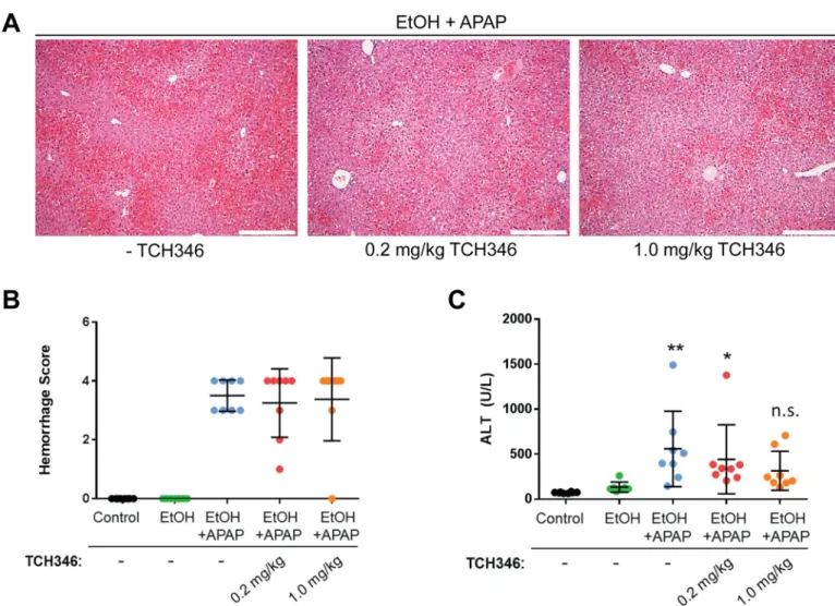
The GAPDH-targeting compound TCH346 partially attenuates
combined ethanol/APAP liver injury relative to control livers
Fig 1. Ethanol and acetaminophen synergize to promote nuclear accumulation of GAPDH in the liver. A. Immunoblots of nuclear fractions (top 3 panels), total tissue lysates (middle panel) and cytoplasmic fractions (bottom 3 panels) from livers of female C57BL/6J mice. The mice were fed either a control or ethanol (EtOH)-containing liquid diet. The amount of EtOH (vol/vol) in the diet was: 0% on days 1–3; 1.67% on day 4; 3.33% on day 5; and 5% on days 6–9. The study was terminated on day 10, following an overnight fast of all mice and a single APAP injection (500mg/kg; during the last 4h), administered to the indicated groups. Note the EtOH/APAP-dependent redistribution of GAPDH in the nuclear compartment. A combinedβ-tubulin and lamin B1 blot was done to validate the separation of the cytoplasmic and nuclear fractions, while pan-actin served as loading control.B. Densitometric quantification of the relative levels of nuclear GAPDH from the three treatment groups shown in panel A.**, p<0.01, two-way ANOVA.
Fig 2. APAP promotes aggregation of cytoplasmic GAPDH in ethanol pre-treated mice. A. Indirect immunofluorescence analysis of GAPDH (green) together with DAPI-stained nuclei (blue) in livers from ethanol pre-treated mice that were given APAP for the indicated time periods. Inset highlights the presence of
TCH346 treatment does not prevent nuclear translocation and
aggregation of GAPDH in the combined ethanol/APAP injury model
The previously reported protective effects of TCH346 treatment were linked to its ability to prevent the nuclear translocation of GAPDH [16], which otherwise leads to the induction of several pro-apoptotic genes [15]. Therefore, we tested whether the decrease in serum ALT lev-els in the TCH346-treated mice, particularly at the higher dose, correlated with a decrease in GAPDH aggregation and nuclear presence. To our surprise, neither parameter was altered by TCH346, regardless of the dose administered (Fig 4). These data suggest that, unlike in brain,
The immunoblots showing GAPDH monomer and high molecular mass (HMM) complexes (arrowheads) were from the same membrane at different exposure times. Shown are 4 representative individual mice from each treatment group, out of 8 total mice/group.
doi:10.1371/journal.pone.0160982.g002
Fig 3. The GAPDH-targeting compound TCH346 partially attenuates combined ethanol and acetaminophen liver injury. A. Representative H&E staining images from livers of EtOH/APAP-treated mice in the absence (-TCH346) or presence of 0.2mg/kg or 1mg/kg TCH346. Scale bar = 200μmB. Histological scoring of the areas of hemorrhage in the different treatment groups.C. Serum ALT levels in mice receiving control liquid diet (Control) or EtOH-containing liquid diet in the absence/presence of 0.2 or 1 mg/kg TCH346, which was co-administered with APAP (500mg/kg for 4hr).*p<0.05;**p<0.01; n.s., not significant, relative to the control group (one-way ANOVA; Dunnet’s multiple comparisons test). There was no statistically significant difference in serum ALT between the EtOH/APAP groups receiving TCH346 as compared to EtOH/APAP alone.
the partial protective effects of TCH346 in the liver are independent of its activity on nuclear GAPDH.
TCH346 treatment preserves mitochondrial Complex 1 activity and
cytochrome c levels
APAP is known to promote hepatocyte necrosis via mitochondrial damage mediated by pro-tein translocation into and out of the mitochondria [30,31]. To test whether TCH346 affected mitochondrial function, we examined activity of mitochondrial Complex I. APAP administra-tion to ethanol pre-treated mice caused ~25% decrease in Complex I activity, which was reversed to control levels by TCH346 treatment (Fig 5A). Immunoblot analysis of isolated liver mitochondria demonstrated that ethanol/APAP-treated mice that also received TCH346 had higher levels of cytochrome c and lower levels of the highly abundant mitochondrial matrix protein carbamoyl phosphate synthase 1 (CPS-1) (Fig 5B and 5C). Since mitochondrial cyto-chrome c levels are sensitive to oxidative stress mediated via p53 [32] and glutathione depletion [33], we assessed the effect of TCH346 on p53 mRNA and glutathione-S-transferase M1 (Gstm1) protein. TCH346 reduces the levels of p53 mRNA (Fig 5D) and increases Gstm1 expression (Fig 5E and 5F), suggesting that the increased levels of mitochondrial cytochrome c in response to TCH346 may be a result of increased resistance to oxidative stress-induced dam-age. Another prominent target of mitochondrial oxidative stress is CPS-1, a component of the urea cycle and a potential serum biomarker of acute mouse and human liver injury [34], which is reduced by TCH346. These data demonstrate that TCH346 alters mitochondrial protein dynamics during ethanol/APAP-induced liver injury, but the exact mechanisms remain to be determined.
Discussion
Formation of reactive metabolites that are damaging to cellular organelles is a common mecha-nism in drug-induced liver injury, including acetaminophen overdose. Understanding the downstream mechanisms of chemically induced hepatotoxicity can reveal potential treatment modalities that may prevent or curtail acute liver failure. The present study provides evidence that the glycolytic enzyme GAPDH is a prominent cellular target in mouse liver injury due to combined ethanol and APAP exposure. We demonstrate that GAPDH undergoes significant nuclear translocation and aggregation into high molecular weight complexes in response to combined ethanol and APAP treatment. Using a pharmacological approach aimed at targeting the nuclear translocation of GAPDH, we demonstrated a modest protective effect of the drug TCH346. However, the hepatoprotective mechanism of TCH346 was independent of nuclear GAPDH translocation and aggregation, in contrast to previous studies on this compound in models of neuroprotection [16,20]. However the TCH346 effects in the liver were likely medi-ated in part via mitochondrial mechanisms. Specifically, TCH346 treatment preserved Com-plex I activity and increased levels of mitochondrial cytochrome c. The latter is known to act as a mediator of APAP-related hepatotoxicity upon its stress-induced release from mitochondria [35,36].
CPS-1 catalyzes the first step of the urea cycle by synthesizing carbamoyl phosphate from ammonia and bicarbonate. Since carbamoyl phosphate is converted to citrulline, CPS-1 also
(non-reducing conditions) of total lysates from livers of 1 each of control and EtOH-treated mice, and 12 EtOH +APAP- treated mice, as labeled at the bottom. The mice in the latter group (last 12 lanes) represent those receiving no additional treatment (-TCH346), 0.2mg/kg TCH346 or 1mg/kg TCH346, as labeled at the top. Shown are 1 or 4 representative individual mice from each treatment group, out of 8 total mice/group.
affects the availability of precursors for NO synthesis [37], which may indirectly affect GAPDH S-nitrosylation. Furthermore, a recent study identified a large number of mitochondrial pro-teins that undergo pathology-promoting nitration in response to APAP mouse liver injury and among these, CPS1 (which contains 36 tyrosines in the mouse isoform) had the greatest num-ber of nitrotyrosine-containing peptides [144] that were detected by mass spectrometry [38]. Although the effect of nitration on CPS1 specifically was not investigated, nitration is known to generally inhibit the activities of metabolic enzymes, leading to mitochondrial dysfunction
Fig 5. TCH346 treatment preserves mitochondrial Complex I activity and cytochrome c levels, and decreases mitochondrial CPS-1 in livers from EtOH+APAP-treated mice. A. Measurement of Complex I activity (rate of NADH oxidation measured as a decrease in absorbance at 340nm) in liver mitochondrial extracts of the designated treatment groups.**p<0.01 compared to control group (one-way ANOVA; Dunnet’s multiple
comparisons test).B. Coomassie gel stains and immunoblots for the indicated proteins in the mitochondrial fractions of control vs. EtOH/APAP (left panels), or—TCH346 versus 1mg/kg TCH346 in EtOH/APAP-treated mice (right panels). TCH346 did not affect the EtOH+APAP-induced increased presence of JNK or Bax, but was associated with increased levels of cytochrome c and decreased levels of the mitochondrial matrix protein CPS-1.C. Densitometric quantification of cytochrome c and CPS-1 levels from panel B (right; EtOH+APAP with or without TCH346).*p<0.05;***p<0.001 when comparing +TCH346 to the -TCH346 group (unpaired t-test).D. Downregulation of p53 mRNA by TCH346.E. Upregulation of the antioxidant protein Gstm1 in livers from TCH346-treated mice.F. Quantification of the immunoblot in panel E.**p<0.01 compared to the -TCH346 group; unpaired t-test. TCH46 dose was 1mg/kg in all experiments shown.
[38,39]. Therefore another possibility is that TCH346 may protect from mitochondrial damage by promoting the release of the highly abundant enzyme CPS-1 that may have been rendered dysfunctional by stress-induced nitration.
GAPDH is one of many protein targets for stress-induced S-nitrosylation, which is the cou-pling of an NO moiety to a reactive cysteine thiol to form nitrosothiol. The function of S-nitrosylation is dependent on the stimulus as well as the protein target. In the case of GAPDH, this post-translational modification promotes GAPDH binding to the E3 ubiquitin ligase Siah 1, which contains a nuclear localization signal and acts as a vehicle to promote the nuclear accumulation of a small pool of GAPDH [17]. Among the known functions of nuclear GAPDH are activation of p300/CREB-binding protein and its target, p53, to induce apoptotic cell death [15,40] as well as trans-nitrosylation of nuclear resident proteins [41]. Given that apoptosis is not thought to be a major contributor to APAP-induced hepatotoxicity [42], the transcriptional pathway of nuclear GAPDH signaling, as related to apoptosis, is unlikely to be involved. One possibility is that GAPDH-mediated trans-nitrosylation of nuclear targets, including the deacetylase sirtuin 1 [41,43] could mediate APAP liver injury, based on the
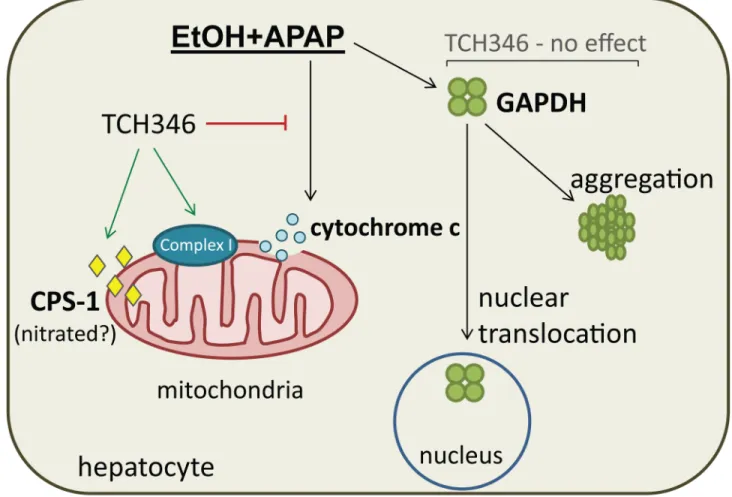
Fig 6. Summary of liver GAPDH changes in response to combined ethanol and APAP exposure and the effects of TCH346 treatment.Combined ethanol and APAP mediated mouse liver injury is associated with a significant increase in GAPDH nuclear translocation and cytoplasmic aggregation. The drug TCH346, which was previously shown to target GAPDH in the brain, attenuates liver injury in association with EtOH+APAP treatment. The mechanism by which TCH346 exerts its effect in this liver injury model are independent of GAPDH nuclear levels and aggregation, but involve mitochondrial events. TCH346 treatment is associated with increased mitochondrial Complex I activity and cytochrome c levels, and decreased CPS-1, which may be nitrated and/or non-functional [38].
known importance of another sirtuin family member, the mitochondrial sirtuin 3, in APAP liver injury [44].
TCH346 was shown to target GAPDH [20] and to prevent its Siah1-mediated translocation [16]. Studies in animal models of neurodegeneration demonstrated TCH346-mediated protec-tion [22,23,45], although this protection did not translate to clinical trials of Parkinson Dis-ease and ALS [46,47]. Nevertheless, given its favorable safety profile that was shown in the clinical studies, there is potential to repurpose TCH346 (also known as CGP 3466 and Omiga-pil) in other disease settings. To that end, TCH346 is currently being considered as a potential treatment in congenital muscular dystrophies in a clinical trial (NCT01805024), based on its effects in preclinical models [48–50]. Our study adds hepatoprotection to the repertoire of other potential uses for TCH346, although its utility in this setting is not as profound as noted in the brain and will need to be validated in additional models of drug-induced liver injury. Dosage and timing of administration are two important considerations for future investigation. This is illustrated by previous studies showing that thein vivoneuroprotective effects of TCH346 are highly dose-dependent and exhibit a bell-shaped curve, with the protective effect being greatest at 0.1mg/kg and lost at or above 1mg/kg (45). The contribution of hepatic metabolism to TCH346 activity in the liver will also need to be examined in more detail. Our findings that TCH346 exerts its partial protective effects by targeting mitochondria are consis-tent with some of the previously reported neuroprotective properties of TCH346. For example, in thepmn/pmn(progressive motor neuropathy) mouse model of ALS, administration of TCH346 extended life-span, prevented weight loss, and slowed the loss of motor neurons by preserving mitochondrial integrity, although the molecular events by which TCH346 protected mitochondria were not delineated [23].
In summary, we report on a novel signaling mechanism involved in ethanol/APAP liver injury that converges on the nuclear translocation and aggregation of GAPDH. The small mol-ecule GAPDH-targeting compound TCH346 did not reverse nuclear accumulation of GAPDH in response to ethanol/APAP treatment, but showed modest hepatoprotection that was poten-tially mediated by mitochondrial mechanisms (Fig 6).
Author Contributions
Conceptualization:NTS MBO.
Formal analysis:JKG.
Funding acquisition:NTS MBO.
Investigation:NTS DAP HHW DM HCM.
Methodology:NTS MBO DAP JKG.
Project administration:MBO.
Resources:NTS MBO.
Supervision:NTS MBO.
Validation:NTS DAP HHW HCM DM.
Visualization:NTS MBO.
Writing - original draft:NTS DAP MBO.
References
1. Gu X, Manautou JE. Molecular mechanisms underlying chemical liver injury. Expert Rev Mol Med. 2012; 14:e4. doi:10.1017/S1462399411002110PMID:22306029; PubMed Central PMCID: PMC3704158.
2. Yuan L, Kaplowitz N. Mechanisms of drug-induced liver injury. Clin Liver Dis. 2013; 17(4):507–18, vii. doi:10.1016/j.cld.2013.07.002PMID:24099014; PubMed Central PMCID: PMC3793205.
3. Nakamura T, Lipton SA. Molecular mechanisms of nitrosative stress-mediated protein misfolding in neurodegenerative diseases. Cell Mol Life Sci. 2007; 64(13):1609–20. doi: 10.1007/s00018-007-6525-0PMID:17453143.
4. Aiken CT, Kaake RM, Wang X, Huang L. Oxidative stress-mediated regulation of proteasome com-plexes. Mol Cell Proteomics. 2011; 10(5):R110 006924. doi:10.1074/mcp.M110.006924PMID: 21543789; PubMed Central PMCID: PMC3098605.
5. Nystrom T. Role of oxidative carbonylation in protein quality control and senescence. EMBO J. 2005; 24(7):1311–7. doi:10.1038/sj.emboj.7600599PMID:15775985; PubMed Central PMCID:
PMC1142534.
6. Chuang DM, Hough C, Senatorov VV. Glyceraldehyde-3-phosphate dehydrogenase, apoptosis, and neurodegenerative diseases. Annu Rev Pharmacol Toxicol. 2005; 45:269–90. doi:10.1146/annurev. pharmtox.45.120403.095902PMID:15822178.
7. Tristan C, Shahani N, Sedlak TW, Sawa A. The diverse functions of GAPDH: views from different sub-cellular compartments. Cell Signal. 2011; 23(2):317–23. doi:10.1016/j.cellsig.2010.08.003PMID: 20727968; PubMed Central PMCID: PMC3084531.
8. Nakajima H, Kubo T, Ihara H, Hikida T, Danjo T, Nakatsuji M, et al. Nuclear-translocated Glyceralde-hyde-3-phosphate Dehydrogenase Promotes Poly(ADP-ribose) Polymerase-1 Activation during Oxida-tive/Nitrosative Stress in Stroke. J Biol Chem. 2015; 290(23):14493–503. doi:10.1074/jbc.M114. 635607PMID:25882840; PubMed Central PMCID: PMC4505517.
9. Leisner TM, Moran C, Holly SP, Parise LV. CIB1 prevents nuclear GAPDH accumulation and non-apo-ptotic tumor cell death via AKT and ERK signaling. Oncogene. 2013; 32(34):4017–27. doi:10.1038/ onc.2012.408. PubMed Central PMCID: PMC3530648. PMID:22964641
10. Guha P, Harraz MM, Snyder SH. Cocaine elicits autophagic cytotoxicity via a nitric oxide-GAPDH sig-naling cascade. Proc Natl Acad Sci U S A. 2016; 113(5):1417–22. doi:10.1073/pnas.1524860113 PMID:26787898; PubMed Central PMCID: PMC4747760.
11. Snider NT, Weerasinghe SV, Singla A, Leonard JM, Hanada S, Andrews PC, et al. Energy determi-nants GAPDH and NDPK act as genetic modifiers for hepatocyte inclusion formation. J Cell Biol. 2011; 195(2):217–29. doi:10.1083/jcb.201102142PMID:22006949; PubMed Central PMCID:
PMC3198167.
12. Samson AL, Knaupp AS, Kass I, Kleifeld O, Marijanovic EM, Hughes VA, et al. Oxidation of an exposed methionine instigates the aggregation of glyceraldehyde-3-phosphate dehydrogenase. The Journal of biological chemistry. 2014; 289(39):26922–36. doi:10.1074/jbc.M114.570275PMID:25086035; PubMed Central PMCID: PMC4175333.
13. Nakajima H, Amano W, Kubo T, Fukuhara A, Ihara H, Azuma YT, et al. Glyceraldehyde-3-phosphate dehydrogenase aggregate formation participates in oxidative stress-induced cell death. The Journal of biological chemistry. 2009; 284(49):34331–41. doi:10.1074/jbc.M109.027698PMID:19837666; PubMed Central PMCID: PMC2797201.
14. Pierce A, Mirzaei H, Muller F, De Waal E, Taylor AB, Leonard S, et al. GAPDH is conformationally and functionally altered in association with oxidative stress in mouse models of amyotrophic lateral sclero-sis. J Mol Biol. 2008; 382(5):1195–210. doi:10.1016/j.jmb.2008.07.088PMID:18706911.
15. Sen N, Hara MR, Kornberg MD, Cascio MB, Bae BI, Shahani N, et al. Nitric oxide-induced nuclear GAPDH activates p300/CBP and mediates apoptosis. Nat Cell Biol. 2008; 10(7):866–73. doi:10.1038/ ncb1747PMID:18552833; PubMed Central PMCID: PMC2689382.
16. Hara MR, Thomas B, Cascio MB, Bae BI, Hester LD, Dawson VL, et al. Neuroprotection by pharmaco-logic blockade of the GAPDH death cascade. Proc Natl Acad Sci U S A. 2006; 103(10):3887–9. 17. Hara MR, Agrawal N, Kim SF, Cascio MB, Fujimuro M, Ozeki Y, et al. S-nitrosylated GAPDH initiates
apoptotic cell death by nuclear translocation following Siah1 binding. Nat Cell Biol. 2005; 7(7):665–74. doi:10.1038/ncb1268PMID:15951807.
19. Sen N, Hara MR, Ahmad AS, Cascio MB, Kamiya A, Ehmsen JT, et al. GOSPEL: a neuroprotective pro-tein that binds to GAPDH upon S-nitrosylation. Neuron. 2009; 63(1):81–91. doi:10.1016/j.neuron. 2009.05.024PMID:19607794; PubMed Central PMCID: PMC2758064.
20. Kragten E, Lalande I, Zimmermann K, Roggo S, Schindler P, Muller D, et al. Glyceraldehyde-3-phos-phate dehydrogenase, the putative target of the antiapoptotic compounds CGP 3466 and R-(-)-depre-nyl. J Biol Chem. 1998; 273(10):5821–8. PMID:9488718.
21. Waldmeier PC, Boulton AA, Cools AR, Kato AC, Tatton WG. Neurorescuing effects of the GAPDH ligand CGP 3466B. J Neural Transm Suppl. 2000;( 60):197–214. PMID:11205140.
22. Andringa G, van Oosten RV, Unger W, Hafmans TG, Veening J, Stoof JC, et al. Systemic administra-tion of the propargylamine CGP 3466B prevents behavioural and morphological deficits in rats with 6-hydroxydopamine-induced lesions in the substantia nigra. Eur J Neurosci. 2000; 12(8):3033–43. PMID: 10971644.
23. Sagot Y, Toni N, Perrelet D, Lurot S, King B, Rixner H, et al. An orally active anti-apoptotic molecule (CGP 3466B) preserves mitochondria and enhances survival in an animal model of motoneuron dis-ease. Br J Pharmacol. 2000; 131(4):721–8. doi:10.1038/sj.bjp.0703633PMID:11030721; PubMed Central PMCID: PMC1572390.
24. Andringa G, Cools AR. The neuroprotective effects of CGP 3466B in the best in vivo model of Parkin-son's disease, the bilaterally MPTP-treated rhesus monkey. J Neural Transm Suppl. 2000;( 60):215– 25. PMID:11205142.
25. Groeneveld GJ, van Muiswinkel FL, de Leeuw van Weenen J, Blauw H, Veldink JH, Wokke JH, et al. CGP 3466B has no effect on disease course of (G93A) mSOD1 transgenic mice. Amyotroph Lateral Scler Other Motor Neuron Disord. 2004; 5(4):220–5. PMID:15799550.
26. Bunchorntavakul C, Reddy KR. Acetaminophen-related hepatotoxicity. Clin Liver Dis. 2013; 17(4):587– 607, viii. doi:10.1016/j.cld.2013.07.005PMID:24099020.
27. Cox B, Emili A. Tissue subcellular fractionation and protein extraction for use in mass-spectrometry-based proteomics. Nat Protoc. 2006; 1(4):1872–8. doi:10.1038/nprot.2006.273PMID:17487171. 28. Frezza C, Cipolat S, Scorrano L. Organelle isolation: functional mitochondria from mouse liver, muscle
and cultured fibroblasts. Nat Protoc. 2007; 2(2):287–95. doi:10.1038/nprot.2006.478PMID:17406588. 29. Nakajima H, Amano W, Fujita A, Fukuhara A, Azuma YT, Hata F, et al. The active site cysteine of the
proapoptotic protein glyceraldehyde-3-phosphate dehydrogenase is essential in oxidative stress-induced aggregation and cell death. The Journal of biological chemistry. 2007; 282(36):26562–74. doi: 10.1074/jbc.M704199200PMID:17613523.
30. Jaeschke H, McGill MR, Ramachandran A. Oxidant stress, mitochondria, and cell death mechanisms in drug-induced liver injury: lessons learned from acetaminophen hepatotoxicity. Drug Metab Rev. 2012; 44(1):88–106. doi:10.3109/03602532.2011.602688PMID:22229890.
31. Seki E, Brenner DA, Karin M. A liver full of JNK: signaling in regulation of cell function and disease path-ogenesis, and clinical approaches. Gastroenterology. 2012; 143(2):307–20. doi:10.1053/j.gastro. 2012.06.004PMID:22705006; PubMed Central PMCID: PMC3523093.
32. Dalleau S, Baradat M, Gueraud F, Huc L. Cell death and diseases related to oxidative stress: 4-hydro-xynonenal (HNE) in the balance. Cell Death Differ. 2013; 20(12):1615–30. doi:10.1038/cdd.2013.138 PMID:24096871; PubMed Central PMCID: PMC3824598.
33. Ghibelli L, Coppola S, Fanelli C, Rotilio G, Civitareale P, Scovassi AI, et al. Glutathione depletion causes cytochrome c release even in the absence of cell commitment to apoptosis. FASEB J. 1999; 13(14):2031–6. PMID:10544186.
34. Weerasinghe SV, Jang YJ, Fontana RJ, Omary MB. Carbamoyl phosphate synthetase-1 is a rapid turn-over biomarker in mouse and human acute liver injury. Am J Physiol Gastrointest Liver Physiol. 2014; 307(3):G355–64. doi:10.1152/ajpgi.00303.2013PMID:24924744; PubMed Central PMCID: PMC4121638.
35. Masubuchi Y, Suda C, Horie T. Involvement of mitochondrial permeability transition in acetaminophen-induced liver injury in mice. Journal of hepatology. 2005; 42(1):110–6. doi:10.1016/j.jhep.2004.09.015 PMID:15629515.
36. Hinson JA, Roberts DW, James LP. Mechanisms of acetaminophen-induced liver necrosis. Handbook of experimental pharmacology. 2010;( 196):369–405. doi:10.1007/978-3-642-00663-0_12PMID: 20020268; PubMed Central PMCID: PMC2836803.
37. Summar ML, Gainer JV, Pretorius M, Malave H, Harris S, Hall LD, et al. Relationship between carba-moyl-phosphate synthetase genotype and systemic vascular function. Hypertension. 2004; 43(2):186– 91. doi:10.1161/01.HYP.0000112424.06921.52PMID:14718356.
60:211–22. doi:10.1016/j.freeradbiomed.2013.02.018PMID:23454065; PubMed Central PMCID: PMC3680365.
39. Agarwal R, MacMillan-Crow LA, Rafferty TM, Saba H, Roberts DW, Fifer EK, et al. Acetaminophen-induced hepatotoxicity in mice occurs with inhibition of activity and nitration of mitochondrial manga-nese superoxide dismutase. J Pharmacol Exp Ther. 2011; 337(1):110–6. doi:10.1124/jpet.110.176321 PMID:21205919; PubMed Central PMCID: PMC3063736.
40. Sawa A, Khan AA, Hester LD, Snyder SH. Glyceraldehyde-3-phosphate dehydrogenase: nuclear translocation participates in neuronal and nonneuronal cell death. Proc Natl Acad Sci U S A. 1997; 94 (21):11669–74. PMID:9326668; PubMed Central PMCID: PMC23578.
41. Kornberg MD, Sen N, Hara MR, Juluri KR, Nguyen JV, Snowman AM, et al. GAPDH mediates nitrosyla-tion of nuclear proteins. Nat Cell Biol. 2010; 12(11):1094–100. doi:10.1038/ncb2114PMID:20972425; PubMed Central PMCID: PMC2972384.
42. McGill MR, Sharpe MR, Williams CD, Taha M, Curry SC, Jaeschke H. The mechanism underlying acet-aminophen-induced hepatotoxicity in humans and mice involves mitochondrial damage and nuclear DNA fragmentation. J Clin Invest. 2012; 122(4):1574–83. doi:10.1172/JCI59755PMID:22378043; PubMed Central PMCID: PMC3314460.
43. Rodriguez-Ortigosa CM, Celay J, Olivas I, Juanarena N, Arcelus S, Uriarte I, et al. A GAPDH-mediated trans-nitrosylation pathway is required for feedback inhibition of bile salt synthesis in rat liver. Gastroen-terology. 2014; 147(5):1084–93. doi:10.1053/j.gastro.2014.07.030PMID:25066374.
44. Lu Z, Bourdi M, Li JH, Aponte AM, Chen Y, Lombard DB, et al. SIRT3-dependent deacetylation exacer-bates acetaminophen hepatotoxicity. EMBO Rep. 2011; 12(8):840–6. doi:10.1038/embor.2011.121 PMID:21720390; PubMed Central PMCID: PMC3147261.
45. Andringa G, Eshuis S, Perentes E, Maguire RP, Roth D, Ibrahim M, et al. TCH346 prevents motor symptoms and loss of striatal FDOPA uptake in bilaterally MPTP-treated primates. Neurobiol Dis. 2003; 14(2):205–17. PMID:14572443.
46. Miller R, Bradley W, Cudkowicz M, Hubble J, Meininger V, Mitsumoto H, et al. Phase II/III randomized trial of TCH346 in patients with ALS. Neurology. 2007; 69(8):776–84. doi:10.1212/01.wnl.0000269676. 07319.09PMID:17709710.
47. Olanow CW, Schapira AH, LeWitt PA, Kieburtz K, Sauer D, Olivieri G, et al. TCH346 as a neuroprotec-tive drug in Parkinson's disease: a double-blind, randomised, controlled trial. Lancet Neurol. 2006; 5 (12):1013–20.
48. Erb M, Meinen S, Barzaghi P, Sumanovski LT, Courdier-Fruh I, Ruegg MA, et al. Omigapil ameliorates the pathology of muscle dystrophy caused by laminin-alpha2 deficiency. J Pharmacol Exp Ther. 2009; 331(3):787–95. doi:10.1124/jpet.109.160754PMID:19759319.
49. Meinen S, Lin S, Thurnherr R, Erb M, Meier T, Ruegg MA. Apoptosis inhibitors and mini-agrin have additive benefits in congenital muscular dystrophy mice. EMBO Mol Med. 2011; 3(8):465–79. doi:10. 1002/emmm.201100151PMID:21674808; PubMed Central PMCID: PMC3377088.