IgG-Containing Isoforms of Neuregulin-1 Are
Dispensable for Cardiac Trabeculation in
Zebrafish
Leigh Ann Samsa1,2, Cade Ellis Ito2,3, Daniel Ross Brown2,3, Li Qian2,3, Jiandong Liu2,3*
1 Department of Cell Biology and Physiology, University of North Carolina at Chapel Hill, Chapel Hill, North Carolina, United States of America, 2 McAllister Heart Institute, University of North Carolina at Chapel Hill, Chapel Hill, North Carolina, United States of America, 3 Department of Pathology and Laboratory Medicine, University of North Carolina at Chapel Hill, Chapel Hill, North Carolina, United States of America
Abstract
The Neuregulin-1 (Nrg1) signaling pathway has been widely implicated in many aspects of heart development including cardiac trabeculation. Cardiac trabeculation is an important morphogenetic process where clusters of ventricular cardiomyocytes extrude and expand into the lumen of the ventricular chambers. In mouse, Nrg1 isoforms containing an immu-noglobulin-like (IgG) domain are essential for cardiac trabeculation through interaction with heterodimers of the epidermal growth factor-like (EGF-like) receptors ErbB2/ErbB4. Recent reports have underscored the importance of Nrg1 signaling in cardiac homeostasis and disease, however, placental development has precluded refined evaluation of the role of this pathway in mammals. ErbB2 has been shown to have a developmentally conserved role in cardiac trabeculation in zebrafish, a vertebrate model organism with completely external development, but the requirement for Nrg1 has not been examined. We found that among the multiple Nrg1 isoforms, the IgG domain-containing, type I Nrg1 (nrg1-I) is the only isoform detectable in the heart. Then, using CRISPR/Cas9 gene editing, we tar-geted the IgG domain of Nrg1 to produce novel alleles, nrg1nc28and nrg1nc29, encoding
nrg1-I and nrg1-II truncations. Our results indicated that zebrafish deficient for nrg1-I
developed trabeculae in an ErbB2-dependent manner. Further, these mutants survive to reproductive adulthood with no overt cardiovascular defects. We also found that additional EGF-like ligands were expressed in the zebrafish heart during development of trabeculae. Together, these results suggest that Nrg1 is not the primary effector of trabeculation and/ or that other EGF-like ligand(s) activates the ErbB2/ErbB4 pathway, either through func-tioning as the primary ligand or acting in a redundant manner. Overall, our work provides an example of cross-species differences in EGF family member requirements for an evolu-tionary conserved process.
a11111
OPEN ACCESS
Citation: Samsa LA, Ito CE, Brown DR, Qian L, Liu
J (2016) IgG-Containing Isoforms of Neuregulin-1 Are Dispensable for Cardiac Trabeculation in Zebrafish. PLoS ONE 11(11): e0166734. doi:10.1371/journal.pone.0166734
Editor: Sheng-Ping Lucinda Hwang, Academia
Sinica, TAIWAN
Received: August 16, 2016 Accepted: November 2, 2016 Published: November 15, 2016
Copyright:©2016 Samsa et al. This is an open access article distributed under the terms of the
Creative Commons Attribution License, which permits unrestricted use, distribution, and reproduction in any medium, provided the original author and source are credited.
Data Availability Statement: All relevant data are
within the paper and its Supporting Information files.
Funding: LAS is supported by National Institutes of
Introduction
Congenital heart diseases (CHD) are highly prevalent birth defects [1] and often feature
pertur-bations in cardiac morphogenesis that arise during development [2–4]. The Nrg1-ErbB2/4
sig-naling pathway has been implicated in many aspects of vertebrate cardiac biology ranging from
heart development to homeostasis and disease [5]. Transmembrane pro-Nrg1 is expressed on
endocardial and microvascular endothelial cells where it is cleaved by extracellular secretases to
release active Nrg1 [6–9]. Once cleaved, Nrg1 binds to cardiomyocyte-expressed ErbB4 via its
epidermal growth factor (EGF) domain. This ligand and receptor interaction subsequently stimulates hetero-dimerization of ErbB4 with its essential co-receptor ErbB2, leading to
activa-tion of ErbB2 tyrosine kinase activity and downstream signaling [10–12]. Recent reports have
underscored the importance of the Nrg1-ErbB2/4 signaling pathway in cardiac repair pro-cesses, and recombinant Nrg1 is currently in clinical trials as a heart failure therapeutic, but the
role of Nrg1 in development is largely unknown [5,13–19]. A refined understanding of this role
could provide insight into CHDs or inform the development of improved therapeutics. Early studies demonstrated that Nrg1, ErbB2 and ErbB4 are each required for proper
cham-ber maturation and cardiac development in mice [7,20–22]. Owing to their rapid
develop-ment, optical clarity, and ease of genetic manipulation, zebrafish (Danio rerio) have emerged as a premier model organism for understanding the molecular and genetic regulation of heart
development [23]. Unlike mammalian models, zebrafish embryos are small enough to meet
oxygen needs by diffusion alone and can survive for days with severe heart malformations
[24–28]. Further, adult zebrafish are highly tolerant of reduced cardiac function [29,30].
The early embryonic zebrafish heart develops into a two-chambered heart within 48 hours post-fertilization (hpf). As the heart matures, it optimizes the ventricular myocardial architec-ture for efficient conduction and contraction. This chamber maturation feaarchitec-tures formation of highly organized luminal myocardial protrusions called trabeculae, which are evident by 72
hpf and comprise the majority of the adult myocardium [31–33]. Failure to initiate
trabecula-tion is embryonic lethal in mice and zebrafish, and trabeculatrabecula-tion defects are often associated
with CHDs [34,35]. Though our previous work demonstrated that ErbB2 is required for
zebra-fish cardiac trabeculation, requirement for Nrg1 in zebrazebra-fish cardiac development has not
been examined [31].
Nrg1 is alternatively spliced to produce a diversity of isoforms [36]. Zebrafishnrg1produces
three major isoforms by alternative splicing,nrg1-I,nrg1-IIa-c, andnrg1-III. The N-terminus
contains either a cysteine-rich domain (nrg1-III) or a unique N-terminal sequence followed by
an IgG-like domain (nrg1-Iandnrg1-II). All isoforms share an EGF-like domain, a
transmem-brane domain, and a C-terminal Neuregulin domain. In mice, genetic deletion of the IgG
domain-containing isoforms (Nrg1-IandNrg1-II) is sufficient to block cardiac trabeculation
[7,21].
To determine the genetic requirement for Nrg1 in zebrafish trabeculation, we used CRISPR/Cas9 targeted nuclease activity to generate frameshift mutations that lead to early
truncation of IgG-containing Nrg1-I and Nrg1-II. The mutant embryos had reducednrg1
transcripts levels, suggesting non-sense mediated decay and absence of previously unanno-tated splice isoforms. Yet, the mutant fish survived to adulthood without cardiac trabecular defects or overt signs of other cardiac malformations. This lack of cardiac phenotypes in zebra-fishnrg1-I/IImutants may be explained by expression of other putative EGF-like ligands expressed in the developing heart. Together, these results suggest that Nrg1 is dispensable for heart development in zebrafish and that additional mutants will need to be generated to deter-mine which ligand(s) have the primary role of regulating trabeculation or if other ligands play compensatory or redundant roles in trabeculation.
Lung, and Blood Institute (R00 HL109079 grant to JL) and American Heart Association (grant 15GRNT25530005 to JL).
Competing Interests: The authors have declared
Materials and Methods
Animal Lines and Care
Embryos and adult fish were reared and maintained at the aquaculture facility of the Univer-sity of North Carolina at Chapel Hill at 28.5˚C on a 14h/10h light/dark cycle in accordance with the University of North Carolina at Chapel Hill Institutional Animal Care and Use
Com-mittee (IACUC) approved protocol [37]. All studies were performed after euthanasia of
zebra-fish and all efforts were made to minimize animal suffering. The zebrazebra-fish lines used in this
study are as follows:nrg1z26[38],nrg1nc28,nrg1nc29,Tg(myl7:dsRed)vc6[39],Tg(kdrl:EGFP)s843
[40], andTg(myl7:GFP)twu26[41].
CRISPR/Cas9 design and injection
Cas9 mRNA wasin vitrotranscribed from pXT7 using the mMessage mMachine kit
(Thermo-Fisher) as previously described with some modifications [42]. CRISPR/Cas9 target site in exon
3 ofnrg1were identified using ZiFit software [43] and zebrafish genomic sequence, build
GRCz9 [44]. Single stranded oligonucleotides corresponding to the targeting sequence were
annealed and cloned into the DR274 vector (Addgene) [45], then transcribedin vitrowith T7
MaxiScript kit (ThermoFisher). Cas9 plasmid was generously provided by Dr. Jing-Wei Xiong. Embryos were injected at the one cell stage with 1–2 nl of a mixture containing 1200 ng Cas9, 50–75 ng gRNA, 10 mM MgCl, and 0.01% phenol red. gRNA targeting efficiency was
determined by High Resolution Melt Analysis (HRMA) [46] as described below using primers
flanking the target site. F1 offspring from F0 founders that carry favorable mutations, deter-mined by DNA sequencing of the target site, were raised to adulthood. Heterozygous F1 fish were interbred to produce homozygous wild type, homozygous mutant, and heterozygous mutant offspring.
Genotyping
Genomic DNA isolation. Genomic DNA was collected from fin clips or embryos in 50 or
25μL lysis buffer containing 10 mM Tris-HCl pH 8.0, 50 mM KCl, 0.3% Tween-20. Samples
were lysed at 95˚C for 10 minutes, and then digested in 0.5μg/mL Proteinase K (Denville
Scientific).
Line-specific genotyping. nrg1z26fish were genotyped by PCR and enzyme digestion as
previously described [38]. HRMA was used to genotype wild type, mutant, and heterozygous
nrg1nc28andnrg1nc29lines (see below). Heterozygous alleles had multiple peaks in the
deriva-tive melt curve. Homozygous wild type and mutant allele melt temperatures differed by at least >1˚C. This genotyping method was verified both by enzyme digestion and by DNA sequence analysis in a subset of samples.
HRMA
High resolution melt analysis (HRMA) was used to validate CRISPR/Cas9 reagents, identify
F1 founders, and genotypenrg1nc28andnrg1nc29fish. Each 10μl reaction contained 0.5μl
genomic DNA (see above), 5μl SYBR Green (ThermoFisher), and 4.5μl primer mix (water
PCR and qRT-PCR
RNA was isolated from whole embryos using Trizol reagent (Thermofisher) and from embry-onic hearts using Qiagen RNAeasy Mini Plus Kit according to manufacturer’s instructions.
Up to 1μg of cDNA was reverse transcribed using Superscript Master Mix (Thermofisher).
GoTaq (Promega) reagents were used for PCR with 10 ng cDNA template as per manufactur-er’s instructions. For qRT-PCR, we used Syber Green chemistry (ThermoFisher) on a ViiA7
qPCR machine in 10μL reactions. Cycle threshold (CT) values were normalized toef1aas a
housekeeping gene and relative expression was calculated comparing average change in CT in
wild type and mutant embryos by the 2^(ΔΔCT)method [47].
Heart isolations
Heart isolations were performed as previously described [48]. Briefly, larvae were euthanized
with 5X Tricaine at 3 dpf (days post-fertilization). Fine forceps were used to manually remove each heart (ventricle, atrium, and bulbous arteriosus) and dissect away non-cardiac tissues. Hearts were transferred to lysis buffer and processed according to manufacturer’s instructions for the RNAeasy Mini Plus kit (Qiagen). A minimum of 30 hearts were pooled for each gene expression replicate.
FACS
Fluorescence activated cell sorting (FACS) of endothelial and myocardial cells was performed
essentially as previously described with minor modifications [49]. Briefly,Tg(kdrl:EGFP)s843or
Tg(myl7:GFP)twu26embryos were dissociated into single cells by enzymatic digestion, then
counterstained with SYTOX Blue dead cell stain (ThermoFisher).Tg(myl7:GFP)twu26embryos
were passed through a 21 gauge needle 100μm cell strainer to enrich for hearts prior to an
abbreviated enzymatic digestion step. For each of 3–6 replicates, 2000–10,000 live, GFP+ cells were sorted with Sony SH800S then processed for qRT-PCR as described above using the Qia-gen RNA Easy Micro kit (QiaQia-gen).
In situ hybridization
In situhybridization was performed as previously described [50].In situhybridization probe
fornrg1was prepared as previously described [8] and synthesized from the pGEMT vector
(Promega) using the DIG RNA labeling kit (Roche). Whole-mount embryo imaging was per-formed on a Leica MZ16F fluorescence stereomicroscope.
Confocal microscopy
Anesthetized larvae 2–5 dpf were embedded in 1% low melt agarose and oriented for optimal viewing of the heart. Immediately prior to imaging, embryos were euthanized with 5-10X Tri-caine (MS-222, Sigma). After cessation of heartbeat, confocal z-stacks were collected using an Olympus Fluoview 1000MPE equipped with a 20X XLPlan water immersion objective (NA 1.0) with 2.5X optical zoom. Fluoview software was used to collect sections through the middle
25–50% of the heart at 512x512 or 1024x1024 pixel resolution and 1–2μm spacing between
z-slices. Fluoview’s brightness correction algorithm was used to account for signal attenuation
with increasing depth. ImageJ [51] was used to process images. For each Z-stack, we selected
as the representative samples from a pool of a minimum of N>12 embryos which were visually inspected for phenotype.
Whole mount microscopy
Adult fish were anesthetized with Tricaine in system water. Fish were imaged alongside a cen-timeter ruler in a minimal volume of water using an Android 13 MP camera. Brightness and contrast were adjusted and images were scaled using ImageJ software.
Mitotracker assay
Supernumerary neuromasts were assayed essentially as previously described [52]. Briefly,
lar-vae were incubated for 5–30 minutes in fish water containing Mitotracker Red (Thermo-Fisher) at a 1:10,000 dilution. Larvae were briefly rinsed with system water and anesthetized with 1X Tricaine, then oriented in a lateral position and epifluorescence images were collected on a Leica M205C fluorescence stereoscope. The number of neuromasts on the lateral line were counted for N>25 embryos. Wild type embryos had 8–12 neuromasts, and we consid-ered 18+ neuromasts to be supernumerary.
Histology
Adult fish were euthanized on ice for 20 minutes. To ensure rapid and complete fixation, each fish was gavaged with 4% paraformaldehyde (PFA) in PBS, then the abdominal cavity was opened by anterior-posterior incision and flushed with 4% PFA. After overnight fixation, the fish were de-calcified with 0.5M EDTA for 3–7 days, dehydrated in 70% ethanol, paraffin
embedded and sectioned at 5μm intervals and stained with hematoxolin and eosin (H&E).
Survival curve
Embryos were obtained from breeding healthy homozygousnrg1WT/WTandnrg1nc28/nc28
adults. For each genotype, 7 tanks containing 10 fish each were raised under standard hus-bandry conditions. Tank order was randomized to minimize hushus-bandry position effects. Sur-vival was recorded weekly at 6–8 day intervals through 12 wpf (weeks post-fertilization).
PD168393 treatment
Embryos were treated with 3.75μM PD168393 (ThermoFisher) in 1% DMSO containing
embryo medium from 2 dpf to 4 dpf. Control embryos were incubated in 1% DMSO in embryo medium.
Results
Molecular features of zebrafish Nrg1 and its expression in the zebrafish
heart
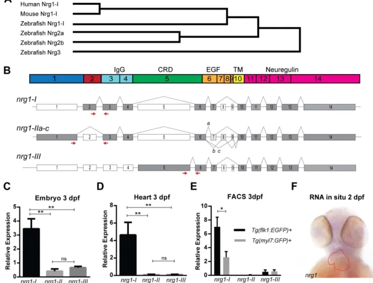
The zebrafish genome encodes several members of the neuregulin family—nrg1,nrg2a,nrg2b,
andnrg3.nrg1, which encodes the putative ligand for cardiac ErbB2 signaling, may be an important regulator of trabeculation in zebrafish. Sequence analysis further indicates that
zeb-rafish Nrg1 is the closest homolog to human NRG1 and mouse Nrg1 (Fig 1A). In the zebrafish
genome,nrg1is located on Chromosome 18 and is predicted to have 14 coding exons
encod-ing several functional domains (Fig 1B). Alternative splicing ofnrg1produces 3 primary
iso-forms (nrg1-I,nrg1-II, andnrg1-III,Fig 1C) that differ primarily in their N-terminal sequence
domain is found in the N-terminus ofnrg1-Iandnrg1-IIwhile a membrane-spanning
cyste-ine-rich domain (CRD) is found in the N-terminus ofnrg1-III(Fig 1B and 1C). In addition, all
isoforms share an epidermal growth factor-like domain (EGF), a transmembrane domain
(TM), and a C-terminal neuregulin domain (Fig 1B and 1C).
To determine which isoform(s) ofnrg1are expressed in the heart, we designed
exon-span-ning primers to assess relative expression levels ofnrg1-I,nrg1-II, andnrg1-IIIat 3 dpf. All
three isoforms were detectable in whole embryo (Fig 1D). However, onlynrg1-Iwas detectable
in cardiac tissue (Fig 1E). We detectednrg1byin situhybridization in the heart and brain of
embryo (Fig 1F). Previous studies suggest that cardiacnrg1expression is confined to
endocar-dial cells in the embryo [35,53,54]. Consistently, using FACS-enriched cells from a preparation
of 3 dpf hearts, we also found that in zebrafish,nrg1-Iwas expressed in the endocardial but not
myocardial cells (Fig 1E).
Fig 1. Zebrafish Neuregulin 1 and the expression of its isoforms. (A) Gene tree from Clustal-Omega multiple alignment comparison. (B) Schematic of Nrg1 domains encoded by Exons 1–14. (C) Schematic of nrg1 gene structure. Exons are drawn to scale; introns are not to scale. Alternative splicing produces three primary isoforms, nrg1-I, nrg1-IIa-c, and nrg1-III. (D-E) Relative expression of nrg1 isoforms in (D) 3 dpf embryos and (E) dissected hearts from 3 dpf embryos normalized to efl1a. (F) In situ hybridization of anti-sense riboprobe targeting nrg1. Heart is outlined in red. Student’s T-test compared to matched control. Error bars are SEM. N3 biological replicates.*p0.05–0.01,**p0.01– 0.001,***p<0.001.
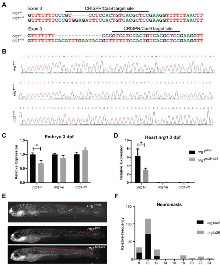
Generation of zebrafish mutant alleles
To investigate the isoform-specific role of Nrg1 in heart development, we used CRISPR/Cas9
gene editing to target its exon 3, which encodes part of the IgG domain shared bynrg1-Iand
nrg1-II. We isolated two frameshift alleles,nrg1nc28andnrg1nc29, carrying a 5 bp insertion and
14 bp deletion in exon 3, respectively (Fig 2A and 2B). These mutations are predicted to
trun-cate Nrg1-I at 55 and 99 amino acids upstream of the receptor binding EGF-like domain (S1
Fig). Interbreeding heterozygous fish for both alleles produced homozygous and heterozygous
alleles at expected Mendelian ratios.
Sincenrg1nc28truncation was predicted to be more severe than thenrg1nc29allele, we
focused our efforts on phenotyping this allele.nrg1mRNA expression levels were dramatically
reduced relative to wild type embryos, suggesting nonsense mediated decay of the mutant
transcript (Fig 2C). To further determine the effect of frameshift mutation onnrg1expression
level,nrg1mRNA expression level was measured via qPCR since loss of function mutant
tran-scripts can have decreased stability through nonsense mediated decay. Transcript level ofnrg1
in the mutant was significantly reduced relative to that in the wild type embryos, indicating a
loss of Nrg1 expression (Fig 2C). Our gene editing strategy is expected to eliminate bothnrg1-I
andnrg1-II, while sparing theNrg1-IIIisoform. It is important to note that functional Nrg1 protein could not be assessed via Western blot due to a lack of available Nrg1 antibody.
Previ-ous report on thenrg1z26allele, which codes for a loss of function mutation in the CRD
domain ofnrg1-III, demonstrated that loss ofnrg1-IIIleads to supernumerary neuromasts in
the developing lateral line as well as later adult lethality [55]. To determine ifnrg1-IIIsignaling
is intact in our mutants, we used a voltage sensitive vital dye (Mitotracker) to label neuromasts
in embryos at 5 dpf [31,56] (Fig 2D). Thoughnrg1z26mutants had supernumerary neuromasts,
extra neuromasts were not observed in offspring of inbrednrg1WT/nc28, indicating thatnrg1nc28
does not disrupt the function ofnrg1-III(Fig 2F).
Trabeculae form in nrg1
nc28mutants in an ErbB2-dependent manner
To address the hypothesis thatnrg1-Iis required for stimulating cardiac trabeculation, we
crossed thenrg1nc28allele onto a transgenic background expressing dsRed in cardiomyocytes.
We used confocal microscopy to examine cardiac trabeculation in thenrg1WT/nc28and
nrg1nc28/nc28fish from 2–5 dpf. Trabeculation has been previously shown to start around 58
hpf [31,57]. At 2 dpf, trabeculae were undetectable in both genotypes, suggesting thatnrg1-I
does not negatively regulate initiation of trabeculae. However, at 3 dpf trabeculae were
detect-able and indistinguishdetect-able between genotypes (Fig 3A and 3B). To verify that trabeculation in
nrg1nc28mutants was not due to escape from requirement of ErbB2 signaling, we incubated
embryos from 2 to 4 dpf with the ErbB2-tyrosine kinase specific inhibitor PD168393 or vehicle
and observed inhibition of trabeculation in bothnrg1WT/nc28andnrg1nc28/nc28fish (Fig 3C–3F).
Together, our data suggest thatnrg1-Iis dispensable for the initiation of cardiac trabeculation
and trabeculation still occurs through the ErbB2 pathway in ournrg1nc28mutants.
Since other EGF-like ligands are predicted to have affinity for ErbB2/ErbB4 heterodimers,
we hypothesized that other EGF-like ligands can compensate for a loss ofnrg1-I. We screened
nrg1WT/WTandnrg1nc28/nc28hearts at 3 dpf for expression of known EGF-like ligands and
receptors (S1 Table). Transcripts for ErbB receptorsegfr1(erbb1),erbb2,erbb3b, anderbb4
were detectable and expressed at comparable levels between all genotypes (Fig 3G). Five
EGF-like ligands,nrg1-I,heparin-binding egf-like receptor a(hb-egfa),neuregulin 2a(nrg2a),
betacel-lulin(btc) andepigen(epgn) were also detected (Fig 3H). As expected for nonsense-mediated
decay,nrg1-Itranscripts were reduced in mutant hearts (Fig 3H). Interestingly,btctranscript
Fig 2. Zebrafish nrg1-I/II mutants. (A) CRISPR/Cas9 gene targeting and validation of nrg1nc28and nrg1nc29alleles showing target site and
mutations. (B) Sanger sequence of nrg1WT, nrg1nc28and nrg1nc29alleles spanning target site in Exon 3. (C) Gel electrophoresis and
neuromasts (red arrows). (E-F) Frequency distribution of the number of neuromasts per embryo. Blue bar marks range of neuromasts found in wild type larvae; red bars mark supernumerary neuromasts. Similar results were obtained with nrg1nc29lines (data not shown). N = 15–20 embryos imaged per pairing; N = 2 biological replicates.
doi:10.1371/journal.pone.0166734.g002
Fig 3. nrg1 mutants require ErbB2 tyrosine kinase activity to form trabeculae. (A-F) Representative confocal optical mid-chamber slice of the ventricle at 3–4 dpf in larvae carrying Tg(myl7:dsRed) cardiomyocyte reporters. Boxes include high-resolution image of the outer curvature. Larvae were examined at (A-B) 3 dpf or (C-F) 4 dpf after treatment with (C-D) 1% DMSO or (E-F) 3.75μM PD168393 from 2–4 dpf. Larvae were genotyped after imaging. Red arrows point to representative trabeculae. N4 larvae for each condition and genotype. Relative expression levels of (G) EGF family receptor genes or (H) EGF family receptor ligand genes from isolated hearts of nrg1WT/WTand nrg1nc28/nc28larvae at 3 dpf. N = 3–5 biological replicates with 30–60 hearts pooled per
condition normalized to efl1a. N = 1 biological replicates with 30–60 hearts pooled for erbb2 normalized to efl1a. Student’s T-test mutant compared to wild type. Error bars are SEM. N3 biological replicates.*p0.05–0.01.
forbtc(Fig 3H). However, additional studies are necessary to distinguish between an absolute requirement and a compensatory role for each of these ErbB2/ErbB4-activating ligand(s) in trabeculation.
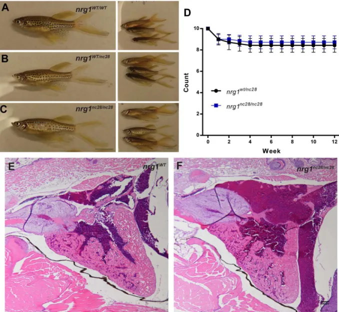
The adult nrg1
nc28mutant does not show overt morphological defect
Though our findings indicate that Nrg1 is dispensable for cardiac morphogenesis through lar-val stages, Nrg1 may be involved in other developmental processes. To address this possibility, we interbred heterozygous fish and followed sibling offspring to adulthood. Homozygous
mutantnrg1nc28fish were indistinguishable from wild type or heterozygous clutch mates at the
gross morphological level (Fig 4A–4C). Similarly, survival from larval to early adulthood was
comparable between genotypes (Fig 4D). To explore the possibility thatnrg1-Iandnrg1-II
Fig 4. nrg1nc28survive to adulthood without overt cardiac abnormalities. (A-C) Representative gross morphology of age-matched (A) nrg1WT/WT, (B) nrg1WT/nc28or (C) nrg1nc28/nc28clutch mates, Standard Length (SL) 25–30. Scale bar is 10mm. (D)
Weekly survival of fish from sibling nrg1WT/nc28and nrg1nc28/nc28. (E-F) Representative cross section of the heart in H&E stained section of formaldehyde-fixed, paraffin embedded (E) nrg1WT/WTand (F) nrg1nc28/nc28adult fish. BA = Bulbous Arteriosus, V = Ventricle, A = Atrium. N = 3 fish per genotype. Scale bar 100μm.
isoforms may modulate cardiac morphology in a non-lethal manner, we examined the H&E
stained heart sections ofnrg1wt/wtandnrg1nc28/nc28adult clutch mates and found no obvious
differences in gross morphology (Fig 4E and 4F). Together, our findings indicate thatnrg1-I
andnrg1-IIis dispensable for development and homeostatic function in zebrafish within the limit of our phenotypic analysis.
Discussion
This study highlights an example of cross-species differences in EGF family member
require-ments for the process of cardiac trabeculation. Previous mouse studies have shown thatnrg1-I
has a critical role in trabecular development [7]. More specifically, knockout studies have
dem-onstrated that loss ofnrg1-Iandnrg1-IIisoforms causes embryonic lethality likely due to a
complete loss of cardiac trabeculation and subsequent defect in cardiac contractility and
func-tion [58]. In contrast, in zebrafish, our study suggests that loss ofnrg1-Iandnrg1-IIfunction
does not have any survival or phenotypic consequences under homeostatic conditions. The
zebrafishnrg1-Iandnrg1-IImutant still develops cardiac trabeculae and survives to adulthood
without overt cardiac abnormalities.
Unfortunately, we were unable to assess functional Nrg1 protein levels via Western blot in this study due to unavailability of Nrg1antibody. Despite this obstacle, our assays of phar-macological inhibition of ErbB2 activity and gene expression data of other EGF-like ligands
suggests cardiac trabeculation occurs in zebrafishnrg1-I/IImutant in an ErbB2-dependent
manner. Pharmacological inhibition of ErbB2 activity in both WT and thenrg1-I/IImutant
completely abolished trabecular formation. These results suggest that othernrg1-I-like
fac-tors could function as the major ligand for ErbB4 receptor, act redundantly withnrg1-Ito
regulate cardiac trabeculation, or may act in a compensatory manner whennrg1-Iis absent.
Except fornrg1-I, we found that another four EGF-like ligands encoded byhb-egfa,btc,epgn
andnrg2awere also expressed in the zebrafish heart during trabeculation stage. The ques-tion arises as to which EGF-like ligand(s) could replace the funcques-tion of or compensate for
the loss ofnrg1-Ito regulate cardiac trabeculation in zebrafish. We suggest that the zebrafish
genomic structure supports divergent evolution of EGF-like ligands in regulating cardiac trabeculation. Therefore, ligands with paralogs in the zebrafish genome are the most likely candidates for this function.
The zebrafish genome contains gene duplications due to the teleost-specific whole genome duplication event during teleost speciation. This type of gene duplication allows one of the
paralogues to evolve new functions while the other retains the gene’s original function [44,59].
The zebrafish genome contains paralogues ofhbegfaandnrg2a,hbegfbandnrg2b, it is thus
possible that eitherhbegfaornrg2aacquired a role in regulating cardiac trabeculation that
could compensate or supersede any role thatnrg1may play in trabeculation. Alternatively,
sincebtctranscript levels were elevated innrg1-I/IImutant, the upregulation ofbtcexpression
may compensate for the loss ofnrg1-Ito regulate cardiac trabeculation in zebrafish.
Neverthe-less, mutants that ablate the function of these Nrg1 like factor(s) will need to be generated to determine whether and which ligand(s) have the primary role of regulating trabeculation and what role Nrg1 plays in the developing zebrafish heart.
Our study highlights the differences and complexity of zebrafishnrg1-Ifunction relative to
other model species, specifically in the process of cardiac trabeculation. In rodent models, loss ofnrg1-Iis developmentally lethal. Conversely, our work demonstrates that zebrafish do not
share the same conserved role fornrg1-I. Additionally, we have highlighted the need to further
define the roles of all Nrg1 isoforms in cardiac development and trabeculation within the
be replaced by EGF-like ligands in the zebrafish genome, the thirdnrg1isoformnrg1-III
appears to play an evolutionary conserved role in Schwann cell development [7,60–62]. In
zeb-rafish, thenrg1z26mutant specifically disruptsnrg1-IIIfunction, leading to severe defects in
Schwann cell migration and proliferation [55]. The differences in phenotypic consequences
upon ablating the function of different Nrg1 isoforms suggest that the function of the different Nrg1 isoforms could evolve independently in different tissue and cell types.
Supporting Information
S1 Fig. Predicted translations ofnrg1-Imutant alleles. (A)nrg1-IWTallele is translated into
599 amino acid (aa), (B)nrg1-Inc28into 55 aa, (C)nrg1-Inc29into 99 aa. (B-C) Amino acids that
differ from wild type are in red. Asterisk indicates stop codon. (TIF)
S1 Table. List of Primers used in this study.
(DOCX)
Acknowledgments
We thank the University of North Carolina Olympus Imaging Research Center for confocal microscope use, and the University of North Carolina Zebrafish Aquaculture Core Facility for fish care and microscope use. Histology services provided by the Histology Research Core Facility in the Department of Cell Biology and Physiology at the University of North Carolina, Chapel Hill NC. L.A.S. is supported by National Institutes of Health T32 grant (HL069768-14; PI, Christopher Mack). D.R.B. is supported by National Institutes of Health/National Institute of General Medical Science grant 5K12GM678-17 (PI, Linda Dyksta). This study was sup-ported by the American Heart Association Scientist Development Grant (13SDG17060010 to L.Q.) and the Ellison Medical Foundation New Scholar Grant (AG-NS-1064-13 to L.Q.). National Institutes of Health/National Heart, Lung, and Blood Institute (R00 HL109079 grant to J.L.) and American Heart Association (grant 15GRNT25530005 to J.L.).
Author Contributions
Conceptualization: LAS LQ JL.
Data curation: LAS CEI DRB LQ JL.
Formal analysis: LAS CEI DRB LQ JL.
Funding acquisition: LQ JL.
Investigation: LAS CEI DRB LQ JL.
Methodology: LAS JL.
Project administration: LQ JL.
Resources: LQ JL.
Software: LAS CEI DRB.
Supervision: LQ JL.
Validation: LAS CEI DRB LQ JL.
Writing – original draft: LAS CEI DRB JL.
Writing – review & editing: LAS CEI DRB LQ JL.
References
1. Parker SE, Mai CT, Canfield MA, Rickard R, Wang Y, Meyer RE, et al. (2010) Updated National Birth Prevalence estimates for selected birth defects in the United States, 2004–2006. Birth Defects Res A Clin Mol Teratol 88: 1008–1016. doi:10.1002/bdra.20735PMID:20878909
2. Moran AE, Roth GA, Narula J, Mensah GA (2014) 1990–2010 global cardiovascular disease atlas. Glob Heart 9: 3–16. doi:10.1016/j.gheart.2014.03.1220PMID:25432106
3. Writing Group M, Mozaffarian D, Benjamin EJ, Go AS, Arnett DK, Blaha MJ, et al. (2016) Heart Disease and Stroke Statistics-2016 Update: A Report From the American Heart Association. Circulation 133: e38–60. doi:10.1161/CIR.0000000000000350PMID:26673558
4. DALYs GBD, Collaborators H, Murray CJ, Barber RM, Foreman KJ, Abbasoglu Ozgoren A, et al. (2015) Global, regional, and national disability-adjusted life years (DALYs) for 306 diseases and injuries and healthy life expectancy (HALE) for 188 countries, 1990–2013: quantifying the epidemiological tran-sition. Lancet 386: 2145–2191. doi:10.1016/S0140-6736(15)61340-XPMID:26321261
5. Rupert CE, Coulombe KL (2015) The roles of neuregulin-1 in cardiac development, homeostasis, and disease. Biomark Insights 10: 1–9.
6. Gemberling M, Karra R, Dickson AL, Poss KD (2015) Nrg1 is an injury-induced cardiomyocyte mitogen for the endogenous heart regeneration program in zebrafish. Elife 4.
7. Meyer D, Birchmeier C (1995) Multiple essential functions of neuregulin in development. Nature 378: 386–390. doi:10.1038/378386a0PMID:7477375
8. Milan DJ, Giokas AC, Serluca FC, Peterson RT, MacRae CA (2006) Notch1b and neuregulin are required for specification of central cardiac conduction tissue. Development 133: 1125–1132. doi:10. 1242/dev.02279PMID:16481353
9. Montero JC, Yuste L, Diaz-Rodriguez E, Esparis-Ogando A, Pandiella A (2000) Differential shedding of transmembrane neuregulin isoforms by the tumor necrosis factor-alpha-converting enzyme. Mol Cell Neurosci 16: 631–648. doi:10.1006/mcne.2000.0896PMID:11083924
10. Vermot J, Forouhar AS, Liebling M, Wu D, Plummer D, Gharib M, et al. (2009) Reversing blood flows act through klf2a to ensure normal valvulogenesis in the developing heart. PLoS Biol 7: e1000246. doi: 10.1371/journal.pbio.1000246PMID:19924233
11. Yarden Y, Sliwkowski MX (2001) Untangling the ErbB signalling network. Nat Rev Mol Cell Biol 2: 127– 137. doi:10.1038/35052073PMID:11252954
12. Yokozeki T, Wakatsuki S, Hatsuzawa K, Black RA, Wada I, Sehara-Fujisawa A (2007) Meltrin beta (ADAM19) mediates ectodomain shedding of Neuregulin beta1 in the Golgi apparatus: fluorescence correlation spectroscopic observation of the dynamics of ectodomain shedding in living cells. Genes Cells 12: 329–343. doi:10.1111/j.1365-2443.2007.01060.xPMID:17352738
13. Fang SJ, Wu XS, Han ZH, Zhang XX, Wang CM, Li XY, et al. (2010) Neuregulin-1 preconditioning pro-tects the heart against ischemia/reperfusion injury through a PI3K/Akt-dependent mechanism. Chin Med J (Engl) 123: 3597–3604.
14. Formiga FR, Pelacho B, Garbayo E, Imbuluzqueta I, Diaz-Herraez P, Abizanda G, et al. (2014) Con-trolled delivery of fibroblast growth factor-1 and neuregulin-1 from biodegradable microparticles pro-motes cardiac repair in a rat myocardial infarction model through activation of endogenous
regeneration. J Control Release 173: 132–139. doi:10.1016/j.jconrel.2013.10.034PMID:24200746 15. Gao R, Zhang J, Cheng L, Wu X, Dong W, Yang X, et al. (2010) A Phase II, randomized, double-blind,
multicenter, based on standard therapy, placebo-controlled study of the efficacy and safety of recombi-nant human neuregulin-1 in patients with chronic heart failure. J Am Coll Cardiol 55: 1907–1914. doi: 10.1016/j.jacc.2009.12.044PMID:20430261
16. Guo YF, Zhang XX, Liu Y, Duan HY, Jie BZ, Wu XS (2012) Neuregulin-1 attenuates mitochondrial dys-function in a rat model of heart failure. Chin Med J (Engl) 125: 807–814.
17. Hill MF, Patel AV, Murphy A, Smith HM, Galindo CL, Pentassuglia L, et al. (2013) Intravenous glial growth factor 2 (GGF2) isoform of neuregulin-1beta improves left ventricular function, gene and protein expression in rats after myocardial infarction. PLoS One 8: e55741. doi:10.1371/journal.pone.0055741 PMID:23437060
favourable acute and chronic haemodynamic responses. Eur J Heart Fail 13: 83–92. doi:10.1093/ eurjhf/hfq152PMID:20810473
19. Liu X, Gu X, Li Z, Li X, Li H, Chang J, et al. (2006) Neuregulin-1/erbB-activation improves cardiac func-tion and survival in models of ischemic, dilated, and viral cardiomyopathy. J Am Coll Cardiol 48: 1438– 1447. doi:10.1016/j.jacc.2006.05.057PMID:17010808
20. Gassmann M, Casagranda F, Orioli D, Simon H, Lai C, Klein R, et al. (1995) Aberrant neural and car-diac development in mice lacking the ErbB4 neuregulin receptor. Nature 378: 390–394. doi:10.1038/ 378390a0PMID:7477376
21. Kramer R, Bucay N, Kane DJ, Martin LE, Tarpley JE, Theill LE (1996) Neuregulins with an Ig-like domain are essential for mouse myocardial and neuronal development. Proc Natl Acad Sci U S A 93: 4833–4838. PMID:8643489
22. Lee KF, Simon H, Chen H, Bates B, Hung MC, Hauser C (1995) Requirement for neuregulin receptor erbB2 in neural and cardiac development. Nature 378: 394–398. doi:10.1038/378394a0PMID: 7477377
23. Brown DR, Samsa LA, Qian L, Liu J (2016) Advances in the Study of Heart Development and Disease Using Zebrafish. J Cardiovasc Dev Dis 3.
24. Bang A, Gronkjaer P, Malte H (2004) Individual variation in the rate of oxygen consumption by zebrafish embryos. Journal of Fish Biology 64: 1285–1296.
25. Chen JN, Haffter P, Odenthal J, Vogelsang E, Brand M, van Eeden FJ, et al. (1996) Mutations affecting the cardiovascular system and other internal organs in zebrafish. Development 123: 293–302. PMID: 9007249
26. Sehnert AJ, Huq A, Weinstein BM, Walker C, Fishman M, Stainier DY (2002) Cardiac troponin T is essential in sarcomere assembly and cardiac contractility. Nat Genet 31: 106–110. doi:10.1038/ng875 PMID:11967535
27. Stainier DY, Fouquet B, Chen JN, Warren KS, Weinstein BM, Meiler SE, et al. (1996) Mutations affect-ing the formation and function of the cardiovascular system in the zebrafish embryo. Development 123: 285–292. PMID:9007248
28. Strecker R, Seiler TB, Hollert H, Braunbeck T (2011) Oxygen requirements of zebrafish (Danio rerio) embryos in embryo toxicity tests with environmental samples. Comp Biochem Physiol C Toxicol Phar-macol 153: 318–327. doi:10.1016/j.cbpc.2010.12.002PMID:21163368
29. Abdallah SJ, Thomas BS, Jonz MG (2015) Aquatic surface respiration and swimming behaviour in adult and developing zebrafish exposed to hypoxia. J Exp Biol 218: 1777–1786. doi:10.1242/jeb.116343 PMID:25944921
30. Rees BB, Sudradjat FA, Love JW (2001) Acclimation to hypoxia increases survival time of zebrafish, Danio rerio, during lethal hypoxia. J Exp Zool 289: 266–272. PMID:11241397
31. Liu J, Bressan M, Hassel D, Huisken J, Staudt D, Kikuchi K, et al. (2010) A dual role for ErbB2 signaling in cardiac trabeculation. Development 137: 3867–3875. doi:10.1242/dev.053736PMID:20978078 32. Minot CS (1901) Notes on Anopheles. J Boston Soc Med Sci 5: 325–329. PMID:19971352
33. Rychter Z, Ostadal B (1971) Fate of "sinusoidal" intertrabecular spaces of the cardiac wall after develop-ment of the coronary vascular bed in chick embryo. Folia Morphol (Praha) 19: 31–44.
34. Jenni R, Rojas J, Oechslin E (1999) Isolated noncompaction of the myocardium. N Engl J Med 340: 966–967. doi:10.1056/NEJM199903253401215PMID:10094647
35. Samsa LA, Yang B, Liu J (2013) Embryonic cardiac chamber maturation: Trabeculation, conduction, and cardiomyocyte proliferation. Am J Med Genet C Semin Med Genet 163C: 157–168. doi:10.1002/ ajmg.c.31366PMID:23720419
36. Honjo Y, Kniss J, Eisen JS (2008) Neuregulin-mediated ErbB3 signaling is required for formation of zeb-rafish dorsal root ganglion neurons. Development 135: 2615–2625. doi:10.1242/dev.022178PMID: 18599505
37. Westerfield M (1993) The zebrafish book: a guide for the laboratory use of zebrafish Danio (Brachyda-nio) rerio. Eugene, OR: Institute of Neuroscience, University of Oregon,.
38. Perlin JR, Lush ME, Stephens WZ, Piotrowski T, Talbot WS (2011) Neuronal Neuregulin 1 type III directs Schwann cell migration. Development (Cambridge, England) 138: 4639–4648.
41. Huang CJ, Tu CT, Hsiao CD, Hsieh FJ, Tsai HJ (2003) Germ-line transmission of a myocardium-spe-cific GFP transgene reveals critical regulatory elements in the cardiac myosin light chain 2 promoter of zebrafish. Dev Dyn 228: 30–40. doi:10.1002/dvdy.10356PMID:12950077
42. Chang N, Sun C, Gao L, Zhu D, Xu X, Zhu X, et al. (2013) Genome editing with RNA-guided Cas9 nuclease in zebrafish embryos. Cell Res 23: 465–472. doi:10.1038/cr.2013.45PMID:23528705 43. Sander JD, Zaback P, Joung JK, Voytas DF, Dobbs D (2007) Zinc Finger Targeter (ZiFiT): an
engi-neered zinc finger/target site design tool. Nucleic Acids Res 35: W599–605. doi:10.1093/nar/gkm349 PMID:17526515
44. Howe K, Clark MD, Torroja CF, Torrance J, Berthelot C, Muffato M, et al. (2013) The zebrafish refer-ence genome sequrefer-ence and its relationship to the human genome. Nature 496: 498–503. doi:10.1038/ nature12111PMID:23594743
45. Hwang WY, Fu Y, Reyon D, Maeder ML, Tsai SQ, Sander JD, et al. (2013) Efficient genome editing in zebrafish using a CRISPR-Cas system. Nat Biotechnol 31: 227–229. doi:10.1038/nbt.2501PMID: 23360964
46. Dahlem TJ, Hoshijima K, Jurynec MJ, Gunther D, Starker CG, Locke AS, et al. (2012) Simple methods for generating and detecting locus-specific mutations induced with TALENs in the zebrafish genome. PLoS Genet 8: e1002861. doi:10.1371/journal.pgen.1002861PMID:22916025
47. Livak KJ, Schmittgen TD (2001) Analysis of relative gene expression data using real-time quantitative PCR and the 2(-Delta Delta C(T)) Method. Methods 25.
48. Samsa LA, Givens C, Tzima E, Stainier DY, Qian L, Liu J (2015) Cardiac contraction activates endocar-dial Notch signaling to modulate chamber maturation in zebrafish. Development 142: 4080–4091. doi: 10.1242/dev.125724PMID:26628092
49. Samsa LA, Fleming N, Magness S, Qian L, Liu J (2016) Isolation and Characterization of Single Cells from Zebrafish Embryos. e53877.
50. Liu J, Stainier DY (2010) Tbx5 and Bmp signaling are essential for proepicardium specification in zebra-fish. Circ Res 106: 1818–1828. doi:10.1161/CIRCRESAHA.110.217950PMID:20413782
51. Schneider CA, Rasband WS, Eliceiri KW (2012) NIH Image to ImageJ: 25 years of image analysis. Nat Methods 9: 671–675. PMID:22930834
52. Lopez-Schier H, Hudspeth AJ (2005) Supernumerary neuromasts in the posterior lateral line of zebra-fish lacking peripheral glia. Proc Natl Acad Sci U S A 102: 1496–1501. doi:10.1073/pnas.0409361102 PMID:15677337
53. Pentassuglia L, Sawyer DB (2009) The role of Neuregulin-1beta/ErbB signaling in the heart. Exp Cell Res 315: 627–637. doi:10.1016/j.yexcr.2008.08.015PMID:18801360
54. Fuller SJ, Sivarajah K, Sugden PH (2008) ErbB receptors, their ligands, and the consequences of their activation and inhibition in the myocardium. J Mol Cell Cardiol 44: 831–854. doi:10.1016/j.yjmcc.2008. 02.278PMID:18430438
55. Perlin JR, Lush ME, Stephens WZ, Piotrowski T, Talbot WS (2011) Neuronal Neuregulin 1 type III directs Schwann cell migration. Development 138: 4639–4648. doi:10.1242/dev.068072PMID: 21965611
56. Lyons DA, Pogoda HM, Voas MG, Woods IG, Diamond B, Nix R, et al. (2005) erbb3 and erbb2 are essential for schwann cell migration and myelination in zebrafish. Curr Biol 15: 513–524. doi:10.1016/j. cub.2005.02.030PMID:15797019
57. Peshkovsky C, Totong R, Yelon D (2011) Dependence of cardiac trabeculation on neuregulin signaling and blood flow in zebrafish. Dev Dyn 240: 446–456. doi:10.1002/dvdy.22526PMID:21246662 58. Lai D, Liu X, Forrai A, Wolstein O, Michalicek J, Ahmed I, et al. (2010) Neuregulin 1 sustains the gene
regulatory network in both trabecular and nontrabecular myocardium. Circ Res 107: 715–727. doi:10. 1161/CIRCRESAHA.110.218693PMID:20651287
59. Glasauer SM, Neuhauss SC (2014) Whole-genome duplication in teleost fishes and its evolutionary consequences. Mol Genet Genomics 289: 1045–1060. doi:10.1007/s00438-014-0889-2PMID: 25092473
60. Newbern J, Birchmeier C (2010) Nrg1/ErbB signaling networks in Schwann cell development and myeli-nation. Semin Cell Dev Biol 21: 922–928. doi:10.1016/j.semcdb.2010.08.008PMID:20832498 61. Taveggia C, Zanazzi G, Petrylak A, Yano H, Rosenbluth J, Einheber S, et al. (2005) Neuregulin-1 type
III determines the ensheathment fate of axons. Neuron 47: 681–694. doi:10.1016/j.neuron.2005.08. 017PMID:16129398