Mixed signature of activation and dysfunction allows
human decidual CD8
+
T cells to provide both
tolerance and immunity
Anita van der Zwana,b, Kevin Bic, Errol R. Norwitzd,e, Ângela C. Crespoa,f, Frans H. J. Claasb, Jack L. Stromingera,1, and Tamara Tilburgsa,1
aDepartment of Stem Cell and Regenerative Biology, Harvard University, Cambridge, MA 02138;bDepartment of Immunohematology and Blood
Transfusion, Leiden University Medical Center, 2333 ZA Leiden, The Netherlands;cDepartment of Pediatric Oncology, Dana-Farber Cancer Institute,
Children’s Hospital, Boston, MA 02115;dDepartment of Obstetrics & Gynecology, Tufts Medical Center, Boston, MA 02111;eMother Infant Research
Institute, Tufts Medical Center, Boston, MA 02111; andfProgram in Cellular and Molecular Medicine, Boston Children’s Hospital, Boston, MA 02115
Contributed by Jack L. Strominger, November 27, 2017 (sent for review August 9, 2017; reviewed by Ana Anderson and Adrian Erlebacher)
Understanding how decidual CD8+T cell (CD8+dT) cytotoxicity is regulated and how these cells integrate the competing needs for maternal–fetal tolerance and immunity to infection is an important research and clinical goal. Gene-expression analysis of effector-memory CD8+dT demonstrated a mixed transcriptional signature of T cell dysfunction, activation, and effector function. High protein expression of coinhibitory molecules PD1, CTLA4, and LAG3, accom-panied by low expression of cytolytic molecules suggests that the decidual microenvironment reduces CD8+dT effector responses to maintain tolerance to fetal antigens. However, CD8+dT degranu-lated, proliferated, and produced IFN-γ, TNF-α, perforin, and gran-zymes upon in vitro stimulation, demonstrating that CD8+dT are not permanently suppressed and retain the capacity to respond to proinflammatory events, such as infections. The balance between transient dysfunction of CD8+dT that are permissive of placental and fetal development, and reversal of this dysfunctional state, is crucial in understanding the etiology of pregnancy complications and prevention of congenital infections.
pregnancy
|
T cell exhaustion|
trophoblast|
cytotoxicity|
placentaT
o establish a successful pregnancy, maternal decidual CD8+ T cells (CD8+dT) at the maternal–fetal interface must in-tegrate the antithetical demands of maternal–fetal tolerance and antiviral immunity (1). The key question is whether CD8+dT have the ability to elicit cytolytic responses to placental, fetal, or viral antigens or are rendered permanently dysfunctional and exhibit impaired effector functions. Among dysfunctional T cells are exhausted CD8+T cells that initially obtain effector functions and become dysfunctional during chronic exposure to antigen (2). Other dysfunctional cells include anergic T cells that fail to gain effector functions due to priming without costimulation and suppressed T cells that may be temporarily inhibited in their effector function after interaction with immune suppressive cells, such as regulatory T cells (Tregs) (2). T cell dysfunction is characterized by loss of IL-2, IFN-γ, and TNF-αproduction, diminished proliferative capacity, and low T cell cytotoxicity. A variety of markers have been implicated to identify dysfunctional T cells but expression of these coinhibitory molecules [e.g., programmed cell death-1 (PD1), T cell Ig mucin-3 (TIM3), and cytotoxic T-lymphocyte–associated protein 4 (CTLA4)] is not exclusive to dysfunctional T cells and is also observed in activated T cells (3–6). The significant overlap of gene-expression profiles and cell-surface markers between dysfunctional and ac-tivated T cells makes functional assessment (e.g., proliferation, cytokine secretion, cytotoxicity) necessary to separate these cell types.T cell dysfunction was first described in chronic lymphocytic choriomeningitis virus (LCMV) infection in mice where LCMV-specific CD8+T cells were unable to control the infection (7, 8). However, infected mice retained antiviral CTL responses and ap-plied selection pressure on the persisting virus (9). In both HIV and
hepatitis-C virus infection, the emergence of viral escape mutants highlights the fact that T cell effector responses are retained re-gardless of the presence of phenotypically dysfunctional T cells (10, 11). Human cytomegalovirus (HCMV)-specific CD8+T cells have low proliferative capacity, low production of IL-2, and express PD1. Despite these signs of dysfunction, they are capable of producing ample amounts of IFN-γand granzyme B (GZMB) when stimu-lated (12). In humans, T cell dysfunction has been demonstrated in a wide variety of cancers, including melanoma and colorectal cancer patients (6, 13) and during chronic infections (5). Fur-thermore, blockade of CTLA4 and PD1 pathways has been associated with improved effector T cell responses in these patients (14, 15). A recent study described gene signatures in dysfunctional tumor infiltrating T cells that can be uncoupled from activation signatures at the single-cell level. This study identified metallothionein-1 (MT1) and MT2 as specific markers for dysfunctional CD8+tumor infiltrating T cells (16). MTs are cysteine-rich zinc chaperones that are involved in zinc regulation and protection against oxidative stress (17). Deletion of MT1 resulted in increased T cell proliferation, loss of T cell dysfunc-tion, and thus reduced tumor growth (16). These clinical and experimental observations provide evidence that immune surveillance
Significance
Successful pregnancy requires establishment of immune toler-ance for invading fetal trophoblasts, as well as immunity to a variety of pathogens that cause placental and congenital infec-tions. Decidual CD8+T cells are key cells for recognition and response to foreign fetal, placental, and viral antigens at the maternal–fetal interface. Thus, regulation of decidual CD8+T cell activation and cytotoxicity is crucial for a healthy pregnancy. Here, we demonstrate that decidual CD8+T cells have a mixed profile of T cell dysfunction, activation, and effector function, which allows for both immune tolerance and immunity. This is of great relevance for understanding the development of preg-nancy complications as well as prevention of congenital infections that occur as result of impaired placental immunity.
Author contributions: A.v.d.Z., J.L.S., and T.T. designed research; A.v.d.Z., Â.C.C., and T.T. performed research; K.B., E.R.N., and F.H.J.C. contributed new reagents/analytic tools; A.v.d.Z., K.B., and T.T. analyzed data; and A.v.d.Z., F.H.J.C., J.L.S., and T.T. wrote the paper.
Reviewers: A.A., Harvard Medical School; and A.E., University of California, San Francisco.
The authors declare no conflict of interest.
Published under thePNAS license.
Data deposition: Microarray data have been deposited in the ArrayExpress database, https://www.ebi.ac.uk/arrayexpress/(accession no.E-MTAB-5890).
1To whom correspondence may be addressed. Email: jlstrom@fas.harvard.edu or tilburgs@fas.harvard.edu.
This article contains supporting information online atwww.pnas.org/lookup/suppl/doi:10. 1073/pnas.1713957115/-/DCSupplemental.
IMMUNOLO
GY
AND
INFLAM
remains active in chronic infections and cancer and that T cell dysfunction is incomplete.
Previous studies have shown that CD8+ dT in first trimester pregnancy have an increased ability to produce cytokines, in-cluding IFN-γ (18, 19). Furthermore, a subset of first trimester PD1+TIM3+ CD8+ dT showed increased proliferation potential and Th2 cytokine production (20). Despite this evidence suggesting increased activation, CD8+dT were also hypothesized to be dys-functional/exhausted because of the high expression of the coin-hibitory molecule PD1 and the ability of PDL1 to modify cytokine secretion (21), as well as the low expression levels of perforin (PRF) and GZMB in term pregnancy CD8+dT (22). Thus far, no comprehensive data has been presented on CD8+ dT function throughout gestation and whether these cells are rendered dys-functional or maintain the ability to generate proinflammatory responses. CD8+ dT are exposed to allogeneic fetal minor and major histocompatibility antigens (mHag and MHC) expressed by fetal HLA-G+HLA-C+extravillous trophoblasts (EVT) through-out gestation (23, 24). CD8+dT make up 2–7% of leukocytes in first trimester decidua and their proportion increases to∼30% at term pregnancy (25). CD8+dT are differentiated effector-memory (EM) cells that express reduced levels of PRF and GZMB (22). In mice, maternal CD8+T cells responded to viral and bacterial an-tigens, but were unable to completely clear the pathogens during pregnancy (26, 27). Activation and expansion of fetus-specific CD4+and CD8+T cells by seminal fluid in mice resulted in high levels of CD4+CD25+(Treg) and activation of fetus-specific CD8+ T cells did not have an influence on pregnancy outcome (28).
In humans, antibody and CTL responses to MHC and mHag (e.g., HY) were detected in maternal blood during and as a result of pregnancy (29, 30). While induction of HLA-A–and HLA-B– specific antibodies did not negatively impact pregnancy outcome, the presence of HLA-C–specific antibodies in women with re-current miscarriage suggested that antibody-mediated rejection may be involved in the origin of unexplained recurrent miscar-riages (31). HCMV seropositivity profoundly influenced the T cell repertoire during pregnancy and led to the accumulation of highly differentiated memory T cells (32). HLA-A–and HLA-B– restricted virus-specific CD8+T cells, as well as CD8+T cells spe-cific for the HY antigen, are present in human decidual tissue (1, 33). Thus, both murine and human studies clearly demonstrate that maternal CD8+T cells respond to viral, fetal, and placental anti-gens during pregnancy. However, regulation of CD8+dT effector function prevents detrimental cytolytic responses to invading fetal EVT and maintains maternal–fetal immune tolerance. In this study, CD8+T cell function was investigated by transcriptome analysis of CD8+dT from human first trimester and term pregnancy. More-over, gene-expression profiles were combined with phenotypic characterization and assessment of CD8+dT effector functions.
Results
CD8+EM dT Have a Mixed Gene-Expression Profile.A significantly
increased percentage of CCR7−CD45RA−EM CD8+T cells and a decrease in CCR7+CD45RA+naïve CD8+T cells was observed in first trimester (6–12 wk) and term (>37 wk) pregnancy decidual tissue when compared with peripheral blood CD8+T cells (CD8+ pT), confirming previous studies (Fig. S1A–D) (22, 34). Within the EM subsets, TEM1cells, defined as CD28+CD27+EM cells, were
significantly increased in the first trimester compared with term pregnancy decidua (Fig. S1E). Furthermore, a small but not sig-nificant increase in CD28−CD27+TEM2and CD28−CD27−TEM3
cells was detected in term compared with first trimester pregnancy decidua. Analysis of the cytolytic molecule PRF also showed re-duced expression in first trimester decidual CD8+effector (Eff) and TEM3cells compared with the same populations in blood, as has
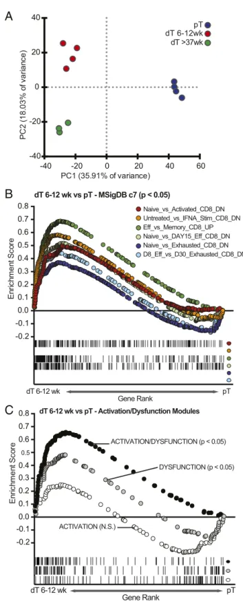
previously been described for term CD8+dT (Fig. S1F) (22). Gene-expression profiles were generated from RNA purified from CD8+CCR7−CD45RA−EM T cells in blood (CD8+ EM pT) and decidua (CD8+EM dT; 6–12 wk and>37 wk). Unsupervised principle component analysis (PCA) separated CD8+ EM dT from CD8+EM pT along the first principal component (35.9% of
variance). PC2 separated first trimester from term pregnancy CD8+ EM dT (18.0% of variance) (Fig. 1A). A transcriptional signature that uniquely defined CD8+ EM dT and EM pT was identified (Fig. S2AandDataset S1). Genes up-regulated in CD8+EM dT compared with EM pT included genes involved in chemotaxis (CCL3, CCL4, IL-8, XCL1), T cell activation (IFN-γ, TNF, FOS, ICOS, NFKB1), and coinhibitory receptors (FASLG, CTLA4, LAG3, TIGIT, CRTAM, and TIM3). An increase in mRNA for gran-zymes, but not the other cytolytic molecules PRF and granulysin, was observed in term CD8+EM dT (Fig. S3A).
CD8+EM dT Have a Mixed Profile of Dysfunction, Activation, and
Effector Function. To further identify transcriptional differences
between first trimester CD8+EM dT and EM pT, gene-set en-richment analysis (GSEA) was performed by comparing the CD8+ EM dT gene set to existing immunological gene sets in the ImmSigDB database (35, 36). GSEA demonstrated a significant enrichment of effector vs. naïve, effector vs. memory, exhausted vs. naïve, and exhausted vs. effector gene sets in CD8+EM dT relative to CD8+EM pT (Fig. 1B). GSEA also revealed that expression of genes associated with both dysfunction and activation states of CD8+T cells are increased in first trimester CD8+EM dT compared with CD8+EM pT. For this analysis, our data were compared with a recently published gene signature where dysfunction and acti-vation gene modules were uncoupled (Fig. 1C) (16). Flow cytometric analysis detecting protein expression of selected activation markers (CD69, HLA-DR, GITR, and CD25) and coinhibitory molecules (PD1, CTLA4, LAG3, and TIGIT) confirmed that expression of markers associated with both activation and dysfunction are increased on CD8+dT compared with pT (Fig. S3C–J).
Term CD8+EM dT Express Methallothioneins, a Signature for Dysfunctional
T Cells.No significantly different gene sets were identified when
gene-expression profiles of first trimester and term CD8+EM dT were compared. However, a striking enrichment of MT1 and MT2 genes, which have recently been associated with dysfunctional T cells, was observed in term CD8+EM dT (Fig. S2BandDataset S2) (16). The presence of MT genes in term CD8+EM dT and not in first trimester suggests that antigenic stimulation throughout the 9 months of pregnancy may gradually increase CD8+EM dT dys-function. Other differences between first trimester and term CD8+ EM dT included increased expression of galectin-8 (LGALS8) and galectin-9 (LGALS9) in term CD8+ EM dT. Galectins have a broad variety of functions including mediation of cell–cell interac-tions, apoptosis, and facilitating the differentiation of regulatory T cells (37). Thus, toward the end of pregnancy CD8+EM dT have also acquired gene signatures associated with immune suppression.
CD8+EM dT Can Acquire Signatures of T Cell Activation.To investigate
a player in microRNA biogenesis, was induced after 72 h of stim-ulation. The proinflammatory cytokines IFN-γand TNF revealed higher expression levels within 12 h after stimulation, whereas expression of the cytolytic molecules PRF, GZMA, GZMB, GZMH, and GNLY were induced within 72 h of stimulation (Fig. S3B). Taken together, stimulation of first trimester CD8+EM dT resulted in up-regulation of genes involved in T cell activation and cytotoxicity.
CD8+dT Are Functional Upon Activation.T cell dysfunction is
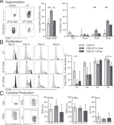
asso-ciated with a limited capacity to degranulate, proliferate, and se-crete cytokines upon stimulation (5, 11). Although, previous studies have shown increased proliferation and cytokine production of first trimester CD8+dT (18–20), no comprehensive study has compared functional aspects of CD8+dT throughout gestation from first tri-mester to term. To determine whether activation of CD8+dT elicits these effector responses, total CD8+ dT and pT were stimulated in vitro. A significantly increased percentage of first trimester CD8+ dT degranulated upon PMA/Ionomycin stimulation compared with CD8+pT (Fig. 3A). Degranulation was predominantly observed in CD8+dTEFF(CD45RA+CCR7−) and dTEM(CCR7−CD45RA−).
Term CD8+dT degranulated significantly less than first trimester CD8+dT, but at a level similar to CD8+pT (Fig. 3A).
Carboxyfluorescein diacetate succinimidyl ester (CFSE) -labeled CD8+pT and dT were stimulated with anti-CD3/28 and analyzed at days 3, 4, 5, and 6 for their capacity to proliferate. At day 3, significantly fewer CD8+dT had proliferated compared with CD8+ pT. However, by days 5 and 6 virtually all first trimester CD8+dT and pT had lost CFSE expression (Fig. 3B). While the pro-liferation index for term CD8+dT was similar to CD8+pT on day 6 (Fig. S4A), there was a significantly lower percentage of term CD8+dT that had divided compared with CD8+pT (median of 65% vs. 89%) (Fig. 3B). No significant differences between the proliferation index of CD8+pT and dT were observed (Fig. S4A). These data demonstrated that CD8+dT required more time to initiate proliferation, yet once they started dividing, first trimester CD8+dT proliferated at a similar rate to CD8+pT.
The percentage of CD8+ dT expressing IFN-γ, TNF-α, and IL-2 upon stimulation with PMA/Ionomycin was comparable to CD8+ pT (Fig. 3C). All effector and EM CD8+ pT and dT produced IFN-γ and TNF-α, whereas the production of these cytokines was in effect absent in naïve CD8+T cells. Among the EM subsets, EM-1 CD8+T cells were the main producers of IFN-γand TNF-α(Fig. S4BandC). No production of IL-10 and IL-17a was observed in CD8+pT or dT.
Next, the expression of PRF and GZMB in activated CD8+dT was analyzed to determine whether the reduced levels of PRF in CD8+ dT is reversible or permanently suppressed. Activation with IL-12 or anti-CD3/28 was sufficient to increase PRF ex-pression in first trimester and term pregnancy CD8+CD28−and CD8+CD28+dT by approximately twofold (Fig. 4AandFig. S5). However, activation of CD8+ dT did not increase the PRF content to levels observed in CD8+pT. Moreover, treatment of CD8+dT with either anti-CD3/28 or the combination of IL-12 and anti-CD3/28 increased GZMB in CD8+dT to levels com-parable or higher than CD8+pT (Fig. 4BandFig. S6). Thus, the majority of CD8+dT in both first trimester and term pregnancy degranulate, proliferate, secrete proinflammatory cytokines, and increase cytolytic molecules upon activation and do not reside in a permanently dysfunctional state.
PRF, but Not GZMB, Is Suppressed in HCMV-Specific CD8+dT.Next,
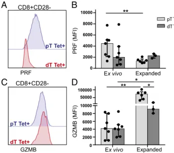
the question whether virus-specific CD8+ dT have suppressed expression of cytolytic molecules that could impair or delay their ability to respond to infections in the placenta was addressed. HCMV is the most common congenital infection and HCMV-specific CD8+dT are present at the maternal–fetal interface (33). First trimester CD8+CD28−dT and pT were stained with HCMV-specific tetramers and analyzed for expression of PRF and GZMB (Fig. 5 andFig. S7A). PRF expression was about twofold lower in HCMV-specific CD8+dT compared with HCMV-specific CD8+ pT (Fig. 5AandB). In contrast, HCMV-specific CD8+dT had
A
-40 -20 0 20 40 60
-40 -20 0 20 40
PC1 (35.91% of variance)
PC
2
(1
8
.0
3
%
o
f v
a
ri
a
n
c
e
)
pT dT 6-12wk dT >37wk
B
-0.2 -0.1 0.0 0.1 0.2 0.3 0.4 0.5 0.6 0.7 0.8
En
ri
c
h
m
e
n
t Sc
o
re
dT 6-12 wk vs pT - MSigDB c7 (p < 0.05)
dT 6-12 wk pT
Gene Rank
C
-0.2 -0.1 0.0 0.1 0.2 0.3 0.4
0.5
0.6 0.7 0.8
En
ri
c
h
m
e
n
t Sc
o
re
dT 6-12 wk vs pT - Activation/Dysfunction Modules
ACTIVATION/DYSFUNCTION (p < 0.05)
DYSFUNCTION (p < 0.05)
ACTIVATION (N.S.)
dT 6-12 wk pT
Gene Rank
Naive_vs_Activated_CD8_DN Untreated_vs_IFNA_Stim_CD8_DN Eff_vs_Memory_CD8_UP Naive_vs_DAY15_Eff_CD8_DN Naive_vs_Exhausted_CD8_DN D8_Eff_vs_D30_Exhausted_CD8_DN
Fig. 1. Transcriptional signatures of CD8+EM dT dysfunction, activation, and effector function are coupled. (A) Unsupervised PCA analysis, using all 54,715 probes, of first trimester (dT 6–12 wk) and term pregnancy (dT>37 wk) CD8+ EM dT and EM pT. (B) GSEA performed with assorted immunological signa-tures, showing positive correlation for an in vitro CD8+activation signature, an effector signature, and a CD8+viral exhaustion signature (MSigDB). (C) GSEA comparing first trimester CD8+EM dT to gene modules of dysfunction, activation, and activation coupled to dysfunction (16).
IMMUNOLO
GY
AND
INFLAM
comparable GZMB levels to CD8+pT (Fig. 5CandD). A ratio of PRF and GZMB expression of each individual donor revealed a significant difference between HCMV-specific CD8+ dT and pT (Fig. S7B). HCMV-specific CD8+dT and pT were expanded and PRF levels in the expanded CD8+ dT did not increase, while PRF significantly decreased in CD8+pT after expansion (Fig. 5B and Fig. S7C). Expansion of HCMV-specific cells significantly increased GZMB content in both CD8+ dT and pT, although GZMB levels in CD8+dT did not reach the same levels as in CD8+ pT (Fig. 5D). Thus, suppression of cytolytic proteins in virus-specific CD8+dT can be partially overcome by T cell activation as may occur during a viral infection in placental tissues.
CD8+ dT Do Not Degranulate in Response to EVT. The
antigen-specificity of CD8+dT and their ability to recognize and respond to fetal antigens expressed by EVT is a key question that is yet to be answered. The potential of CD8+ T cells to degranulate in response to EVT was determined by culturing CD8+T cells alone, or in the presence of EVT or anti-CD3/28 beads for 12 h. Co-culture of EVT with CD8+dT from the same pregnancy sample (dT sample-matched), a different pregnancy sample (dT nonmatched), or from unrelated blood donors (pT nonmatched) did not induce degranulation by any of the CD8+T cells (Fig. S8). Addition of anti-CD3/28 increased degranulation by all CD8+ T cells, dem-onstrating T cell viability. Thus, similar to EVT and decidual NK
cell (dNK) cocultures (39), EVT do not directly elicit degranula-tion by CD8+T cells.
Discussion
Numerous gaps remain in our understanding of how pregnancy affects adaptive immunity and, in particular, how CD8+dT in-tegrate protective immunity against pathogens with immune tolerance to invading EVT. In this study, expression analysis of decidual CD8+EM T cells demonstrated a mixed transcriptional signature of T cell dysfunction, activation, and effector function. The enriched gene signature for T cell dysfunction in CD8+dT together with the increased expression of coinhibitory molecules PD1, CTLA4, and LAG3, and low expression of the cytolytic molecule PRF suggests that the decidual microenvironment re-duces CD8+dT effector responses, possibly to maintain immune tolerance to fetal antigens. However, the ability of CD8+dT to up-regulate PRF and GZMB expression, degranulate, proliferate, and secrete proinflammatory cytokines upon activation suggests that CD8+ dT are not permanently suppressed and retain the capacity to respond to proinflammatory events, such as infections. This also confirms the uncoupling of coinhibitory receptors from the dysfunctional T cell phenotype (16).
RNA sequencing on single cells or small population level of CD8+ dT combined with functional assessment of the CD8+ T cell subpopulations will determine whether gene modules for activation and dysfunction are intertwined or uncoupled. It is of high clinical relevance to determine whether there are CD8+EM dT populations responsible for maternal immune tolerance that
A
DegranulationB
ProliferationDay 3 Day 4 Day 5 Day 6
CFSE
pT
dT
6-12wk
dT
>37wk
C
Cytokine ProductionCD8 dT >37wk CD8 pT CD8 dT 6-12wk
0 20 40 60 80 100
Total CD8+ T cells divided (%)
* *
D3 D4 D5 D6
0 40 60 80 100
n=16 n=15 n=7 20
IFNγ
IFNγ (%)
0 40 60 80 100
20
TNF
α (%)
TNFα
0 20 40 60 80 100 IL2
IL2 (%)
CD8
CD107a+
pT
dT 6-12wk dT >37wk Control
Total CD8 dT (6-12wk)
IgG IFNγ
TNF
α
IL2
0 20 40 60 80 100
Total CD8+CD107a+ (%)
Total CD8+
** *
TN TCM TEFF TEM
**
n=16 n=15 n=7 n=16 n=15 n=7
** *
CD107a+ (%)
0 20 40 60 80 100
Fig. 3. CD8+dT are functional upon activation. (A) FACS plots (Left) and percentages (Right) of CD107a+CD8+pT and dT after stimulation with PMA/ Ionomycin (1μg/mL) for 6 h. Percentage CD107a+cells are depicted within total CD8+T cells and the four CD8+T cell subpopulations (as percentage of total CD8). (B) Representative histograms (Left) and percentage divided cells (Right) of CFSE-labeled total CD8+T cells at days 3, 4, 5, and 6 after stimu-lation with anti-CD3/28 (2μL/mL) and 50 U/mL IL-2. (C) FACS plots (Left) and percentages (Right) of expression of intracellular IFN-γ, TNF-α, and IL-2 in total CD8+pT and dT stimulated with PMA/Ionomycin (1μg/mL) for 6 h. Bars represent the median with interquartile range; data are representative of three to six independent experiments; *P≤0.05, **P≤0.01.
0h
Cluster 1
Cluster 2
Cluster 3
Cluster 4
Cluster 5
12h 72h
−1 0 1
0 12 72
Hours after stimulation CXCL3 HLAE IL11 IL6ST IL7R Cluster 3
Cluster 4
−1.0 −0.5 0.0 0.5 1.0 1.5
BATF CDK6 CXCL10 IL2RA PRDM1 PRDX1 TNFAIP1
Cluster 5
−1 0 1 2
CCL28 CXCL11 DROSHA GZMB IL10RB IL13
IL9 IRF2 LAG3 LGALS3 RUNX1 TGFBR1
Mean relative expression
Row Max
Mean relative expression
n
oi
s
s
er
p
x
e
e
vit
al
er
n
a
e
M
A
B
0 12Hours after stimulation 72
0 12Hours after stimulation 72
Row Min
are distinct from a subset of activated CD8+EM dT with effector functions ready to confront incoming pathogens. A major challenge here is to connect gene-expression analysis at the single-cell level with accurate functional assessment of T cell function. This is par-ticularly challenging for human T cell subtypes that proliferate in vivo but not in vitro. Further identification and validation of markers that can separate dysfunctional T cells from activated T cells such as the recently proposed MT1 and MT2 genes is re-quired (16). Consistent with prolonged antigen stimulation of CD8+ dT at term pregnancy, enrichment of MT1 and MT2 genes was observed in term CD8+EM dT compared with first trimester CD8+ EM dT. However, phenotypic and functional assessment of first trimester and term CD8+EM dT revealed only minor differences. The reduced capacity of CD8+dT to initiate proliferation and the failure of term CD8+dT to fully degranulate and proliferate at day 6 suggests that a subset of CD8+dT gradually becomes dysfunc-tional through exhaustion or suppression over the course of preg-nancy, while the majority retains the capacity to respond. A recent study in mice observed that the priming of maternal naïve T cells by fetal antigens resulted in the differentiation of long-lived PD1+ CD8+T cells with selective silencing of effector function in a sub-sequent pregnancy. This dysfunction was reversed during skin trans-plantation (40). In the same manner, the integration of the competing needs for maternal immune tolerance in concert with immunity to placental infections may be achieved by the proficiency of the de-cidual microenvironment to dampen the activation state of CD8+ dT while not exclusively inducing their dysfunctional state.
dNK biology and interactions with MHC molecules expressed by EVT has been a major focus of research for over two decades.
Despite their abundance of cytolytic granules, dNK degranulate significantly less than blood NK cells in response to classic target cells (41, 42). However, when activated with a low dose of IL-15, dNK degranulate and produce proinflammatory cytokines to HCMV-infected decidual stromal cells (41, 43), yet are unable to respond to HCMV-infected EVT (39). The inability of dNK to kill infected EVT may make placental tissue more dependent on CD8+dT responses to clear infections. Activation of CD8+dT increased both PRF and GZMB mRNA and protein expression. The high levels of PRF mRNA in the absence of PRF protein in term CD8+dT (22) and the induction of DROSHA, as presented here, suggests that posttranscriptional regulation mediated by miRNAs may allow for a rapid increase in cytolytic proteins and cytolytic capacity upon stimulation (44). Thus, although dNK and CD8+dT both require additional activation by cytokines or receptor-ligand interactions to display their full cytotoxicity, the mechanisms that inhibit or delay their cytotoxic response are inherently different. HLA-A– and HLA-B–restricted virus-specific CD8+ T cells are enriched in decidual tissue at term pregnancy (33). Here, we demonstrated that HCMV-specific CD8+ dT in first trimester pregnancy have reduced levels of PRF protein compared with HCMV-specific CD8+pT, whereas their GZMB protein levels are comparable. After expansion of HCMV-specific CD8+ dT, PRF levels remained the same and GZMB levels significantly increased, although levels remained lower than in CD8+pT. HCMV-specific CD8+dT and pT are capable of cytotoxicity regardless of low PRF levels (33). Our data imply a general suppression of PRF trans-lation in all CD8+dT, including virus-specific CD8+dT. However, this suppression can be overcome by T cell activation as might be the case during viral infections of the placenta. A and HLA-B are not expressed by EVT, and further investigation is needed to determine whether HLA-C–restricted pathogen-specific responses by CD8+dT can provide immunity when EVT are infected. In-vestigation into whether HLA-C–restricted T cells with specificity for placental or fetal antigens cause detrimental immune responses and contribute to the development of pregnancy complications is required. Here, we demonstrated that CD8+dT do not degranu-late during coculture with healthy EVT. Additional research should determine whether the failure of CD8+dT to respond to EVT is due to their dysfunction or to a lack of antigen specificity to EVT antigens. Furthermore, investigation into how the interactions of EVT and other immune modulatory placental cells (e.g., decidual
A
C
PRFpT Tet+
dT Tet+
GZMB pT Tet+
dT Tet+
Expanded
PRF (MFI)
0 2000 4000 6000 8000
10000 **
Ex vivo
0 2000 4000 6000 8000 10000 50000 100000
GZMB (MFI)
** * *
CD8+CD28-Expanded Ex vivo
pT Tet+ dT Tet+
B
D
Fig. 5. PRF, but not GZMB, is suppressed in HCMV-specific CD8+CD28−dT. Histograms and MFI of PRF (AandB) and GZMB (CandD) expression in tet-ramer-positive CD8+CD28−pT and dT, directly ex vivo and after in vitro ex-pansion. Bars depict the median with interquartile range; *P≤0.05, **P≤0.01.
A
pT
dT 6-12wk
dT >37wk
PRF
B
pT
dT 6-12wk
dT >37wk
GZMB
IL2
TNFα
IL12
CD3/28
IL12+ CD3/28
Fig. 4. Decidual CD8+CD28−T cells increase PRF and GZMB upon activation. Histograms of intracellular PRF (A) and GZMB (B) expression in CD8+CD28− pT and dT cultured for 6 d in the presence of IL-2 (50 U/mL), TNF-α(20 ng), IL-12 (20 ng), anti-CD3/28 (2μL/mL), or a combination of anti-CD3/28 and IL-12. The vertical line signifies the PRF and GZMB mean fluorescence intensity (MFI) after addition of a nonactivating concentration of IL-2.
IMMUNOLO
GY
AND
INFLAM
Treg and decidual macrophages) with CD8+dT changes CD8+dT function is needed to determine if CD8+ dT are rendered dys-functional through exhaustion or suppression and what molecular pathways are involved. Another key question is to determine if immune tolerance by CD8+dT in response to EVT is maintained during placental infections as was recently shown for dNK (39).
Coordinated interaction between many different cell types present at the maternal–fetal interface establishes immune tol-erance and allows allogeneic fetal trophoblasts to invade maternal tissues. How infections or other inflammatory responses destabi-lize the tolerogenic placental environment, increase dNK and CD8+dT cytotoxicity, and contribute to placental pathology is central to understanding the development of pregnancy complications, such as miscarriages and preterm birth.
Materials and Methods
Decidua parietalis was obtained from healthy women after uncomplicated pregnancy at term (gestational age>37 wk) delivered by elective cesarean section or uncomplicated spontaneous vaginal delivery at Tufts Medical Center. Discarded first trimester human placental and decidual materials (gestational age 6–12 wk) were obtained from women undergoing elective pregnancy termination at a local reproductive health clinic. Peripheral blood leukocytes were isolated from leuko packs from unrelated healthy blood donors at Massachusetts General Hospital in Boston. All human tissue used for this research was de-identified, discarded clinical material. The committee on the use of human subjects (Harvard Institutional Review Board) determined that this use of placental and decidual material is not human subject’s re-search. Term placental tissue was collected under a protocol approved by Tufts Health Sciences Institutional Review Board. Decidual lymphocytes and
peripheral blood leukocytes were isolated as previously described (19). In short, first trimester, villous, and decidual tissues were macroscopically iden-tified and separated. Decidua parietalis from term pregnancy was collected by removing the amnion and delicately scraping the decidua parietalis from the chorion. Collected decidual tissues were washed, minced, and digested with 0.1% collagenase type IV and 0.01% DNase I (Sigma-Aldrich) for 60–75 min at 37 °C. After digestion, cells were washed and filtered through 100-, 70-, and 40-μm sieves (BD, Labware). Lymphocytes were dissolved in 20 mL 1.023 g/mL Percoll (GE Healthcare) and layered on a Percoll gradient (10 mL 1.080 g/mL; 15 mL 1.053 g/mL) for density gradient centrifugation (30 min, 800×g). Lymphocytes were isolated from the 1.080–1.053 g/mL interface, washed twice, and directly stained for sorting on a BD FACS ARIA-II (Fig. 1). Peripheral CD8+T cells were isolated using RosetteSep (StemCell Technologies) followed by Ficoll (GE Healthcare) gradient centrifugation (20 min, 800×g). Additional details on methods for RNA preparation and microarray hybridization; com-putational analysis; isolation of EVT; flow cytometry; cell culture; pro-liferation, degranulation, and cytokine assays; generation of HCMV-specific CD8+T cell lines and clones; and statistical analyses used are described inSI Materials and Methods.
ACKNOWLEDGMENTS.We thank Joyce Lavecchio and Silvia Ionescu for help with cell sorting; Chiara Gerhardinger and Curtis Warren for technical advice on RNA isolations; Tove Lekva for assisting with the cell isolations; Heleen van den Heuvel and Ellen van der Meer-Prins for advice on expansion of virus-specific T cells; Cees van Kooten for critical review of the manuscript; and all past and current J.L.S. laboratory members for their helpful discussions. This work was supported by National Institutes of Health Grant AI053330 and March of Dimes Grant 6-FY14-453. A.v.d.Z. was also supported by VSBfonds (2013) and Studiefonds Ketel1 (2014). Â.C.C. was also supported by the Por-tuguese Foundation for Science and Technology–FCT (SFRH/BD/33885/2009).
1. Tilburgs T, Strominger JL (2013) CD8+effector T cells at the fetal-maternal interface, balancing fetal tolerance and antiviral immunity.Am J Reprod Immunol69:395–407. 2. Schwartz RH (2003) T cell anergy.Annu Rev Immunol21:305–334.
3. Virgin HW, Wherry EJ, Ahmed R (2009) Redefining chronic viral infection.Cell138:30–50. 4. Wherry EJ, et al. (2007) Molecular signature of CD8+T cell exhaustion during chronic
viral infection.Immunity27:670–684.
5. Zhang JY, et al. (2007) PD-1 up-regulation is correlated with HIV-specific memory CD8+T-cell exhaustion in typical progressors but not in long-term nonprogressors. Blood109:4671–4678.
6. Ahmadzadeh M, et al. (2009) Tumor antigen-specific CD8 T cells infiltrating the tumor express high levels of PD-1 and are functionally impaired.Blood114:1537–1544. 7. Zajac AJ, et al. (1998) Viral immune evasion due to persistence of activated T cells
without effector function.J Exp Med188:2205–2213.
8. Gallimore A, et al. (1998) Induction and exhaustion of lymphocytic choriomeningitis virus-specific cytotoxic T lymphocytes visualized using soluble tetrameric major his-tocompatibility complex class I-peptide complexes.J Exp Med187:1383–1393. 9. Johnson S, et al. (2015) Protective efficacy of individual CD8+T cell specificities in
chronic viral infection.J Immunol194:1755–1762.
10. Draenert R, et al. (2004) Immune selection for altered antigen processing leads to cytotoxic T lymphocyte escape in chronic HIV-1 infection.J Exp Med199:905–915. 11. Timm J, et al. (2004) CD8 epitope escape and reversion in acute HCV infection.J Exp
Med200:1593–1604.
12. Vieira Braga FA, Hertoghs KM, van Lier RA, van Gisbergen KP (2015) Molecular characterization of HCMV-specific immune responses: Parallels between CD8(+) T cells, CD4(+) T cells, and NK cells.Eur J Immunol45:2433–2445.
13. Wu X, et al. (2014) PD-1(+) CD8(+) T cells are exhausted in tumours and functional in draining lymph nodes of colorectal cancer patients.Br J Cancer111:1391–1399. 14. Wykes MN, Lewin SR (October 9, 2017) Immune checkpoint blockade in infectious
diseases.Nat Rev Immunol, 10.1038/nri.2017.112.
15. Baumeister SH, Freeman GJ, Dranoff G, Sharpe AH (2016) Coinhibitory pathways in immunotherapy for cancer.Annu Rev Immunol34:539–573.
16. Singer M, et al. (2016) A distinct gene module for dysfunction uncoupled from acti-vation in tumor-infiltrating T cells.Cell166:1500–1511.e9.
17. Hamer DH (1986) Metallothionein.Annu Rev Biochem55:913–951.
18. Scaife PJ, Bulmer JN, Robson SC, Innes BA, Searle RF (2006) Effector activity of de-cidual CD8+T lymphocytes in early human pregnancy.Biol Reprod75:562–567. 19. Tilburgs T, et al. (2015) Human HLA-G+extravillous trophoblasts: Immune-activating
cells that interact with decidual leukocytes.Proc Natl Acad Sci USA112:7219–7224. 20. Wang SC, et al. (2015) PD-1 and Tim-3 pathways are associated with regulatory CD8+
T-cell function in decidua and maintenance of normal pregnancy.Cell Death Dis6:e1738. 21. Taglauer ES, Trikhacheva AS, Slusser JG, Petroff MG (2008) Expression and function of
PDCD1 at the human maternal-fetal interface.Biol Reprod79:562–569.
22. Tilburgs T, et al. (2010) Human decidual tissue contains differentiated CD8+ effector-memory T cells with unique properties.J Immunol185:4470–4477.
23. King A, et al. (2000) Surface expression of HLA-C antigen by human extravillous trophoblast.Placenta21:376–387.
24. Holland OJ, et al. (2012) Minor histocompatibility antigens are expressed in syncy-tiotrophoblast and trophoblast debris: Implications for maternal alloreactivity to the fetus.Am J Pathol180:256–266.
25. Bulmer JN, Morrison L, Longfellow M, Ritson A, Pace D (1991) Granulated lymphocytes in hu-man endometrium: Histochemical and immunohistochemical studies.Hum Reprod6:791–798. 26. Constantin CM, et al. (2007) Normal establishment of virus-specific memory CD8 T cell
pool following primary infection during pregnancy.J Immunol179:4383–4389. 27. Clark DR, et al. (2014) Perinatal Listeria monocytogenes susceptibility despite
pre-conceptual priming and maintenance of pathogen-specific CD8(+) T cells during pregnancy.Cell Mol Immunol11:595–605.
28. Moldenhauer LM, et al. (2009) Cross-presentation of male seminal fluid antigens elicits T cell activation to initiate the female immune response to pregnancy.J Immunol182: 8080–8093.
29. van Kampen CA, et al. (2001) Pregnancy can induce long-persisting primed CTLs specific for inherited paternal HLA antigens.Hum Immunol62:201–207.
30. Verdijk RM, et al. (2004) Pregnancy induces minor histocompatibility antigen-specific cytotoxic T cells: Implications for stem cell transplantation and immunotherapy.Blood 103:1961–1964.
31. Meuleman T, et al. (2016) HLA-C antibodies in women with recurrent miscarriage suggests that antibody mediated rejection is one of the mechanisms leading to re-current miscarriage.J Reprod Immunol116:28–34.
32. Lissauer D, et al. (2011) Cytomegalovirus sero positivity dramatically alters the ma-ternal CD8+T cell repertoire and leads to the accumulation of highly differentiated memory cells during human pregnancy.Hum Reprod26:3355–3365.
33. van Egmond A, van der Keur C, Swings GM, Scherjon SA, Claas FH (2016) The possible role of virus-specific CD8(+) memory T cells in decidual tissue.J Reprod Immunol113:1–8. 34. Romero P, et al. (2007) Four functionally distinct populations of human
effector-memory CD8+T lymphocytes.J Immunol178:4112–4119.
35. Subramanian A, et al. (2005) Gene set enrichment analysis: A knowledge-based approach for interpreting genome-wide expression profiles.Proc Natl Acad Sci USA102:15545–15550. 36. Godec J, et al. (2016) Compendium of immune signatures identifies conserved and
species-specific biology in response to inflammation.Immunity44:194–206. 37. Wu C, et al. (2014) Galectin-9-CD44 interaction enhances stability and function of
adaptive regulatory T cells.Immunity41:270–282.
38. Paiva P, et al. (2007) Interleukin-11 promotes migration, but not proliferation, of hu-man trophoblast cells, implying a role in placentation.Endocrinology148:5566–5572. 39. Crespo AC, Strominger JL, Tilburgs T (2016) Expression of KIR2DS1 by decidual natural killer cells increases their ability to control placental HCMV infection.Proc Natl Acad Sci USA113:15072–15077.
40. Barton BM, Xu R, Wherry EJ, Porrett PM (2017) Pregnancy promotes tolerance to future offspring by programming selective dysfunction in long-lived maternal T cells. J Leukoc Biol101:975–987.
41. Tilburgs T, Evans JH, Crespo AC, Strominger JL (2015) The HLA-G cycle provides for both NK tolerance and immunity at the maternal-fetal interface.Proc Natl Acad Sci USA112: 13312–13317.
42. Kopcow HD, et al. (2005) Human decidual NK cells form immature activating synapses and are not cytotoxic.Proc Natl Acad Sci USA102:15563–15568.
43. Siewiera J, et al. (2013) Human cytomegalovirus infection elicits new decidual natural killer cell effector functions.PLoS Pathog9:e1003257, and erratum (2013) 9:10.1371. 44. Trifari S, et al. (2013) MicroRNA-directed program of cytotoxic CD8+T-cell