1
GENETIC AND BIOCHEMICAL CHARACTERIZATION OF DROSOPHILA GEN DURING DNA REPAIR AND RECOMBINATION
1
Stephanie Patricia Bellendir
1
A dissertation submitted to the faculty of the University of North Carolina at Chapel Hill in partial fulfillment of the requirements for the degree of Doctor of Philosophy in the
Curriculum in Genetics and Molecular Biology.
1
Chapel Hill 2015
1
Approved by: Jeff Sekelsky Shawn Ahmed Gregory Copenhaver Dorothy Erie
© 2015
1
ABSTRACTStephanie Patricia Bellendir: Genetic and Biochemical Characterization of Drosophila Gen during DNA Repair and Recombination
(Under the direction of Jeff Sekelsky)
Holliday junction (HJ) resolvases maintain genome stability by processing DNA intermediates that arise during DNA repair. While human GEN1 and several orthologs possess HJ resolvase activity in vitro, in vivo studies indicate that GEN1 (and S. cerevisiae Yen1) is secondary to the Mus81–Mms4/Eme1 nuclease. Prior work suggests that this relationship is reversed in Drosophila; however, a full characterization of Gen has yet to be performed. Here we confirm that Gen is the primary HJ resolvase in Drosophila somatic cells and reveal key
elements of its biochemistry, including that it preferentially cuts 5’ flaps and exists in a
1
1
ACKNOWLEDGEMENTS
I would like to thank my advisor, Jeff Sekelsky, for never giving up on me. Thank you for your unending support and guidance and for letting me test my wings. I could not have asked for a better advisor.
I would also like to thank my “second advisor,” Dorothy Erie. Thank you for always making time for me and for giving me confidence to do all those 5 sec time points. I value our conversations about enzyme kinetics and life.
I would like to thank Bill Walton. It was truly a pleasure working with you, and most of this dissertation would not be possible without you.
Thank you to my committee for helping me to see the bigger picture. I value your critiques and the care you put into helping me develop into a better scientist.
To my family, Mom, Dad, Jimmy, and Jenny, thank you for always believing in me and for your prayers. I could not have done this without your love, support, and the adorable pictures of my niece and nephews that kept me smiling.
Finally, Jim and Rusty Roo Bodippity Bojangles, thank you for all your support, unlimited hugs and kisses, and nightly hot chocolates and/or mochas. This is for you.
CONTRIBUTIONS
Much of the information in Chapters 2 and 3 will be submitted as a primary research article entitled “Gen is the Predominant Mitotic Holliday Junction Resolvase in Drosophila but Preferentially Cleaves 5’ Flaps.” Thank you to all the wonderful, talented people I had the pleasure of working with to accomplish this research in this dissertation. The
immunofluorescence microscopy images (Fig. 3) were obtained by Lydia Morris, with the assistance of the Tony Perdue of the UNC Biology Microscopy Facility. I had the pleasure of working with Grzegorz Zapotoczny to perform the S. pombemus81 experiments (Fig. 4). Ashutosh Tripathy of the UNC Mac-in-Fac core facility performed the SEC-MALS (Fig. 6). I worked closely with William Walton of Matthew Redinbo’s lab to purify recombinant Gen (Fig. 7). Danielle Rogers, under the direction of Dorothy Erie, performed the Atomic Force
1
TABLE OF CONTENTS
LIST OF TABLES ... x
LIST OF FIGURES ... xi
LIST OF ABBREVIATIONS ... xiii
CHAPTER 1. INTRODUCTION ... 1
Genome Stability ... 1
Double strand break repair (DSBR) model ... 2
Holliday junction structure ... 3
Structure-selective endonucleases ... 4
Gen orthologs ... 7
Regulation of HJ processing ... 9
Studying the Gen paradox in Drosophila ... 10
Scope of this work ... 11
CHAPTER 2. GEN GENETICS AND REGULATION ... 12
Introduction ... 12
Results ... 15
Gen mutants are more sensitive to DNA damage than mus81 mutants ... 15
Gen is not required for meiotic recombination ... 17
Gen localizes to the cytoplasm of early embryos and S2 cells ... 19
Materials and Methods ... 24
Drosophila stocks and genetics ... 24
Expression of Gen in S. pombe and sensitivity analysis ... 25
Immunofluorescence Microscopy ... 26
CHAPTER 3. GEN IS A CANONICAL HJ RESOLVASE BUT PREFERS 5’ FLAPS ... 28
Introduction ... 28
Results and Discussion ... 31
The C terminus of Gen is disordered ... 31
Gen exists in a monomer-dimer equilibrium ... 32
Proteins purified ... 33
Gen is a Holliday junction resolvase and a 5’ flap endonuclease ... 36
Gen cleaves 5’ flaps faster than Holliday junctions ... 40
The rate-limiting step of HJ0 cleavage is assembly of a productive dimer complex on the substrate ... 44
Dimerization of Gen on 5’ flaps stimulates its cleavage activity ... 48
Conclusion ... 52
Materials and Methods ... 54
Purification of full-length and truncated Gen (1-518) from E. coli ... 54
Size Exclusion Chromatography and Multiangle Light Scattering (SEC-MALS) ... 56
Nuclease assays ... 56
DNA-Binding Assays ... 57
Highlighted Findings ... 60
Future Directions ... 62
APPENDIX. PRELIMINARY STUDIES OF THE REGULATION OF DROSOPHLA GEN ... 68
Introduction ... 68
Results and Discussion ... 69
Preliminary attempts to localize Gen after DNA damage ... 69
Preliminary attempts to identify post-translational modifications ... 70
Putative regulatory regions ... 71
FEN-1 family of monomeric 5’ flap endonucleases ... 73
Determination of the dimerization interface ... 75
Conclusion ... 76
Materials and Methods ... 78
Protein sequence analysis ... 78
Immunofluorescence Microscopy ... 78
LIST OF TABLES
Table 1. X chromosome non-disjunction (NDJ) in Gen mutants ... 18
Table 2. Meiotic crossing over in Gen and Gen mus312 mutants ... 19
Table 3. Strains and plasmids used in this study. ... 26
Table 4. Summary of cleavage rates from all kinetics experiments ... 44
LIST OF FIGURES
Figure 1. Model for the repair of DSBs via HR ... 3
Figure 2. Drosophila Gen mutants are more sensitive to DNA damaging agents than mus81 mutants ... 17
Figure 3. Gen localizes to the cytoplasm in early embryos and cultured Drosophila S2 cells ... 21
Figure 4. Gen expression rescues the DNA damage sensitivity of S. pombe mus81 mutants ... 23
Figure 5. The C terminus of GEN is predicted to be disordered ... 32
Figure 6. SEC-MALS analysis of Gen shows both monomers and dimers in solution ... 33
Figure 7. Proteins purified ... 34
Figure 8. Controls for Gen nuclease assays ... 35
Figure 9 Gen is a Holliday junction resolvase and 5’ flap endonuclease ... 37
Figure 10. GEN cuts HJs symmetrically to produce two nicked duplexes ... 39
Figure 11 Gen cleaves 5’ flaps faster than HJ0s under conditions of excess substrate ... 41
Figure 12 Gen cleaves 5’ flaps faster than HJs under conditions of excess protein ... 43
Figure 13 The rate-limiting step of the HJ0 reaction is formation of a productive dimer-DNA complex ... 46
Figure 14. Model of Drosophila Gen function on HJs ... 47
Figure 15. Gen dimerizes on the 5’ flap and HJ0 ... 49
Figure 16. Direct visualization of GEN monomers and dimers by AFM ... 51
Figure 17. Model of Drosophila Gen function on 5’ flaps ... 54
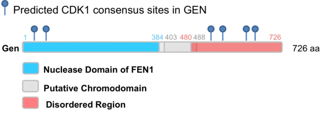
Figure 19. Predicted CDK1 consensus sites ... 71 Figure 20. Model of Gen ... 72 Figure 21. Putative chromodomain identified by Phyre2 ... 73 Figure 22. Summary of human FEN-1 family substrates and conserved
active site features ... 75 Figure 23. Sequence alignment between Gen and orthologs to try to
LIST OF ABBREVIATIONS
AFM – atomic force microscopy BLEO – bleomycin
CPT – camptothecin CO – crossover
dHJ – double Holliday junction DSB – double-strand break
DSBR – double-strand break repair HhH – helix hairpin helix
HJ – Holliday junction
HR – homologous recombination HU – hydroxyurea
ICL – interstrand crosslink IR – ionizing radiation
MMS – methyl methanesulfonate NCO – non-crossover
NDJ – non-disjunction NH2 – nitrogen mustard nt – nucleotide
RF – replication fork
SDSA – synthesis-dependent strand annealing ssDNA – single-stranded DNA
1
CHAPTER 1 INTRODUCTION Genome StabilityDNA provides all the information needed for cellular processes; therefore its fidelity is essential for the survival of an organism, and the faithful transmission of the genetic information from one organism to another is essential for the survival of a species. Because damage is encountered through many exogenous and endogenous processes, cells have developed numerous mechanisms to repair our genomes depending on the type of damage encountered. These mechanisms are highly conserved from yeast to humans, and several were the subjects of this year’s Nobel Prize in Chemistry. When these DNA repair pathways are not intact, they often lead to cancer predispositions. For example, nucleotide excision repair (NER) removes damage caused by UV light through the actions of the protein XPG. In the absence of XPG, patients develop xeroderma pigmentosum, which is characterized by extreme light-sensitivity and early onset skin cancer. DNA damage can also occur as a result of normal endogenous cellular process, such as during replication, or as the result of exogenous chemicals in the environment. For example, the nitrogen mustard mechlorethamine (HN2) can cause intrastrand and interstrand crosslinks (ICLs) (Wijen et al., 2000). Additionally, hydroxyurea (HU) inhibits ribonucleotide reductase, leading to decreased dNTP pools and fork slowing and stalling (Alvino et al., 2007).
ultimately lead to cancer. Surprisingly, despite their toxicity, DSBs are generated deliberately by cells to initiate the process of meiotic recombination. One of the primary mechanisms through which double strand breaks (DSBs) are repaired is homologous recombination (HR).
Double strand break repair (DSBR) model
Our current understanding of DSB repair comes mainly from studies in S. cerevisiae
meiosis. Like DNA repair, meiotic recombination initiates with a double strand break. In the canonical model (Fig. 1) proposed by Szostak et al. (reviewed in Kohl and Sekelsky, 2013; Szostak et al., 1983), processing of DSBs begin with the resection of the DNA ends to leave 3’ overhangs, which invade a homologous template, forming a dissociation loop (D-loop). The free 3’ end then primes synthesis. The other resected end can then anneal to the D-loop, and
Figure 1. Model for the repair of DSBs via HR. In the Szostak et al. model (Szostak et al., 1983), processing of a DSB begins with the resection of the DNA ends to leave 3’ overhangs, which invade a homologous template, forming a dissociation loop (D-loop). Further processing of the D-loop generates a double HJ (dHJ) intermediate, which can be resolved to produce either COs and NCOs with equal
probability. Alternatively, two NCO-only pathways were described: synthesis-dependent strand annealing (SDSA), in which the invading strand is dissociated; and dHJ dissolution, in which two HJs are
decatenated by a topoisomerase. See text for details.
Holliday junction structure
existence of a HJ, they were visualized by electron microscopy during recombination in bacteriophage and E. coli (Bell and Byers, 1979; Benbow et al., 1975; Doniger et al., 1973). More recently, HJs were visualized on 2D gels, showing that they are indeed intermediates of meiotic and mitotic recombination (Bzymek et al., 2010; Schwacha and Kleckner, 1995), although mitotic HJs were detected at 10-fold lower levels than meiotic HJs.
In the 1980’s, scientists began to generate HJs in vitro using the RecA recombinase to generate HJs using two circular double stranded plasmids (Cunningham et al., 1980; West et al., 1983). Soon afterward, formation of a synthetic HJ was achieved by annealing four oligos (Kallenbach et al., 1983); these four-way junctions have been one of the most useful tools for characterizing HJ structure and the enzymes that process them. Surprisingly, depending on the conditions, HJs can adopt several different conformations. In the presence of a divalent ion, free HJs can adopt an antiparallel structure, in which the two DNA helices run antiparallel to each other with the crossing strands adopting a U-shaped conformation (Duckett et al., 1988; reviewed in Lilley, 2000). Alternatively, in the absence of ions, the junction forms an open, symmetric, square-planar formation due to the repulsion of the DNA backbone. Interestingly, the structure-selective enzymes that process HJs, create a conformational change in the DNA
junction upon binding, stabilizing its opening to correctly position the DNA in the protein active site (reviewed in Lilley, 2000).
Structure-selective endonucleases HJ resolvases
migrated toward each other by and dissolved by a helicase and topoisomerase, alternatively two strands of a HJ can be cleaved by an endonuclease. The specific cleavage is achieved by a group of structure-selective endonucleases called Holliday junction resolvases.
Canonical resolvases in prokaryotes
Surprisingly, most HJ resolvases share little to no sequence similarity, but remarkably, they function very similarly (reviewed in West, 2009; White et al., 1997). The first biochemical evidence of HJ resolution came from studies of T4 endonuclease VII and T7 endonuclease I from bacteriophage (de Massy et al., 1987; Mizuuchi et al., 1982). These enzymes recognized specific branched DNA structures, in particular HJs, and cleaved them with a high degree of specificity. E. coli RuvABC was the first identified cellular HJ resolvase (Connolly et al., 1991; Connolly and West, 1990; Dunderdale et al., 1994; Iwasaki et al., 1991; Sharples and Lloyd, 1991; Takahagi et al., 1991). Interestingly, T4 endo VII, T7 endo 1, and RuvC share a similar mechanism of action on HJs. They first bind the HJ as a dimer with high specificity, bend the junction DNA into a specific configuration, and finally, introduce nicks symmetrically across the junction onto strands of the same polarity (reviewed in Lilley, 2000). The products of the
cleavage reaction can then be ligated without further processing. These HJ resolvases became known as “canonical” HJ resolvases, and subsequent searches to identify eukaryotic HJ
Search for the eukaryotic resolvases
As a result of the independent evolution of the HJ resolvases, it was difficult to identify their functional eukaryotic counterparts. An activity termed ResA, which displayed the
properties of a canonical HJ resolvase, was identified from calf thymus tissue (Elborough and West, 1990) and nuclear tissue culture extracts (Constantinou et al., 2001). However, this activity eluded identification until 2008.
MUS81 orthologs
In the meantime, Mus81-Eme1 from S. pombe, and later Mus81-Mms4 from S.
cerevisiae, was the first nuclear HJ resolvase identified in eukaryotes (Boddy et al., 2001; Chen et al., 2001). Both the catalytic Mus81 subunit and its requisite non-nucleolytic S. pombe subunit Eme1, or S. cerevisiae Mms4, belong to the XPF/ERCC4 family of endonucleases which
participate in DNA repair and recombination (reviewed in Schwartz and Heyer, 2011). During meiotic recombination, S. pombe mus81 mutants exhibit severe meiotic defects, including an 85% decrease in CO formation compared to wild-type, as well as increased nondisjunction and spore inviability. Additionally, overexpression of the E. coli RusA HJ resolvase rescues the meiotic phenotypes of mus81 mutants (Boddy et al., 2001). Similarly, S. cerevisiae mus81
the intact HJ results in a flapped and a gapped product, which cannot be ligated without further processing, leading investigators to question whether Mus81 is in fact a “canonical” resolvase.
Consistent with doubts that Mus81 was a canonical resolvase, during DNA repair, mutations in mus81 in most organisms cause sensitivity to DNA damaging agents which block replication forks, including MMS, HN2, and CPT (reviewed in Schwartz and Heyer, 2011). Additionally, loss of Mus81 in mammalian cells increases the number of genome-scale rearrangements and chromosome abnormalities during cell division (Abraham et al., 2003; Dendouga et al., 2005). These data suggested that Mus81 played a role in RF maintenance and restart and perhaps favored other in vivo substrates than HJs.
Gen orthologs
In 2008, the ResA activity that had eluded researchers for almost 20 years was finally identified as GEN1. Human GEN1 and its S. cerevisiae ortholog, Yen1, were discovered based on their ability to cleave HJs in vitro (Ip et al., 2008). For human GEN1, researchers followed intact HJ resolvase activity through extensive fractionation of HeLa cell extracts. The proteins were later renatured, and mass spectrometry (MS) identified a 60 kDa N terminal fragment of an uncharacterized protein. Interestingly, MUS81 was also identified in this screen, which was quite puzzling at the time because MUS81 had shown only weak activity on intact HJs. Similarly difficult was the identification of S. cerevisiae Yen1; thousands of proteins from a TAP-tagged library were immunoprecipitated and assayed for nuclease activity (Ip et al., 2008). Remarkably, GEN1 and Yen1 turned out to be orthologs.
contain conserved FEN-1 nuclease domains and a helix-hairpin-helix motif (Ip et al., 2008). The catalytic domain of this protein has been hypothesized to take part in branch migration, and assays using model DNA substrates in vitro revealed that GEN1/Yen1 display canonical HJ resolvase activity, preferentially cleaving HJs but also cleaving 5’ flaps, RFs, and nHJs with a high degree of specificity (Ip et al., 2008; Rass et al., 2010). Surprisingly, in ensuing genetic studies, yen1 mutants and siRNA knockdown of GEN1 in HeLa cells exhibited no overt mutant phenotype (Blanco et al., 2010; Ho et al., 2010; Svendsen et al., 2009). Rather, mutations in yen1
enhanced the DNA damage sensitivity and meiotic phenotypes of mus81 mutants (Blanco et al., 2010) indicating partial functional redundancy between Mus81 and GEN1/Yen1. Likewise, mutations in yen1 further decrease the meiotic CO formation defects in mus81 mutants (Zakharyevich et al., 2012), and spore viability is drastically reduced (Agmon et al., 2011). Interestingly, the CO reduction and the resulting spore inviability are not observed when the mutations in yen1 are combined with mutations in any other resolvase. Thus, Yen1 and Mus81 function in partially overlapping pathways in S. cerevisiae, with Yen1 specifically substituting for Mus81 in its absence. Evidence for an in vivo HJ resolution function for GEN1 was
Regulation of HJ processing
Processing of HJs are under tight regulation. In somatic S. cerevisiae cells, the STR complex (Sgs1-Top3-Rmi1) preferentially processes joint molecules into NCOs. However, in the absence of Sgs1, Mus81 and Yen1 resolve these structures, resulting in an increase in aberrant mitotic COs (Ira et al., 2003). A similar dynamic exists between the BTR complex (BLM-TopIIIa-RMI1-RMI2) in human cells and MUS81 and GEN1 (Wechsler et al., 2011); mutations in BLM result in Blooms Syndrome, which is characterized by increased sister chromatid exchange (SCE) and a general increase in cancers. Thus, the BTR/STR complexes are somatic cells’ first line of defense in dealing with DNA damage and replication problems, with the HJ resolvases providing a failsafe.
Disruption of HJ regulatory mechanisms results in genome instability. For example, premature activation and nuclear localization of S. cerevisiae Yen1 results in sensitivity to the DNA damaging agent MMS and a significant increase in mitotic CO formation at the expense of NCOs and increasing loss of heterozygosity (LOH) (Blanco et al., 2014). Similarly, premature nuclear import of human GEN1 results in significant increases in sister chromatid exchanges (SCEs) (Chan and West, 2014).
into the nucleus and its affinity for DNA increases (Blanco et al., 2014; Kosugi et al., 2009). Human MUS81–EME1 appears to be regulated similarly to yeast Mus81; whereas, human GEN1 is primarily regulated via a functional nuclear export signal (NES), which drives active nuclear exclusion until nuclear envelope breakdown during mitotic entry. At telophase, GEN1 is shuttled back into the cytoplasm via CRM1-mediated nuclear export (Blanco et al., 2014; Chan and West, 2014). In conclusion, the two major pathways are responsible for removing joint molecules during mitotic recombination: the first, mediated by STR/BTR specializes in the removal of HJs through NCO-promoting pathways, such as HJ dissolution; and the second, is mediated by the CO-generating SSEs, with Mus81 activated first and Yen1/GEN1 acting as a “backup.” Why GEN1/Yen1, the simplest and most robust canonical HJ resolvase, was relegated to a backup role for Mus81, which has limited activities on intact HJs, presents a paradox.
Studying the Gen paradox in Drosophila
We were prompted to conduct this work for several reasons. First, the Gen mutation was identified in a screen for DNA repair mutants that exhibited sensitivity to the DNA damaging agents HN2 and MMS (Laurençon et al., 2004); Gen mutants were hypersensitive to both. These overt mutant sensitivities indicated that Drosophila was the ideal organism in which to study this protein because we would not have to perform our work in a mus81 mutant background; yet
mus81 is present in Drosophila, below.
Furthermore, previous genetic studies suggested that Gen may be the more predominant enzyme in Drosophila. S. cerevisiae, mus81 sgs1 double mutants are inviable, but yen1 sgs1
mitotic resolvase with Yen1 acting as a backup. In Drosophila, mutations in mus81 and Gen are both synthetically lethal with mutations in Blm (the Drosophila ortholog of SGS1) (Andersen et al., 2011; Trowbridge et al., 2007); yet Gen Blm double mutants die much earlier in development than mus81 Blm mutants (Andersen et al., 2011; Trowbridge et al., 2007), suggesting that Gen may be the primary HJ resolvase in Drosophila.
Scope of this work
This dissertation describes work I have done to characterize Gen’s role in double-strand break repair (DSBR) and Holliday junction resolution during DNA repair and meiosis. To understand how this protein carries out its functions, I took both in vivo and in vitro approaches. First, I asked what role Gen has in DNA damage repair and what the relationship between MUS81-MMS4 and Gen is in Drosophila (Chapter 2). I showed that Gen mutants have more severe sensitivities to DNA damaging agents that block replication forks and create DSBs than
mus81 mutants and that this difference from its orthologs is not simply due to Gen’s subcellular localization. Next I asked if the sensitivity of Gen mutants is due to a defect in processing Holliday junctions using in vitro techniques (Chapter 3). I determined that Gen is a flap endonuclease and also a canonical HJ resolvase. Surprisingly, I found that the kinetics with which Gen cleaves these structures is different form its orthologs; Gen cleaves 5’ flaps
1
CHAPTER 2GEN GENETICS AND REGULATION
Introduction
More than 50 years ago, Robin Holliday proposed a four-stranded DNA structure that now bears his name – the Holliday junction (HJ) – as a key intermediate in recombination (Holliday, 1964). He suggested that “at the points where strands exchange partner precise breakage and reunion of non-complementary strands can occur so that there is no deletion or duplication of material.” It was more than 25 years before the identification of the first endonuclease with specificity for HJs was identified: E. coli RuvC (Connolly et al., 1991). RuvC resolves HJs by making symmetric nicks on non-complementary strands, resulting in nicked duplexes that can be ligated without further processing, as in Holliday’s model (Bennett et al., 1993).
It was another ten years before the first good candidate for a eukaryotic nuclear HJ
for most meiotic crossovers, and S. cerevisiae Mus81–Mms4 is required for a substantial
minority of crossovers (Boddy et al., 2001; de los Santos et al., 2003; Smith et al., 2003). Models in which Mus81–Eme1 generates crossovers by cleaving nicked structures offered one possible solution to this apparent paradox (Osman et al., 2003), but electron microscopy studies suggested that meiotic crossovers in S. pombe are produced from ligated HJs (Cromie et al., 2006). More recently, in vitro experiments demonstrated that HJ resolution can be achieved through
collaboration between the human nucleases SLX1, which nicks HJs, and MUS81–EME1, which makes the second nick, in a reaction orchestrated by the SLX4 nuclease scaffolding complex (Castor et al., 2013; Garner et al., 2013; Wyatt et al., 2013). Again, this does not seem to be a complete answer to the paradox, since S. cerevisiaeslx1 mutants do not have reduced meiotic crossovers like mus81 mutants.
In 2008, Ip et al. (Ip et al., 2008) reported that human GEN1 and the S. cerevisiae ortholog Yen1 have HJ resolvase activity with properties similar to that of RuvC. At first, it seemed that GEN1/Yen1 could be long sought-after nuclear HJ resolvase; however, genetic studies
subsequently found that yen1 mutants do not have measurable defects in meiotic crossovers and are not hypersensitive to DNA damaging agents (Blanco et al., 2010; Ho et al., 2010; Tay and Wu, 2010). These same studies found that mus81 yen1 double mutants have more severe phenotypes than mus81 single mutants. S. pombe does not have a GEN1/Yen1 ortholog, but expression of human GEN1 rescues defects caused by loss of Mus81–Eme1 (Lorenz et al., 2010). Together, these results suggested that Yen1 functions primarily as a backup to Mus81.
al., 2012; Matos et al., 2011). Yen1 is kept inactive and cytoplasmic by phosphorylation until anaphase, when it is dephosphorylated by Cdc14 (Blanco et al., 2014; Eissler et al., 2014). Therefore, Mus81–Mms4 is activated prior to Yen1 activation. Human MUS81–EME1 and GEN1 appear to be regulated in a similar fashion (Blanco et al., 2014; Chan and West, 2014).
The first GEN1/Yen1 ortholog to be described was Drosophila GEN (Ishikawa et al., 2004). GEN was shown to cleave 5’ flaps and model replication forks in vitro, but no HJ resolvase activity was detected (Kanai et al., 2007). The first genetic studies of Gen reported spontaneous apoptosis and a rough eye phenotype in mus81Gen double mutants, indicating an interaction between GEN and MUS81–MMS4 like that in budding yeast (Andersen et al., 2011). Synthetic lethality between Gen and Blm was also reported. This is different from S. cerevisiae, where sgs1 yen1 mutants are viable (Blanco et al., 2010). There is synthetic lethality between
mus81 and sgs1 mutations in S. cerevisiae (Fricke and Brill, 2003; Kaliraman et al., 2001), and likewise between mus81 and Blm mutations in Drosophila (Trowbridge et al., 2007).
Interestingly, Gen Blm mutants die much earlier in development than mus81 Blm mutants (Andersen et al., 2011; Trowbridge et al., 2007). This observation, combined with the finding that Drosophilamus81 mutants are not hypersensitive to most DNA damaging agents
repair are due to protein localization, and found that similar to its orthologs, Gen localized to the cytoplasm of Drosophila embryos and S2 cells. Finally, we show that Gen is able to rescue the DNA damage phenotypes of S. pombe mus81 mutants. Together, these results suggest that Gen is the primary mitotic HJ resolvase in Drosophila and that HJs are an in vivo substrate; however the genetic differences are not attributable to protein localization.
Results
Gen mutants are more sensitive to DNA damage than mus81 mutants
To assess the relationship between Gen and MUS81 in Drosophila, we examined the sensitivity of single and double mutants to a variety of DNA damaging agents (Fig. 2). The agents we used were (a) camptothecin (CPT), a topoisomerase I poison that results in replication-associated DSBs (Liu et al., 2000); (b) methyl methanesulfonate (MMS), an alkylating agent that induces lesions that can block replication forks (Groth et al., 2010); (c) the nitrogen mustard mechlorethamine (HN2), which generates base adducts and interstrand crosslinks (Wijen et al., 2000); (d) hydroxyurea (HU), which inhibits ribonucleotide reductase, leading to decreased dNTP pools and fork slowing and stalling (Alvino et al., 2007); and (e) ionizing radiation (IR), for which the most toxic damage is double-strand breaks (DSBs). At the doses used in our experiments, HN2 was the only agent to which mus81 single mutants were hypersensitive (Fig. 2c). In contrast, Gen mutants are hypersensitive to all of the agents tested. Furthermore, mus81 Gen double mutants are significantly more hypersensitive to the damaging agents than Gen
Our results with mus81 mutants were mostly similar to those of a previous study
(Trowbridge et al., 2007) except that we did not detect hypersensitivity (i.e., decreased relative survival) of mus81 mutants to CPT (Fig. 2a) possibly due to a difference in experimental design (see Materials and Methods). Additionally, the previous study reported significant
hyposensitivity (i.e., greater relative survival) of mus81 mutants to HU, suggesting that the presence of MUS81 is detrimental to survival in the presence of this drug. In results reported here, the mean relative survival of mus81 mutants was elevated after treatment with HU, consistent with hyposensitivity, but the difference was not statistically significant (Fig. 2d). Regardless, Gen mutants do worse after treatment with HU than mus81 mutants.
In summary, Gen mutants are more sensitive to a range of DNA damaging agents than
Figure 2. Drosophila Gen mutants are more sensitive to DNA damaging agents than mus81
mutants. Graphs show survival of mutants relative to control siblings (see Materials and Methods). (a) 0.025 mM camptothecin (CPT); (b) 0.04% methyl-methane sulfonate (MMS); (c) 0.004% nitrogen mustard (HN2); (d) 70 mM hydroxyurea (HU); (e) 2000 rads ionizing radiation (IR). Each point corresponds to one vial; means and 95% confidence intervals are shown. Dotted lines indicate 100% relative survival (note that Y axes differ between treatments). Paired t-tests between mutant and control individuals were done to evaluate sensitivity of mutants to each treatment; statistical significance of sensitivity is indicated below each genotype. Differences between genotypes were assessed by one-way ANOVA and are indicated above each graph. n.s. = not significant (p > 0.05); ** = p < 0.01; *** = p < 0.001; **** = p < 0.0001.
Gen is not required for meiotic recombination
MUS81 is required for a subset of meiotic crossovers in S. cerevisiae, Arabidopsis, and mice, and most or all meiotic crossovers in S. pombe (Berchowitz et al., 2007; de los Santos et al., 2003; Holloway et al., 2008; Smith et al., 2003). In yeast, yen1 mutants exhibit WT levels of NDJ and CO; however, mus81 yen1 mutants fail to produce viable spores. In Drosophila,
segregation, we first examined Gen’s meiotic role by measuring X chromosome NDJ in the Gen
mutant as a readout for meiotic CO defects (Table 1). We observed WT levels of NDJ in the Gen
mutant. We next directly measured CO levels in the Gen mutant to determine if there was a subtle decrease in CO levels.
Table 1. X chromosome non-disjunction (NDJ) in Gen mutants
Allele Progeny X Nondisjunction
(%) Normal Nullo-X Diplo-X
+/Df 3,452 1 1 0.11
GenZ4235/Df 1,656 1 0 0.12
GenZ5997/Df 1,290 0 1 0.15
In Drosophila, most meiotic crossovers are generated by the presumptive resolvase MEI-9–ERCC1 and the scaffolding protein MUS312 (Radford et al., 2005; Sekelsky et al., 1995; Yıldız et al., 2002). Crossovers are reduced by about 90% in mei-9 or mus312 single mutants (Baker and Carpenter, 1972; Green, 1981). Trowbridge et al. (2007) found that mus81 mei-9
mutants were no worse than mei-9 single mutants. To determine whether Gen produces the crossovers that remain when MEI-9–ERCC1–MUS312 is missing, we measured crossing over in
As a result of the inviability of the triple mutant with the net – cn second chromosome, we will score recombination frequency between two markers, st and e, on the third chromosome, which also contains the mutations of Gen and mus81. We are currently building stocks with the appropriate genetic markers. We therefore conclude that Gen is not required for the generation of meiotic COs, even in the absence of the major meiotic resolvase. It will be of great interest to determine whether any COs exist in the mus81 Gen mus312 triple mutant.
Table 2. Meiotic crossing over in Gen and Gen mus312 mutants Genetic distance (MU)
Genotype net−dppho dppho−dp dp−b b−pr Total % of
WT n
wild-type* 5.1 7.5 27.3 3.5 43.4 100 2320
Gen 2.42 5.21 26.01 9.41 43.05 99 2072
mus312** 0 0.28 3.06 0.56 3.9 9 359
mus312 Gen 0.23 0.23 1.15 0.69 2.3 5 433
mus81 mus312
Gen — — — — — — —
*
Kuo et al (2014) **
Yildiz et al (2002)
Gen localizes to the cytoplasm of early embryos and S2 cells
Figure 3. Gen localizes to the cytoplasm in early embryos and cultured Drosophila S2 cells. (a-b) 2-3 hr old Drosophila embryos were stained with DAPI (blue) and antibodies to Gen (green) (c-h) Full-length Gen-His was expressed in Drosophila S2 cells from the CuSO4-inducible metallothionein
promoter. Cells were treated with (c-e) or without (f-h) CuSO4 for three days, then fixed and stained with DAPI (blue) and antibodies to Gen (green) and to the His tag (red). Both endogenous Gen and
overexpressed Gen-His were detected in the cytoplasm but not in the nucleus.
Gen rescues the DNA-damage sensitivity of S. pombemus81 mutants
Heyer, 2000). S. pombe lacks a Yen1 ortholog, but truncated human GEN1 expressed in S. pombe rescues phenotypes caused by loss of Mus81 (Lorenz et al., 2010). Consequently, we expressed two forms of Drosophila Gen in S. pombemus81Δ mutants (Fig. 4): full-length protein and a truncated form (residues 1-518) that is similar to truncated human GEN1 that was expressed in S. pombe (Lorenz et al., 2010). Truncated Gen rescues the hypersensitivity of the
mus81Δ mutant to MMS, CPT, HU, and the radiomimetic drug bleomycin (BLEO), and this rescue is dependent on Gen nuclease activity (Fig. 4a). Full-length Gen did not rescue any of these hypersensitivities. Expression of both proteins was confirmed by Western blot (Fig. 4b), but it is possible the full-length protein was misfolded or excluded from the nucleus. We
Figure 4. Gen expression rescues the DNA damage sensitivity of S. pombemus81 mutants. (a)Effect of Gen (1-518) overexpression mimics that of the canonical resolvase RusA. Serial dilutions on EMM2 plates were supplemented with drug. All proteins were expressed from the thiamine-repressible nmt1
promoter in mus81Δ or WT strains. Empty pREP41 plasmid was used as a negative control. (b) Western blot showing expression of full-length Gen and Gen (1-518) in S. pombe mus81Δmutants. FL = full-length Gen; 518 = 1-518 aa.
Conclusion
Together, these data confirm that Gen is the primary mitotic resolvase in Drosophila and is partially genetically redundant with MUS81; however, our data raise questions as to the why the relationship between Gen and MUS81 is different in Drosophila versus other organisms. To try to identify the origin of these differences, we therefore examined the biochemical properties of Gen, which we discuss in Chapter 3.
Materials and Methods
Drosophila stocks and genetics
All stocks were maintained at 25°C on standard media. The following null mutations were described previously: GenZ5997 (Andersen et al., 2011), which was made hemizygous by putting it over Df(3L)6103; and mus81NheI (Trowbridge et al., 2007).
To analyze nondisjunction, virgin females were crossed to y cv v f / T(1:Y)BS males. There are four classes of exceptional progeny, which exhibit X chromosome NDJ. These are: Bar-eyed females and WT-eyed males, which are viable; and triplo-X and nullo-Y flies, which are inviable. The total number of exceptional progeny was calculated by multiplying the number of viable exceptional progeny by 2. Percent non-disjunction was calculated by dividing the total number of exceptional progeny by sum of the total number of progeny scored plus the inviable progeny.
To analyze meiotic crossing over, virgin females of various genetic backgrounds were heterozygous for markers on chromosome 2L (net dppd-ho dp Sp b pr cn) and were crossed to net dppd-ho dp b pr cn tester males, and progeny were scored for 5 days. Sensitivity to DNA
in DMSO and diluted in 10% ethanol and 0.2% Tween. Control larvae were mock treated with DMSO dissolved in 10% ethanol and 0.2% Tween. For IR, vials with 3rd instar larvae were irradiated with 2000 rads from a 137Cs source (Gammacell GG10). Progeny were scored for five days after eclosion began. Relative survival was calculated as the ratio of mutant to control flies per vial and normalized to the ratio in untreated vials. Statistical analyses were done using GraphPad Prism.
Expression of Gen in S. pombe and sensitivity analysis
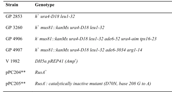
Table 3. Strains and plasmids used in this study.
(courtesy of Dr. Gerry Smith, Fred Hutchinson Cancer Research Center)
Strain Genotype
GP 2853 h+ ura4-D18 leu1-32
GP 3260 h+ mus81::kanMx ura4-D18 leu1-32
GP 4906 h- mus81::kanMx ura4-D18 leu1-32 ade6-52 ura4-aim tps16-23
GP 4907 h+ mus81::kanMx ura4-D18 leu1-32 ade6-3034 arg1-14
V 1982 DH5α pREP41 (Ampr)
pPC204** RusA+
pPC205** RusA-: catalytically inactive mutant (D70N, base 208 G to A)
**Both have an additional mutation at base 363 A to C. pPC205 has a mutation at base 329 T to C.
Immunofluorescence Microscopy
Polyclonal antibodies were raised to residues 236 to 335 of Gen and affinity-purified by Genomic Antibody Technology (SDIX, Newark, DE). All imaging was done with a laser-scanning confocal microscope (710, Carl Zeiss) and analyzed with ImageJ.
For embryo staining, 2-3 hr old embryos were dechorionated, fixed in equal volumes 7%
formaldehyde:heptane, devittelinized, then stained. The primary antibody was rabbit anti-Gen-N (1:1,000), which was visualized with goat anti-rabbit IgG (H+L)-Alexa Fluor 488 (1:500, Life Technologies). DNA was detected by staining with DAPI (1:1000) for 2 min at room
temperature.
1
CHAPTER 3GEN IS A CANONICAL HJ RESOLVASE BUT PREFERS 5’ FLAPS
Introduction
A suite of structure-selective endonucleases (SSEs) has evolved to process branched DNA structures such flaps, bubbles, replication forks, and Holliday junction (HJs), which are four-stranded intermediates in recombination pathways. The first family of such nucleases identified in eukaryotes was initially defined by FEN-1 (flap endonuclease and 5’ exonuclease 1) and XPG (xeroderma pigmentosum group G) (Lieber, 1997). These enzymes share conserved nuclease domains related to the 5’-to-3’ exonucleases of prokaryotic DNA polymerases, but they have divergent activities and functions. FEN-1 processes Okazaki fragments during replication, whereas XPG nicks the damaged strand at the 3’ end of a bubble during nucleotide excision repair. Exo1 (exonuclease 1), an enzyme with numerous repair and recombination functions (Tran et al., 2004), was later found to be a member of this family.
GEN1 exhibits canonical HJ resolvase activity, making symmetric nicks on non-complementary strands of a HJ thereby yielding nicked duplexes that can be directly ligated (Rass et al., 2010). Surprisingly, genetic studies in S. cerevisiae failed to find recombination and repair defects in yen1 mutants and instead suggested that Yen1 is a backup to another SSE, Mus81 (Blanco et al., 2010; Ho et al., 2010; Tay and Wu, 2010). Mus81 has been implicated in diverse processes across a number of organisms, including replication fork repair and meiotic recombination (reviewed in Schwartz and Heyer, 2011). Although some of these functions can be explained by HJ resolvase activity, in in vitro assays, Mus81 (together with its non-catalytic partner Mms4/EME1) cleaves 3’ flaps and nicked HJs well but has limited ability to cut intact HJs (Ehmsen and Heyer, 2008; Oğrünç and Sancar, 2003). Why the robust, canonical HJ
resolvase Yen1/GEN1 has been relegated to a backup role for Mus81, which has limited activity on intact HJs, presents a paradox.
Studies of the regulation of Mus81–Mms4 and Yen1 help to explain the mechanism for how Mus81–Mms4 acts before Yen1. Both proteins are regulated by cell cycle-dependent phosphorylation and dephosphorylation, such that Mus81–Mms4 is available and active in the nucleus prior to Yen1 (Blanco et al., 2014; Eissler et al., 2014); the human orthologs are
a complete answer to the GEN1 paradox, since S. cerevisiaeslx1 mutants do not have reduced meiotic crossovers like mus81 mutants, and slx1 mutants have a different phenotype than mus81
mutants (Fricke and Brill, 2003; Zakharyevich et al., 2012).
To gain insight into these questions, we conducted genetic and biochemical analyses of
Drosophila Gen. Previous genetic studies suggest that Gen may be the more predominant enzyme in Drosophila. In S. cerevisiae, mus81 sgs1 double mutants are inviable, but yen1 sgs1
double mutants are viable (Blanco et al., 2010; Fricke and Brill, 2003; Kaliraman et al., 2001; Mullen et al., 2001). These results are consistent with S. cerevisiae Mus81 being the primary mitotic resolvase with Yen1 acting as a backup. In Drosophila, mutations in mus81 and Gen are both synthetically lethal with mutations in Blm (the Drosophila ortholog of SGS1) (Andersen et al., 2011; Trowbridge et al., 2007); yet Gen Blm double mutants die much earlier in development than mus81 Blm mutants (Andersen et al., 2011; Trowbridge et al., 2007), suggesting that Gen may be the primary HJ resolvase in Drosophila.
In Chapter 2, we showed that Gen mutants are more sensitive to a broad range of DNA damaging agents than are mus81 mutants and that like its fungal and human orthologs, Gen is primarily cytoplasmic during interphase. In this chapter, we show that Gen exhibits robust HJ resolving activity, a result that contrasts with a previous report on Gen (Kanai et al., 2007) but is similar to data from fungal and human orthologs (Freeman et al., 2014; Rass et al., 2010).
electrophoretic mobility shift assays (EMSAs) with a 5’ flap suggest that dimerization of Gen enhances the cleavage activity of Gen on 5’ flaps. Finally, we directly show that a productive dimer-DNA complex and a DNA conformational change are the rate-limiting steps of the HJ cleavage reaction, providing insight into the mechanism of HJ resolution that can likely be extended to all Gen orthologs.
Results and Discussion
The C terminus of Gen is disordered
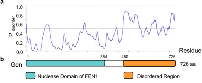
Truncated versions of human GEN1 and C. thermophilium GEN1 were identified and shown to be canonical HJ resolvases (Freeman et al., 2014; Ip et al., 2008; Rass et al., 2010); however, the full-length versions of these proteins were not purified due to the instability of the C terminus. These studies suggested that the disordered C terminus may play important
regulatory roles in controlling protein activity. This suggestion is backed up by evidence that the C terminus of EXO1, another member of the FEN-1 family of repair endonucleases to which GEN1 belongs, is also disordered and plays a role in the negative regulation of enzyme activity (Orans et al., 2011). We used to in silico tools, Phyre2 and metaPrDOS, to ask whether the C terminus of Gen was disordered (Fig. 5). Both programs showed that similar to other orthologs, the N terminal nuclease domain of Gen is highly conserved, whereas the C terminus is highly disordered. We therefore expressed and purified both full-length and truncated (1-518 aa) forms of the protein to assess whether the disordered C terminus played any role in substrate
Figure 5. The C terminus of GEN is predicted to be disordered. (a) The line graph depicts the disorder tendency (i.e. the average probability of intrinsic disorder) predicted by metaPrDOS along the length of GEN. Residues above the line are predicted to be disordered. (b) Structural domains were identified by Phyre2. The “Nuclease Domain of FEN1” corresponds to Protein Data Bank fold 1UL1. The “Catalytic core of RAD2 (complex 1)” was also identified by Phyre2 and corresponds to Protein Data Bank fold 4Q0R. The “Disordered Region” indicates a region lacking secondary structure that was predicted by the DISOPRED2 Disorder Prediction program as part of the Phyre2 analysis.
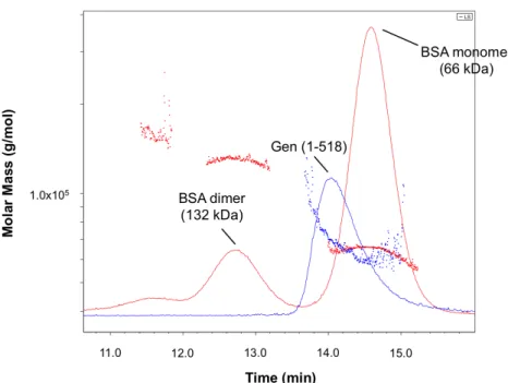
Gen exists in a monomer-dimer equilibrium
Human GEN1, S. cerevisiae Yen1, and C. thermophilium GEN1 all purify as monomers in solution (Freeman et al., 2014; Ip et al., 2008; Rass et al., 2010). We analyzed Gen (1-518) by SEC-MALS after purification by affinity chromatography and ion exchange chromatography and compared it to a BSA standard (Fig. 6). BSA eluted from the column in three distinct peaks, with the monomer (66 kDa) eluting between 14 and 15 min. The truncated Gen (1-518) peak partially overlapped the BSA monomer peak, exhibiting an average molecular weight of 69 kDa
Figure 6. SEC-MALS analysis of Gen shows that both monomers and dimers exist in solution. GEN (1-518)-His is indicated in blue. The BSA standard is indicated in red.
Proteins purified
Previous in vitro experiments using an N-terminal 6xHis-tagged full-length Drosophila
Gen did not detect activity on HJs (Kanai et al., 2007); however, studies of Gen orthologs with C-terminal tags have demonstrated activity (Freeman et al., 2014; Ip et al., 2008; Rass et al., 2010). Thus, we expressed and purified both N- and C-terminal tagged Gen in full-length and truncated (1-518) forms (Fig. 7). The nuclease activity of the N-terminal tagged proteins is weak, but the C-terminal tagged versions of Gen show high activity (Fig. 8b), with no evidence of contaminating nuclease activity (Fig. 8c).
It was previously reported that the O. sativa OsGEN-L ortholog was acutely sensitive to the salt concentration in the nuclease buffer, only showing activity on 5’ flaps at KCl
concentrations. HJ cleavage activity was not observed under the same conditions as the 5’ flap but only at higher salt concentrations (150 mM). In addition, previous reports with Drosophila
Gen showed that it preferentially cleaved 5’ flaps between 50-75 mM KCl; they did not observe HJ cleavage under these conditions. Importantly, they did not ask whether HJ cleavage was observed at higher concentrations of KCl. We optimized cleavage conditions for Drosophila Gen (Fig. 8a), and observed robust cleavage of both the 5’ flap and HJ0 under all salt concentrations tested.
Figure 7. Proteins purified. (a) Schematic of recombinant proteins. Domains were determined using Phyre2. The arrow shows the location of the Z5997 mutation present in Gen mutant flies used in Chapter 2. (b) Coomassie-stained SDS-PAGE gels showing C-terminally His-tagged full-length Gen and
mixture. In that study, the 5’ flap was efficiently cleaved at KCl concentrations below 100 mM, and activity dropped off rapidly at higher KCl concentrations. In contrast, HJ cleavage activity was not observed under the same conditions as the 5’ flap. Rather, high salt (150 mM) was critical for HJ cleavage. (b) The N-terminal His tag interferes with the nuclease activity of Gen. Nuclease assays were done with Gen (FL = full-length; 518 = 1-518; or N = full-length with N-terminal tag). N OE = The gel was overexposed to show residual nuclease activity with N-terminally tagged Gen. Arrow = This band results from extra breathing at the 5’ flap ss-dsDNA junction due to an extra A added to the 3’ end of Oligo 992 (see Table 1). Nuclease assays contained 50 nM protein and 1 nM DNA. (c) Substrate
cleavage is dependent upon Gen nuclease activity. Nuclease-dead Gen had mutations in two glutamic acid residues in the catalytic domain: E143A and E145A. Assays contained 20 nM protein and 1 nM DNA. All assays were done at 22 for 30 min. were incubated at 22°C for 30 minutes. Products were analyzed by denaturing PAGE.
Gen is a Holliday junction resolvase and a 5’ flap endonuclease
To examine substrate specificity, we incubated Gen with radiolabeled DNA substrates (Fig. 9). Both full-length and truncated C-terminal His-tagged Gen exhibit robust cleavage of 5’ flaps, RFs, and fixed, mobile, and nicked HJs. Similar to other Gen orthologs, denaturing polyacrylamide gel electrophoresis (PAGE) demonstrates that the predominant cut sites are one nucleotide (nt) 3’ of the junction branch point on the 5’ flap, RF, and immobile HJs (HJ0 and nHJ0) (Fig. 9). The secondary cut observed with the HJ0 and nHJ0 are likely due to substrate breathing or weak sequence preference. Finally, we observe multiple cut sites on the mobile HJ12 substrate, which contains a 12 bp homologous core within which the junction can migrate; all cut sites on this structure are within the 12 bp core. We do not detect cleavage of unbranched dsDNA or nicked duplex DNA. We noted weak cleavage of the 3’ flap substrate, but the
To map HJ cleavage sites on each strand, we alternately labeled each strand of the immobile HJ0 structure (Fig. 10). The major cuts on each strand are located one nt 3’ to the junction branch point. As discussed above, on strand A there was a secondary cleavage site two nt 3’ to the junction, possibly due to weak sequence preference (Fig. 9 and 10). These studies show that Drosophila Gen, like its fungal and human orthologs, retains the ability of the FEN-1 nuclease family to cut 5’ flaps one nt 3’ of the branch point. Additionally, like its orthologs,
Gen cleaves 5’ flaps faster than Holliday junctions
Figure 11 Gen cleaves 5’ flaps faster than HJ0s under conditions of excess substrate. (a-b) Time courses of Gen progression under conditions of excess substrate: (a) 5’ flap (b) HJ0. For each time course, aliquots were taken at various time points (note that the time scales differ in each panel). The intensity of each cleavage product was quantified by ImageQuant, and the data were normalized to the expected amount of detectable product (see Materials and Methods). Each dot represents the mean of three experiments, except in (a), which is the mean of two experiments. Error bars indicate standard error of the mean. (c) Selwyn tests to examine nuclease progression curves in (a-b). The percentage of
substrate cleaved was plotted against the initial enzyme concentration multiplied by time for various enzyme concentrations to examine perturbations to the nuclease reaction. Overlapping curves indicate that there were no perturbations to the reaction (for example, enzyme death or substrate and/or product inhibition).
when different enzyme concentrations are multiplied by time and plotted as function of percent substrate cleaved. For the 5’ flap reactions, the progress curves with different concentrations of protein overlapped, indicating that progress is governed solely by the reactants (Fig. 11c). However, curves for the HJ0 reactions did not overlap, but fell into three distinct groups (Fig. 11d). The progressive decrease in protein activity with increasing substrate is indicative of substrate inhibition. Human GEN1, which is a monomer in solution but must dimerize on an HJ for cleavage, shows similar inhibition of cleavage when the HJ substrate is in excess of GEN1 (Rass et al., 2010). In this scenario, excess substrate molecules bind GEN1 monomers, thereby reducing the concentration of dimer available to cleave the HJ. Given that Drosophila Gen is in a monomer-dimer equilibrium (Fig. 6), a likely source of the reduced plateau levels is substrate inhibition. In addition, these data, along with the observation that the HJ0 is always cut on both sides (Fig. 9 and 10), indicate that dimerization is required for HJ cleavage but not for 5’ flap cleavage, similar to the Gen orthologs (Freeman et al., 2014; Rass et al., 2010)
Figure 12 Gen cleaves 5’ flaps faster than HJs under conditions of excess protein. (a) Time courses of Gen progression on 5’ flaps and HJ0s under conditions of excess protein. For each time course, aliquots were taken at various time points (note that the time scales differ in each panel). The intensity of each cleavage product was quantified by ImageQuant, and the data were normalized to the expected amount of detectable product (see Materials and Methods). Each dot represents the mean of three experiments. (b) Each individual replicate from (Fig. 11 and 12) was fit to a single exponential curve to obtain the cleavage rate, and mean cleavage rates were plotted as a function of protein concentration. Note that the first point (3 nM protein) was performed with 5 nM 5’ flap or HJ0, whereas the rest of the experiments (20, 60, 100, and 200 nM protein) were performed with 2 nM DNA. The 5’ flap and HJ0 data from these and additional experiments (not shown) were fit to a hyperbolic binding curve given by the equation y=m1*x/(m2+x), where m1 = maximum rate at saturating protein concentrations and m2 = the equilibrium dissociation constant. With the 5’ flap, m2 = 62.12 nM, and with the HJ0, m2 = 656.46 nM.
Because the cleavage rates of both the HJ0 and 5’ flap increased with increasing
Table 4. Summary of cleavage rates from all kinetics experiments.
Substrate DNA (nM) Gen (nM) Average Rate SE
5’ Flap 5 0.5 0.70 0.01
5 0.75 1.43 0.29
5 1 2.52 0.77
5 3 4.11 0.71
2 20 11.56 0.32
2 60 22.24 0.88
2 100 29.01 2.88
2 200 35.44 0.56
HJ0 5 0.5 0.12 0.01
5 0.75 0.13 0.01
5 1 0.23 0.03
5 3 0.26 0.02
2 20 1.53 0.17
2 60 2.16 0.50
2 100 4.12 0.51
2 200 7.17 0.14
The rate-limiting step of HJ0 cleavage is assembly of a productive dimer complex on the substrate
be a result of weak binding of the dimer to the HJ0, or it could represent a pre-equilibrium step if the rate-limiting step is a conformational change after binding. In the experiments described above (Fig. 11 and 12), the DNA, Gen, and Mg++ are added simultaneously, so it is not possible to determine whether the limiting step is after binding. To assess the possibility that the rate-limiting step is a conformational change after binding, we pre-incubated Gen with both the 5’ flap and HJ0 in the absence of Mg++, allowing time for the dimer to assemble on the DNA, and then initiated cleavage by the addition of Mg++ (Fig. 13). If conformational change/assembly of the dimer on the substrate is rate-limiting, then cleavage will be significantly more rapid in the pre-incubation experiment than in the simultaneous addition experiments. We were unable to determine the rate-limiting step with the 5’ flap because residual Mg++ in the DNA buffer was sufficient to promote robust cleavage (Fig. 13b), but in pre-incubation experiments with HJ0 we observed a burst of cleavage before the first time point (5 seconds) followed by a slow rate of cleavage similar to that seen in the simultaneous addition experiment (Fig. 13a). This
observation strongly suggests that given sufficient time, Gen can cooperatively assemble into a productive complex on the HJ0. This suggestion is supported by data on the Gen ortholog
Figure 13 The rate-limiting step of the HJ0 reaction is formation of a productive dimer-DNA complex. (a) To determine whether the rate-limiting step of the HJ0 reaction is binding and/or a
conformational change, we pre-incubated 3 nM Gen with 5 or 10 nM HJ0 before starting the time course experiment with Mg++2. (b) To determine whether the rate-limiting step for the 5’ flap reaction was formation of a productive complex, we pre-incubated 1 or 3 nM Gen (1-518)-His with 5 nM 5’ flap before starting the reaction with Mg++. Unfortunately, contaminating divalent ion in the DNA storage buffer was sufficient to initiate cleavage and turnover before addition of Mg++ (indicated by 45-60% of the substrate cleaved at time 0) and prevented us from determining the rate-limiting step.
In conclusion, the pre-incubation experiments indicate tight binding of a Gen dimer to the HJ0 with only a small amount of substrate inhibition (Fig. 13a); whereas, the simultaneous addition experiments yield a very weak K1/2 (660 nM) and exhibit significant substrate inhibition, suggesting a monomer of Gen binds tightly to the HJ0 (Fig. 11 and 12). Taken
together, these data lead us to suggest a model (see Fig. 14 and Chapter 3 Conclusion) in which a monomer binds tightly to the HJ0 followed by a second monomer binding to form a
nonspecific dimer on the HJ0 with a weak binding affinity. Next, this nonspecific dimer-HJ0 complex undergoes a conformational change to a productive complex followed by rapid
cleavage. For human GEN1, a conformational change triggered by formation of a dimer on HJs was proposed to explain the observation that human GEN1 does not nick HJs, but always cuts both strands (Rass et al., 2010). Other HJ resolvases, including the RuvC and T4endo7
conformation and then alter the conformation prior to cleavage (Fogg and Lilley, 2000; Pohler et al., 1996). Our results are consistent with the idea that a prerequisite for the Gen dimer to bind tightly to the HJ0 is that the DNA has to be in the proper conformation. Consequently, if the HJ0 is not in a conformation that presents a proper dimer interface, it could promote the dissociation of one monomer of the dimer.
one monomer likely dissociates from the HJ, allowing other Gen proteins access to the junction. (top right box) It is also possible that a DNA conformational change occurs prior to the second monomer binding; however, given our observation that production of a productive dimer-DNA complex is the rate-limiting step, this is unlikely to represent a main pathway.
Dimerization of Gen on 5’ flaps stimulates its cleavage activity
In contrast to human and fungal GEN1 (Freeman et al., 2014; Rass et al., 2010),
Drosophila Gen cleaves the 5’ flap more rapidly than the HJ0 at all concentrations tested, and the rate of flap cleavage increases with increasing protein concentration with a K1/2 ~60 nM (Fig. 12b). Consistent with our SEC-MALS result that shows that, unlike human and fungal GEN1 (Freeman et al., 2014; Rass et al., 2010), Drosophila Gen exists in a monomer-dimer equilibrium (Fig. 6), we wondered whether the increasing cleavage rate of the 5’ flap with increasing Gen concentration resulted from dimerization of Gen on the 5’ flap. To examine this possibility, we used electrophoretic mobility shift assays (EMSAs) to monitor binding to the 5’ flap and atomic force microscopy (AFM) to directly observe the oligomerization state of the protein. We
cleavage on the flap determined from the kinetics assays (Fig. 11 and 12). Performing the EMSAs in the absence of EDTA results in complete cutting of the 5’ flap at all concentrations of protein, and no shifted bands are observed (Fig. 15b). These results indicate that glutaraldehyde crosslinking does not lead to accumulation of non-specific protein-DNA complexes and
therefore that the bands observed with the 5’ flap are the result of specific interactions of a monomer and dimer of Gen interacting with the 5’ flap.
polyacrylamide gel. (b) In the absence of EDTA, no shifts are observed on the 5’ flap or HJ0, suggesting that complexes formed in (a) are not an artifact of glutaraldehyde crosslinking.
To garner additional evidence for the dimerization of Gen, we used AFM (Fig. 16) to analyze Gen at concentrations similar to those used in our kinetics experiments (Fig. 11 and 12). Previous studies show that there is a linear relationship between the molecular mass of a protein and its observed volume in AFM images, which allows oligomerization state and the association constants of the protein-protein complexes to be determined (Ratcliff and Erie, 2001; Yang et al., 2003). At 20 nM and 37 nM Gen, we observe two major populations of peak volumes: one consistent with the volume of a Gen monomer and the other consistent with the volume of a dimer of Gen (Fig. 16). From these studies, we estimated the protein dissociation constant to be within the 60 nM to the µM range. Quantitative assessment is not possible because of
mica and imaged with tapping mode AFM in air. The gradient bar represents 0-1.2 nm height above the mica surface. Yellow arrows denote Gen dimers. (b-c) Representative 1x1 µm AFM images of truncated Gen at (a) 20 nM and (b) 37 nM. Bar represents 100 nm. (d-e) AFM volume analysis of particles in (b, c), respectively. Particles from at least 7 images for each concentration were analyzed for volume (nm3). Resulting volumes were binned in 30 bins and graphed as a histogram. The volume calculated is directly proportional to molecular size. The predicted molecular mass of proteins from the AFM-derived volume was based on Equation 1 in Materials and Methods. Note that observed volumes are slightly larger than predicted volumes of Gen based upon molecular size. Brackets indicate volumes representing dimers.
Conclusion
In this chapter, we show that Drosophila Gen, a unique member of the Class IV monomeric FEN-1/XPG endonucleases, is a key SSE during the repair of DNA damage. Gen exhibits the characteristic substrate preferences of its yeast and human orthologs; namely, it cleaves 5’ flaps and RFs with a high degree of specificity and is a bona fide HJ resolvase. Notably, we show that Gen displays dramatically altered nuclease kinetics on these substrates relative to other orthologs that have been studied, cleaving 5’ flaps substantially faster than HJs
in vitro. Further, this enhanced rate is due to the ability of the protein to dimerize in solution and on the 5’ flap substrate. Finally, while it has been suggested that a conformational change is the rate-limiting step to cleavage on an HJ, our studies provide direct evidence that a conformational change occurs after the second monomer of Gen binds to the HJ0. Together, these data allow us to propose two models regarding Gen’s mechanism of action on the HJ and 5’ flap (Fig. 14 and 17, respectively).
opposing strands of the junction in a cooperative and symmetric manner, yielding two nicked duplexes. It is possible that a Gen dimer can form in solution prior to binding the HJ (Fig. 14,
bottom left box); however, our results indicate that the HJ0 must be in the proper conformation for the Gen dimer to bind tightly. Given the affinity of the monomer for DNA, the HJ0 may promote the dissociation of one monomer of the non-productive dimer-DNA, allowing other Gen proteins access to the junction. It is also possible that a DNA conformational change occurs prior to a second Gen monomer binding the HJ (Fig. 14, top right box). Our observation that the production of a productive dimer-DNA complex is the rate-limiting step, coupled with
observations that the human GEN1 monomer does not nick the HJ (Rass et al., 2010), indicates that this is unlikely to represent a major mechanistic pathway.
The mechanism of action of Gen on the 5’ flap is similar to its orthologs in that a Gen monomer can bind the 5’ flap, which undergoes a conformational change to position the DNA in the Gen active site thereby allowing the protein to rapidly cleave the flap strand one nt 3’ of the junction branch point (Fig. 17, top). Our studies elucidate a Drosophila-specific pathway (Fig. 17, bottom). Gen can dimerize in solution, and a dimer can bind the 5’ flap (Fig. 17, (i)). Alternatively, two monomers can sequentially bind the 5’ flap with a predicted “Kd” ~ 60 nM (Fig. 17, (ii)). We hypothesize that the additional DNA binding sites provided by the second monomer help to constrain the DNA, facilitating the DNA conformational change that positions the flap strand in the active site of the other Gen monomer. Cleavage of this 5’ flap-dimer complex is much faster than cleavage by the 5’ flap-monomer complex.
In light of our biochemical data, this work reveals two fundamental differences between
action of the protein on flaps and replication fork intermediates as opposed to HJs. Second, the ability of Gen to dimerize in solution and on the 5’ flap suggests that this function underlies the increased activity on these substrates. The implications of these apparent differences in substrate preference may be that while Gen still plays a role in the resolution of HJs in vivo, its main role may reside outside of HJ cleavage. We speculate that other DNA substrates may also represent relevant repair intermediates for human GEN1 and yeast Yen1.
Figure 17. Model of Drosophila Gen function on 5’ flaps. (See text for more details.) (top) Gen
monomer binds the 5’ flap, triggering a DNA conformational change and rapid cleavage of the flap strand one nt 3’ of the junction branch point. (bottom) A Drosophila-specific pathway is depicted in the box. (i) A pre-formed dimer can bind the 5’ flap. (ii) Alternatively, two monomers can subsequently bind the 5’ flap with a predicted “Kd” ~ 60 nM. The additional DNA contacts provided by the second monomer facilitate 5’ flap cleavage.
Materials and Methods
Purification of full-length and truncated Gen (1-518) from E. coli
which carries a C-terminal hexahistidine tag. The nuclease-dead mutations E143A E145A, previously described by (Kanai et al., 2007), were made by QuikChange site-directed mutagenesis (Agilent Technologies). Gen-His was expressed in RDK cells (Richard D.