STRUCTURE-FUNCTION STUDIES OF LATE STAGES OF E. COLI MMR: INTERACTION OF DNA HELICASE II WITH SINGLE-STRANDED DNA BINDING
PROTEIN (SSB) AND MUTL
Junghoon In
A dissertation submitted to the faculty of the University of North Carolina at Chapel Hill in partial fulfillment of the requirements for the degree of Doctor of Philosophy in the
Curriculum in Applied and Material Sciences
Chapel Hill 2008
Approved by:
Dr. Dorothy Erie
ABSTRACT
Junghoon In
Structure-Function studies of late stages of E. coli MMR:
Interaction of DNA helicase II with Single-stranded DNA binding protein (SSB) and MutL (Under the direction of Dr. Dorothy Erie)
DNA mismatch repair (MMR) is the post-replicative mechanism by which errors made during DNA replication and recombination are corrected. E. coli UvrD (DNA helicase II) is involved in mismatch repair, where it unwinds dsDNA to correct DNA errors following DNA incision by the combined activation of MutS-MutL-MutH. This dissertation focuses on the interaction of UvrD with SSB on DNA substrates, with each other, and with other protein (ie: MutL) in the MMR pathway.
facilitates UvrD-catalyzed unwinding of nicked DNA in a 3'→5' direction. These findings suggest that the stimulatory effect of SSB on UvrD helicase activity is not solely due to SSB’s binding function to ssDNA, but due to the specific interaction of SSB with UvrD.
I have shown that UvrD also interacts with MutL. The interaction of UvrD and MutL facilitates unwinding of DNA in the presence of ATP onto duplex DNA including 3-nt 3' overhang, suggesting that oligomerization of MutL and UvrD on DNA may facilitate DNA unwinding.
ACKNOWLEDGEMENTS
I am most especially grateful to my wonderful husband, mother, and all my family members for their encouragement, support and extraordinary dedication.
I cannot thank enough Dorothy who was very generous in giving me invaluable feedback and input. They are the countless reviews that Dorothy gave me that made me think and my work stay on track. My sincere appreciation also goes to all members of my dissertation committee for their mentorship, support and commitment to my dissertation.
Chapter 1………...……… 1
Introduction………...……… 1
DNA mismatch repair (MMR)………...……… 1
MMR pathway in Escherichia coli (E. coli)………...……… 2
Initiation of repair by MutS and MutL………...……… 5
Helicases………...……… 10
The helicase superfamily………...……… 10
DNA helicase II (UvrD gene)………...……… 12
UvrD structure………...……… 13
Single-stranded binding protein (SSB)………...……… 19
SSB binding modes………...……… 19
Interaction with other proteins………...……… 21
C-terminus of SSB mediates interactions with proteins………...… 22
Atomic Force Microscopy (AFM)………...……… 22
References………...……… 26
Chapter 2………...……… 38
Structure-Function studies of the interaction of E. coli UvrD and SSB………38
Introduction………...………38
Results………...………40
Wild-type SSB stimulates UvrD helicase activity………...……… 48
Apparent Binding of UvrD-SSB to DNA………...………51
C-terminus of SSB is required for its interaction with UvrD and the stimulation of UvrD unwinding of DNA……… 54
Discussion……… 60
SSB stimulates UvrD unwinding activity by physical interaction……… 60
Function of SSB-C terminus on UvrD helicase activity……… 61
Conservation of UvrD-SSB binding……… 62
Possible Functional role of UvrD and SSB in overall mismatch repair system………… 63
Conclusions……… 66
Materials and Methods……… 66
DNA substrates……… 66
Protein Purification……… 67
Atomic Force Microscopy……… 68
Helicase activity assay………68
References……… 69
Chapter 3……….……… 74
SSB recruits UvrD onto nicked DNA……….……… 74
Introduction……….……… 74
Results……….……… 75
WtSSB reduces UvrD binding to DNA ends and 3' overhangs……….………… 75
SSB recruits UvrD onto nick within the DNA substrates……….……… 80
UvrD helicase unwinding 3'->5' direction……….……… 93
Discussion………..……….……… 98
WtSSB loads UvrD onto nicked DNA……….……… 98
SSB facilitates UvrD catalyzed DNA unwinding…….……….……… 99
The functional role of E. coli SSB……….………100
Conclusions……..……….………102
Materials and Methods……….………102
DNA substrates……….………102
Nicked 400 and 600 bp DNA-AFM……….……… 102
Labled 24 bp DNA-bandshift assays……….…………102
Atomic force microscopy……….……… 103
Position distribution……….……… 103
General methods……….………103
Data analysis……….……… 103
Identification of position distributions of protein-DNA complexes……… 104
Electrophoretic mobility shift assay……….……… 104
References...……….………106
Chapter 4……….……… 110
MutL loads UvrD onto the DNA ……….………110
Introduction……….………110
Results……….………112
Conformational change of MutL is nucleotide-dependent...……….………112
Binding activity of UvrD alone and MutL and UvrD together ...……….……122
Large complexes of UvrD and MutL……….………125
Binding of UvrD-MutL along DNA ……….………129
Discussion……….………134
ATP induces Conformational changes in E. coli MutL……….………134
Oligomerization of MutL and UvrD on DNA may facilitate DNA unwinding.…………135
Conclusions and future directions ……….………136
Materials and Methods……….………137
DNA substrates……….………137
Protein Purification……….………137
Atomic force microscopy……….……… 137
Figure 1.1 Schematic of Bi-directional MMR in E. coli……… 3
Figure 1.2 Crystal structure of Taq MutS (Obmolova 2000)……… 6
Figure 1.3 Crystal structure of MutL……….…… 9
Figure 1.4 Structure of the PcrA helicase ………..……… 15
Figure 1.5 Crystal structures of UvrD-DNA complexes (Lee 2006)………. 17
Figure 1.6 SSB binding mode in the presence of DNA (Roy 2007)……….. 20
Figure 1.7 Schematic of AFM……… 24
Figure 2.1 Representative of AFM images……… 42
Figure 2.2 Histogram of volume……… 46
Figure 2.3 Helicase activity assays of UvrD and UvrD-wtSSB at different substrates……. 49
Figure 2.4 Binding of UvrD and UvrD-wtSSB to DNA substrates……… 52
Figure 2.5 Representative of AFM images and volume of UvrD and SSBΔC10……… 56
Figure 2.6 Helicase activity assays of UvrD and SSBΔC10……….. 58
Figure 2.7 Structure of UvrD………. 65
Figure 3.1 Band shift assays with UvrD, SSB, and UvrD-SSBs……… 78
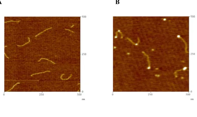
Figure 3.2 Representative AFM images………. 82
Figure 3.3 Illustration of protein-DNA binding and representative AFM images…………. 83
Figure 3.4 Histogram of position distributions of UvrD and/or SSB………. 85
Figure 3.5 Representative of AFM images……… 88
Figure 3.6 Length distribution of UvrD and/or wtSSB in the absence and presence of ATP……… 91
Figure 3.8 Distribution of the lengths of the DNA arms for UvrD and UvrD-wtSSB on
nicked DNA……… 96 Figure 4.1 Representative AFM images of MutL……….. 113 Figure 4.2 Histogram of conformational change of MutL………. 115 Figure 4.3 The volume histogram of MutL and UvrD alone vs MutL and UvrD together… 119 Figure 4.4 Representative AFM images……… 123 Figure 4.5 Binding of UvrD and/or MutL to the 817 bp DNA containing a 3′ ssDNA tail.. 126 Figure 4.6 Histogram of volume of UvrD and/or MutL on the DNA……… 128 Figure 4.7 Representative AFM images of MutL-UvrD bound to internal site on the
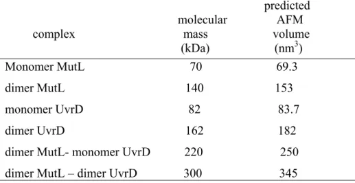
Table 1.1 Members of helicase families……… 11 Table 2.1 Predicted AFM volumes for Uvrd and SSB……… 44 Table 4.1 Predicted AFM volumes for MutL and UvrD……… 121 Table 4.2 UvrD binding occupancies on the DNA in the absence and presence of
nucleotides……… 124 Table 4.3 MutL and UvrD binding occupancies on the DNA in the presence of
List of Abbreviations
3-D Three-dimensional
A Alanine
ADP adenosine diphosphate
AFM atomic force microscopy
AID activation induced cytidine deaminase
Ala Alanine
AMPPPNP 5’-adenylyl-β-γ-imidodiphosphate
ATP adenosine triphosphate
bp Base pair
C- carboxy
χ chi subunit of DNA polymerase III
ddH2O doubly deionized water
DNA Deoxyribonucleic acid
dsDNA double stranded DNA
DTT dithiothreitol
E. coli Escherichia coli
EDTA Ethylamine diamine tetraacetic acid EMSA Electromobioity gel shift assay
ExoI Exonuclease I
FRET Fluorescence resonance energy transfer g gram
H Histidine
HEPES (4-(2-hydroxyethyl)-1-piperazineethanesulfonic acid
His Histidine
HNPCC hereditary non-polyposis colorectal cancer
hsp90 Family of 90 kDa heat shock proteins found in the cytoplasm & endoplasmic reticulum
IDL insertion-deletion loop
KC1 Potassium chloride
λ wavelength
L Liter
Li Lithium
μm micron
μM micromolar
M molar
Mg Magnesium
MgCl Magnesium Chloride
min minute
mL milliliter
Mlh MutL homolog
mM millimolar
mmol millimole
MMR DNA mismatch repair
MW Molecular weight
N Structured N-terminal domain of either MutL or MutS
N- amino
Na Sodium
NaCL Sodium chloride
NaOH Sodium hydroxide
nM nanomolar
nm nanometer
nm3 Cubic nanometers
nt nucleotide
OD595 Optical density at λ=595
OH- base
P Proline
PcrA Plasmid copy number reduction, DNA helicase, SF1 PCNA proliferating cell nuclear antigen
PMSF Phenylmethylsufonylfluoride
Polymin P Poly(ethylenimine)
Rep DNA helicase, SF1
RNA ribonucleic acid
RPA Replication protein A
rpm Revolutions per minute
SDS Sodium dodecyl sulfate
ssDNA single stranded DNA
T Threonine
T Thymine
Taq Thermus auaticus
TBE Tris-borate-EDTA buffer
T4 gene 32 protein gene 32 protein of bacteriophage T4 Tris 2-amino-2-hydroxymethyl-1,3-propanediol
U uracil
UvrD DNA helicase II
W Adenine or Thymine
Wt wild-type
X Any amino acid
Chapter 1 Introduction
DNA mismatch repair (MMR)
DNA mismatch repair (MMR) is one of several DNA repair systems conserved
from bacteria to humans (Au 1992; Buermeyer 1999; Jiricny 2003). MMR is the primary
mechanism by which DNA-synthesis errors are corrected post-replicatively. MMR
recognizes and repairs insertion/deletion loops (IDLs) and non-Watson Crick base pairs, e.g.,
G:T, which can arise during DNA replication and recombination (Buermeyer 1999; Kolodner
1999; Genschel 2000; Hsieh 2001). Non-Watson-crick base pairs, or mismatches, within the
DNA helix can result from nucleotide misincorporation during DNA synthesis. The error of
base-pairing and editing is corrected by mismatch repair, further elevating fidelity of DNA
replication 50 to 1,000-fold (Kolodner 1996; Modrich 1996; Jiricny 1998; Buermeyer 1999;
Kolodner 1999; Schofield 2003). Inactivation of the human mismatch repair system is
associated with > 85% of occurrences of hereditary non-polyposis colon cancer (HNPCC)
and has been implicated in the development of a subset of sporadic tumors that occur in a
variety of tissues (Eshleman 1995; Peltomaki 2001; Peltomaki 2003; de la Chapelle 2004;
MMR pathway in Escherichia coli (E. coli)
Escherichia coli (E. coli) provides the best understood mismatch repair system and
serves as a model system for the more complicated, but homologous, eukaryotic systems
(Modrich 1996; Harfe 2000). In E. coli, the DNA mismatch repair system takes advantage of
the transient post–replicative hemi-methylation of the adenosine in GATC sites to
discriminate between parent and daughter DNA strands (Modrich 1996). The mismatch
repair pathway is bi-directional (Figure 1.1). MutS, MutL and MutH are responsible for the
initiation of MMR in E.coli (Su 1986; Su 1988; Grilley 1989; Modrich 1996). MMR is
initiated by the recognition of DNA mismatches or insertion deletion loops (IDL) by MutS
(Su 1986; Su 1988). Subsequently, MutL binds to the MutS-DNA complex in a
ATP-dependent manner (Grilley 1989). This MutS-MutL-mismatch complex activates MutH,
which incises the newly-synthesized strand at a hemimethlylated GATC site (Modrich 1987;
Welsh 1987; Au 1992). This incision confers strand specificity of MMR, directing repair
exclusively to the newly synthesized strand containing the error. DNA helicase II (UvrD)
unwinds the DNA toward the mismatch and the appropriate exonucleases. The excision
depends on the position of the mismatch relative to the GATC site, a 5' to 3' exonuclease
(RecJ or Exo VIII) or a 3' to 5' (Exo I, Exo X or Exo VIII) (Cooper 1993; Grilley 1993;
Yamaguchi 1998; Mechanic 2000). The DNA polymerase III holoenzyme resynthesizes the
=SSB
Figure 1.1 Schematic of Bi-directional MMR in E. coli
The MMR process is bi-directional. The pathway includes steps of mismatch recognition,
Initiation of repair by MutS and MutL
The crystal structures of MutS-DNA complexes have been determined for Thermus aquaticus (Taq) MutS binding to a number of different mismatched DNA bases and IDLs
(Obmolova 2000; Natrajan 2003) (Figure 1.2), and for E.coli MutS bound to a G: T
mismatch and ADP (Lamers 2000). The MutS homodimer contains a carboxyl-terminal
ATPase domain, as well as a mismatch base recognition domain (Haber 1991; Wu 1994). A
sharp 60° kink in the DNA at the mismatch site is observed with MutS only 2 residues of
MutS interacting with the mismatched base. A phenylalanine, conserved in the MutS family
(Phe 36 in E.coli, Phe 39 in Taq), stacks with one of the mismatched bases: this base also
forms a hydrogen bond to a conserved glutamic acid (Glu 38 in E.coli, Glu 41 in Taq).
There are three models that describe the initiation events in DNA MMR. One model,
the translocation model, proposes that the DNA-bound MutS moves along the helix and that
this movement is coupled to adenosine triphosphate binding and hydrolysis (Blackwell 1998).
In the molecular switch model, mismatch recognition by the MutS·ADP complex promotes
exchange of ADP for ATP and MutS slides away from the mismatch (Gradia 1999). The
third model, the trans-activation model, suggests that upon recognizing and binding to a
mismatch or insertion/deletion loop, MutS remains in the vicinity of the mismatch. The
MutS-MutL-mismatch ternary complex is postulated to interact with MutH at the strand
discrimination signal site by DNA bending and protein-protein interactions (Junop 2001;
Schofield 2001). In this model, the MutS-MutL complex may provide the signal for
termination of the excision step involving the DNA helicase II and exonucleases (Schofield
A
B
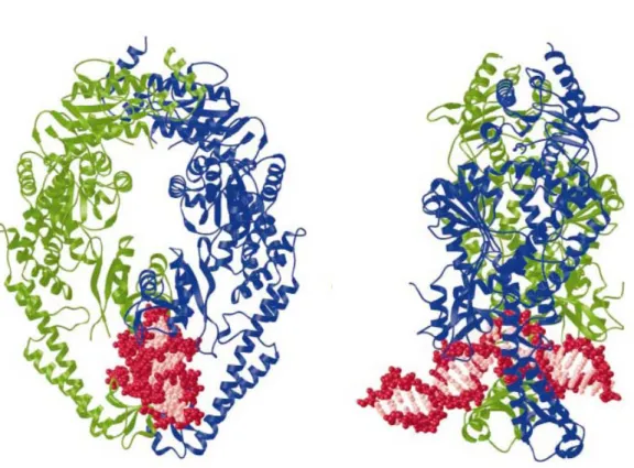
Figure 1.2 Crystal structure of Taq MutS (Obmolova 2000)
(A) Taq MutS bound to DNA. Two subunits are represented by ribbon (blue and green) and
DNA is shown in red and pink. The backbone of DNA is pink. (B) Side view of the Taq
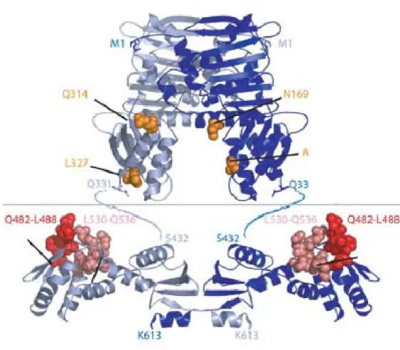
MutL plays an essential role in coupling of mismatch recognition by MutS to the
activation of MutH and DNA helicase II. In E. coli, MutL mediates initiation steps and
downstream repair events in MMR. Previous crystallographic and biochemical studies have
shown that MutL contains an N-terminal ATPase region (residues 1-349) and a C-terminal
dimerization region (residues 432-615) (Ban 1998; Ban 1999) (Figure 1.2). The N-terminal
ATPase domain is conserved in all members of the MutL family (Ban 1998), but the
C-terminal region, which is responsible for dimerization, is diverse among MutL homologs
(Pang 1997; Drotschmann 1998) The C-terminal domain of MutL crystallizes as a V-shaped
dimer, while the N-terminal domain forms a saddle-shape dimmer in the presence of
AMPPNP. The N-terminal ATPase fragment is monomeric in the absence of nucleotide. In
the presence of the nonhydrolyzable ATP analog AMPPNP, the ATPase fragment is dimeric
and is fully folded (Ban 1998; Ban 1999). MutL and MutL homologs are conserved from
prokaryotes to eukaryotes (Modrich 1996). In eukaryotes, MutL homologs are called MLH
and PMS. Like MutL, MutL homologs catalyze a weak ATPase activity, which is necessary
in DNA repair (Ban 1998; Ban 1999; Spampinato 2000).
Conformational changes that occur upon ATP binding and hydrolysis have been
proposed to allow MutL to coordinate various protein-protein interactions during the
mismatch repair process (Ban 1999). In the absence of MutS and a mismatch, MutL can
activate MutH endonuclease to a relatively low level in an ATP binding-dependent, yet ATP
hydrolysis-independent, manner (Ban 1998). Yeast two hybrid assays suggest that MutL
physically interacts with UvrD (Hall 1998). The C-terminal 219 amino acids (residues
398-615) of MutL are adequate to interact with UvrD, indicating that this region may contain the
facilitates the loading of UvrD onto DNA (Mechanic 2000). Helicase activity assays have
shown that MutL stimulates UvrD helicase unwinding (Dao 1998; Yamaguchi 1998), and
DNA binding assays have revealed that MutL increases UvrD binding to partial duplex DNA
Figure 1.3Crystal structure of MutL
The ribbon diagram of the homodimeric N-terminal domain and C-terminal domain aligned.
Subunits are colored in light blue and dark blue. Residues of N-terminal domain shown to be
bound to MutH are indicated by orange spheres. The highly conserved surface of C-terminal
Helicases
The helicase superfamily
Helicases have been identified and characterized from different organisms, including bacteria,
yeast and humans. Based on sequence alignments, helicases can be classified into six
families: three superfamilies (I, II, III), DnaB-like family, Rho-like family and a branch in
the AAA+ family (Gorbalenya 1993; Lyer 2004) (Table 1.1). SF1 and SF2 members share
seven conserved sequence motifs (Hodgman 1988; Gorbalenya 1993). PcrA helicase from
Bacillus stearothermophilus, which is in the SF1 family and shares 42 % sequence similarity
with UvrD, is a 3' → 5' helicase (Bird 1998; Petit 1998; Soultanas 1999) (Table1.1). PcrA is
involved in DNA repair and rolling circle plasmid replication (Petit 1998; Soultanas 1999). E. coli Rep helicase shares 37 % sequence homology with UvrD and is also in the SF1 family
(Lohman 1996). Rep helicase participates in the replication of E. coli bacteriophages M13
and ΦΧ174 (Takahashi 1979). The Rep protein is monomer in the absence of DNA (Lohman
1989), but in vitro, Rep monomers are inactive as helicases (Cheng 2001). While the
monomers of Rep protein can translocate on ssDNA in the 3' → 5' direction, the dimeric
form appears to be required to unwind dsDNA in vitro (Wong 1992; Cheng 2001; Ha 2002;
Brendza 2005). Wong and Lohman (1992) have proposed that Rep helicase unwinds and
translocates along DNA by an “active, rolling” mechanism, in which Rep requires at least
Superfamily
I Superfamily II Superfamily III
DnaB-like helicase
Rho-like family UvrD
(E. coli,
DNA repair)
RecQ
(E. coli, DNA
repair)
LTag
(Simian Virus 40, replication)
dnaB
(E. coli,
replication)
Rho
(E. coli,
transcriptio n termination
)
Rep
(E. coli,
DNA replication) eIF4A (Baker's Yeast, RNA translation) E1 (human papillomavirus, replication) gp41 (bacteriophag e T4, DNA replication)
PcrA
(Staphylococ cus aureus,
recombinatio n) WRN (human, DNA repair) Rep (Adeno-Associated Virus, replication, viral integration, virion packaging) T7gp4 (bacteriophag e T7, DNA replication)
Dda
(bacteriopha ge T4,
replication initiation) NS3 (Hepatitis C virus, replication) TRCF
(Mfd) (E.coli,
transcription-repair coupling)
DNA helicase II (UvrD gene)
UvrD catalyzes unwinding of duplex DNA, which is necessary to form ssDNA
intermediates during DNA replication, recombination and repair (Lohman 1996). UvrD is a
motor protein that translocates along single-stranded (ss)- and unwinds double-stranded (ds)
DNA using the energy derived from ATP hydrolysis (Matson 1990; von Hippel 2003). E. coli UvrD (DNA helicase II) is involved in mismatch repair, where it unwinds dsDNA to
correct DNA errors, following DNA incision by the combined activation of
MutS-MutL-MutH (Modrich 1994).
E. coli UvrD helicase, which is in the SF1 family of helicases, is essential for both
MMR and nucleotide excision repair (NER) (Sancar 1994; Modrich 1996). UvrD is a 82
kDa protein and binds both ss- and dsDNA (Abdel-Monem 1977). UvrD unwinds duplex
DNA with a 3'→ 5' direction with respect to the DNA strands on which UvrD is bound
(Matson 1986). There is a controversy regarding the functional states of UvrD. Yeast two
hybrid and biochemical assays identified a mutant, UvrDΔ40C, that is defective in
oligomerization, but whose ATPase and unwinding activities are indistinguishable from
wtUvrD (Mechanic 1999).
In addition, crystallographic studies suggest that one helicase molecule is bound at a
ss-dsDNA junction (Lee 2006). Although mutational and structural studies suggest that the
monomeric state is functional (Mechanic 1999; Lee 2006), systematic DNA unwinding
kinetic studies suggested that the dimeric form is required for helicase activity, at least on
some substrates (Ali 1999; Maluf 2003). Pre-steady state quenched-flow studies suggest that
demonstrated that UvrD is predominantly dimeric when binding to ssDNA (Runyon 1993)
and that UvrD becomes oligomerized at the junction of ss-dsDNA region (Matson 1987).
UvrD structure
The first helicase structure solved was that of Bacillus stearothermophilus PcrA
protein (Subramanya 1996). PcrA complexed with DNA and SO42- or DNA and AMPPNP
showed that the enzyme crystallizes as a monomer that consists of two domains (domains
1and 2) (Figure 1.4). Each domain contains two subdomains (A and B). Subdomain 1A
carries the Walker A and B motifs, and subdomain 2A is similar in structure to 1A. Both
subdomains contain a large insertion within the polypeptide sequences (Velankar 1999).
Given that the 2B domain of PcrA undergoes conformational changes upon binding to
ss-dsDNA junction, it is likely that 2B domain is essential for binding and unwinding of duplex
DNA (Soultanas 2000).
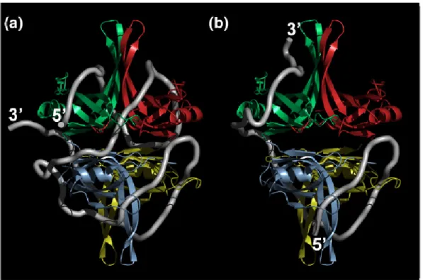
Lee and Yang (2006) reported crystal structures of E. coli UvrD showing distinct
steps during ATP hydrolysis: (1) UvrD and DNA (Figure 1.5A), (2) UvrD with a
nonhydrolyzable ATP analog (AMPPNP) (Figure 1.5B), and (3) UvrD with an ATP
hydrolysis intermediate (ADP-MgF3). In these crystal structures, each ds-ss DNA junction is
bound by one UvrD monomer. UvrD contains four structural domains, like PcrA, which
include 1A, 2A, 1B and 2B:1A (1-89, 215-280 aa), 2A (281-377, 551-647 aa), 1B (90-214
aa) and 2B (378-550 aa). Domain 1A and 2A are responsible for ATP binding and hydrolysis
(Figure 1.4B) and Domain 1B and 2B interact with DNA (Lee 2006). The 3' ssDNA tail is
bound across domains 1A and 2A, and duplex DNA interactes with 1B and 2B (Figure 1.5).
In the presence of AMPPNP molecule or ADP-MgF3 complex, domain 2B is reoriented and
forms an open conformation. The binding of ATP induces a closed conformation between
domain 2A and the rest of the protein, which leads to separation of the first base pair at the
ss-dsDNA junction. ATP hydrolysis leads to domain 2B opening and translocation of 1bp,
Figure 1.4 Structure of the PcrA helicase
(A) apo PcrA (B) PcrA-DNA (C) PcrA-DNA-AMPPNP: Domain 1A (green), 2A (red), 2B
(blue), 1B (orange in (a) or yellow). The bound DNA is magenta. The AMPPNP and the
Figure 1.5 Crystal structures of UvrD-DNA complexes (Lee 2006)
(A) Binary complex of UvrD-DNA, (B) UvrD-DNA-AMPPNP. UvrD contains four
structural domains 1A, 1B, 2A and 2B. Domain 1A and 2A are responsible for ATP binding
Single-stranded binding protein (SSB)
SSB plays a critical role in replication, is involved recombination and repair, and
interacts with other proteins involved in DNA metabolism (Meyer 1990; Lohman 1994).
SSB protein binds preferentially and cooperatively to ssDNA depending on the salt
concentration. In the absence of DNA, the protein exists as a homotetramer (Chase 1986;
Lohman 1994) and there is no evidence for distinct higher-order forms (Meyer 1990). Each
subunit of SSB possesses an oligonucleotide/oligosaccharide binding (OB) fold, which has
potential ssDNA binding sites (Roy 2007).
SSB binding modes
SSB binds to DNA in several different binding modes with different ‘site sizes’,
defined as the average number of nucleotides occluded by the bound protein. In the two
main modes of DNA binding by SSB, there are “35 base” and “65 base” modes. In the
(SSB)35 binding mode, in which the average site size per tetramer is 35 nucleotides, only two
SSB subunits interact in a highly cooperative fashion with ssDNA, and long protein tracts
form along the DNA. Since more SSB proteins can bind to DNA in the (SSB)35 as compared
to the (SSB)65 mode, high protein concentrations favor the (SSB)35 mode, which is stable at
Figure 1.6 SSB binding mode in the presence of DNA (Roy 2007)
Model of the binding mode derived from the crystal structure of a chymotrypsin truncated
SSB tetramer (missing 42 C-terminal residues) (Raghunathan 2000) (A) (SSB)65 mode.
ssDNA interacts with all four subunits of SSB (B) In the (SSB)35 mode, only two subunits of
the SSB tetramer interact with ss DNA. Each SSB subunit is color coded. DNA is shown in
The (SSB)65 binding mode, in which ssDNA interacts with all four SSB subunits, is
stable at high salt, [NaCl] ≥ 0.2 M. This binding mode displays “limited” type cooperativity,
in which the SSB cluster size is limited to clusters of two SSB tetramers (Lohman 1994;
Raghunathan 2000). There is another minor binding mode of SSB, which is the (SSB)56
mode and is grouped with (SSB)65 mode by molecular similarity. In the (SSB)56 binding
mode observed at intermediate [NaCl], all four subunits interact with DNA, but the details of
the interaction remain unknown (Kuil 1990) (Figure 1.6). The concentration of monovalent
and divalent cations, protein concentrations, pH, and temperature influence the stability of
these modes. Recently, single molecule fluorescence resonance energy transfer (smFRET)
experiments (Ha 2001; Ha 2001) determined the direct dynamics of structural changes
between the two binding modes, “35” and “65” (Roy 2007) (Figure 1.6). These smFRET
experiments revealed the two major binding modes, (SSB)35 and (SSB)65, as well as a new
binding configuration, (SSB)35b, which can be formed from the (SSB)35 mode without protein
dissociation (Roy 2007).
Interaction with other proteins
It has been known that a major function of SSB is to protect ssDNA from nuclease
degradation in vitro (Delagoutte 2003); however, SSB also interacts with several other
proteins and modulates their activity. SSB containing a single amino acid substitution (a
proline to serine) in the C-terminus still binds ssDNA with similar affinity as wild-type
protein, but it does not interact with the χ subunit of DNA polymerase III (Kelman 1998).
Recently, Cadman and McGlynn (2000) have revealed that SSB physically interacts with the
SSB stimulates PriA unwinding of branched DNA substrates (Cadman 2004). PriA helicase
activity and binding assays using a number of different DNA substrates have shown that SSB
significantly stimulates PriA helicase at branched DNA substrates including the lagging
strand duplex and the single-stranded leading strand, but not other substrates (Cadman 2004).
SSB also interacts with RecQ helicase, which is critical in replication fork maintenance,
DNA damage checkpoint signaling, and recombination regulation (Bachrati 2003; Bennett
2004; Ozgenc 2005; Shereda 2007). Helicase and electrophoretic mobility shift assays have
revealed that SSB stimulates RecQ unwinding of 70-nt 3' overhang DNA (Shereda 2007).
The observation of interactions between SSB and these helicases lead to the question of
whether or not SSB interacts with SSB.
C-terminus of SSB mediates interactions with proteins
The last 5 amino acids of E. coli SSB are negatively charged, making the C-terminus
highly acidic. This region is suggested to be involved in interactions with other accessory
proteins in DNA replication, recombination and repair (Lohman 1994; Curth 1996; Kelman
1998; Genschel 2000; Handa 2001). The SSB C-terminal domain is required for interactions
with proteins, including ExoI (Genschel 2000), the χ subunit of DNA polymerase III
(Kelman 1998; Witte 2003), PriA DNA helicase (Cadman 2004) and RecQ helicase (Shereda
2007).
Atomic Force Microscopy (AFM)
Since its invention in 1986, Atomic Force Microscopy (AFM) has been developed as
1995; Fotiadisa 2002). The use of AFM has extended to studies of DNA, RNA, proteins,
lipids, carbohydrates, biomolecular complexes, and cells (Ratcliff 2001; Yang 2003).
In AFM, a cantilever is oscillated as it scans the surface of a sample (X- and Y-
directions). The deflection of the cantilever, which is caused by forces of interaction
between the sample and the tip, is monitored by a laser beam that is reflected into a
photodiode. A feedback loop between the piezo and photodiode keeps the amplitude of the
cantilever constant through vertical motion of the piezo (Z). A topographic image of the
sample is generated by plotting the vertical movement of the piezo (Z-direction) over X, Y
Laser Light Mirror
4-Segment Photodiode
Cantilever
Tip
Feedback Loop
Piezo
X Y Z
Sample
Figure 1.7Schematic of AFM
The sample is placed on a piezo, which is scanned in the X and Y directions by a tip attached
to a cantilever. The laser monitors the tip over the sample and measures the vertical
deflection of the cantilever. The feedback loop keeps the cantilever deflection constant. The
AFM was developed as a powerful tool for studing protein-protein and protein-DNA
complexes, mainly with its relative simplicity and reliability in sample preparation and
imaging processes, contributing to the majority of the AFM studies of biological assemblies.
It has been demonstrated that there is a linear relationship between AFM scan volume
and molecular weight of proteins which allows the stoichiometries and the oligomerization
states of proteins and a multi-protein complex to be determined. In addition, it is possible to
estimate protein-protein and protein-DNA binding constants (Ratcliff 2001; Yang 2005), as
well as visualize conformational changes including bending and wrapping of a protein and
protein-DNA complexes (Bustamante 1996; Rivetti 1999; Rivetti 2003; Yang 2003),
suggesting that specific recognition sites of proteins on DNA, i.e. the binding specificity of
protein to a site in DNA, can be observed. As the mismatch repair machinery corrects
mismatches in base-pairing, AFM imaging is a useful technique for structure-function
References
Abbel-Monem, M., Channal, M. C. and Hoffmann-Berling, H. (1977). "DNA unwinding enzyme II of Escherichia coli. 2. Characterization of the DNA unwinding activity." Eur. J. Biochem. 79: 39-45.
Abdel-Monem, M., Channal, M. C. and Hoffmann-Berling, H. (1977). "DNA unwinding enzyme II of Escherichia coli. 1. Purification and characterization of the ATPase activity." Eur. J. Biochem. 79: 33-38.
Ali, J. A. M., N. K. and Lohman, T. M. (1999). "An oligomeric form of E. coli UvrD is
required for optimal helicase activity " J. Mol. Biol. 293: 815-834.
Ali, M. M., Roe, S. M., Vaughan, C. K., Meyer, P., Panaretou, B., Piper, P. W., Prodromou, C., and Pearl, L. H. (2006). "Crystal structure of an Hsp90-nucleotide-p23/Sba1 closed chaperone complex." Nature 440: 1013-1017.
Allen, D. J., Markhow, A., Grilley, M., Taylor, J., Thresher. R., Modrich, P. and Griffith, J. D. (1997). "MutS mediates heteroduplex loop formation by a translocation mechanism." EMBO J. 16: 4467-4476.
Amaratunga, M. a. L., T. M. (1993). "E.coli Rep Helicase Unwinds DNA by an Active
Mechanism." Biochmistry 32: 6815-20.
Au, K. G., Welsh, K. et al. (1992). "Initiation of methyl-directed mismatch repair." J. Biol. Chem. 267(17): 12142-8.
Bachrati, C. Z., and Hickson, I. D. (2003). " RecQ helicases: suppressors of tumorigenesis and premature aging" Biochem. J. 374: 577-606.
Ban, C., Junop, M. and Yang, W. (1999). "Transformation of MutL by ATP binding and hydrolysis: a switch in DNA mismatch repair." Cell 97: 85-97.
Ban, C. a. Y., W. (1998). "Crystal structure and ATPase activity of MutL: implications for DNA repair and mutagenesis." Cell 95: 541-552.
Bennett, R. J., and Keck, J. L. (2004). " Structure and function of RecQ DNA helicases" Crit. Rev. Biochem. Mol. Biol. 39: 79-97.
Bird, L. E., Subramanya, H. S. and Wigley, D. B. (1998). "Helicases, a unifying structural theme?" Curr. Opin. Struct. Biol. 8: 14-18.
Blackwell, L. J., Martik, D., Bjornson, K. P., Bjornson, E. S. and Modrich, P. (1998). "Nucleotide-promoted release of hMutSα from heteroduplex DNA is consistent with an ATP-dependent translocation mechanism." J. Biol.Chem. 273: 32055-32062.
Brendza, K. M. e. a. (2005). "Auto-inhibition of E. coli Rep monomer helicase activity by its
2B sub-domain." Proc. Natl. Acad. Sci. 102: 10081.
Buermeyer, A. B., Deschenes, S. M., Baker, S. M., Liskay, R. M. (1999). "Mammalian DNA Mismatch Repair." Annu. Rev. Genet. 33: 533-564.
Bustamante, C. a. K., D. (1995). "Scanning Force Microscopy in Biology." Physics Today
48: 32-38.
Bustamante, C. a. R., C. (1996). "Visualizing protein-nucleic acid interactions on a large scale with the scanning force microscope." Annu. Rev. Biophys. Biomol. Struct 25: 395-429.
Cadman, C. J., and Mcglynn, P. (2004). "PriA helicase and SSB interact physically and functionally." Nucleic Acids Res. 32: 6378-6397.
Caron, P. R., Kushner, S. and Grossman, L. (1985). "Involvement of helicase Ii (uvrD gene product) and DNA polymerase I in excision mediated by the UvrABC protein complex." Proc. Natl. Acad. Sci. U. S. A. 82: 4925-4929.
Caruthers, J. M., and McKay, D. B. (2002). "Helicase structure and mechanism." Curr. Opin. Struct. Biol. 12: 123-133.
Chao, K., Lohman, T. M. (1991). "DNA-induced dimerization of the Escherichia coli Rep
helicase." J. Mol. Biol. 221: 1165-81.
Chase, J. W., L'Italien, J. J., Murphy, J. B., Spicer, E. K. and Williams, K. R. (1984). "Characterization of the Escherichia coli SSB-113 mutant single-stranded DNA-binding protein. Cloning of the gene, DNA and protein sequence analysis, high pressure liquid chromatography peptide mapping, and DNA-binding studies" J. Biol. Chem. 259: 805-814.
Chase, J. W. W., K. R. (1986). "Single-stranded DNA binding proteins required for DNA replication." Annu. Rev. Biochem. 55: 103-136.
Cheng, W., Hsieh, J., Brendza, K. M. and Lohman, T. M. (2001). "E.coli Rep oligomers are
required to initiate DNA unwinding in vitro." J. Mol. Biol. 310: 327-350.
Chu, F., Maynard, J. C., Chiosis, G., Nicchitta, C. V., and Burlingame, A. L. (2006). "Identification of novel quaternary domain interactions in the Hsp90 chaperone, GRP94." Protein Sci. 15: 1260-1269.
Corbett, K. D., and Berger, J. M. (2003). "Structure of the topoisomerase VI-B subunit: implications for type II topoisomerase mechanism and evolution." EMBO J. 22: 151-163.
Curth, U., Genschel,J., Urbanke,C. and Greipel,J. (1996). "In vitro and in vivo function of the C-terminus of Escherichia coli single-strandedDNA binding protein. ." Nucleic Acids Res.
24: 2706–2711.
Dabrowski, S., Olszewski, M., Piatek, R., Brillowska-Dabrowska, A., Konopa, G. and Kur, J. (2002). "Identification and characterization of single-stranded DNA binding proteins fro
Thermus thermophilus and Thermus aquaticus-new arrangement of binding domains."
Microbiology 148: 3307-3315.
Dao, V. a. M., P. (1998). "Mismatch-, MutS-, MutL-, and Helicase II-dependent unwinding from the single-strand break of an incised heteroduplex." J. Biol. Chem. 273: 9202-9207.
de la Chapelle, A. (2004). "Genetic predisposition to colorectal cancer." Nat. Rev. Cancer 4:
769-780.
Delagoutte, E. a. v. H., Peter H. (2003). "Helicase mechanisms and the coupling of helicases within macromolecular machines. Part II." Q. Rev. Biophys. 36: 1-69.
Dollins, D. E., Immormino, R. M., and Gewirth, D. T. (2005). "Structure of unliganded GRP94, the endoplasmic reticulum Hsp90. Basis for nucleotide-induced conformational change." J. Biol. Chem. 280: 30438-30447.
Drotschmann, K., Aronshtam, A., Fritz, H. J. and Marins, M. G. (1998). "The Escherichia coli MutL protein stimulates binding of Vsr and MutS to heteroduplex DNA." Nucleic Acids
Res. 26: 948-953.
Dutta, R., and Inouye, M. (2000). "GHKL, an emergent ATPase/kinase superfamily. ." Trends Biochem. Sci. 25: 24-28.
Eggington, J. M., Haruta, N., Wood, E. A. and Cox, M. M. (2004). "The single-stranded DNA binding protein of Deincoccus radiodurans." BMC Microbiol 4: 2.
Eshleman, J. R., Markowitz, S. D. (1995). "Microsatellite instability in inherited and sporadic neoplasms." Curr. Opin. Oncol. 7: 83-89.
Fay, P. J., Johanson, K. O., MaHenry, C. S. and Bambara, R. A. (1982). "Size classes of products synthesized processively by two subassemblies of Escherichia coli DNA
polymerase III holoenzyme." J. Biol. Chem. 257: 5692-99.
Fedorov, R., Witte, G., Urbanke, C., Manstein, D. J. and Curth, U. (2006). "3D structure of
Thermus aquaticus single-stranded DNA-binding protein gives insight into the functioning of
Ferrari, M. E. B., W. and Lohman, T. M. (1994). "Co-operative binding of Escherichia coli
SSB tetramers to single-stranded DNA in the (SSB)35 binding mode." J. Mol. Biol. 236:
106-123.
Fotiadisa, D., Scheuringa, S., Müllera, S. A., Engela, A. and Müller, D. J. (2002). "Imaging and manipulation of biological structures with the AFM " Micron 33: 385-397.
Genschel, J., Curth,U. and Urbanke,C. (2000). "Interaction of E. coli single-stranded DNA binding protein (SSB) with exonuclease I. The carboxy-terminus of SSB is the recognition site for the nuclease." Biol. Chem. 381: 183–192.
Gorbalenya, A. E. a. K., E. V. (1993). "Helicases: amino acid sequence comparisons and structure-function relationships." Curr. Opin. Struct. Biol. 3: 419-429.
Gradia, S., Subramanian, D., Wilson, T., Acharya, S., Makhov, A., Griffith, J. and Fishel, R. (1999). "hMSH2-hMSH6 forms a hydrolysis-independent sliding clamp on mismatched DNA." Mol. Cell. Biol 3: 255-261.
Grilley, M., Griffith, J., and Modrich, P. (1993). "Bidirectional excision in methyl-directed mismatch repair." J. Biol. Chem. 268: 11830-11837
Grilley, M., Welsh, K. M., Su, S.-S., and Modrich, P. (1989). "Isolation and Characterization of the Escherichia coli mutL Gene Product." J. Biol. Chem. 264: 1000-1004
Guarne, A., Junop, M. S., and Yang, W. (2001). "Structure and function of the N-terminal 40 Da fragment of human PMS2: a monomeric GHL ATPase." EMBO J. 20: 5521-5531.
Gulbis, J. M., Kazmirski, S. L., Finkelstein, J., Kelman, Z., O'Donnell, M., and Kuriyan, J. (2004). "Crystal structure of the chi:psi subassembly of the Escherichia coli DNA
polymerase clamp-loader complex." Eur. J. Biochem. 271: 439-449.
Ha, T. (2001). "Single-mlecule fluorescence resonance energy transfer." Methods 25: 78-86.
Ha, T. (2001). "Single-molecule fluorescence methods for the study of nucleic acids." Curr. Opin. Struct. Biol. 11: 287-292.
Ha, T. e. a. (2002). "Initiation and reinitiation of DNA unwinding by the Escherichia coli
Rep helicase." Nature 419: 638-641.
Hall, M. C., Jordan, J. R. and Matson, S. W. (1998). "Evidence for a physical interaction between the Escherichia coli methyl-directed mismatch repair proteins MutL and UvrD."
EMBO J. 17: 1535-1541.
Hall, M. C., Matson, S. W. (1999). "The Escherichia coli MutL protein physically interacts with MutH and stimulates the MutH-associated endonuclease activity." J. Biol. Chem. 274:
1306-1312.
Handa, P., Acharya,N. and Varshney,U. (2001). "Chimeras between single-stranded DNA-binding proteins from Escherichia coli and Mycobacterium tuberculosis reveal that their C-terminal domains interact with uracil DNA glycosylases." J. Biol. Chem. 276: 16992–16997.
Hansma, H. G., Sinsheimer, R. L., Groppe, J., Bruice, T. C., Elings, V., Gurley, G., Bezanilla, M., Mastrangelo, I. A., Hough, P. V. and Hansma, P. K. (1993). "Recent advances in atomic force microscopy of DNA." Scanning 15: 296-299.
Harfe, B. D. a. J.-R., S. (2000). "Mismatch repair porteins and mitotic genome stability." Mutat. Res. 451(1-2): 151-167.
Harmon, F. G. a. K., S. C. (2001). "Biochemical characterization of the DNA helicase activity of the Escherichia coli RecQ helicase." J. Biol. Chem. 276(1): 232-243.
Hodgman, T. C. (1988). "A new superfamily of replicative proteins." Nature 333: 22-23.
Hsieh, P. (2001). "Molecular mechanisms of DNA mismatch repair." Mutat. Res. 486(2):
71-87.
Hu, X., Machius, M., and Yang, W. (2003). "Monovalent cation dependence and preference of GHKL ATPases and kinases." FEBS lett. 544: 268-273.
Hurley, J. M., Chervitz, S. A., Jarvis, T. C., Singer, B. S. and Gold, L. (1993). " Assembly of the bacteriophage T4 replication machine requires the acidic carboxy terminus of gene 32 protein." J. Mol. Biol. 229: 398-418.
Husain, I., van Houten, B., Thomas, D. C., Abdel-monem, M. and Sangar, A. (1985). "Effect of DNA polymerase I and DNA helicase II on the turnover rate of UvrABC excision nuclease." Proc. Natl. Acad. Sci. U. S. A. 82: 6774-6778.
Immormino, R. M., Dollins, D. E., Shaffer, P. L., Soldano, K. L., Walker, M. A. and Gewirth, D. T. (2004). "Ligand-induced conformational shift in the N-terminal domain of GRP94, an Hsp90 chaperone." J. Biol. Chem. 279: 46162-46171.
Jiang, H., Giedroc, D. and Kodadek, T. (1993). " The role of protein-protein interactions in the assembly of the presynaptic filament for T4 homologous recombination" J. Biol. Chem.
Jiricny, J. (1998). "Replication errors: cha(lle)nging the genome." EMBO J. 17(6427-6436).
Jiricny, J. a. G. M. (2003). "DNA repair defects in colon cancer." Curr. Opin. Genet Dev.
13(1): 61-69.
Jones, J. M. a. N., H. (1999). "Duplex opening by primosome protein PriA for replisome assembly on a recombination intermediate." J. Mol. Biol. 289: 503-516.
Jones, J. M. a. N., H. (2001). "Escherichia coli PriA Helicase:fork binding orients the
helicase to unwind the lagging strand side of arrested replication forks." J. Mol. Biol. 312:
935-947.
Junop, M. S., Obmolova, G., Rausch, K., Hsieh, P. and Yang, W. (2001). "Composite active site of an ABC ATPase MutS uses ATP to verify mismatch recognition and authorize DNA repair." Mol. Cell 7(1): 1-12.
Kelman, Z., Yuzhakov,A., Andjelkovic,J. and O’Donnell,M. (1998). "Devoted to the lagging strand-the c subunit of DNA polymerase III holoenzyme contacts SSB to promote processive elongation and sliding clamp assembly." EMBO J. 17: 2436–2449.
Kolodner, R. (1996). "Biochemistry and genetics of eukaryotic mismatch repair." Genes Dev.
10: 1433-1442.
Kolodner, R. D., Marsischky, G.T. (1999). "Eukaryotic DNA mismatch repair." Curr. Opin. Genet Dev. 9(1): 89-96.
Kosinskia, J., Steindorfa, I., Bujnickib, J. M., Giron-Monzona, L., andFriedhoff, P. (2005). "Analysis of the Quaternary Structure of the MutL C-terminal Domain." J. Mol. Biol. 351(4):
895-909.
Kuhn, B. M., Abdel-Monem, M., and Hoffmann-Berling, H (1978). "DNA helicases." Cold Spring Harbor Symp. Quant. Biol. 43: 63-67.
Kuhn, B. M., Abdel-Monem, M., Krell, H., and Hoffmann-Berling, H (1979). "Evidence for two mechanisms for DNA unwinding catalyzed by DNA helicases " J. Biol. Chem. 254:
11343-11350.
Kuhn, H., Protozanova, E., and Demidov, V. (2002). "Monitoring of single nicks in duplex DNA by gel electrophoretic mobility-shift assay." Electrohporesis 23: 2384-2387.
Kumura, K., Sekiguchi, M., Steinum, A. L. and Seeberg, E. (1985). "Stimulation of the UvrABC enzyme-catalyzed reactions by the UvrD protein (DNA helicase II)." Nucleic Acids Res. 13: 1483-1492.
Lahue, R. S., Au, K. G., and Modrich, P. (1989). "DNA mismatch correction in a defined system." Science 245: 160-164.
Lamers, M. H., Perrakis, A., Enzlin, J. H., Winterwerp, H. H. K., de Wind, N. and Sixma, T. K. (2000). "The crystal structure of DNA mismatch repair protein MutS binding to a G•T mismatch." nature 407: 711-717.
LeBowitz, J. H. a. M., R. (1986). "The Escherichia coli dnaB replication protein is a DNA
helicase." J. Biol. Chem. 261: 4738-4748.
Lee, J. Y., and Yang, W. (2006). "UvrD helicase unwinds DNA one base pair at a time by a two-part power stroke." Cell 127: 1349-1360.
Lohman, T. M. (1992). "E.coli DNA Helicases: Mechanisms of DNA Unwinding." Mol.
Microbial. 6: 5-14.
Lohman, T. M. (1993). "Helicase-catalyzed DNA Unwinding." J. Biol. Chem. 268: 2269-72.
Lohman, T. M., Chao, K., Green, J. M. Sage, S. and Runyon, G. T. (1989). "Large-scale purification and characterization of the Escherichia coli rep gene product." J. Biol. Chem. 264: 10139-10147.
Lohman, T. M. a. B., K. P. (1996). "Mechanisms of helicase-catalyzed DNA unwinding." Annu. Rev. Biochem. 65: 169.
Lohman, T. M. F., M. E. (1994). "Escherichia coli single-stranded DNA-binding protein:
Multiple DNA-binding modes and cooperativites." Annu. Rev. Biochem. 63: 527-570.
Lyer, L. M., Leipe, D. D., Koonin, E V. and Aravind, L. (2004). "Evolutionary history and higher order classification of AAA+ATPases." J. Struct. Biol. 146: 11-31.
Maluf, N. K., Fischer, C. J. and Lohman, T. M. (2003). "A dimer of Escherichia coli UvrD is the active form of the helicase in vitro." J. Mol. Biol. 325: 913–935.
Maluf, N. K., Lohman, T.M. (2003). "Self-association equilibria of Escherichia coli UvrD
helicase studied by analytical ultracentrifugation." J. Mol. Biol. 325: 889-912.
Matson, S. W. (1986). "Escherichia coli helicase II (urvD gene product) translocates unidirectionally in a 3' to 5' direction." J. Biol. Chem. 261: 10169-10175.
Matson, S. W. a. G., J. W. (1987). "DNA helicase II of Escherichia coli. Characterization of
the single-stranded DNA-dependent NTPase and helicase activities." J. Biol. Chem. 262:
2066-2076.
Matson, S. W. a. K.-R., K. A. (1990). "DNA helicases." Annu. Rev. Biochem. 59: 289–329.
Mechanic, L. E., Frankel, B. A. and Matson, S. W. (2000). "Excherichia coli MutL loads
DNA helicase II onto DNA." J. Biol. Chem. 275: 38337-38346.
Mechanic, L. E., Hall, M. C. and Matson, S. W. (1999). "Escherichia coli DNA helicase II is active as a monomer." J. Biol. Chem. 274: 12488–12498.
Meyer, R. R. L., P.S. (1990). "The single-stranded DNA-binding protein of Escherichia coli." Microbiol. Rev. 54: 342-380.
Modrich, P. (1987). "DNA Mismatch Correction." Annu. Rev. Biochem. 56: 435-466
Modrich, P. (1994). "Mismatch repair, genetic stability, and cancer." Science 266: 1959-1960.
Modrich, P. a. L., R. (1996). "Mismatch repair in replication fidelity, genetic recombination, and cancer biology." Annu. Rev. Biochem. 65: 101-133.
Molineux, I. J. a. G., M. L. (1974). " Properties of the Escherichia coli DNA Binding (Unwinding) Protein: Interaction with DNA Polymerase and DNA" Proc. Natl. Acad. Sci. U. S. A. 71: 3858-62.
Moore, K. J. M. a. L., T. M. (1994). "Kinetic Mechanism of Adenine Nucleotide Binding to the E. coli Rep Monomer. II. Application of a Kinetic Competition Approach." Biochmistry 33: 14565-78.
Moore, K. J. M. a. L., T. M. (1994). "Kinetic Mechanism of Adenine Nucleotide Binding to and Hydrolysis by the E. coli Rep Monomer. I. Use of Fluorescent Nucleotide Analogues."
Biochmistry 33: 14550-64.
Natrajan, G., M. H. Lamers, et al (2003). "Structures of Escherichia coli DNA mismatch repair enzyme MutS in complex with different mismatches: a common recognition mode for diverse substrates." Nucleic Acids Res. 31: 4814-4821.
Obmolova, G., Ban, C., Hsieh, P. and Yang, W. (2000). "Crystal structure of mismatch repair protein MutS and its complex with a substrate DNA." Nature 407: 703-710.
Orren D. K., S., C. P., Hearst, J. E. and Sancar, A. (1992). "Post-incision steps of nucleotide excision repair in Escherichia coli. Disassembly of the UvrBC-DNA complex by helicase II
and DNA polymerase I." J. Biol. Chem. 267: 780-788.
Ozgenc, A., and Loeb, L. A. (2005). " Current advances in unraveling the function of the Werner syndrome protein" Mutat. Res. 577: 237-251.
Pang, Q., Prolla, T. A. and Liskay, M. (1997). "Functional domains of the Saccharomyces cerevisiae Mlh1pand Pms1p DNA mismatch repair protein and their relevance to human hereditary nonpolyposis colorectal cancer-associated mutations." Mol. Cell. Biol 17:
4465-4473.
Peltomaki, P. (2001). " DNA mismatch repair and cancer" Mutat. Res. 488(77-85): 77.
Peltomaki, P. (2003). " Role of DNA Mismatch Repair Defects in the Pathogenesis of Human Cancer" J. Clin. Oncol. 21: 1174-1179.
Petit, M. A., Dervyn, E., Rose, M., Entian, K. D., McGovern, S., Ehrich, S. D. and Bruand, C (1998). "PcrA is an essential DNA helicase of Bacillus subtilis fulfilling functions in repair and rolling-circle replication." Mol. Microbiol. 29: 261-273.
Prolla, T. A., Pang, Q., Alani, E., Kolodner, R. D. and Liskay, R. M. (1994). "Interactions between the MSH2, MLH1 and PMS1 proteins during the initiation of DNA mismatch repair." Science 265: 1091-1093.
Raghunathan, S., Kozlov, A. G., Lohman, T. M. and Waksman, G. (2000). "Structure of the DNA binding domain of E. coli SSB bound to ssDNA." Nature Struct. Biol. 7: 648-652.
Ratcliff, G. C., and Erie, D. A. (2001). "A novel single-molecule study to determine protein-protein association constants." J.Am. Chem. Soc. 123: 5632-5635.
Rivetti, C., Codeluppi, S., Dieci, G. and Bustamante, C. (2003). "Visualizing RNA extrusion and DNA wrapping in transcription elongation complexes of bacterial and eukaryotic RNA polymerases." J. Mol. Biol. 326(5): 1413-1426.
Rivetti, C., Guthold, M. and Bustamante, C. (1996). "Scanning force microscopy of DNA deposited onto mica: equilibration versus kinetic trapping studied by statistical polymer chain analysis." J. Mol. Biol. 264(5): 919-932.
Rivetti, C., Guthold, M. and Bustamante, C. (1999). "Wrapping of DNA around the E. coli
RNA polymerase open promoter complex." EMBO J. 18(16): 4464-4475.
Rowley, P. T. (2005). " Inherited susceptibility to colorectal cancer" Annu. Rev. Med. 56:
539-554.
Roy, R., Kozlow, A. G., Lohman, T. M., and Ha, T. J. (2007). "Dynamic structural rearrangements between DNA binding modes of E. coli SSB protein." J. Mol. Biol. 269:
1244-1257.
Runyon, G. T., Wong, I. and Lohman, T. M. (1993). "Overexpression, purification, DNA binding , and dimerization of the Escherichia coli uvrD gene product (helicase II)."
Biochmistry 32: 602-612.
Sacho, E. J., Kadyrov, F., Modrich, P., Kuncel, T. A. and Erie, D. A. (2008). "Direct visualization of asymmetric adenine nucleotide-induced conformational changes in
MutL." Mol. Cell 29: 112-121.
Sancar, A. (1994). "Mechanisms of DNA excision repair. ." Science 266: 1954-1956.
Sandler, S. J. a. M., K. J. (2000). "Role of PriA in replication fork reactivation in Escherichia coli." J. Bacteriol. 182: 9-13.
Schofield, M. J., Hsidh, P. (2003). "DNA mismatch repair: Molecular mechanisms and biological fuction." Annu. Rev. Microbiol. 57: 579-608.
Schofield, M. J., Nayak, S., Scott, T. H., Du, C. and Hsieh, P. (2001). "Interaction of
Escherchia coli MutS and MutL at a DNA mismatch." J. Biol. Chem. 276: 28291-28299.
Shereda, R. D., Bernstein, D. A. and Keck, J. L. (2007). "A central role for SSB in E.coli RecQ DNA helicase function." J. Biol. Chem. 282: 19247-19258.
Shiau, A. K., Harris, S. F., Southworth, D. R., and Agard, D. A. (2006). "Structural analysis of E. coli hsp90 reveals dramatic nucleotide-dependent conformational rearrangements." Cell 127: 329-340.
Sigal, N., Delius, H., Kornberg, T., Gefter, M. L. and Alberts, B. M. (1972). " A DNA-Unwinding Protein Isolated from Escherichia coli: Its Interaction with DNA and with DNA Polymerases" Proc. Natl. Acad. Sci. U. S. A. 69: 3537-41.
Soultanas, P., Dillingham, M. S., Papadopoulos, F., Philips, S. E. V., Thomas, C. D. and Wigley, D. B. (1999). "Plasmid replication initiator protein RepD increases the processivity of PcrA DNA helicase." Nucleic Acids Res. 27: 1421-1428.
Spampinato, C. a. M., P. (2000). "The MutL ATPase Is Required for Mismatch Repair." J. Biol. Chem. 275: 9863-9869.
Su, S.-S., Lahue, R. S., Au, K. G., and Modrich, P. (1988). "Mispair Specificity of Methyl-directed DNA Mismatch Correction in Vitro." J. Biol. Chem. 263(14): 6829-6835.
Su, S.-S. a. M., P. (1986). "Escherichia coli mutS-encoded protein binds to mismatched DNA base pairs." Proc. Natl. Acad. Sci. U. S. A. 83(14): 5057-5061.
Subramanya, H. S., Bird, L. E., Brannigan, J. A., and Wigley, D. B. (1996). "Crystal structure of a DE xx box DNA helicase." Nature 384: 379-383.
Takahashi, S., Hours, C., Chu, A., and Denhardt, D. T. (1979). "The rep mutation. VI. Purification and properties of the Escherichia coli rep protein, DNA helicase III." Can. J. Biochem. 57: 855-866.
Umezu, K. a. N., H. (1993). "RecQ DNA helicase of Escherichia coli characterization of the
helix-unwinding activity with emphasis on the effect of single-stranded DNA-binding protein." J. Mol. Biol. 230: 1145-1150.
Velankar, S. S., Soltanas, P., Dillingham, M. S., Subramanya, H. S. and Wigley, D. B. (1999). "Crystal structures of complexes of PcrA DNA helicase with a DNA substrate indicate an inchworm mechanism." Cell 97: 75-84.
von Hippel, P. H. a. D., E. (2003). "Macromolecular complexes that unwind nucleic acids." Bioessays 25: 1168-1177.
Wang, H., Yang, Y., Schofield, M. J., Du, C., Fridman, Y., Lee, S. D., Larson, E. D., Drummond, J. T., Alani, e., Hsieh, P., and Erie, D. A., (2003). "DNA bending and unbending by MutS govern mismatch recognition and specificity." PNAS 100(25): 14822-14827.
Welsh, K. M., Lu, A. L., Clark, S., and Modrich, P. (1987). "Isolation and characterization of the Escherichia coli mutH gene product " J. Biol. Chem. 262: 15624-15629
Williams, K. R., Spicer, E. K., LoPresti, M. B., Guggenheimer, R. A. and Chase, J. W. (1983). J. Biol. Chem. 258: 3346-3355.
Witte, G., Urbanke, C. & Curth, U. (2003). "DNA polymerase III chi subunit ties single-stranded DNA binding protein to the bacterial replication machinery." Nucleic Acids Res.
31: 4434-4440.
Witte, G., Urbanke, C. and Curth, U. (2005). "Single-stranded DNA binding protein of Deinococcus radiodurans: a biophysical characterization." Nucleic Acids Res. 33: 1662-1670.
Wong, I., Amaratunga, M. and Lohman, T. M. (1993). "Heterodimer formation between
Escherichia coli Rep and UvrD proteins." J. Biol. Chem. 268: 20386-20391.
Wong, I., and Lohman TM. (1992). "Allosteric Effects of Nucleotide Cofactors on E. coli
Rep Helicase-DNA Binding." Science 256: 350-55.
Wong, I., Chao, K. L., Bujalowski, W. and Lohman, T. M. (1992). "DNA-induced Dimerization of the E. coli Rep Helicase: Allosteric Effects of Single Stranded and Duplex
DNA." J. Biol. Chem. 267: 7596-610.
Wu, T. H., and Marinus, M. G. (1994). "Dominant negative mutator mutations in the MutS gene of Escherichia coli." J. Bacteriol. 176: 5393-5400.
Yamaguchi, M., Dao, V. and Modrich, P. (1998). "MutS and MutL activate DNA helicase II in a mismatch-dependent manner." J. Biol. Chem. 273: 9197-9201.
Yang, Y., Sass, L. E., Du, C., Hsieh, P., Erie, D. (2005). "Determination of protein-DNA binding constants and specificities from statistical analyses of single molecules: MutS-DNA interactions." Nucleic Acids Res. 33: 4322-4334.
Yang, Y., Wang, H., and Erie, D. A. (2003). "Quantitative characterization of biomolecular assemblies and interactions using atomic force microscopy." Methods 29: 175-187.
Chapter 2
Structure-Function studies of the interaction of E. coli UvrD and SSB
Introduction
Helicases participate in various cellular processes requiring the manipulation of DNA, including replication, transcription, repair and recombination (Caruthers 2002; Delagoutte 2003). Helicases, which work to separate nucleic acid strands using the energy derived from hydrolysis of ATP, are found in both prokaryotes and eukaryotes (Matson 1994; Lohman 1996). The unwinding of DNA by helicase yields single-stranded (ss) sequences for correcting DNA errors by other proteins, such as DNA polymerases. Thus, helicases play an essential role in genomic maintenance and stability in biological systems.
characterized the oligomeric state of UvrD, some discrepancy exists as to the oligomerization state of the active form. Mechanic et al. (Mechanic 1999) identified a mutant of UvrD that is in monomer, even at high concentration of UvrD, and this monomeric form exhibits helicase activity indistinguishable from wtUvrD. Crystal structures of UvrD also suggeste that the monomeric UvrD is the active form (Lee 2006). However, cross-linking studies have shown that UvrD dimers are stabilized by binding to ssDNA (Runyon 1993), and DNA unwinding kinetic studies suggest that UvrD dimers or higher oligomers are the active forms (Ali 1999; Maluf 2003).
Previous studies have suggested that UvrD interacts with other MMR proteins involved in MMR (Hall 1998; Yamaguchi 1998). In the absence of other proteins, UvrD helicase unwinds DNA with a specific 3' to 5' directionality (Matson 1986), however, in MMR, UvrD can unwind DNA in either direction. A interaction between UvrD and MutL has been shown, in which MutL strongly stimulates UvrD helicase activity (Hall 1998; Yamaguchi 1998) Their interaction leads us to the questionary whether UvrD interacts with other proteins that could assist in the loading of UvrD and/or could increase its processivity.
SSB and other helicases, we set out to investigate any potential interactions between SSB and UvrD using AFM and other biochemical techniques.
In this study, we demonstrate that UvrD interacts with SSB using AFM. This interaction stimulates UvrD helicase unwinding of DNA. In contrast, SSBΔC10, which does not contain the final 10 residues of C-terminus, neither interacts with UvrD nor stimulates the function of UvrD.
Results
Physical interaction of UvrD and wtSSB
From AFM images, it is possible to determine the stoichiometries and conformational properties of protein-protein and protein-DNA complexes, as well as protein-protein and protein-DNA binding constants (Yang 2005). AFM has been used to investigate structure-function relationships of the proteins involved in DNA mismatch repair (Rivetti 1996; Wang 2003; Yang 2003). There is a linear relationship between AFM volume and the molecular weight of proteins defined by the equation V = (1.2 MW) – 14.7, where V is the volume measured by AFM and MW is the molecular mass. Using this equation, the stoichiometry of proteins can be determined (Ratcliff 2001; Yang 2003). To quantify the volume of the molecules, we counted all molecules in the images and analyzed molecular volume.
A
UvrD SSB
D
B
E
C
F
Figure 2.1 0 5 10 15 20 25
0 100 200 300 400 500
P er cen t o f p rot ei ns ( % )
Molecular Volume (nm3
monomer 0 5 10 15 20 25
0 100 200 300 400 500
P e rc ent of pr ot ein s (% )
Molecular volume (nm3)
tetramer
dimer
Figure 2.1 Representative of AFM images.
(A) 500 nm x 500 nm images of E. coli UvrD deposited in the absence of nucleotide. 20 nM UvrD were deposited onto the mica surface. (B) The image of surface plot of (A) under a same condition. (C) Volume histogram for UvrD (20 nM, n=834). The peaks show the volume for monomer (~78 nm3) and dimer (~160 nm3). (D) Representative image of wtSSB. 20 nM in tetramer were deposited. We refer the concentration of SSB as tetramer in all reactions (E) Surface plot of (C). (F) Histogram of wtSSB (20 nM in tetramer, n=505). The
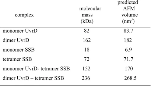
predicted molecular AFM complex mass volume (kDa) (nm3) monomer UvrD 82 83.7 dimer UvrD 162 182 monomer SSB 18 6.9 tetramer SSB 72 71.7 monomer UvrD- tetramer SSB 152 170 dimer UvrD – tetramer SSB 236 268.5
Table 2.1 Predicted AFM volumes for UvrD and SSB
Predicted volumes were calculated by the equation V = (1.2 MW)-14.7, where V is the olume measured by AFM and MW is the molecular mass.
To investigate if UvrD and SSB interact with one another, UvrD and SSB were incubated together, deposited and imaged, these images were compared to those of UvrD or SSB deposited alone. If UvrD interacts with wtSSB, the volume of proteins observed will be larger due to the physical association of the two proteins. The image of UvrD and SSB shows larger volume of complexes compared to those of UvrD or SSB alone (compare Figure 2.1 A and D with 2.2A). Incubation of SSB with UvrD results in a shift of the protein sizes to larger volumes (Figure 2.2C). To compare depositions of UvrD and SSB together with those from deposition of either UvrD or SSB alone, the individual volume histograms for UvrD (Figure 2.1A) and SSB (Figure 2.1D) were summed and divided by 2, and the resultant volumes were plotted for reference in the histogram (Figure 2.2D). The volume distribution would be expected to be similar if there were no interaction between UvrD and SSB. The volume of UvrD and SSB together results in an increase in the population of protein with higher volumes (Figure 2.2D). There is a new peak in the histogram (Figure 2.2D) with a volume of ~180 nm3, which is consistent with a tetramer of SSB interacting with a monomer of UvrD. This volume would also to be consistent with a dimmer of UvrD but since there is no significant population of this volume in the absence of SSB, it is likely that this peak infact represent a tetramer of SSB and monomer of UvrD. There is also a significant increase in the population of higher volume species that would be consistent with a tetramer of SSB interacting with a dimer of UvrD (Table 2.1). Roughly 30% of the UvrD and SSB proteins appear to be in complexes with one another. This extent of oligomerizatin is consistent with a SSB-UvrD binding constant around 20 nM to 50 nM. These results show
at UvrD and SSB interact absence of nucleotides.
A
B
C
D
0 5 10 15 20
0 100 200 300 400 500
P er ce nt of pr ot e in s (% )
Molecular volum (nm3)
Figure 2.2
5 10 15 20
UvrD & SSB ref. UvrD & SSB
P er ce n t of p ro tei ns ( % ) 0
0 25 50 75 100 125 150 175 200 225 250 275 300 325 350 375 400 425 450 475
Molecular volume (nm3)
UvrD + SSB
UvrD1-SSB
Figure 2.2 Histogram of volume
(A) AFM image of UvrD and wtSSB. 16 nM UvrD and 15 nM SSB were deposited under a same condition. (B) Surface plot of (A). All concentration of SSB is in tetramer. (C) Histogram of volume for UvrD (16 nM) and wtSSB (15 nM) in the absence of nucleotide. UvrD and wtSSB (n=795) were analyzed. The peaks around 180 and 265 nm3 represent the binding of UvrD to wtSSB. The binding of UvrD-SSB are shown (D) Comparison of reference volume and UvrD-SSB volume. Reference histogram of volume for UvrD and wtSSB: summed (A) and (B) data points and divided by 2. The red bars represent the reference volume (A and B) and the blue bars represent (C). The bars are shifted by the interaction of UvrD with wtSSB.
Wild-ty
dies is a circular double-nicked
nitial rate is similar for all reactions, but the extent of unwinding is different (Figure 2.3B).
pe SSB stimulates UvrD helicase activity
Our observation that UvrD physically interacts with SSB suggests that SSB may play an important role in the function of helicase activity. To test this idea, the effects of E. coli SSB on UvrD helicase activity on double nicked-circular DNA were analyzed. Although nicked duplex DNA is the natural substrate for UvrD, the helicase activity of UvrD on nicked DNA has not studied in detail previously. Thus, we designed this nicked DNA substrate for UvrD helicase reaction. The DNA substrate employed in these stu
DNA with a 37-nt fragment labeled on the 5'-end with 32P.