Original Research Article
Study on usual presentation of unusual neck masses
in paediatric population
Kamalpreet Singh*, Amrindarjeet Kour
INTRODUCTION
A neck mass is defined as an abnormal lesion (congenital or acquired) that is visible, palpable, or seen on an imaging study. Neck masses are common in adults, but often the underlying etiology is not easily identifiable. While infections cause most of the neck masses in children, most persistent neck masses in adults are neoplasms. Malignant neoplasms far exceed any other etiology of adult neck mass.1-3 Neck masses are a common complaint in children worldwide and constitute major indication for surgical consultation in many pediatric surgical centres.4,5 It is a cause for anxiety as chances of malignancy are there.6 Etiology is divided into three groups– inflammatory or infectious, abnormal embryonic development and neoplastic.7 Fortunately
benign masses are more frequently encountered which are reaction to the upper airway infection.8-10 A proper history, followed by detailed clinical examination, and the use of diagnostic imaging modalities such as ultrasonography, computerized tomography and other investigations such as fine needle aspiration cytology, Histopathological examination of the excised tissue can be utilized to arrive at a diagnosis. Surgical excision is the optimal choice of treatment in neck lesions, for aesthetic reasons and for the prevention of recurrent infections in addition to the potential danger of malignancy.11 The most common congenital pediatric neck lesions are thyroglossal duct remnant, followed by branchial cleft anomalies.12,13 The rational of doing this study is related to the demographic characteristics of patients presented with neck masses differed with the
ABSTRACT
Background: A neck mass is defined as an abnormal lesion (congenital or acquired) that is visible, palpable, or seen on an imaging study. Neck masses are common in adults, but often the underlying etiology is not easily identifiable.
Methods: Total of 36 patients (24 males and 12 females) who fulfill the above criteria were collected over 1-year and enrolled in this study. The clinical history was obtained from the parents or the proxy of the patients. Each patient was physically examined and a proper laboratory and/or radiological investigations were carried on to achieve the definite diagnosis.
Results: According to the aetiology, the inflammatory category was the main group accounting for 16 cases (44.4%), followed by the congenital category 9 (25%), neoplastic 8 (22.2%), and then the non-inflammatory non neoplastic 3 (8.3%). Thyroglossal duct cyst was the most common congenital mass observed in 4 (11.1%) cases, followed by branchial cleft cyst, cystic hugroma, hemangioma and finally dermoid.
Conclusions: The differential diagnosis of the pediatric neck mass includes a wide array of congenital, inflammatory, benign and malignant lesions.
Keywords: Neck mass surgery, Congenital, Inflammatory, Neo-plastic
Department of ENT, 166 Military Hospital, Jammu, Jammu and Kashmir, India
Received: 29 March 2019
Revised: 14 April 2019
Accepted: 15 April 2019
*Correspondence:
Dr. Kamalpreet Singh, E-mail: kprsingh5@gmail.com
Copyright: © the author(s), publisher and licensee Medip Academy. This is an open-access article distributed under the terms of the Creative Commons Attribution Non-Commercial License, which permits unrestricted non-commercial use, distribution, and reproduction in any medium, provided the original work is properly cited.
location of the population. Therefore, the aim of this study was to assess the usual presentation of unusual neck masses in paediatric population.
METHODS
This present study was conducted in the Department of ENT of 166 Military Hospital, India, during the period from 2016 to 2019. Only patients in the pediatric age group, ranging from 0 years to 15 years were included in the study. The patients attended the ENT OPD presented with neck mass seeking for management. The eligible patients were below 15 years of both genders. The criteria of inclusions included neck mass of whatever origin or site that caused discomfort to the patient or his/her parents and ended with surgical intervention either to treat or reach definite cause. The criteria of exclusions included thyroid gland enlargement or goiter, oral diseases and acute infections complicated with lymphadenopathy associated acute infections that responded to medical therapy, e.g., tonsillitis, otitis media, etc. A total of 36 patients (24 males and 12 females) who fulfill the above criteria were collected over 1-year and enrolled in this study. The clinical history was obtained from the parents or the proxy of the patients. Each patient was physically examined and a proper laboratory and/or radiological investigations were carried on to achieve the definite diagnosis. All the cases were subjected to ultrasonographic evaluation of neck masses, followed by fine needle aspiration cytology (FNAC). In cases where the diagnosis based on ultrasonography and cytology was inconclusive, excision biopsy was done and histopathological examination of the specimen was carried out using haematoxylin and Eosin staining. Routine blood examinations, including haemoglobin estimation, total leucocyte count, differential leucocyte count with peripheral blood smear study was carried out in all the cases. In cases of thyroid enlargement, estimation of thyroid hormone level in blood was carried out. In cases where pus was aspirated from neck masses, gram staining and Ziehl-Neelsen staining of the aspirate was done along with culture and sensitivity. The neck masses are categorizing into the following categories:
congenial, inflammatory, non-neoplastic
non-inflammatory conditions, and neoplastic. All surgical cases were prepared and planned for appropriate surgical management according to the diagnosis. Patients subjected to the surgical interventions were followed-up
for 6-18 months. Medical cases including
lymphoproliferative disorders, reactive lymphadenopathy or metastatic lymphadenopathy, were referred to the medical consultation clinic for proper management. The results are expressed as number, percent and whenever possible as a range and mean.
RESULTS
Over a period of one and half years, Out of 36 children 24 (66.7%) were male and 12 were female (33.3%) shown in Figure 1. Sex ratio was 2:1. According to the
aetiology, the inflammatory category was the main group accounting for 16 cases (44.4%), followed by the congenital category 9 (25%), neoplastic 8 (22.2%), and then the noninflammatory non neoplastic 3 (8.3%) (Table 1). Thyroglossal duct cyst was the most common congenital mass observed in 4 (11.1%) cases, followed by branchial cleft cyst, cystic hugroma, hemangioma and finally dermoid.
Figure 1: No. of cases.
Table 1: Etiology of neck masses in the study.
Etiology Number of cases
N (%) Congenital
Thyroglossal duct cyst 4 (11.1)
Branchial cleft anomalies 2 (5.5)
Cystic hugroma 1 (2.8)
Dermoid cyst 1 (2.8)
hemangioma 1 (2.8)
Inflammatory
Abscess (suppurative
lymphadenitis) 5 (13.9)
Reactive nonsuppurative
lymphadenitis 10 (27.8)
Tuberculous adenitis 1 (2.8)
Non-inflammatory benign
Fibromatosis colli (sternomastoid
tumour) 2 (5.5)
Epidermal inclusion cyst 1 (2.8)
Neoplastic Benign
Lipoma 1 (2.8)
Malignant
Lymphoma 2 (5.5)
Burkitts 1 (2.8)
Teratoma 1 (2.8)
Metastatic 1 (2.8)
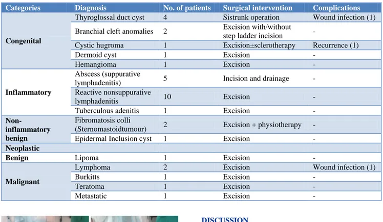
Table 2 shows the main cause of neck mass in patients under 15 years old was reactive (non-suppurative) lymphadenitis that accounts for 10 out of 36 patients (27.8%). Surgical interventions were done as a part of
67% 33%
No. of cases (%)
Male
excision-diagnostic or as a part of management. During the follow-up, recurrence of cystic hygroma is observed in one out of three patients. Wound infections observed in
two patients; one followed excised lymph node for biopsy and the other presented with wound infection after thyroglossal cyst excision.
Table 2: Surgical interventions to the patients presented with neck mass.
Categories Diagnosis No. of patients Surgical intervention Complications
Congenital
Thyroglossal duct cyst 4 Sistrunk operation Wound infection (1)
Branchial cleft anomalies 2 Excision with/without
step ladder incision -
Cystic hugroma 1 Excision±sclerotherapy Recurrence (1)
Dermoid cyst 1 Excision -
Hemangioma 1 Excision -
Inflammatory
Abscess (suppurative
lymphadenitis) 5 Incision and drainage -
Reactive nonsuppurative
lymphadenitis 10 Excision -
Tuberculous adenitis 1 Excision -
Non-inflammatory benign
Fibromatosis colli
(Sternomastoidtumour) 2 Excision + physiotherapy -
Epidermal Inclusion cyst 1 Excision -
Neoplastic
Benign Lipoma 1 Excision -
Malignant
Lymphoma 2 Excision Wound infection (1)
Burkitts 1 Excision -
Teratoma 1 Excision -
Metastatic 1 Excision -
Figure 2 (A and B): Intraoperative picturess of excision of cyst (excision without spillage is key).
Figure 3 (A and B): Excised specimen of epidermoid cyst.
DISCUSSION
Swellings in the head and neck region are very common in children and may be due to a variety of causes. The results of this study show that the most common aetiological cause of pediatrics neck masses is inflammatory. Al-Mayoof et al found inflammatory category was the main group accounting 57.8%, reactive non-suppurative lymphadenitis 40.6%, then suppurative lymphadenitis 15.6%, followed by the congenital category 25%, thyroglossal duct cyst 9.3%, then branchial cleft anomalies 7.8%, neoplastic 12.5%, and then the non-inflammatory non neoplastic 4.7%.14 Ragesh et al studied neck masses in children in India they found inflammatory category was the main group accounting 54%, tuberculous lymphadenitis 28%, reactive lymph node hyperplasia 20%, and chronic non-specific lymphadenitis 6%, followed by the congenital category 30% and neoplastic 16%.15 Lucumay et al studied pediatric neck masses in Northwestern Tanzania they found inflammatory category was the main group accounting 43.9%.16 Most common lesions are reactive lymph node hyperplasia 28.3%, followed by the congenital category 38.5% cystic hygroma most common 18.2% then thyroglossal cyst 14.9%, neoplastic 14.9%, and traumatic 2.1%.
In our study the most common masses are inflammatory 44.4% reactive nonsuppurative lymphadenitis10 (27.8%), abscess (suppurative lymphadenitis) 5 (13.9%) and
A B
Tuberculous adenitis1 (2.8%) and congenital 25%, thyroglossal duct cyst most common congenital mass 11.1%, then branchial cleft cyst 5.5%, cystic hureoma 2.8, hemangioma 2.8%, and dermoid 2.8% and malignant tumor 25.8%. The most common malignant tumor lymphoma 5.5%. Ragesh et al in their study noticed that 64% cases were males and the rest 36% were females.15 Lucumay et al found 71.6% were males and 28.4% were females.16 In our study male was 66.6% while females are 33.3%. Recently Meier and Grimmer categorized the pediatrics neck masses into three categories: Developmental, inflammatory/reactive, or neoplastic and mentioned that the most common causes of inflammatory /reactive category are reactive lymphadenopathy, infectious lymphadenitis (viral, staphylococcal, and mycobacterial infections; cat-scratch disease), or Kawasaki disease.17 The percent of neoplastic category of pediatric neck masses that reported in this study is 16.6% that is similar to that reported by Goins and Beasley which accounted to 11-15%.18 Moreover, the percent of the congenital category that reported in this study (25%) is in agreement with other studies all over the world 22% and 30%.19,20 In this study the majority of cases are male, with a male:female ratio 2:1 which is the same result observed by Osifo and Ugiagbe study included 35 children with neck masses, but different from other studies in which the male:female ratio was 1:1.2 and 1:1.19,21,22 In this study, the surgical intervention included specific carried upon thyroglossal duct cyst, branchial cleft and cystic hygroma, and non-selective that included excision, and/or drainage for the conditions that is listed in Table 2. The approach and results of surgical intervention have similarity and discrepancy to those reported by others.23-26 The most marked limitation of this study is its retrospective design. Decisions regarding haematological analysis, choice of antibiotherapy and duration, preferred imaging modality, and surgical biopsy were made by more than one physician. The preoperative clinical evaluation of the patients was based on the collaborative approach of ENT specialists, pediatricians, and family medicine specialists.
CONCLUSION
In conclusion, the differential diagnosis of the pediatric neck mass includes a wide array of congenital, inflammatory, benign and malignant lesions. The initial evaluation is the history and physical which should be used to place the mass into one of these categories if a definitive diagnosis is not possible. Pediatrics neck masses are distributed in categories that are similar in pattern and distribution in the world except the infectious/inflammatory category that shows variation in distribution in respect to the socioeconomic status. The surgical intervention and procedures are related to the facility as well as to the experience. Diagnosis is made based on a detailed anamnesis and the findings of the physical examination. Most masses are easily identified due to a typical anamnesis and clinical presentation of the patient.
Funding: No funding sources Conflict of interest: None declared
Ethical approval: The study was approved by the Institutional Ethics Committee
REFERENCES
1. Olsen KD. Evaluation of masses in the neck. Prim Care. 1990;17:415-35.
2. Beenken SW, Maddox WA, Urist MM. Workup of a
patient with a mass in the neck. Adv Surg. 1995;28:371-83.
3. Gray SW, Skandalakis JE, Androulakis JA. Non-thyroid tumors of the neck. Contemp Surg. 1985;26:13-24.
4. Turkington JR, Paterson A, Sweeney LE, Thornbury GD. Neck masses in children. Br J Radiol. 2005;78:75-85.
5. Tracy TF, Muratore CS. Management of common head and neck masses. Semin Peiatr Surg. 2007;16:3-13.
6. Osifo OD, Ugiagbe EE. Neck masses in children: Etiopathology in a tertiary centre. Niger J Clin Pract. 2011;14:232-6.
7. Gosche JR, Vick L. Acute, subacute and chronic cervical lymphadenitis in children. Semin Pediatr Surg. 2006;15(2):99-106.
8. Bull P, Cavinatto JN. Head and Neck Tumors in children. In: Sih T, Chinski A, Eavey R, Godinho R. VII Manual of Pediatric Otolaryngology, IAPO. 7th ed. Sao Paulo: Life and Consciuosness. 2008: 106-112.
9. Prathima S, Suresh TN, Harendra KML, Krishnappa J. Fine Needle Aspiration Cytology in Pediatric Age Group with Special Reference to Pediatric Tumors: A Retrospective Study Evaluating Efficacy and its Diagnostic Role. Ann Med Health Sci Res. 2014;4(1):44-7.
10. Thorell EA, Chesney PJ. Cervical Lymphadenitis and Neck Infections. In: Kliegman RM, Jenson HB, Behrman RE, Stanton BF, Nelson WE, eds. Nelson Treaty of Pediatrics. 18th ed. Rio de Janerio: Elsevier; 2009: 143-155.
11. Erikci V, Hosgör M. Management of congenital neck lesions in children. J Plast Reconstr Aesthet Surg. 2014;67:217–22.
12. Al-Khateeb TH, Al Zoubi F. Congenital neck masses: A descriptive retrospective study of 252 cases. J Oral Maxillofac Surg. 2007;65:2242–7. 13. Foley DS, Fallat ME. Thyroglossal duct and other
congenital midline cervical anomalies. Semin Pediatr Surg. 2006;15:70–5.
14. Al-Mayoof, Ali F. Neck masses in paediatric population: An experience with children attended the Central Teaching Hospital of Pediatrics in Baghdad 2008-2009. Afr J Paediatr Surg. 2015;12:136.
15. Ragesh KP, Chana RS, Varshney PK, Naim M.
clinicopathological study. Indian J Otolaryngol Head Neck Surg. 2002;54(4):268-71.
16. Lucumay EM, Gilyoma JM, Rambau PF, Chalya PL. Paediatric neck masses at a University teaching hospital in northwestern Tanzania: a prospective analysis of 148 cases. BMC Res Notes. 2014;7(1):772.
17. Meier JD, Grimmer JF. Evaluation and management of neck masses in children. Am Fam Physician. 2014;89:353–8.
18. Goins MR, Beasley MS. Pediatric neck masses. Oral Maxillofac Surg Clin North Am. 2012;24:457–68. 19. Ayugi JW, Ogeng’o JA, Macharia IM. Pattern of
congenital neck masses in a Kenyan paediatric population. Int J Pediatr Otorhinolaryngol. 2010;74:64–6.
20. Ragesh KP, Chana RS, Varshney PK, Naim M.
Head and neck masses in children: A
clinicopathological study. Indian J Otolaryngol Head Neck Surg. 2002;54:268–71.
21. Osifo OD, Ugiagbe EE. Neck masses in children: Etiopathology in a Tertiary Center. Niger J Clin Pract. 2011;14:232–6.
22. Al-Khateeb TH, Al Zoubi F. Congenital neck masses: A descriptive retrospective study of 252 cases. J Oral Maxillofac Surg. 2007;65:2242–7. 23. Narayana Moorthy S, Arcot R. Thyroglossal duct
cyst-more than just an embryological remnant. Indian J Surg. 2011;73:28–31.
24. Chan KC, Chao WC, Wu CM. Surgical
management of first branchial cleft anomaly presenting as infected retroauricular mass using a
microscopic dissection technique. Am J
Otolaryngol. 2012;33:20–5.
25. Charabi B, Bretlau P, Bille M, Holmelund M. Cystic hygroma of the head and neck – A long-term follow-up of 44 cases. Acta Otolaryngol Suppl. 2000;543:248–50.
26. Mawn LA. Infantile hemangioma: Treatment with surgery or steroids. Am Orthopt J. 2013;63:6–13.