0095-1137/93/040911-06$02.00/0
Copyright © 1993, AmericanSocietyforMicrobiology
Identification of Group-Common Linear
Epitopes in
Structural and Nonstructural Proteins of Enteroviruses
by Using Synthetic Peptides
JERONIMO
CELLO,`*
AGNETA SAMUELSON,2PERSTALHANDSKE,3
BOSVENNERHOLM,1
STIG
JEANSSON,l
ANDMARIANNE FORSGREN2Departmentof ClinicalVirology, University ofGoteborg, GuldhedsgatanJOB, 5-41346
Goteborg,
DepartmentofVirology, CentralMicrobiological Laboratory ofStockholm County Council, S-107 26Stockholm,2andDepartmentofFood Hygiene, Swedish Universityfor
AgriculturalSciences, S-75007Uppsala,3 Sweden
Received 8 September1992/Accepted 21 January 1993
Synthetic peptideswereemployedinenzyme-linked immunosorbent assaystoidentifygroup-commonlinear epitopesinthestructuralandnonstructuralproteins of enteroviruses. Nine linearepitopeswererecognized by usingserafrompatients with
heterotypic
immunoglobulin G antibodyresponses toenterovirusinfections. The most-reactive peptides were derived from conserved regions ofthe amino-terminal part ofVP1, whereas peptidesrepresenting sequences from other conservedregions of VP1,aswell asVP2, VP3, and VP4, and froma nonstructural region showed no or poor
reactivity.
Thesefindings may be useful in the development of serological testsforthediagnosis of infections causedbyabroad range ofenteroviruses.Enteroviruses include 69 serotypes: polioviruses,
cox-sackieviruses, echoviruses, and enterovirus types 68 to 72 (35). Infections due to enteroviruses range in
severity
from the asymptomatictoasepticmeningitis, encephalitis,
paral-ysis, pneumonia, and myocarditis(24).
The enterovirusesaresmall(24to30nm), nonenveloped,
single-stranded
RNA viruses, made up of 60copies of fourproteins, VP1toVP4(35).
Knowledge of theprimarystructureandgenetic organiza-tion ofenteroviruseshasincreased
recently
(31, 35, 40),
and it has been shown that conserved sequencescanbefound in structural and nonstructuralproteins
among enteroviruses. Considerableantigenic homology
between individual sero-types has been shown byserological
cross-reactivity in assays such as theenzyme-linked
immunosorbent assay (ELISA) (36, 42), the radioimmunoassay (28, 41),comple-ment fixation (15), the hemadsorption technique (13), gel double diffusion
(6,
37,38),
and theimmunoblotting
tech-nique (4, 25, 32,33).
Enterovirus group-commondetermi-nants areexposedindefective, heated, ordisruptedvirions (7, 8, 20, 22, 23,29). Ithas been shownbythe immunoblot-ting technique thatcross-reactive immunoglobulin G (IgG) antibodies reactedonlywithepitopesofcapsidprotein VP1, which is notpresentonthe surface of intact virusparticles (25, 33). Accordingto some authors (4, 25), cross-reactive enterovirus IgM antibodies reacted exclusively with VP1, although reactions with VP2 and VP3 have subsequently been demonstrated elsewhere
(33).
Information about the amino acid sequences and theexact
location of group-common
epitopes
is limited(34).
Since heterotypicantibody
responsesareobserved inpatients
with enterovirus infections, there is considerable interest in ob-tainingabroadlyreactiveantigen
thatcandetectantibodies againstawide range of enterovirus serotypes. Identification of the group-common epitopes eliciting cross-reactiveanti-*Correspondingauthor.
911
bodies has potentialvalue for serodiagnosis of enterovirus infections.
Synthetic peptides have been used to study antigen-antibody interactions(11) andtomapimmunogenicdomains
orlinear epitopes onvirus proteins (14, 18, 26). They have also been found tobespecificandsensitive forthe detection ofantibodies when used as antigens in different diagnostic
tests (2, 18, 39).
In the present report, we describe the use of synthetic peptides for the identification of group-common linear epitopes in the structural and nonstructural proteins of enteroviruses. Two panels of serum samples, one from patients showing arise in the level of heterologous entero-viral IgG antibodies and another frompatientswith hetero-typic enteroviral IgM responses, were used to characterize thereactivity of different peptides by ELISA.
MATERIALSANDMETHODS
Patients and sera. To evaluate the IgG reactivities of the syntheticpeptides,wetestedacute-andconvalescent-phase
sera from 11 patients with aseptic meningitis due to the following enteroviruses: echovirus 6 (1 patient [n = 1]), echovirus24 (n = 1), echovirus 30 (n = 7), coxsackievirus B4 (n = 1), and coxsackievirus B5 (n = 1) (confirmed by isolation fromcerebrospinal fluidorfeces). Acute-phase sera
were obtained within 7 days, and convalescent-phase sera
wereobtained 11to38 days after the onset of illness. All of these paired serum samples showed demonstrable rises in thetiters ofheterologous IgG byan enteroviral IgG ELISA based onheated virions asantigen (described below).
Anotherserumpanel from 12patients with culture-proven enteroviral infections was used for evaluation of the IgM reactivities of the synthetic peptides. The patients were infected with echovirus 4 (n = 2), echovirus 9 (n = 2), echovirus 11 (n = 1), echovirus 30 (n = 1), coxsackievirus
A9(n= 1), coxsackievirus B3(n = 1),coxsackievirus B4(n
= 2), or coxsackievirus B5 (n = 2). By an enteroviral IgM-capture ELISA based on heated, peroxidase-labeled
on January 8, 2021 by guest
http://jcm.asm.org/
Downloaded from
on January 8, 2021 by guest
http://jcm.asm.org/
Downloaded from
on January 8, 2021 by guest
http://jcm.asm.org/
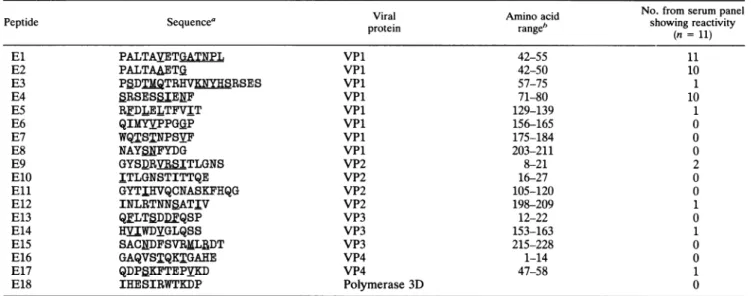
TABLE 1. Sequences and localizations of enteroviruspeptides andreactivitywith 11convalescent-phaseserumsamplesfrom patients infectedwithenteroviruses of differentserotypes
Viral Amino acid No. from serum panel
Peptide Sequencea protein range" showingreactivity
(n = 11)
El PALTAVETGATNPL VPl 42-55 11
E2 PALTAAETG VPl 42-50 10
E3 PBDTMQTR V RSES VPl 57-75 1
E4 SRSESSIENF VPl 71-80 10
E5 RFDLELTFVIT VPl 129-139 1
E6 QIMYVPPGGP VPl 156-165 0
E7 WQTSTNPSVF VPl 175-184 0
E8 NAYSNFYDG VPl 203-211 0
E9 GYS_RVfRSITLGNS VP2 8-21 2
E10 ITLGNSTITTQE VP2 16-27 0
Eli GYTIHVQCNASKFHQG VP2 105-120 0
E12 INLRTNNSATIV VP2 198-209 1
E13 QFLTSDDFQSP VP3 12-22 0
E14 HVIWDYGLQSS VP3 153-163 1
E15 SACNDFSVRMLRDT VP3 215-228 0
E16 GAQVSTQKTGAHE VP4 1-14 0
E17 QDPSKFTEPVKD VP4 47-58 1
E18 IHESIRWTKDP Polymerase 3D 0
aThe underlined amino acids indicate variation amongdifferententerovirustypesincludedinthisstudy (seeMaterials andMethods).
bAminoacidnumbering accordingtoalignmentwithpoliovirustype 1Mahoney (GenBank release67[accession number,J02281]).
virions asantigen(described below),allserashowed hetero-typic IgM responses.
Enteroviral ELISA forheterologous-antibody detection. (i)
Enteroviral cross-reactive IgG ELISA. The indirect ELISA
test used for the characterization of the heterologous IgG responseofour serum panelhas been described elsewhere (36). Briefly, three different serotypes of enteroviruses, echoviruses 9 and 30 and coxsackievirusB5,wereprepared by differential centrifugation, heated at 56°C, and used as
group-reactive antigens. The plates were coated with anti-gens of the three different serotypes. After theplates were
washed three times, patientserum diluted 1/500was added
to duplicate wells. Following incubation overnight at room
temperature, boundIgGwasdetected byrabbit anti-human IgGconjugatedwith alkalinephosphatase(incubationfor 90 min at 37°C). Substrate was added and incubated at room
temperaturefor 45min, and
theA405
wasread.Aheterotypic rise in titer ofIgG antibody was defined as an absorbance difference between acute- and convalescent-phase sera of >0.200againstaserotype(s)
other than theoneisolated. Of the 11patientstested, 1 showedarise in titer ofantibodytoantigenofoneserotype,fivepatientsshowedarise in titer of
twoof theantigens,andfivepatientsshowedarise intiter of antibodytoall threeantigens.
(ii) Enteroviral cross-reactive IgM ELISA. The ,i-capture ELISAusedtocharacterizetheIgMactivities ofourserum panel hasbeen described elsewhere (36). Briefly, two
sero-types ofenteroviruses, echovirus 6 and coxsackievirus A9,
wereprepared bydifferentialcentrifugation, heatedat56°C, and then conjugated with horseradish peroxidase.
Polysty-rene microtiterplates (Maxisorp; NuncAS, Roskilde, Den-mark)werecoated with rabbit anti-human IgM(Dakopatts, Copenhagen,
Denmark).
Aftertheplateswerewashed, 100 ,u ofpatient serum, diluted 1/200, was added. After being incubated for1 h at37°C,theplateswerewashed, 100 ,ul of peroxidase-conjugated antigen was added, and the plateswere further incubated for 2 h at room temperature. After theplateswerewashed and after the substrate step,theA490
wasread.
Cutoffserumrepresenting2.5times the absorbance value of that ofthenegative control was included in each test. A positive reaction was defined as an optical density (OD) value abovethe cutoff value. An IgM response was consid-ered heterotypic when a positive reaction against a sero-type(s) other than theoneisolatedwas detected.
Peptidesynthesis. The peptide sequences from the struc-tural and nonstrucstruc-tural proteins were selected from regions that are conserved among enteroviruses according to the sequencedata for different enteroviruses (GenBank release 67),coxsackievirusA9(1),and coxsackievirus A21(19). The sequenceofonepeptide(El)waschosen from theliterature (34). Thepeptide sequences are given in Table 1.
Thesolid-phasepeptide synthesiswas performedwith an Applied Biosystems 430A peptide synthesizer (Applied Bio-systems, Foster City, Calif.). An amino-terminal cysteine
was added to all peptides to facilitate coupling to carrier protein. Thepeptides were synthesized by using the t-Boc synthesis protocol as suggested by the manufacturer. All solvents were obtained from Applied Biosystems. Side chain-protected amino acids used were from NovaBiochem (Laufelfingen, Switzerland) and Applied Biosystems. As
solid phase, the polymer p-methylbenzhydrylamine resin (PeptidesInternational, Louisville, Ky.)wasused. Follow-ing each amino acid coupling, a sample was taken and a quantitativeninhydrinassaywasperformed.
Aftercompletionofsynthesis,peptideswerecleaved from the resin by treatment with trifluoromethane sulfonic acid (Applied Biosystems), and amino acid side chains were deprotected by acidic hydrolysis with anisole and ethanedithiol (Merck, Darmstadt, Germany)asscavengers. Theamino acid sequence of eachpeptidewas confirmedby sequencing with Applied Biosystem's protein sequencer
473A.
Solid-phase peptideELISA forantibody detection. (i)
Pep-tide ELISA for detection ofIgG antibodies. Synthetic pep-tides dissolvedat aconcentration of 1 mg/mlin 10% acetic acid were covalently coupled to bovine serum albumin (BSA) fraction V (Boehringer, Mannheim, Germany) at a
on January 8, 2021 by guest
http://jcm.asm.org/
10:1 (peptide-BSA)molar ratio with N-succinimidyl 3-(2-pyridyldithio)propionate (SPDP; Pharmacia, Uppsala, Swe-den) as described bythe manufacturer. Polystyrene micro-titer plates (Maxisorp; Nunc AS) were used in the ELISAs. Forcoating of the plates, 4 p.g of peptide-BSA conjugate per ml ina volume of 100,ul was added to each well and allowed tobind at 4°C overnight. Remaining free-binding sites in the wells were blocked for 1 h at
37°C
with 1% BSA in phosphate-buffered saline (PBS).Toidentify the reactive peptides, convalescent-phase sera from patients showing a rise in the titer of heterologous IgG antibody were used. Sera diluted 1:50 in PBS containing 1% BSA and 0.05% Tween 20 (100,ul per well) were tested against each peptide. After incubation for 2 h at
37°C
in a humidifier, the plates were washed four times in PBS con-taining 0.05% Tween 20. Subsequently, 100,ul of horseradish peroxidase-conjugated goat-anti-humanIgG (Jackson Labo-ratory, Bar Harbor, Maine), diluted 1/30,000, was added and allowed to react for 90 min at37°Cin a humidifier. Following sixwashes, 100pl
ofortho-phenyl-diamine was added. After 5 min at room temperature, the substrate reaction was stopped by the addition of 50p,l
of 2 MH2SO4and the plates were read at 490nm. All sera were run in duplicate, and the mean OD values were used for further calculations. The background level was the mean OD value obtained with the 11 convalescent-phase serum samples tested against an unrelated synthetic peptide (a peptide from human T-cell leukemia-lymphotropicvirus type II [HTLV-II]). A reaction wasconsidered positive when the OD value was equal to or exceeded three standard deviations of the mean background level established. The mean OD values of the unrelated peptide tested against convalescent-phase sera in 10 peptide ELISAs were calculated to be 0.114 to 0.140, and the standard deviations were found to range from 0.032 to 0.047. (ii) Peptide ELISA for detection of IgM. For the indirect IgM ELISA, the serum samples from patients with hetero-typic enteroviral IgM responses were assayed against the different synthetic peptides. The sera were treated with Protein A-Sepharose (Pharmacia) (41). Subsequently, the treated sera were tested at dilutions of 1/50 and 1/200. Goat anti-human IgM conjugated to peroxidase (Jackson) was used as detector antibody. The net absorbance of each sample was the absorbance to the indicated peptide minus the absorbance to BSA. A cutoff serum with a net absor-bance value 2.5 times higher than the net absorabsor-bance value of thenegative control was included in each test. A positive reaction was defined as anOD value above the cutoff value. Inall other respects, the assay was similar to the peptide IgG ELISAdescribed above.Adsorption test. Since El and E2 are similar except for an additional sequence on the carboxy terminus ofEl(Table 1), we wanted to know whether El and E2 contain the same epitope. Three patient serum samples that reacted against both peptides were diluted from 1/50 to 1/6,400. For adsorp-tion, 100 p.l of each dilution was added to an ELISA plate coated with El and another plate coated with E2, at a concentration of 4
p.g
ofpeptide-BSA conjugate per ml, and theplates wereincubated for 30 min at room temperature for adsorption of specific antibodies. The adsorbed sera were transferred for testing of IgG activity against El and E2 as described above in the solid-phase peptide ELISA. To control nonspecific adsorption, the sera were adsorbed un-derconditions identical to those described above with a plate coated with an unrelated peptide (HTLV-II); after that, the adsorbed sera were transferred for testing of IgG activity against El and E2. As a second control, the titers of IgGS
0
0 0
0)
1.0 1.5
oo0.0
,t
__,>_n
III
11
v11
!
1 2 3 4 5 6 7 8 9 10 1 1
Patients' sera
FIG. 1. Serum reactivity profiles obtained with convalescent-phase sera from11patients with heterotypic IgG antibody responses to enterovirus infections as measured by ELISA at an OD of 490 nm against the El, E2, and E4 peptides. Absorbance values obtained with sera at a dilution of
1/50
and with coated peptide-BSA conju-gate at a concentration of 4,ug/ml
are presented.against measles virus were measured by ELISA before and after adsorption of the sera with the El and E2 peptides, respectively.
Nucleotide sequence accession numbers. The GenBank re-lease 67 accession numbers for the different enterovirus sequences included in this study are as follows: poliovirus type 1, J02281; poliovirus type 2, M12197; poliovirus type 3, K01392; coxsackievirus Bi, M16560; coxsackievirus B3 M16572; and coxsackievirus B4, D00149.
RESULTS
Eighteen peptides representing different conserved re-gions of the structural and nonstructural proteins of entero-viruses were synthesized. The reactivities of synthetic pep-tides were determined by an ELISA with peptide-BSA adsorbed to solid phase. Sera from patients with culture-proven enterovirus infections and heterotypic enteroviral antibody responses were used for evaluation of the peptides. In the IgG ELISA, all serum samples reacted with the
El
peptide, whereas both E2 and E4 were recognized by 10 of 11 serum samples (Table 1). The
OD
values obtained with these three immunoreactive peptides were between 0.4 and 1.13. The absorbance profiles are shown in Fig. 1. The mean absorbance value for the serum panel was 0.889 forEl,0.765 for E2, and 0.723 for E4. One peptide (E9) reacted with two serum samples, whereas five peptides (E3,E5,
E14, E12, and E17) were recognized by only one serum sample (Table 1). However, the absorbance values obtained against these peptides were low, ranging betweenOD
values of 0.300 and 0.600. No reactivity could be detected with the remaining peptides.The most-reactive serum sample was from one of the patients infected with echovirus 30; it exhibited reactivity to six peptides(El,E2, E3, E4, E9, and E12). A serum sample
on January 8, 2021 by guest
http://jcm.asm.org/
1.2
1.0
0.8
0.6
0.4
0.2
0.0
_/ VE2
/ *E4
V E13
OHTLV II
0.i 1 10ii01 1111 H- l
0.1 1 10 100
PEPTIDE-BSA CONJUGATE (big/ml)
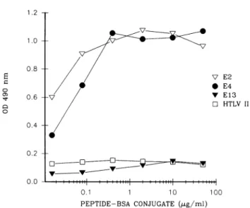
FIG. 2. IgG reactivity of a convalescent-phase serum from a
patient with heterotypic IgG antibody response to enterovirus infection againstthreeenteroviruspeptides (E2, E4, andE13)and
oneunrelatedpeptide (HTLV-II) analyzed byELISA. Absorbance
values obtained with theserumatadilutionof 1/50 and with coated peptide-BSA conjugateat fivefolddilutions from 0.016to50 pLg/ml
arepresented.
from another patient infected with echovirus 30 reacted
againstfive peptides (El, E2, E4,E5, and E17).The serum
sample from one patient infected with coxsackievirus B4
reacted only with the El peptide. The rest of the serum
samples recognized El, E2, and E4; moreover, sera from three patients (two infected with echovirus 30 and one infectedwithechovirus 6) recognized one morepeptide.
The influence of the concentration of the peptide-BSA conjugates (4 ,ug/ml) used in the assays was evaluated. A concentration range between0.016 and 50 ,ugof each
con-jugate permlwasused. Apeptide from HTLV-IIwasused to evaluate nonspecific binding. Three convalescent-phase
serum samples frompatients that showed a rise in titer of antibody to all three antigens by our enteroviral cross-reactiveIgGELISAwereused to test thereactivityofallour
peptides. Figure 2 shows OD values as a function of the peptide-BSA conjugate concentrations forone serum
sam-ple, three synthetic enterovirus peptides, and the control peptide.Atconjugateconcentrations in therangeof 0.4 to 10 ,ug/ml, the absorbance values reached a plateau. Similar findings were obtained with other sera and peptides. As shown in Fig. 2, an increase in the concentration of the peptide-BSA conjugate neither influenced the activity of alreadyreactivepeptides, suchasE2andE4,norenhanced the reactivityofthe nonreactive peptide, such asE13. The differences inreactivitiesamongthepeptidesareattributable not toquantitativebutto qualitativedifferences.
When acute- and convalescent-phase sera weretested in
parallel againstthehighlyreactivepeptides El, E2, andE4, significantincreasesinIgG activitywere seenformostof the patients (8of 11 patients;datanot shown).
Forcomparisonandevaluation ofthe epitopesin El and E2, patient serareactive with bothpeptideswere adsorbed withonepeptideand testedbyELISAagainstbothpeptides.
After adsorption with either peptide, there was a fourfold decrease in the titer of antibody against both antigens,
whereas no change was observed when the sera were absorbedwith the HTLV-IIpeptide (datanotshown).
More-over, IgG titers of antibody against measles virus were the
samebefore and afteradsorption with either El or E2. These results indicate thatEl and E2 contain the same epitope.
Thepeptides were also tested by indirect IgM assays with thesame formatasthat used for IgG. However, the exper-imental system suffered frombackground problems, which
weredifficultto eliminate, although several different block-ing procedureswere tried. No specific activity with the 12 serumsamples known to be highly reactive by the ,t-capture enteroviral IgM test wasdiscernible.
DISCUSSION
Inthis report,arangeofimmunoreactivities was observed whensynthetic peptides representing conserved regions of enterovirus proteinswere testedby ELISA with serafrom patients withaheterotypic enteroviral IgG response. Two of
our selectedpeptides fromVP1, as well as the El peptide chosen from the literature(34), showed consistent reactivity withmostof theserainourpanel. It seems likely that these peptidesrepresentepitopeslocatedonVP1 which have been shownto contain cross-reactive determinants by the immu-noblotting technique (25,32, 33). In contrast, the number of
serareactingwith the otherpeptides representing regionsof VP2, VP3, and VP4capsid proteinswasverylow, and the absorbance valuesobtainedwerelower than those obtained with the most-immunoreactive peptides of VP1. Our results
areconsistent withthe data frompreviousstudiesperformed by Western blot showing that the cross-reactivity of IgG antibodies is directed almost exclusively to VP1 antigens (25, 33). Other investigators using peptide-scanning tech-niques to identify antigenic regions of poliovirus type 3 (Sabin) have found that a large number ofpeptides repre-senting regionsinVP1, VP2,andVP3capsidproteins bound significantamountsofIgG antibodies.However, alsoin this study, regionswith the highest activity werefound in VP1 (34). AlthoughVP1seems tohaveapredominantly antigenic role, antigensfromVP2, VP3, and VP4 are also capable of eliciting an IgGresponse. This wasshown inour study by the reactivity of some sera to E9, E12, E14, and E17
synthetic peptides.
The extent ofcross-reactivity observed inpatientseraand the number ofepitopesinvolved may beproportional
tothe number ofenterovirusinfections experi-enced by the individual(21).
It can also be expected that because of the genetic variability of the immune system, antibodies inseraderived from different individuals maynotbe directedtothesameepitopeand maydiffer inqualityand
quantity.
Our resultsarein agreementwith those of Roivainenetal. who detected the presence of thehighesttiters of antibodyto
theepitope consistingof residues 40to53 of VP1 that has the
sameamino acid sequenceasthe El peptide.The patternof reactivityof E2wassimilartothat ofEl.Sinceits sequence, PALTAAETG, is very similarto thefirst stretch of that of El, PALTAVETG, it may be inferred that the heterotypic antibodiesaredirectedtothis conservedlinearepitope.The adsorptionresults support thisassumption. Thus,thefinding that serum from one patient (serum sample no. 7; Fig. 1) reactedonlywiththe Elpeptideandnotwith the E2peptide mightbeexplained byalower sensitivityofthe E2peptide fordetectingantibodies.
Besides,theamino acid sequence ofElnearthecarboxy terminus, GATNPL(Table 1), is the less-conserved partof this peptide, which speaks against the possibilitythat this part of the peptide is directly involved in the induction of heterologousantibodies.
S
0
0)
0
Vo
on January 8, 2021 by guest
http://jcm.asm.org/
The peptide E4, which was recognized by most sera, is similar to peptides resembling the amino-terminal part of VP1 of poliovirus type 1 previously reported to prime or induce neutralizing antibody response in experimental ani-mals (3, 5). Also, a moderate reactivity in the region of poliovirus type 3 (Sabin) coinciding with the E4 sequence wasdetected by using the peptide-scanning technique (34). It is known that after attachment of poliovirus type 1 to the cell, the amino terminus of VP1, which is
internal
in the native virion (16), is exposed and may be available for immune recognition (9). From our results, it seems that a similar conformational change occurs with other enterovi-ruses during the infection of humans, since a significant increase of IgG activity against three peptides corresponding to conserved parts of the amino terminus of VPl was detected in our panel of sera.To demonstrate that antibodies directed to our reactive peptides were specific to enterovirus structures, affinity chromatography was used to obtain antipeptide antibodies from human gamma globulin. By indirect ELISAs, these purified antipeptide antibodies were individually tested against the following antigens: poliovirus types 1, 2, and 3; coxsackievirus types B1, B3, B5, A9, and A16; echovirus types 6, 9, 15, 17, 19, and 21; cytomegalovirus; herpes simplex virus; varicella virus; and measles virus. Reactivity against all enterovirus antigens was detected, whereas no reactivity to the other viral antigens was observed (data not shown).
Sinceantibodies directed to the El, E2, and E4 peptides were part of the human response to a variety of infecting enteroviral serotypes, these epitopes or very similar se-quences seem to be part of the structures of many
enterovi-ruses that have not yet been sequenced.
The E3 peptide, located between El and E4, was recog-nized by only one serum. By the scanning technique, pep-tides similar to the E3 sequence were found to be reactive, showing several peaks. The scanning technique, in which a peptide length of 14 amino acids was used, also demon-strated activity for regions of VP1, VP2, and VP3 covered by E5, E7, E8, E9, E10, Ell, E12, and E15 (34). These peptides, however, were poorly reactive or nonreactive by ourELISA. This disagreement can be explained in different ways. First, the formats of the tests were different. Second, although the peptides have the same critical amino acids, shorter or longer peptides may adopt a disadvantageous conformation for binding of specific antibodies. Third, the detection of a low level of cross-reactivity is augmented when thepeptide-scanning method is used (43).
On the other hand, the sequences of the peptides selected in this study varied in some residues compared with the differententerovirus sequences reported. It has been shown that achange in a single amino acid can dramatically alter the reactivity of a peptide (12). Therefore, we cannot rule out the possibility of having chosen some poorly reactive or nonre-active peptides. A set of analogous peptides, in which the variable residue is replaced by the alternative amino acid(s), could besynthesized in order to find other possible reactive peptides.
The lack ofreactivity of some of our peptides cannot be due to a low concentration of peptide-carrier conjugate on the solid phase since, as shown in Fig. 2, an increase in the conjugate concentration above 4,g/ml did not improve the sensitivity of the ELISA. A small number of peptide mole-cules on thecarrier protein may also cause low sensitivity. However, a ratio of 5 to 20 mol of peptide per mol of carrier
protein is considered suitable for the detection of antibodies byELISA (30).
Although antigenicity prediction methods are popular for predicting the locations of viral epitopes (17, 27), the selec-tion of linear epitopes in our study was done only by choosing conserved regions of enteroviruses. There is evi-dence that none of the prediction methods achieves a high level of correct prediction (10,44). Besides, it was found that none of three predictive algorithms predicted better than randomly at a peptide length of less than 15 residues (10).
The rise in titer of antibody against the El, E2, and E4 peptides observed with sera from patients with enteroviral infections and the heterotypic rise of IgG antibody, plus the fact that affinity-purified human antibodies against these peptides recognizedifferent serotypes of enteroviruses, con-firm that these epitopes are part of conserved structures which elicit a heterotypic IgG response. However, this approach was not successful for the measurement of
IgM
activity. Other formats for the assay may be attempted, e.g.,a
,u-capture
assay with biotinylated peptide.In conclusion, our attempt to characterize broadly reac-tive peptides may,if extended, have serodiagnostic implica-tions, since there is a demand for a test that could detect antibodies against a wide range of enterovirus infections.
ACKNOWLEDGMENTS
We thank Bo Johansson for expert assistance with computer searching and Nancy Nenonen for correcting the English and for many helpful suggestions. We also thank Eva Skoog for her excel-lent technical assistance.
This work was supported by the Medical Faculty of the Univer-sity ofGoteborg, the Karolinska Institute, and Syntello AB.
REFERENCES
1. Chang, K. A., P.Auvinen, T. Hyypia, and G. Stanway. 1989.
The nucleotide sequence of coxsackievirus A9; implications for receptor binding and enterovirus classification. J. Gen. Virol.
70:3269-3280.
2. Chiodi, F., J. A. von Gegerfeldt, E. M.
Fenyo,
H. Gaines, M. vonSydow, G. Biberfeld, E. Parks, and E. Norrby. 1987. Site-directed ELISA with synthetic peptides representing the HIV transmembrane glycoprotein. J. Med. Virol. 23:1-9.
3. Chow, M., R. Yabrov, J. Bittle, J. Hogle, and D. Baltimore. 1985. Synthetic peptides from four separate regions of the
poliovirus type 1 capsid protein VP1 induce neutralizing anti-bodies. Proc. Natl. Acad. Sci. USA 82:910-914.
4. Dorries, R., and V. Ter Meulen. 1983. Specificity of IgM
antibodies in acute human coxsackievirus B infections analyzed by indirect solid phase enzyme immunoassay and immunoblot technique. J. Gen. Virol. 64:159-167.
5. Emini, E. A., B. A. Jameson, and E.Wimmer. 1983. Priming for and induction of anti-poliovirus neutralizing antibodies by syn-thetic peptides. Nature (London) 304:699-703.
6. Forsgren, M. 1968. Studies of echovirus antigens in immuno-diffusion. 3. Antibody response against echovirus 6 antigens in human infections with homologous and heterologous entero-viruses. Acta Pathol. Microbiol. Scand.74:611-623.
7. Forsgren, M. 1972. Relationship between poliovirus and echo-virus antigens. I. Immunization experiments in guinea pigs. Arch. Gesamte Virusforsch.39:108-120.
8. Forsgren, M. 1972. Relationship between poliovirus and echo-virus antigens. II. Antibodypatternsin sera from patients with poliovirus infections. Arch. Gesamte Virusforsch.39:108-120.
9. Fricks, C. E., and J. M. Hogle. 1990. Cell-induced conforma-tional change in poliovirus: externalizationof the amino termi-nus of VP1 is responsible for liposome binding. J. Virol. 64:1934-1945.
10. Getzoff, E. D., J. A. Tainier, R. A. Lerner, and H. M. Geysen.
1988. The chemistry and mechanism of antibody binding to
on January 8, 2021 by guest
http://jcm.asm.org/
protein antigens. Adv. Immunol. 43:1-98.
11. Geysen, H. M. 1985. Antigen-antibodyinteractions atthe
mo-lecularlevel:adventuresinpeptide synthesis.Immunol. Today 6:364-369.
12. Geysen, H. M., T. J. Mason, and S. J. Roda. 1991. Peptides as
specific recognition devices, p. 111-119. In A. Vaheri, R. C. Tilton,and A.Balows(ed.), Rapid methods and automation in
microbiology and immunology. Springer-Verlag, Berlin. 13. Glimaker, M., A. Ehrnst, L. Magnius, P. Berglund, M.
Fors-gren, T. Vikerfors, and P. Olcen. 1990. Early diagnosis of enteroviralmeningitis by asolid-phasereverseimmunosorbent
testandvirus isolation.Scand. J. Infect. Dis. 22:519-526.
14. Gnann,J. W.,Jr., J. A.Nelson, and M. B. A.Oldstone. 1987. Fine mapping ofanimmunodominant domain inthe
transmem-brane glycoproteinof humanimmunodeficiency virus.J.Virol. 61:2639-2641.
15. Halonen, P., L. Rosen, and R. J. Huebner. 1959.Homologous and heterologous complement fixing antibody in persons in-fected with echo, coxsackie and poliomyelitis viruses. Proc.
Soc.Exp. Biol.Med. 101:236-241.
16. Hogle, J. M., M. Chow, and D. J. Filman. 1985. Three-dimen-sional structure of poliovirus at 2.9 A resolution. Science 229:1358-1365.
17. Hopp, T.1986.Protein surface analysis:methodsforidentifying antigenicdeterminantsand otherinteractionsites. J.Immunol.
Methods88:1-18.
18. Horal, P., W. W. Hall, B. Svennerholm, J.Lycke, S. Jeansson, L.Rymo, M. H.Kaplan, and A.Vahlne. 1991. Identification of
typespecific linear epitopes in the glycoproteinsgp46andgp2l of humanT-cell leukemia virusestype I(HTLV-1)and type II
(HTLV-II) using synthetic peptides. Proc. Natl. Acad. Sci. USA 88:5754-5758.
19. Hughes,P.J., C.North, P. D.Minor,andG.Stanway. 1989. The nucleotide sequence of coxsackievirus 21. J. Gen. Virol. 70:
2943-2952.
20. Hummeler, K., and V. V. Hamparian. 1958. Studies on the complement fixing antigens of poliomyelitis. I. Demonstration oftypeandgroup specific antigens tonative andheated viral preparations. J. Immunol. 81:499-505.
21. Kapsenberg, J. 1988. Picornaviridae: the enteroviruses (polio-viruses, coxsackieviruses, echoviruses), p. 692-722. In E. H.
Lennette, P. Halonen, and F. A. Murphy (ed.), Laboratory diagnosis of infectiousdiseases, principlesandpractice, vol.II.
Viral, rickettsial, and chlamydial diseases. Springer-Verlag,
NewYork.
22. Katze, M.G., and R. L.Crowell. 1980. Indirectenzyme-linked immunosorbentassay(ELISA)fordetection of coxsackievirus
groupBantibodies. J.Gen. Virol.48:225-229.
23. Katze, M. G., and R. L. Crowell. 1980.Immunological studiesof
the group Bcoxsackieviruses bythe sandwich enzyme-linked immunosorbent assay (ELISA) and immunoprecipitation. J. Gen. Virol. 50:357-367.
24. Melnick, J. 1990. Enteroviruses: polioviruses, coxsackie-viruses, echoviruses, andnewerenteroviruses, p. 549-605.In B. N.Fields(ed.), Virology,2nded.,vol. 1. RavenPress, New York.
25. Mertens,T.H.,U.Pika,andH.J. Eggers. 1983. Cross
antige-nicity among enteroviruses as revealed by immunoblot
tech-nique.Virology 129:431-442.
26. Middeldorp, J.M., and R. H. Meloen. 1988. Epitope-mapping ontheEpstein-Barr virus major capsid proteinusingsystematic synthesis ofoverlapping oligopeptides. J. Virol. Methods 21:
147-159.
27. Modrow, S.,B. H.Hahn, G. M.Shaw,R.C.Gallo,F.
Wong-Staal,and H. Wolf. 1987. Computer assisted analysis of enve-lope proteinsequencesof sevenhumanimmunodeficiencyvirus isolates: prediction of antigenic epitopes in conserved and
variable regions. J.Virol.61:570-578.
28. Morgan-Capner, P., and C. McSorley. 1983. Antibodycapture
radioimmunoassay (MACRIA)forcoxsackieB4 andB5 IgM.J.
Hyg. 64:159-167.
29. Muir, P., and J. E. Banatvala. 1990. Reactivity of
enterovirus-specific IgM with infective anddefective coxsackieBvirionsin
patientswithmonotypicandmultitypic IgMresponses.J. Virol. Methods29:209-224.
30. Muller, S., S. Plaue, M.Couppez,and M.H. V. van
Regenmor-tel. 1986. Comparisonofdifferentmethods forlocalizing
anti-genicregions in histoneH2A.Mol.Immunol.23:593-601.
31. Palmenberg, A. C. 1989. Sequencealignments ofpicornaviral capsid proteins,p.211-241. In B. L. Selmer andE. Ehrenfeld
(ed.),Molecular aspects ofpicornavirus infection anddetection.
American Society forMicrobiology, Washington,D.C. 32. Pozzetto, B., 0. G. Gaudin, F. R. Laucht, J. Hafid, and A. Ros.
1990. DetectionofimmunoglobulinG,M, andAantibodiesto
enterovirus structural proteins by immunoblot technique in
echovirus type 4-infected patients. J. Virol. Methods 29:143-156.
33. Reigel, F., F. Burkhardt, and U. Schilt. 1985. Cross-reactions of
immunoglobulin M and G antibodies withenterovirus-specific viral structuralproteins. J.Hyg. 95:469-481.
34. Roivainen, M., A. Narvanen, M. Korkolainen, M.-L. Huhtala, and T. Hovi.1991.Antigenicregions of poliovirustype3/Sabin capsidproteinsrecognized by humanserain thepeptide
scan-ningtechnique. Virology 180:99-107.
35. Rueckert, R. R. 1990. Picornaviridae and their replication, p. 507-548. In B. N. Fields(ed.), Virology,2nded.,vol. 1. Raven
Press,NewYork.
36. Samuelson,A., E.Skoog,andM.Forsgren. 1990.Aspectsonthe
serodiagnosis of enterovirusinfections byELISA. Serodiagn.
Immunother. Infect. Dis. 4:395-406.
37. Schmidt,N.J., and E. H. Lennette.1962. Geldouble diffusion studies with group B and groupA,type9 coxsackieviruses. I. Thetechniqueand reactionsobtainedwithhyperimmuneanimal seraandhumansera.J. Immunol. 89:85-95.
38. Schmidt,N.J.,andE. H. Lennette.1962. Gel double diffusion studies withgroupB and groupA,type9coxsackie viruses.II.
Serologic diagnosis of coxsackie virus infections by the gel double diffusiontechnique.J.Immunol. 89:96-105.
39. Smith,R.S., R. B. Nago, J.Rosen,A.Whalley,Y.-L.Hom,K.
Hoey,C. J.Kennedy, J.A.McCutchan,S. A.Spector,and D. D. Richman.1987.Antibodyto asynthetic oligopeptideinsubjects at risk for human immunodeficiencyvirus infection. J. Clin. Microbiol. 25:1498-1504.
40. Stanway,G. 1990.Structure, functionandevolution of
picorna-viruses. J. Gen. Virol.71:2483-2501.
41. Torfason, E. G., G. Frisk, and H. Diderholm.1984. Indirect and
reverse radioimmunoassays and their apparent specificitiesin
the detectionofantibodies toenteroviruses in humansera. J. Med. Virol. 13:13-31.
42. Torfason, E. G., R. Galindo, and H. L. Keyserling. 1988.
Comparison of five ELISA assays for IgG antibody against
coxsackievirusB1. J. Med. Virol. 25:53-60.
43. Trifilieff, E., M. C. Dubs,and M.H. V.vanRegenmortel. 1991.
Antigenic cross-reactivity potentialofsyntheticpeptides
immo-bilizedonpolyethylene rods.Mol.Immunol. 28:889-896. 44. van Regenmortel, M. H. V. 1989. Structural and functional
approaches to the study of protein antigenicity. Immunol.
Today 10:266-272.
on January 8, 2021 by guest
http://jcm.asm.org/
AUTHOR'S
CORRECTIONS
Genomic
Fingerprinting
of Borrelia
burgdorferi
Sensu Lato
by
Pulsed-Field Gel
Electrophoresis
J.BELFAIZA, D. POSTIC, E.BELLENGER, G.BARANTON,ANDI. SAINTGIRONS UnitedeBacteriologie MoleculaireetMedicaleInstitutPasteur, 25 Rue du docteur Roux,
75724ParisCedex15, France
Volume 31,no. 11, p. 2877, column 1,line 1 of
Acknowledgments:
Should read"...by
agrant from the G. Harold and Leila Y.Mathers Charitable Foundation to...."Identification
of
Group-Common
Linear
Epitopes
in Structural and
Nonstructural Proteins
of Enteroviruses
by
Using
Synthetic
Peptides
JERONIMOCELLO, AGNETASAMUELSON,PER
STALHANDSKE,
BOSVENNERHOLM, STIG JEANSSON,ANDMARIANNEFORSGREN
DepartmentofClinicalVirology, UniversityofGoteborg,Guldhedsgatan 10B, S-413 46
Goteborg,
DepartmentofVirology, CentralMicrobiological Laboratory of Stockholm County Council, S-107 26Stockholm, and Departmentof Food Hygiene, Swedish University forAgriculturalSciences, S-750 07 Uppsala, Sweden
Vol. 31, no. 4, p. 911-916: On the basis of recentexperiments, we report that new syntheses of peptide E4 with the amino acid sequenceSRSESSIENF were not reactive (A. Samuelson, M. Forsgren, B. Johansson, B. Wahren, and M.
Sallberg,
Clin. Diagn. Lab.Immunol.1:336-341, 1994).Careful analysis of the original E4 material used in the study showed it to be a mixture of peptidesE2 and E4. This explains the overlap of reactivities between peptides E2 and E4 presented in our study. The contamination occurredwhen the peptides wereaccidentally mixed after the amino acid sequencing of each peptides was done. The new syntheses ofpeptide E2 as well as those of the other peptides tested in our study showed the same reactivity reported in our paper, which validates the original data presented.