Selection of our books indexed in the Book Citation Index in Web of Science™ Core Collection (BKCI)
Interested in publishing with us?
Contact book.department@intechopen.com
Numbers displayed above are based on latest data collected.For more information visit www.intechopen.com Open access books available
Countries delivered to Contributors from top 500 universities International authors and editors
Our authors are among the
most cited scientists
Downloads
We are IntechOpen,
the world’s leading publisher of
Open Access books
Built by scientists, for scientists
12.2%
122,000
135M
TOP 1%
154
Intensive Care Management
of the Traumatic Brain Injury
Akarsu Ayazoglu Tülin
1and Özden Nihan
21
Chief Asistant Kartal Kosuyolu Highly
Specialized Education and Training Hospital
İ
stanbul
2
Göztepe Education and Training Hospital Istanbul
Turkey
1. Introduction
Traumatic brain injury has been major cause of mortality and morbidity worldwide, especially in children and young adults and it has been continuing a difficult problem in intensive care units.
Brain trauma can be caused by a direct impact or by acceleration alone. In addition to the damage caused at the moment of injury, brain trauma causes secondary injury, a variety of events that take place in that minutes and/or days following the injury
Secondary brain injury is attributable to a decrease in cerebral oxygen delivery as a result of hypertension, hypoxia, cerebral oedema, intracranial hypertension or abnormalities in cerebral blood flow. Although the severity of primary brain injury cannot be reduced, secondary brain injury can be minimised if appropriate therapies are implemented in time. The main aim in the traumatic brain injured patients must be to maintain a good result from primary injury caused by trauma and/or as a result of direct effect of trauma.
The second aim must be to prevent secondary brain injury caused by as results of the complications. The basic principle in the care and treatment of traumatic brain injury is to describe and begin the treatment these complications that worsen the primary injury and lead to secondary brain injury.
The main targets in these aims are:
1. Maintain the cerebral energy metabolism by maintaining needed systemic support, 2. Maintain cerebral perfusion pressure (CPP) in normal limits,
3. Maintain ICP in normal limits as possible.
The intensive care for traumatic brain injury should consist beside the control of ICP, respiratory system, central nervous system, and cirulatory system, it should also consist monitoring of metabolism especially glucose metabolism, temperature and electrolite balance in short intervals. With these invasive and noninvasive monitoring, all the precausions for the problems should be ready.
Beside heavy brain injury may result in a permanant neurologic sequale, it may also give a good results especially in young patients that aggresivelly lowered inreased ICP levels and optimized CPP and cerebral oxygenation by multidisiplinary approach with neurointensivist, neuroanesthesist and neurosurgeon.
In this chapter we will discuss the intensive care management of severe TBI with emphasis on the specific measures directed for prevention and/or treatment of secondary brain injury
2. Indication for admission to ICU
The role of an intensive care unit is to maintain a patient’s normal physiological homeostasis while actively treating the underlying cause of any physiological derangement. Discussion will be targeted towards a number of areas; respiratory system, cardiovascular system, alimentary system, nasocomial infection and infection surveillance, anticoagulation, patient comfort. Indication for admission to ICU include
Impaired level of consciousness,
Impaired airway protection
Progressive respiratory impairment or the need for mechanical ventilation
Seizures
Clinical or computed tomographic (CT) evidence of raised ICP caused by a space occupying lesion , cerebral edema or haemorrhagic conversion of a cerebral infarct.
General medical complications (for example, hyper/hypotension, fluid and electrolyte disturbances, aspiration pneumonia,sepsis, cardiac arrhytmias, pulmonary embolism)
Monitoring (for example level of conciousness, respiratory function, ICP continuous electroencephalography(EEG)
Specific treatments(for example , neurosurgical intervention, intravenous or arterial trombolysis )
Mechanical ventilation
Most patients admitted to neuro-intensive care require respiratory support because of hypoxaemia, ventilatory failure or due to treatment modalities requiring respiratory support. The support may range from oxygen therapy by face mask, through non-invasive techniques such as continuous positive airways pressure, to full ventilatory support with endotracheal intubation.
Oxygen is usually given by face mask, although nasal prongs or cannulas may be well tolerated.
If the patient remains hypoxaemic on high flow oxygen (15 l/min) continuous positive airways pressure (CPAP) may be used. The continuous positive airways pressure mask often becomes uncomfortable and gastric distension may occur. Patients must therefore be cooperative, able to protect their airway, and have the strength to breathe spontaneously and cough effectively.
In patients with acute brain lesions at risk for cerebral ischemia, maintenance of adequate cerebral perfusion pressure (CPP), artificial ventilation for prevention of hypercapnia and deep sedation are all major determinants for actual strategies of a cerebroprotective therapy (1).
The patient who has an altered level of consciousness (GCS <8 ) and loss of gag/cough reflex often has deficits in a number of airway protection mechanisms or exhaustion need ventilatory support.
The goals of mechanical ventilation of acute severely brain injured patients are to improve gas exchange, to minimize intrathoracic pressure, to reduce the work of breathing and to avoid complications. These patients are also in the risk of developing neurogenic pulmonary edema, aspiration of oropharyngeal contents, pneumonia, and atelectasis.
Criteria for starting mechanical ventilation are difficult to define and the decision is made clinically. It is decided according to respiratory status.
Neurologic indications
Altered level of consciousness (GCS <8 ) /airway protection.
Brainstem dysfunction.
Intracranial hypertension.
Anticipated neurologic deterioration. Respiratory indications
Respiratory rate >35 or <5 breaths/ minute
Exhaustion, with laboured pattern of breathing
Hypoxia - central cyanosis, SaO2 <90% on oxygen or PaO2 < 8kPa
Hypercarbia - PaCO2 > 8kPa
Tidal volume < 5ml/kg or Vital capacity <15ml/kg
Activity Score Eye Opening
None 1= Even to supra-orbital pressure
To pain pressure 2=Pain from sternum/limb/supra-orbital To speech 3=Non-specific response, not necessarily to
command
Spontaneous 4=Eyes open, not necessarily aware
Motor Response
None 1=To any pain; limbs remain flaccid
Extension 2=Shoulder adducted and shoulder and forearm internally rotated
Flexor response 3=Withdrawal response or assumption of hemiplegic posture
Withdrawal 4=Arm withdraws to pain, shoulder abducts Localizes pain 5=Arm attempts to remove supra-orbital/chest
pressure
Obeys commands 6=Follows simple commands
Verbal Response
None 1=No verbalization of any type Incomprehensible 2=Moans/groans, no speech
Inappropriate 3=Intelligible, no sustained sentences Confused 4=Converses but confused, disoriented Oriented 5=Converses and oriented
Intracranial physiology and mechanical ventilation
The goals of positive-pressure ventilation (PPV) in patients with multitrauma with head trauma are improving oxygenation and controlling arterial CO2 tension to minimise intracranial hypertension. PPV increases functional residual capacity (FRC) by improving alveolar recruitment, thus optimising oxygenation.
On the other hand, increased intrathoracic pressure (ITP) increases intracranial pressure (ICP) by these mechanisms:
Direct transmission of ITP to the intracranial cavity via the neck.
Increased ITP decreases venous return to the right atrium, and increases jugular venous pressure, thereby increasing cerebral blood volume (CBV) and ICP.
Decreased venous return decreases cardiac output and mean arterial pressure (MAP). This results in decreased cerebral perfusion pressure (CPP) leading to compensatory cerebral vasodilation, increased CBF and potentially increased ICP, if cerebral autoregulation is impaired.
Mechanical ventilatory strategies(2): conventional ventilation
Current practice guidelines for ventilatory management advocate protective lung strategies to prevent volutrauma, barotrauma, atelectrauma and biotrauma (3-5). The principles are to
use low tidal volumes (Vt) (5-6 ml/kg ideal body weight), maintenance of low mean airway pressures ≤ 30 cmH2O, judicious use of positive end-expiratory pressure (PEEP) with ∆ pressure ≤ 18 cmH2O, higher respiratory rates and permissive hypercapnia. This is in direct conflict with the previous “brain-directed” ventilatory strategies that used Vt of 10 ml/kg, high FiO2 and low PEEP or zero end-expiratory pressure. There is proven mortality benefit
with the use of low Vt, but permissive hypercapnia may precipitate intracranial hypertension(3,6,7). Animal studies indicate a higher incidence of severe pulmonary oedema
and haemorrhage after exposure to injurious ventilation in the presence of brain trauma. High Vt independently predicts ALI/ARDS and poor outcome in brain trauma patients(8).
Haemodynamic fluctuations induced by mechanical ventilation may be detrimental in the brain with impaired autoregulation. That's why with starting mechanical ventilation, intravascular expansion and vasopressor may be necessary.
The role of PEEP
Lung protection strategy permising hypercapnia induces the development of cranial hyperemia and hypertension. On the other hand, “aggressive” ventilation with high tidal volume may aggravate lung injury and provoke ventilator-associated lung damage(9).
In mechanical ventilation treatment, PEEP improves oxygenation by recruitment of atelectatic alveolar units, improving FRC and preventing atelectrauma. Also it may have detrimental neurologic effects in certain clinical circumstances(10). In a recent study in
patients with traumatic brain injury shows that increasing PEEP up to 15 cm H2O to optimize oxygenation has not been associated with reduced cerebral perfusion pressure or acute intracranial hypertension (11)
In normal pulmonary compliance, PEEP is associated with increased ITP, decreased right atrial volume, decreased MAP and thus compromised CPP. This situation is not similar to non-compliant lungs, where there is a comparatively low ITP transmission to
the cranium, therefore lesser effects on cerebral blood flow (CBF) and ICP. CPP may be indirectly affected by systemic effects of PEEP, but these effects still remain quantitatively modest. PEEP is therefore safe to apply as part of a ventilatory strategy to improve oxygenation.
Alveolar overdistension should be avoided and stable haemodynamic parameters should be maintained. Head position also needs attention. At least 30º head elevation promotes intracranial venous drainage via anterior neck veins, as well as the vertebral venous system - which is not majorly affected by ITP. Jugular veins collapse and act as resistors to some of the ITP transmitted. Tight endotracheal tube ties around the neck and extremes of neck rotation should be avoided
The Role of PaCO2 control
Arterial CO2 tension is a powerful modulator of cerebral vascular calibre, CBF and ICP (12-15.)
While the mechanisms are incompletely understood, CO2 relaxes pial arterioles by interactions between the endothelium, vascular smooth muscle, pericytes, adjacent neurons and glial cells. Studies supported that cerebral vessels are sensitive to changes in extracellular pH, rather than a direct response to CO2 or bicarbonate. In the limits of physiological PaCO2, 20-60 mmHg, the relationship between PaCO2 and CBF is linear. Therefore, increased PaCO2 results in vasodilation of cerebral vessels and this leads to increase CBF, increase CBV, decrease intracranial compliance and increase ICP. The reverse mechanism is also true for low CO2 tension. This has been the reason for inducing hyperventilation in the patients with intracranial hypertension, but there is a risk for cerebral vasoconstriction precipitating cerebral ischaemia because pericontusional areas are sensitive to hyperventilation-induced ischaemia. The Brain Trauma Foundation management guidelines do not recommend hyperventilation for initial management of raised ICP, unless ICP is unresponsive to first-line therapy or hyperventilation is for very brief periods of time. Maintaining normocarbia is recomended.
Role of brain monitoring during ventilatory support in brain injury
It is essential to monitor intracranial pressure, CPP, and brain oxygenation during ventilatory support in the patients with traumatic brain injury. Brain oxygenation monitoring technics are jugular venous saturation monitoring, near-infrared spectroscopy and microdialysis catheters. Availability and cost of these devices are limiting factors to their use. The studies on brain trauma patients shows that there is no proven mortality benefit in continuous ICP monitoring.
Non-conventional ventilatory strategies
There are some ventilatory strategies that may be used for proper patients. These are prone position, recruitment manouvres, high frequency oscillatory ventilation (HFOV) and newer technics like extracorporeal CO2 removal (ECCO2R), pumpless extracorporeal lung assist (pECLA) and nitric oxide.
Prone ventilation(15- 18) Benefits are:
Recruitment of atelectatic lung units.
Improved drainage of secretions.
Even distribution of mechanical ventilatory forces.
ICP and brain tissue oxygenation (PbtO2) monitoring are recomended. There are conflicting results on the effects of prone ventilation on ICP and CPP, but there are clear data on benefits for respiratory mechanics and oxygenation. Present studies shows that there is no mortality benefit to prone positioning.
Recruitment manoeuvres
In neurointensive patients with acute lung injury, achieving the goal of lung protection without threatening cerebral perfusion is very difficult. In patients with more refractory raised intracranial pressure, the optimal balance between brain and lung may not be well established. Multiple strategies are used to recruit atelectatic alveoli and improve oxygenation. Among them incremental levels of PEEP and high intermittent tidal volumes should require extend brain physiological monitoring.
High frequency oscillatory ventilation (HFOV) (18,19)
High frequency oscillatory ventilation (HFOV) forms high mean airway pressure with very small Vt of 1-5 ml/kg at a rapid rate. It's aim is to recruit alveoli, while preventing overdistension. Some studies have supported that HFOV is safe and effective in preventing ventilator-induced lung injury (VILI) and improving oxygenation in severe ARDS. There no sufficient studies supporting HFOV for the improvement of intracranial compliance.
Extracorporeal CO2 removal (ECCO2R) (20)
Extracorporeal membrane oxygenators have been attempted in brain-injured patients to improve oxygenation. By using ECMO increased intracranial pressure may decrease in a normal limits and CPP is maintained. But anticoagulation requirement in that technic increases the risk of intracranial bleeding.
Pumpless extracorporeal lung assist (pECLA) (21)
pECLA has recently been utilised in small case series with promising results. Protective respiratory care can be maintained while CO2 removal is optimised. Patients treated by pECLA must be hemodynamically stable. So cardiovascular instability and shock are contraindications for pECLA. Anticoagulation is as for thrombo prophylaxis in immobilised patients. The risk of the device clotting is not entirely eliminated by impregnation with anticoagulant in the filter. Vascular injury, exsanguination and limb ischaemia are some of the recognised complications.
Nitric oxide
Nitric oxide improves oxygenation in ALI/ARDS with no survival benefit. There is potential to cause harm. There is no data for its use in ARDS with the patients with brain taruma. Weaning (22-30)
Without resolving underlying pathological condition, weaning must not be thought. With prolongation ventilatory support, the respiratory muscles become weaken and atrophy of this muscles is inevitable. As a consequence, the duration of weaning period is often related
to the duration and mode of ventilation. As possible as using assisted modes of ventilation and good nutritional support are essential to prevent atrophy of the respiratory muscles. Critical illness polyneuropathy is seen in patients recovering from prolonged critical illness. In this condition, there is both respiratory and peripheral muscle weakness, with reduced tendon reflexes and sensory abnormalities. There is evidence that long-term administration of some aminosteroid muscle relaxants (such as vecuronium) may cause persisting paralysis. No absolute treatment is used for it except supportive therapy.
The plan for disengagement of the patient from mechanical ventilation should be made at initiation of ventilation therapy. The recognition of when mechanical ventilatory support should be reduced and ultimately discontinued is so important. Appropirate time for disengagement from ventilation has the following advantages:
Decreased airway injury
Decreased risk of VILI.
Decreased risk of VAP.
Decreased sedation requirements.
Decreased delirium.
Shortened ICU length of stay.
Assessment for extubation criteria:
Respiratory criteria.
Haemodynamic criteria.
Neurologic criteria. This includes stable neurological status, ICP ≤ 20 mmHg, CPP ≥ 60 mmHg.
Premature weaning and extubation may cause respiratory muscle fatigue, gas exchange failure and loss of airway protection.
There is clear benefit to weaning according to protocol. There should be frequent assessment of ventilatory support requirement and re-evaluation of factors contributing to ventilator dependence before ventilation is discontinued.
Indications for weaning
Improving of underlying illness
Respiratory function:
Respiratory rate < 35 breaths/minute FiO2 < 0.5, SaO2 > 90%, PEEP <10 cmH2O
Tidal volume > 5ml/kg Vital capacity > 10 ml/kg Minute volume < 10 l/min
Absence of infection or fever
Cardiovascular stability, optimal fluid balance and electrolyte replacement
Prior to trial of weaning, there should be no residual neuromuscular blockade and sedation should be stoped or decreased in appropirate level so that the patient must be awake, cooperative and in a semirecumbent position. Weaning is likely to fail if the patient is confused, agitated or unable to cough.
Modes of weaning
There are several different approaches for the weaning that are not superior to others.
Unsupported spontaneous breathing trials. The machine support is withdrawn and a T-Piece (or CPAP) circuit can be attached intermittently for increasing periods of time, thereby allowing the patient to gradually take over the work of breathing with shortening rest periods back on the ventilator.
Intermittent mandatory ventilation (IMV) weaning. The ventilator delivers a preset minimum minute volume which is gradually decreased as the patient takes over more of the respiratory workload. The decreasing ventilator breaths are synchronised to the patient's own inspiratory efforts (SIMV).
Pressure support weaning. In this mode, the patient initiates all breaths and these are 'boosted' by the ventilator. This weaning method involves gradually reducing the level of pressure support, thus making the patient responsible for an increasing amount of ventilation. Once the level of pressure support is low (5-10 cmH2O above PEEP), a trial
of T-Piece or CPAP weaning should be commenced. Failure to wean
During the weaning process, the patient should be observed for early indications of fatigue or failure to wean. These signs include distress, increasing respiratory rate, falling tidal volume and haemodynamic compromise, particularly tachycardia and hypertension. At this point it may be necessary to increase the level of respiratory support as, once exhausted, respiratory muscles may take many hours to recover.
It is sensible to start the weaning process in the morning to allow close monitoring of the patient throughout the day. In prolonged weaning, it is common practice to increase ventilatory support overnight to allow adequate rest for the patient.
Tracheostomy in the intensive care unit (31-33)
The commonest indication of tracheostomy in an ICU setting is to facilitate prolonged artificial ventilation and the subsequent weaning process. Tracheostomy allows a reduction in sedation and thus increased cooperation to the weaning process. It also allows effective tracheobronchial suction in patients who are unable to clear pulmonary secretions either due to excessive secretion production or due to weakness following critical illness. Tracheostomy can be performed as a formal surgical procedure in theatre or at the bedside in the intensive care unit using a percutaneous method. Tracheostomy placement leads to earlier liberation from mechanical ventilation, but without any mortality benefit or effect on pulmonary infection rates.
Other indications for tracheostomy are to bypass an upper airway obstruction, protect the lungs from soiling if the laryngopharygeal reflexes are depressed or as part of a surgical or anaesthetic technique eg larygectomy.
Advantages of tracheostomy is summerized as decreased risk of self-extubation; decreased sinusitis; decreased airway resistance, dead space and breathing work ;better tolerance; less sedative requirements; potentially-reduced duration of mechanical ventilation.
Risks of tracheostomy is summerized as surgical site infection, airway haemorrhage, pneumothorax, oesophageal perforation.
Sedation in the Neuro-ICU(34-67)
Sedation is the important factor in comfort of brain trauma patients. Insuffient sedation causes hypertension, tachycardia, hypoxia, hypercapnia and uncomfortable with ventilator. On the other hand excess sedation causes hypotansion, bradycardia, coma, respiratory depresion, ileus, renal insufficiency, veinous stasis and immunosupression.
For the patients in the critical care unit firstly nonpharmacological method should be experinced for sedation. The patients should be frequently oriented. Sleep-awake cycling, proper enveriomental temperature, control of the noise aroused from alarms must be arranged.
Calling the family members, the relexing exercises, musical therapy, masaj and sitting exercises are important in control of anxiety and ajitation of patients.
Safe and effective management of the pain and anxiety needs a delicate balance for analgesia and sedation protocols while managing delirium status.
The weaning of patients from mechanical ventilation is often hampered by the sedation that they receive. Additionally, coordinated daily interruption of sedative infusions with objective re-titration in critically ill patients has been shown to decrease the durations of mechanical ventilation and length of ICU stay.
Consequences of agitation include self-extubation, removal of IV catheters, dyssynchrony with mechanical ventilation, and, perhaps, a long-term risk of psychiatric problems, such as delirium and posttraumatic stress disorder can be prevented by a proper sedation. Prolonged and excessive sedation are problematic too, interfering with weaning from mechanical ventilation and leading to increased rates of nosocomial pneumonia, prolonged ICU stays, and difficulty identifying new problems, such as myocardial infarction or stroke.
Sedation Indications in the Neuro-ICU
Patient comfort
Decreases anxiety and agitation
Relieve fear
Risk of self-injury or injury of others
Withdrawal from alcohol or drugs
Risk of self-extubation or removal of invasive monitors
Suppreses stres response
Increases the tolerance of ventilatory support
Facilitates the cares like aspiration, invasive prosedures and dressing the wound
Control of pain
Facilitate mechanical ventilation
Reduce oxygen extraction/ utilization in ARDS and Sepsis
Brain protection (seizure control, decrease cerebral metabolism , control ICP)
Blunting adverse outcome
Amnesia during paralysis with muscle relaxants
During interventions (line insertion, tracheostomy)
To prevent movement (during imaging and transfering of the patient)
Facilitate sleep
Facilitate nursing management
Properties of an ideal agent for neurointensive care sedation:
Rapid onset and rapid recovery so that a neurologic evaluation can be conducted
Predictable clearance independent of end-organ function, avoiding the problem of drug accumulation
Easily titrated to achieve adequate levels of sedation
Reduces intracranial pressure by cerebral blood volume reduction or cerebral vasoconstriction
Reduces cerebral blood flow and cerebral metabolic rate of oxygen consumption, maintaining their coupling
Maintains cerebral autoregulation
Permits normal cerebral vascular reactivity to changes in arterial carbon dioxide tension
Minimal cardiovascular depressant effects
Easy control respiratory side-effects
Inexpensive
Adapted with permission.
Both sedative and analgesic
Lack of respiratory depression
No tolerance over time
Inactive or non harmful metabolites
No interactions with other ICU drug
Rapid onset and rapid recovery so that a neurologic evaluation can be conducted General expectational situations:
Equipment and personnel to intubate and mechanically ventilate must be readily available
Decreased level-of-consciousness or obtundation
Poor airway protection
Respiratory depression, hypercarbia, and increased intracranial pressure (ICP)
Impairment of neurological exam
Hemodynamic instability
Sedation should be performed according to protocols standarized with scales. For this reason Ramsay Sedation Scale (RSS), Riker Sedation-Agitation Scale (SAS) and Richmond Agitation-Sedation Score (RASS) are used for planing treatment. For many years, the Ramsay Sedation Scale was the most commonly used tool to monitor sedation in the ICU. However, it cannot distinguish different levels of agitation, making it less useful than other available scales. Currently, two of the most commonly used techniques are the Riker Sedation-Agitation Scale (SAS) and the Richmond Agitation-Sedation Score (RASS).
Score Term Descriptor
1. Unarousable – Minimal or no response to noxious stimuli, does not communicate or follow commands
2. Very Sedated – Arouses to physical stimuli but does not communicate or follow commands, may move spontaneously
3. Sedated – Difficult to arouse, awakens to verbal stimuli or gently shaking, but drifts off again, follow simple commands
4. Calm and Cooperative – Calm, awakens easily, follows commands
5. Agitated – Anxious or mildly agitated, attempting to sit up, calms down to verbal stimuli
6. Very Agitated – Does not calm despite frequent verbal reminding of limits, biting ET 7. Dangerous Agitation – Pulling ET, trying to remove catheters, climbing over bedrails, striking at staff, thrashing side to side
Guidelines for SAS Assessment
1. Agitated patients are scored by their most severe degree of agitation, as described. 2. If patient is awake or awakens easily to voice (“awaken” means responds with voice or head shaking to a question or follows commands), that is a SAS 4 (same as calm and appropriate might even be napping).
3. If more stimuli such as shaking is required but patient eventually does awaken, that is a SAS 3.
4. If patient arouses to stronger physical stimuli (may be noxious) but never awakens to the point of responding yes/no or following commands, that is a SAS 2.
5. Little or no response to noxious physical stimuli is a SAS
6. This helps separate sedated patients into those you can eventually awaken (SAS 3), those you can not awaken, but can arouse (SAS 2), and those you can not arouse (SAS 1). Table 2. Riker Sedation-Agitation Scale (SAS) (SAS Target Sedation = 3 to 4).
Score Description
+4Combative Overtly combative, violent, immediate danger to staff +3 Very Agitated Pulls or removes tube(s) or catheter(s),aggressive +2 Agitated Frequent non-purposeful movement, fights ventilator +1 Restless Anxious but movements not aggressive vigorous 0 Alert and Calm
-1 Drowsy Not fully alert, but has sustained awakening (>10 seconds) (eye-opening/eye contact) to voice
-2 Light Sedation Briefly awakens with eye contact to voice (<10 seconds) -3 Moderate Sedation Movement or eye opening to voice (but no eye contact) -4 Deep Sedation No response to voice, but movement or eye opening to physical stimulation
Procedure for RASS Assessment: The basis of the RASS assessment is to see what amount of stimulation is necessary to evoke a respons and evaluate sedation.
Observe patient.
a. Patient is alert, restless, or agitated. (Score 0 to +4)
If not alert, state patient’s name and say “open eyes and look (speaker).” b. Patient awakens with sustained eye opening and eye contact (Score –1)
c. Patient awakens with eye opening and eye contact, but not sustained (Score –2) d. Patient has any movement in response to voice but no eye contact (Score –3)
When no response to verbal stimulation, physically stimulatepatient by shaking shoulder and/or rubbing sternum.
e. Patient has any movement to physical stimulation (Score –4) f. Patient has no response to any stimulation (Score –5).
Table 3. Richmond Agitation Sedation Scale (RASS) ( RASS Target Sedation = 0 to -3).
Even if the sedative strategy in the NICU shares the same general aims as general intensive care, the characteristics of the patients in the NICU present other unique challenges and specific indications, including intracranial pressure control, cerebral oxygen consumption and seizure reduction . Analgesic and sedative agents are used both to prevent undesirable increases in intracranial pressure and to reduce cerebral metabolic requirements. Intracranial pressure control cerebral autoregulation may be impaired in the traumatic brain injury. Therefore, agitation and associated blood pressure elevations directly determine intracranial pressure surges. Moreover, severe agitation increases intrathoracic pressure, reducing jugular venous outflow and increases cerebral metabolism with concomitantly increased cerebral blood flow (CBF). These potentially deleterious phenomena can lead to increase in intracranial pressure. This can trigger an additional cerebral vasodilator cascade, as cerebral perfusion pressure (CPP) is reduced.
Sedatives decrease the cerebral metabolic rate of oxygen consumption (CMRO2), and because the coupling of CBF and CMRO2 is usually maintained with these agents, CBF is reduced by increasing cerebral vascular resistance. The reduction in CBF results in a reduction of cerebral blood volume and, consequently, a decrease in intracranial pressure. In order to maintain adequate oxygen availability and energy production at the cellular level, treatment is directed to increase oxygen delivery by optimizing systemic hemodynamics and reduce cerebral metabolic demand. Sedative drugs confer a protective effect by reducing oxygen demand and increasing oxygen delivery (through improvement of central perfusion pressure and by inhibiting deleterious pathologic intracellular processes). The pharmacologic reduction in CMRO2 depresses either the basal or the activation components of cerebral metabolism. The metabolic suppression is dose dependent until the electroencephalogram becomes isoelectric. Beyond this level, no further suppression of cerebral oxygen consumption or blood flow occurs because energy expenditure, associated with electrophysiologic activity, has been reduced to close to zero, and the minimal consumption for cellular homeostasis persists unchanged.
There is no appropirate ratio for seizure activity in patients with brain trauma, seizures are a frequent complication in the NICU. Sedation appears to be an attractive option in reducing seizures in the NICU. Benzodiazepines increase the seizure threshold and are useful anticonvulsants. There are conflicting data on propofol, and, consequently, its ability to
protect against seizures is less certain. Pharmacologic properties rapid onset and rapid recovery of hypnosis are the most important pharmacokinetic properties to consider when comparing different hypnotic alternatives.
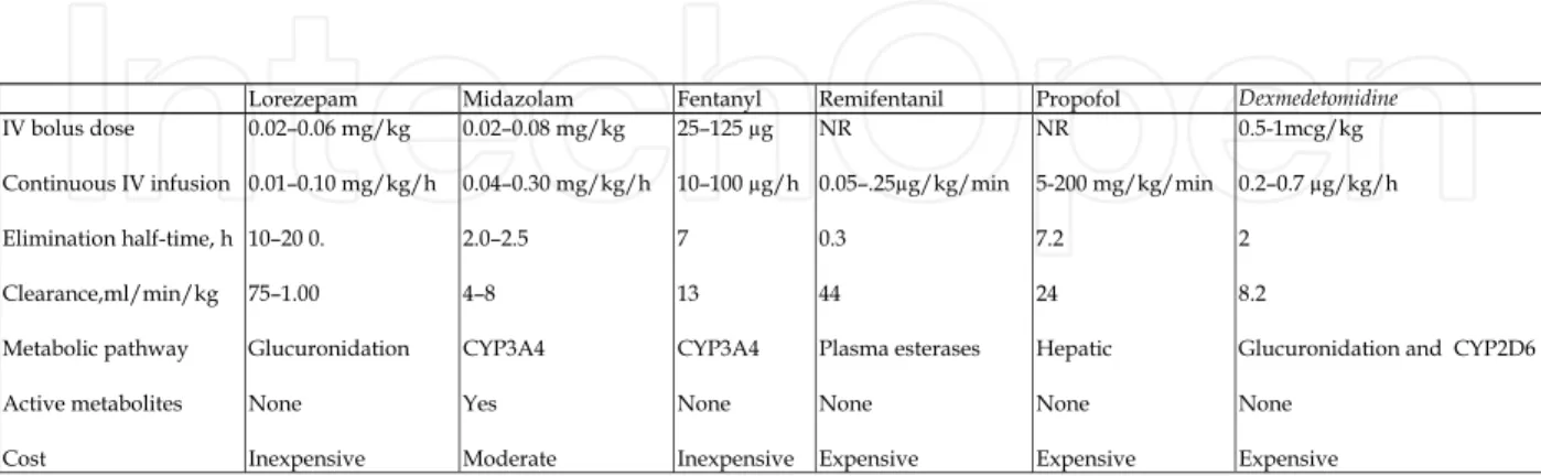
The drug used for sedation in intensive care summarized in table below.
NR: Not recommended
Table 4. Pharmacokinetic parameters, dosing, and cost of sedative and analgesic agents. For patients that are mechanically ventilated for three days or less, short acting agents should be used such as Propofol or Midazolam. For longer periods of mechanical ventilation, longer acting agents such as Lorazepam should be used. Patients that have been ventilated for long periods using the long acting agent Lorazepam may need to be switched to a shorter acting agent such as Propofol for optimal weaning purposes.
Rapid onset Fast recovery Easily titrated ICP reduction CBF reduction CMRO2 reduction MAP
Propofol +++ +++ +++
Midazolam +++
++ ++
Lorazepam + + +
Fentanyl +++
++ ++ /
Remifentanyl +++ +++ +++ /
↑, modest increase; ↑↑, pronounced increase; , no clear effect; , modest decrease; , pronounced decrease; +++, very favorable; ++, favorable; +, not favorable; CBF, cerebral blood flow; CMRO2, cerebral metabolic rate of oxygen consumption; ICP, intracranial pressure; MAP, mean arterial pressure.
Barbiturate, benzodiazepins, propofol and narcotics are all used as a sedative agent.
Propofol is very lipid soluble, has a large volume of distribution, and can be given for prolonged periods of time without significant changes in its pharmacokinetic profile. Because propofol has no active metabolites, the termination of its clinical effect is dependent solely on redistribution to peripheral fat tissue stores. When the infusion is discontinued, the fat tissue stores redistribute. Because of its pharmacokinetics and specific effects on cerebral hemodynamic variables, preserving autoregulation and vasoreactivity to carbon dioxide, propofol approximates the ideal sedative. Intravenous bolus administration produces a dose-dependent, coupled decrease in CBF and CMRO2 similar to that described after barbiturate administration. The effects on CBF are probably secondary to a reduction in CMRO2. A strong linear correlation between CBF and CMRO2 has been demonstrated. The major cardiovascular effect of propofol is a profound decrease in mean arterial pressure, resulting from a decrease in systemic vascular resistance, cardiac contractility, and preload. A bolus dose of 2 to 2.5 mg/kg propofol results in a 25 to 40% reduction in systolic blood pressure. This potent effect on mean arterial pressure may affect CPP by one of two mechanisms. If autoregulation is intact, a reduction in mean arterial pressure will produce reflex cerebral vasodilatation and a possible increase in intracranial pressure. Alternatively, if autoregulation is impaied hypotension may produce a critical decrease in CPP and CBF. The risk of hypotension is greatest in the presence of hypovolemia.
Evaluations of the effects of benzodiazepines on cerebral physiology have involved primarily diazepam and midazolam. Both cause dose-dependent decreases in CMRO2 and CBF, and by increasing cerebral vascular resistance, decreases in intracranial pressure have been observed. Usually CPP is not compromised. Moreover, a “ceiling effect” in CBF and CMRO2 reduction has been described, suggesting a saturation of the benzodiazepine receptors. It appears that benzodiazepines are safe to administer to patients with intracranial hypertension without respiratory depression and associated increases in arterial carbon dioxide tension.
The cerebral physiologic effects of opioids are controversial. Morphine-related increases in CBF, as described in early reports, were probably secondary to an increaseing arterial carbon dioxide tension resulting from respiratory depression. In general, opioids slightly reduce CMRO2, CBF, and intracranial pressure, as long as normocapnia is maintained by mechanical ventilation. Opioids can produce short-lasting, mild decreases in mean arterial pressure and then, subsequently, can produce decreases in CPP. Furthermore, remifentanyl may cause decreases in both cerebral metabolic rate and intracranial pressure, with minimal changes in CPP and cerebral blood flow. Opioids lead to dose-dependent, centrally mediated respiratory depression. The carbon dioxide response curve is shifted to the right, and the ventilatory response to hypoxia is obliterated. For this reason, in spontaneously ventilating NICU patients, if opioids are administered, strict end-tidal carbon dioxide monitoring or frequent blood gas analysis must be implemented to identify rapid onset of respiratory depression. When opioids and benzodiazepines are administered concomitantly, they may exhibit a synergistic effect on hemodynamics. The reasons for this synergy are not entirely clear.
Hypnotics can decrease mean arterial blood pressure by both cardiac depression and peripheral vasodilatation. The decrease in blood pressure can cause an increase in
intracranial pressure as a result of autoregulatory compensation (vasodilatory cascade) and consequently, can cause a reduction in CPP. The hemodynamic effects are usually dose dependent. Therefore, to avoid, or at least to limit, decreases in blood pressure, the patient should be euvolemic before the infusion is started, and slow boluses or continuous infusions are preferred.
Because no single drug can achieve all of the requirements for sedation and analgesia in the ICU, use of a combination of drugs, each titrated to specific end points, is a more effective strategy. This allows lower doses of individual drugs and reduces the problems of drug accumulation. In the acute phase (ie, first 48–72 hours or when intracranial hypertension is not controlled), a continuous infusion of a combination of propofol (1.5–6 mg/kg/h) and fentanyl (0.5–1.5 µg/kg/h) should be initiated.
In the subacute phase (ie, after 72 hours or when intracranial pressure is normalized), intermittent infusion of lorazepam (0.05 mg/kg every 2–6 h) should be initiated.
It is essential to contiue sedation until ventilatory support is required, and then sedation is stoped step by step to prevent withdrawal symptoms in 24 to 48 hours. No neuromuscular blocking drugs are routinely added, except in cases of severe, uncontrollable intracranial hypertension.
Neuromuscular blockers in the Neuro-ICU (68-71)
The routine use of neuromuscular blockers varies between centres. The routine use of muscle relaxants should be avoided, they can be useful to prevent peaks in ICP induced by the patient coughing or “straining” or in the face of patient-ventilator dysynchrony. However, muscle paralysis makes it clinically impossible to recognise and treat seizures. Prolonged administration of neuromuscular blockers by continuous infusion can also lead to significant long-term problems, such as critical illness polyneuropathy and myopathy.
Neuromuscular blockers indications
Facilitation of intubation
Facilitate mechanical ventilation
To eliminate spontaneous breathing and promote mechanical ventilation
During interventions (line insertion, tracheostomy)
To prevent movement (during imaging and to permit transfer of the patient)
Severe refractory intracranial hypertension
Severe pulmonary disease with inability to mechanically ventilated
Cause a pharmacologic restraint so patients do not harm themselves General Precautions
Patient must be intubated for mechanically ventilated
Transient increase in ICP with depolarizing blockade (succinylcholine)
Unexpected prolongation of neuromuscular blockade (e.g.,enzyme deficiencies, hepatic or renal dysfunction)
Agent Onset of action Duration of action ED90-95*(mg/kg)
Agent Onset of action Duration
of action ED90-95*(mg/kg) Short Acting
Mivacurium (Mivacron)
2.5 min 15 - 20 min 0.07
Rapacuronium (Raplon)
Mean: 90 seconds (35 - 219 sec)
Mean: 15 min
(6 - 30 min) 1.03
Rocuronium
(Zemeron) 1 - 3 min
31 min
(15 - 85 min) 0.3
Succinylcholine 30 - 60 seconds 5 - 8 min 0.3 Intermediate acting
Atracurium
(Tracrium) 2.5 - 5 min 20 - 45 min 0.2
Cisatracurium
(Nimbex) 2 - 3 min 30 - 40 min 0.05
Pancuronium
(Pavulon) 2 - 3 min 60 - 90 min 0.06
Vecuronium
(Norcuron) 2 - 3 min 25 - 40 min 0.05
Long Acting Doxacurium
(Nuromax)
6 min (2.5 - 13)
100 min
(39 - 232) 0.025
Pipecuronium
(Arduan) 2.5 - 5 min
75 min
(35 - 175) 0.07
Tubocurarine 3 - 5 min 70 - 90 min 0.05
ED90-95 = Dose required to produce 90-95% suppression of muscle response
Table 6. Neuromuscular Blocking Agents.
Atracurium: initially 0.4 -0.5 mg/kg IV bolus, Maintenance infusion rates of 5 to 9 mcg/kg/min are usually adequate (Range:2 to 15mcg/kg/min). Toxic metabolite (laudanosine) may accumulate in renal failure
Doxacurium: initially, 0.05 mg/kg -0.08 mg/kg IV bolus, Maintenance, 0.005 mg/kg and 0.01 mg/kg IV to provide neuromuscular blockage for an average of 30 min and 45 min, respectively.
Rocuronium: initially, 0.6-1.2 mg/kg IV bolus. Maintenance, 0.1-0.2 mg/kg IV repeated as needed. Maintenance (continuous IV infusion): 0.01-0.012 mg/kg/minute.
Pancuronium: initially 0.1 to 0.2 mg/kg (usually 0.1) bolus, followed by 1 to 1.7 mcg/kg/min or 0.06 to 0.1 mg/kg/hr
Vecuronium: Initially 0.08 to 0.1 mg/kg IV bolus. (Higher initial doses-up to 0.3 mg/kg-may be used for rapid onset. Continuous infusion: 1 mcg/kg/min infusion, usual range: 0.8 to 1.2 mcg/kg/min).
Succinylcholine: Initially 0.3–1.1 mg/kg, bolus Infusion is 2.5–4.3 mg/min (cause refractory intracranial hypertension Succinylcholine should be avoided due to increased ICP)
Nutrition
Nutrition could also have an impact on the posttraumatic stress response, which is associated with adverse outcomes from TBI. The posttraumatic stress response is charaterized by increased blood levels of glucose, lactate, catecholamines, and cortisol. Nutrition support should be initiated as soon as possible following the head injury. Adequate nutrition is to provide the body healing. Early enteral nutrition (EN) support has been shown to attenuate the catabolic response and improve immune function and is associated with improved neurologic outcome(73,74) .If the digestive tract is functional,
enteral nutrition, or nutrition given via a tube placed into the stomach, is the preferred route. Parenteral nutrition (PN) should be reserved for those patients with impaired gastrointestinal function or those who cannot meet their nutritional needs via EN alone. The goal of nutritional support in patients who are critically ill is to provide protein and caloric replacement while attenuating a negative nitrogen balance. Incidence of malnutrition in hospitalized patients ranges between 30% to 55%. Delaying in the initiation of nutritional support may result in muscle and gastrointestinal atrophy, inability of weaning from ventilatory support, heart failure, impaired immunity, increase in the incidence of sepsis, length of hospital stay, morbidity and mortality and all of these resulte increase in costs (72).
At the begining, all patients admitted to the critical care unit should be screened for risk or presence of malnutrition. Critically ill patients with neurologic impairment often require specialized nutrition support because of needing intubation, dysphagia, or altered mental status.
The provision of adequate nutritional support is an essential component of caring the critically ill patient. During starvation, homeostatic mechanisms are designed to burn fat rather than protein as an energy source until the fat stores are significantly depleted.
Especially in the begining of the infection, a catabolic state causing significant protein loss
progresses. Plasma and urine levels of catecholamines and cortisol are elevated. Hyperglycaemia is frequently seen and causes ketone production and lactic acid production leading acidosis in brain cells. The studies show that the severity and duration of hyperglycaemia following head injury correlate with longer term outcome.
Enteral nutrition should be preferred in the critically ill patients(75,76). This can be achieved
by either nasal tube feeding or via a percutaneous gastrostomy (PEG) if prolonged feeding is envisaged. Standard enteral feeding regiments aim to provide 1500–2500 kcal in 24 hours with 70 g protein in a volume of 1.5–2 L.
A patient may require more calories for healing for the TBI. Patients with a GCS of 8 to 12 will require approximately 30 to 35 calories per kilogram of body weight per day. Patients with a GCS of 6 to 7 require 40 to 50 calories per kilogram of body weight per day.
The main goal of nutrition should be preserve muscle mass, and provide adequate fluids and strict electrolyte and glucose monitoring should be recommended.
It is prudent to consider postpyloric feeding in patient with neurological catastrophies, because gastric atony increases the risk of aspiration. Enteral feeding should be preferably done by continuous infusion with a volumetric pump.
In the average patient in the intensive care unit who has no contraindications to EN or PN, the choice of route for nutritional support may be influenced by several factors. Because EN
and PN are associated with risks and benefits(77,78)
Advantages associated with enteral rather than parenteral nutrition include:
Maintenance of mucosal integrity and prevention of villous atrophy
Reduced infection rate
Absence of requirement for central venous line
Better maintenance of fluid balance
Reduced cost
Contraindications to enteral feeding are few, particularly in the patient with isolated intracranial pathology, but include abdominal sepsis, obstruction, acut malabsorption and inflammatory syndromes and enteric fistulae. Only a short segment of small intestine (30
cm) is required for adequate absorption since hypertrophy will occur in response to lumenal nutrients. Neither bowel sounds nor flatus are required for successful enteral feeding.
Enteral feeding should be started if gastric aspirates are less than 400 ml/day and there are no obvious contraindications. Commence with standard enteral feed at 25 ml/h, increasing the rate every 12 hours until 100 ml/h is achieved. Aspirate residual volume and rest for 1 hour in every 6 hours of feeding, and rest continuously for 8 hours overnight.
Complications of enteral feeding (79).:
Large residual gastric volumes.
Regurgitation and aspiration.
Diarrhoea.
Ulceration of nares.
Contamination of feed (rare).
Gastric atony and delayed emptying may be seen during enteral feeding. For treating this unwanted result pro-kinetic agents can be used. Also nasojejunal tube bypassing pylorus should be inserted. The passage of nasal feeding tubes should be avoided in patients with facial injuries and basal skull fracture. Instate oragastric replacement should be used. Diarrhoea also may be seen during enteral feeding and may resolve with using a different formula of feed. But persistent diarrhoea should be thought infection with Clostridium difficile , particularly in patients receiving multiple antibiotics. A specimen should always
be sent for microbiological culture.
Total parenteral nutrision (TPN) is prefered especialy enteral nutrision is imposible(80).
Protein calorie requirements are more easily met by parenteral nutrition comparing enteral nutrition. Excessive calorie intake, particularly consisting excessive carbohydrate increases oxygen consumption, carbon dioxide production, the respiratory quotient (RQ) and lipogenesis.
The calorie:nitrogen ratio for TPN should be 150:1 and it must be contain lipid, carbonhyrates, amino acids, electrolytes, trace elements and vitamins as much as needed. Lipids are essential for cell wall integrity, prostaglandin synthesis and the action of fat-soluble vitamins, but should provide no more than 33% of the energy requirements. Intralipid, mixture of refined olive oil (approximately 80%) and refined soya oil (approximately 20%) ( which may rarely cause severe hypersensitivity reactions), is an isotonic emulsion of soyabean oil with egg phosphatides and lecithin. The particle size of the emulsion is similar to a chylomicron, and the lipidis handled in a similar manner. The energy yield from fat is 9 cal/g, but the presence of the egg phosphatides increase the caloric value of intralipid to 11 cal/g. The lipid load should be decreased in the presence of sedation with propofol, severe jaundice ,severe hypoxamia, thrombocytopaenia and hypothermia .
Carbohydrate should be consist of two-thirds of the energy requirement and is in the form of glucose having 4cal/g energy. An insulin sliding scale will frequently be required to tightly control plasma glucose levels.
Protein is usually omitted from caloric calculations. A wide range of amino acids are supplied as the L-isomer in commercial preparations. Protein requirements increase in sepsis and burns and are 12–17 g nitrogen/day (1 g nitrogen=6.25 g protein).
Daily electrolyte requirements of sodium, potassium, calcium, phosphate, magnesium and chloride should be met by TPN. Trace elements essential for homeostasis include zinc, copper, manganese, iron, cobalt, chromium, selenium, molybdenum and iodine. Commercially prepared vitamin supplements contain most water-soluble and fat-soluble vitamins (A, D and E) with the exception of folic acid, vitamin B12 and vitamin K (fat-soluble).
Schedule Enteral Parenteral
Baseline Electrolytes, BUN, Cr, Ca, Mg, PO4, glucose, albumin
Electrolytes, BUN, Cr, Ca, Mg, PO4, glucose, liver function
tests, triglycerides, cholesterol, albumin
Daily Intake and output, weight Intake and output, weight Daily until stable; then 2 to 3
times/week Electrolytes, BUN, Cr, glucose Electrolytes, BUN, Cr, glucose Every other day until stable;
then 1 to 2 times/week Ca, Mg, PO4 Ca, Mg, PO4
Every 10-14 days Albumin Liver function tests, albumin, triglycerides
Weekly PT, prealbumin PT, prealbumin
BUN, blood urea nitrogen; PT, prothrombin time; Cr, creatinine; PO4, phosphate Table 7. Recommended monitoring guidelines for enteral and parental nutrition.
Increasingly, hospital pharmacies are supplying pre-mixed ‘big bag’ TPN containing the complete 24-hour nutritional requirements.
While receiving enteral or parenteral nutrition, the patient must be monitored for changes in body composition, blood chemistry, blood glucose, triglycerides, and protein synthesis. Electrolytes (Na, K, Cl, Mg, Phosphate, and Ca) and markers of renal function (blood urea nitrogen and creatinine) should be monitored routinely. Daily weight and the total volume of the patient's intake and output need to be monitored in addition to assessing other markers of hydration and volume status.
Management and treatment of intracranial hypertension
The principle focus of critical care management for traumatic brain is to limit secondary brain injury (SBI). On admission to the NICU, all patients after trauma are at risk of increasing ICP, and standard systemic monitoring; pulse oximetry, invasive arterial blood pressure with regular analyses of arterial blood gases and blood glucose and central venous access with central venous pressure monitoring must be established. End-tidal carbon dioxide monitoring is invaluable in this group of patients because it enables early correction of hypercapnia-induced rises.
For the treatment and prevention of SBI, using a neuroprotective strategy to maintain cerebral perfusion and maintaining intracranial pressure within normal limits are important. Beside this optimizing oxygenation and blood pressure are needed and temperature, glucose, seizures, and other potential secondary brain insults management are essential. If GCS of the patients equal and below the 8, there are clinical symptoms like unilateral or bilaterally fixed and dilated pupils suggesting possible impending herniation from elevated ICP and/or decorticate or decerebrate posturing, bradycardia, hypertension, and respiratory depression progress, the treatment of head elevation, hyperventilation, and osmotic therapy (mannitol 1 g/kg iv) is planed urgently. With the treatment neuroimaging and other assessments are organized. The evaluation and management of increased ICP are discussed in detail below.
In traumatic brain injury, ICP and low CPP cause mass effect in the brain and is associated with more severe symptoms and more abrupt onset and poor outcome. The increased ICP is caused by increases in tissue volume, cerebral blood volume, or cerebrospinal fluid (CSF)
volume. The pathophysiology and management of increased ICP is based on the Monro-Kellie doctrine.
Cerebral blood flow is maintained through adequate CPP, which is determined by the mean systemic arterial pressure minus intracranial pressure (CPP = MAP – ICP). Normal CPP values 80 mm Hg for adults, CPP > 50–60 mm Hg for children and CPP > 40–50 mm Hg for infants/toddlers.
If the CPP reduces below the value of 70 mmHg, it assosiated with high mortality and poor outcome(81-83) Especially the value reduces to less than 50 mmHg, metabolic evidence of
ischaemia, reduced electrical activity and, ultimately, brain death are expected.
Normal values of ICP are within the range of 10 to 15 mm Hg for adults and older children, 3 to 7 mm Hg for young children, and 1.5 to 6 mm Hg for term infants( 84). Intracranial
hypertension (ICH) is defined when the ICP values ebow 20 mm Hg. The values greater
than 20 to 25 mm Hg require treatment in most circumstances. Sustained values of greater than 40 mm Hg indicate severe, life-threatening intracranial hypertension(76). During
intracranial hypertension, systemic hypotension or relative hypotension (insufficient to maintain adequate CPP due to increased ICP) lead to poor cerebral perfusion and ischemic insults and worse outcomes(81,85) and treatment is recommended (81,85,86) .
Sustained intracranial hypertension has a negative effect on cerebral blood flow and cerebral
perfusion pressure and can cause direct compression of vital cerebral structures and lead to herniation. Appropriate management of intracranial hypertension begins with stabilization of the patient and simultaneous assessment of the level of sensorium and the cause of it. Stabilization is initiated with securing the airway, ventilation and circulation(87). The
management of the patient involves the maintenance of an adequate CPP, prevention of intracranial hypertension and optimization of oxygen delivery. In patients with severe coma, signs of herniation or acutely elevated intracranial pressure, treatment should be started prior to plan imaging technics or invasive monitoring.
Indications for ICP monitoring, varing from unit to unit, may include traumatic brain injury, anoxic-ischaemic brain injury, intracerebral and subarachnoid haemorrhage, hydrocephalus, brain oedema after large strokes, hypoxic brain injury, central nervous system infections or fulminant hepatic failure(83,85).
The primary goals of ICP monitoring are identification of intracranial pressure trends and evaluation of therapeutic interventions. Intracranial hypertension compromises the relationship between systemic blood pressure and the resistance that must be overcome to accomplish cerebral perfusion. When cerebral perfusion pressure (CPP) falls below 50 mm Hg, secondary brain ischemia, herniation, and, ultimately, brain death occur. ICP monitoring allows for early detection of intracranial hypertension and subsequent aggressive management.
ICP monitoring helps the earlier detection of intracranial mass lesions and can limit the indiscriminate use of therapies to control ICP which themselves can be potentially harmful. It can also reduce ICP by CSF drainage and thus improve cerebral perfusion and helps in determining prognosis.
Currently, accurate monitoring requires invasive devices. Parenchymal pressure monitors (eg, Camino or Codman) measure ICP via a fiber-optic monitor placed in the subarachnoid space. Intraventricular catheters placed via ventriculostomy allow for both measurements of ICP and drainage of CSF. This device offers a therapeutic advantage over subarachnoid monitors but carries a higher incidence of infection(83,85).
Maintenance of adequate CPP(84,86, 89,90) is accomplished by reducing the ICP and ensuring
adequate MAP. Adequate CPP and reducing ICP Interventions are used step by step.
The first step typically includes the use of analgesia and sedation, elevation of head, airway and ventilatory management and preferably with concomitant monitoring of jugular venous saturation(86,88-90) Obtunded patients, especially those with a GCS ≤ 8 require intubation for
airway protection. Mechanical ventilation will also facilitate deep sedation and hyperventilation. During mechanical ventilation hypercarbia should be avoided because it causes vasodilation and may further increase ICP. Therapeutic hyperventilation decreases ICP via vasoconstriction cerebral vessels. Also it must be thought that hyperventilation is rapidly effective but may lead to reduce CBF and may not be a reasonable long-term
strategy. Therefore, it is recommended that maintaining the PaCO2 at the low end of normal
level is more definitive strategies for reducing ICP.
The second step includes using mannitol or hypertonic saline infusions for refractory intracranial hypertension. In the third step rescue therapies such as high-dose barbiturate infusions and possibly decompressive craniectomy or hypothermia may be needed. Thus, interventions are traditionally chosen in the order of an increasing risk of complications. The goal for patients presenting with raised ICP is reduce ICP immediately ( Table 8).
1. Assessment and management of ABC’s (airway, breathing, circulation)
2. Early intubation if; GCS <8, evidence of herniation, apnea, inability to maintain airway
3. Mild head elevation of 15–30° (Ensure that the patient is euvolemic)
4. Hyperventilation: Target PaCO2:30–35 mm Hg (suited for acute, sharp increases in ICP or signs of impending herniation)
5. Mannitol: Initial bolus: 0.25–1 g/kg, then 0.25–0.5g/kg,q 2–6 has per requirement, up to 48 h
6. Hypertonic Saline: Preferable in presence of hypotension, hypovolemia, serum osmolality >320 mOsm/kg, renal failure, dose: 0.1–1 ml/kg/hr infusion, target Na+ 145–155 meq/L.
7. Steroids: Intracranial tumors with perilesional edema, neurocysticerocosis with high lesion load, pyomeningitis, abscess
8. Adequate sedation and analgesia
9. Prevention and treatment of seizures: use lorazepam or midazolam followed by phenytoin as initial choice.
10. Avoid noxious stimuli: use lignocaine prior to ET suctioning [nebulized (4% lidocaine mixed in 0.9% saline) or intravenous (1–2 mg/kg as 1% solution) given 90 sec prior to suctioning]
11. Control fever: antipyretics, cooling measures
12. Maintenance IV Fluids: Only isotonic or hypertonic fluids (Ringer lactate, 0.9% Saline, 5% D in 0.9% NS), No Hypotonic fluids
13. Maintain blood sugar: 80–120 mg/dL 14. Refractory raised ICP:
Heavy sedation and paralysis
Barbiturate coma
Hypothermia
Decompressive craniectomy
Table 8. Treatment to reduce intracranial pressure. Osmotic Diuresis
Mannitol
The optimal dosing of mannitol is not known. Mannitol is used as a rapid and effective method for reducing ICP. It is given as a 0.5- to 1g/kg bolus and repeat every 6-8 hours to maintain serum osmolarity ebow the value of 310 mOsm/L. Mannitol results osmotic
diuresis. The altered osmolar gradient facilitates fluid shifts and reduces cerebral edema. Similarly, furosamide increases intravascular oncotic pressure via hypoosmolar diuresis, which reduces cerebral edema and CSF production and is synergistic with mannitol. These agents should be used with caution because hypovolemia and hypotension may result, further impairing CPP(84,91,92). Urine output should be matched with crystalloid replacement
to maintain intravascular volume.
Attention has to be paid to the fluid balance so as to avoid hypovolemia and shock. There is also a concern of possible leakage of mannitol into the damaged brain tissue potentially leading to “rebound” rises in ICP (93). For this reason, when it is time to stop mannitol, it
should be tapered and its use should be limited to 48 to 72 h. Mannitol can also lead to hypokalemia, hemolysis and renal failure.
Hypertonic Saline
Hypertonic saline has an obvious advantage over mannitol in children who are hypovolemic or hypotensive. These may be preferred are renal failure or serum osmolality >320 mosmol/Kg too. It has been found effective in patients with serum osmolality of up to 360 mosmol/Kg(94). The expected complications with the use of hypertonic saline are
bleeding, rebound rise in ICP, hyperchloremic acidosis and hypokalemia, central pontine myelinolysis, acute volume overload, renal failure, cardiac failure or pulmonary edema (95-97). In different studies the concentration of hypertonic saline used has varied from 1.7% to
30% (98). There are also a variety of application method and evidence-based
recommendations are difficult. It would be reasonable to administer hypertonic saline as a continuous infusion at 0.1 to 1.0 mL/kg/hr, to target a serum sodium level of 145–155 meq/L (99,100) Serum sodium level and neurological status should be closely monitored
during treatment. While finishing hypertonic saline treatment, serum sodium should be slowly returned to normal values (hourly decline in serum sodium of not more than 0.5 meq/L) to avoid complications related to fluid shifts(101). Monitoring of serum sodium and
serum osmolality should be done every 2–4h till target level is reached and then followed up with 12 hourly estimations. Under careful monitoring, hypertonic saline has been used for up to 7 days (102).
Sedatives, analgesics, and neuromuscular blocking agents
Agitation and the painful stimuli may significantly increase ICP in traumatic brain injury patients, and therefore, the use of sedative agents is important in ICP management. A variety of pharmacological agents have been suggested to treat agitation. But no optimal sedative regimen has been exactly identified.
Sedatives, analgesics, and neuromuscular blocking agents are commonly used in the management of traumativ brain injury. This agents are used for two purposes, emergency intubation and management including control of ICP in the intensive care unit (ICU).
Pain and agitation produce sympathetic hyperactivity, resulting in an increase in heart rate, stroke volume, myocardial oxygen consumption and ICP and should be avoided. Analgesia and sedation reduce the neuro-endocrine response to stress by resulting an increase in adrenocorticotrophic hormone (ACTH), cortisol, antidiuretic hormone (ADH), growth hormone (GH) and glucagon. The stress response reduces insulin uptake, causing hyperglycaemia, relative glucose intolerance and insulin resistance, and promotes
catabolism of proteins and lipids and a decrease in gastrointestinal motility and urine output.
Benzodiazepines
The benzodiazepines are the favoured group of drugs for sedation and are divided into short-acting, intermediate-acting and long-acting drugs. Among them midazolam, lorazepam and diazepam are mostly used.
Midazolam (Dormicum) is short-acting benzodiazepine and 2-3 times more potent than diazepam, and is available in three strengths. Midazolam is well known for its idiosyncratic effects. It has active metabolites, and can accumulate. Its duration of action is approximately 45 minutes. The adult loading dose is 2 mg intravenously over 30 seconds. The maximum effect is in 3 minutes. After 3 minutes, increments of 1 mg can be given every 3 minutes until the desired effect is achieved. Midazolam may cause respiratory depression. Elderly patients should be given half of the above recommended dosage. Midazolam can be used orally, rectally, intranasally, or intravenously in children. The recommended paediatric doses are as follows: intranasal 0.4 mg/kg, oral/rectal 0.5 mg/kg, intravenous 0.1 mg/kg.
Lorazepam (Ativan) is an intermediate-acting benzodiazepine which has no metabolites. It is useful in acute anxiety, acute phsychosis and status epilepticus. The maximum safe adult dosage is 4 mg and the minimum effective dose should be titrated to the individual patient’s needs. The intravenous dose for children is 0.1 mg/kg.
Diazepam (Valium) is a long-acting benzodiazepine which is less potent than the previous two. It has active metabolites, and a sclerosing effect on veins. The intravenous dosage of 0.1 mg/kg applies to both adults and children, and the rectal dosage is 0.5 mg/kg.
α-2 receptors agonists
Sedative and hypnotic effects of these drugs are due to the action on 2-receptors in the locus ceruleus. Analgesic effect is maintened by an action on 2-receptors within the locus ceruleus and the spinal cord. These are dextmedetomidine and clonidine.
Dexmedetomidine (Precedex) is a new selective -2 adrenoreceptor agonist drug. It has both anxiolytic and analgesic properties. It does not cause respiratory depression, and patients maintain cognitive function and are easily rousable. The bradycardia and hypotension may be seen as side effects. There is no effect on ICP, but CPP may decrease by virtue of decreasing MAP.
Anaesthetics
Propofol and thiopentone are used for sedation and lowering ICP in traumatic brain injuried patients.
Propofol
- Mechanisms of actions:
- Acts on GABA receptors in the hippocampus. - Inhibition of NMDA receptors.
- IOP, ICP & CMRO2
Propofol is widely used in the ICU to sedate intubated neurolojic patients with increased ICP(94). Propofol has been shown to maintain or reduce ICP while maintaining an adequate
CPP(95). Propofol has also neurovascular, neuroprotective, and electroencephalographical
effects that are salutory in the patient in neurocritical care. The short half-life of propofol facilitates serial neurologic exams and is the prefered agent. However, excessive sedation and analgesia may mask examination findings and prolong the course of mechanical ventilation(82,88,104).
Propofol will be initiated at 25 mcg/kg/min and titrated to achieve an ICP < 20 mm Hg (up to a maximum of 75 mcg/kg/min). Propofol will be continued for up to 48 h or until removal of the ICP or when mechanical ventilation is discontinued.
Propofol is employed as a first-line sedative agent in neurosurgecal patients due to its favorable pharmacokinetic profile (105) . However, some patients require prolonged infusions
and high rates of propofol. This has been shown increase their risk for development of a severe propofol-related infusion syndrome, which can be fatal. Propofol infusion syndrome is expected in ebow 5 mg/kg/hr infusion more than 48 hours. Its clinical features consist of cardiomyopathy with acute cardiac failure, myopathy, metabolic acidosis, hiperkalemia and hepatomegaly. Mechanism of this syndrome is by though inhibition of free faty acids entry into mitochondria and failure of its metabolism. Both barbiturates and propofol have favorable effects on cerebral oxygen balance, but propofol is more potent in this regard. Benzodiazepines and propofol have the added benefit of increasing the seizure threshold. Barbiturates act on the GABA receptors and its mechenism of action in zero order kinetics. It provides a cerebral protection effect and uses in neurointensive care units in patients with high ICP and status epilepticus.
Barbiturates are less effective than mannitol for lowering ICP. Thiopentone can be used for this purpose and the dosing of the drug is adjusted to a target ICP as monitored on an ICP monitor. The drug is titrated to a 90% burst suppression (2–6 bursts per minute) using an EEG monitor. Monitoring in barbiturate coma should include EEG, ICP monitoring, invasive hemodynamic monitoring (arterial blood pressure, central venous pressure, SjvO2) and frequent assessment of oxygenation status. The complication rate of barbiturate therapy is high and includes hypotension, hypokalemia, respiratory complications, infections, hepatic dysfunction and renal dysfunction (106).
Opiods:
Morphine, fentanyl and remifentanil can be used not only pain management but also sedation effect. Morphine and fentanyl are the two most commonly used opioids for the analgesia of critically ill patients(107,108). They are routinely used in patients with severe head
injury as part of the management of increased intracranial pressure (ICP). However, the cerebrovascular effects of such drugs remain controversial. Studies in laboratory animals and humans have shown increases, decreases or no change in ICP after opioid administration(109-120). Most of these studies find a concomitant decrease in systemic arterial
pressure and, recently, it has been suggested that reduced MAP could in fact be responsible for the increases in ICP observed after the administration of potent opioids such as sufentanil(107). In patients with low intracranial compliance and intact autoregulation,
reduced MAP would be expected to result in vasodilation, increased blood volume, and thus increased ICP.
Remifentanil is a selective mu-opioid agonist with a context-sensitive half-time of 3 to 5 minutes, independent of dose or administration duration of. Its metabolism by nonspecific