S H O R T R E P O R T
Open Access
The mechanism of (+) taxifolin
’
s protective
antioxidant effect for
•
OH-treated bone
marrow-derived mesenchymal stem cells
Xican Li
1,2*†, Hong Xie
1,2†, Qian Jiang
1, Gang Wei
1,2, Lishan Lin
1, Changying Li
1, Xingmei Ou
1, Lichan Yang
1,
Yulu Xie
1,2, Zhen Fu
3,4, Yamei Liu
3,4and Dongfeng Chen
4** Correspondence:lixican@126.com;
lixc@gzucm.edu.cn;
CDF27212@21cn.com;
chen888@gzucm.edu.cn †Equal contributors 1
School of Chinese Herbal Medicine, Guangzhou University of Chinese Medicine, Waihuang East Road No. 232, Guangzhou Higher Education Mega Center, Guangzhou 510006, China
4
The Research Center of Integrative Medicine, Guangzhou University of Chinese Medicine, Guangzhou 510006, China
Full list of author information is available at the end of the article
Abstract
The natural dihydroflavonol (+) taxifolin was investigated for its protective effect on Fenton reagent-treated bone marrow-derived mesenchymal stem cells (bmMSCs). Various antioxidant assays were used to determine the possible mechanism. These included•OH-scavenging, 2-phenyl-4, 4, 5, 5-tetramethylimidazoline-1-oxyl-3-oxide radical-scavenging (PTIO•-scavenging), 1, 1-diphenyl-2-picryl-hydrazl radical-scavenging (DPPH•-scavenging), 2, 2′-azino-bis (3-ethylbenzo-thiazoline-6-sulfonic acid) radical-scavenging (ABTS+•-scavenging), Fe3+-reducing, and Cu2+-reducing assays. The Fe2
+
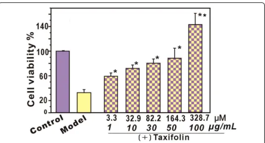
-binding reaction was also investigated using UV-Vis spectra. The results revealed that cell viability was fully restored, even increasing to 142.9 ± 9.3% after treatment with (+) taxifolin. In the antioxidant assays, (+) taxifolin was observed to efficiently scavenge•OH, DPPH•and ABTS+•radicals, and to increase the relative Cu2+- and Fe3+-reducing levels. In the PTIO•-scavenging assay, its IC50values varied with pH. In the Fe
2+
-binding reaction, (+) taxifolin was found to yield a green solution with two UV-Vis absorbance peaks:λmax= 433 nm (ε=5.2 × 10
2
L mol−1cm−1) andλmax= 721 nm (ε= 5.1 × 10 2
L mol−1cm−1). These results indicate that (+) taxifolin can act as an effective• OH-scavenger, protecting bmMSCs from•OH-induced damage. Its•OH-scavenging action consists of direct and indirect antioxidant effects. Direct antioxidation occurs via multiple pathways, including ET, PCET or HAT. Indirect antioxidation involves binding to Fe2+.
Keywords:(+) Taxifolin; bmMSCs,•OH damage, Antioxidant mechanism, Electron transfer, Fe2+binding
Background
Antioxidant supplementation has been suggested as a means to reduce the DNA dam-age and relieve oxidative stress during the expansion and proliferation of bone marrow-derived mesenchymal stem cells (bmMSCs) [1]. This oxidative stress is a re-sult of the imbalance between ROS production and diminished endogenous antioxi-dant protection. Accumulative ROS (especially•OH with a half-life of 10−9s) not only have the potential to damage all types of biomolecules (such as DNA, proteins, lipids and carbohydrates), but can also inhibit MSC immunomodulation, thus increasing sen-escence and reducing ex vivo expansion, which is critical for clinical application off the cells [2]. Effective antioxidants that could protect MSCs from oxidative stress are a de-sirable focus of research.
From the perspective of free radical biology, plants also encounter serious oxidative stress from strong UV-Vis light, atmospheric ROS, temperature changes, and the pro-cesses of oxygen consumption for photosynthesis. Notably, some plants, such as pine, have what could be considered a strong vital force and a long history of survival. They have successfully resisted oxidation from complicated ecological environments and may serve as a library of efficient phenolic antioxidants [3].
Pine grows on the Sharon Plain in Israel and in mountains and highlands around the world. Notable species and varieties are Pinus pinaster (French maritime pine) [4],
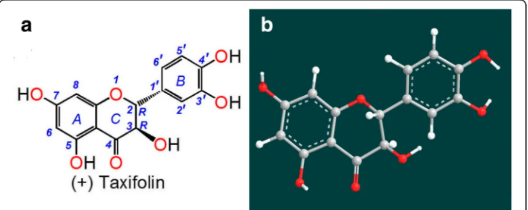
Pseudotsug amenziesii[5],Pinus massonianaLamb [6],Pinus sylvestris var.mongovica Litvin [7] and Larix olgensis Henry var. Koreana Nakai [8]. Pine has survived for ap-proximately 1.9 hundred million years, suggesting that it possesses strong defenses, probably including a strong antioxidant defense with numerous antioxidant compo-nents. In fact, extract from the bark of French maritime pine has been developed as an antioxidant supplement known commercially as Pycnogenol, which has a bioactive component named (+) taxifolin (2R,3R–dihydroquercetin, Fig. 1) [4, 9].
As shown in Fig. 1a, (+) taxifolin is actually a dihydroflavonol that exists in the afore-mentioned pine types. It was reported to inhibit free radical formation at key stages of apoptosis in cellular mitochondria [10] and to correct cerebral ischemia-reperfusion in-jury [11]. Recently, (+) taxifolin was found to exhibit anticancer and neuroprotective ef-fects [12–14].
This indicates that (+) taxifolin has potential as an antioxidant for protecting MSCs against oxidative stress damage. However, no study has reported on the protective ef-fects of (+) taxifolin towards•OH-treated bmMSCs.
Here, we applied the methyl thiazolyl tetrazolium (MTT) assay to assess the protect-ive effects of (+) taxifolin on •OH-treated bmMSCs. We then explored the possible mechanisms for this effect.
Methods
Chemicals and animals
The chemicals (+) taxifolin (CAS number: 480–18-2, 98%), dihydromyricetin (CAS number: 27,200–12-0, 98%), and 4’-O-methyltaxifolin (CAS number: 70,411–27-7, 98%) were obtained from Chengdu Biopurify Phytochemicals Ltd. Catechol (CAS
number: 120–80-9, 99.5%) and DNA sodium salt (fish sperm) were purchased from Aladdin Chemistry Co. The 1, 1-diphenyl-2-picryl-hydrazyl radical (DPPH•), (±)-6-hy-droxyl-2, 5, 7, 8-tetramethylchromane-2-carboxylic acid (trolox), 2, 9-dimethyl-1, 10-phenanthroline (neocuproine), 3-(2-pyridyl)-5, 6-bis (4-phenylsulfonicacid)-1, 2, 4-triazine (ferrozine), 2, 4, 6-tripyridyltriazine (TPTZ), 2-phenyl-4, 4, 5, 5-tetramethylimidazoline-1-oxyl-3-oxide radical (PTIO•), and methyl thiazolyl tetrazolium (MTT) were purchased from Sigma-Aldrich Shanghai Trading Co. The CCK-8 (BB-4221-2) kits were from Best-Bio Inc. (NH4)2ABTS [2, 2′-azino-bis (3-ethylbenzo-thiazoline-6-sulfonic acid
diammo-nium salt)] was obtained from Amresco Chemical Co. Dulbecco’s modified Eagle’s medium (DMEM), fetal bovine serum (FBS) and trypsin were purchased from Gibco. All other reagents were of analytical grade.
Sprague-Dawley (SD) rats (4 weeks old) were obtained from the Animal Center of Guangzhou University of Chinese Medicine. The protocol was performed under the supervision of the Institutional Animal Ethics Committee at the Guangzhou University of Chinese Medicine.
MTT assay to assess the protective effect against•OH-induced damage
The bmMSCs were cultured according to our previous report [15] with slight modifica-tions. In brief, bone marrow was obtained from the femur and tibia of the rats. Marrow samples were diluted with low-glucose DMEM containing 10% FBS. MSCs were pre-pared by gradient centrifugation at 900×gfor 30 min on 1.073 g/ml Percoll. The pre-pared cells were detached by treatment with 0.25% trypsin and passaged in culture flasks at 1 × 10⁴/cm2. At passage 3, bmMSCs were evaluated for cell homogeneity using CD44 detection via flow cytometry. These cells were used for the subsequent experiments.
The protective effect of (+) taxifolin against •OH-induced bmMSC damage was in-vestigated based on the method described in [16, 17] with slight modifications. Briefly, bmMSCs were seeded at 5000 cells per well into 96-well plates. After adherence for 24 h, bmMSCs were divided into control, model and sample [(+) taxifolin] groups.
In the control group, bmMSCs were incubated for 24 h in DMEM. In the model and sample groups, bmMSCs were incubated in the presence of FeCl2(100 μM) followed
by H2O2(50μM). After incubation for 20 min, the mixture of FeCl2and H2O2was
re-moved. The bmMSCs in the model group were incubated for 24 h in DMEM, while bmMSCs in the sample group were incubated for 24 h in DMEM with the indicated (+) taxifolin concentrations.
After incubation, 20μl MTT (5 mg/ml) was added, and the culture was incubated for an additional 3 h. The culture medium was discarded and replaced with 150μl DMSO. Absorbance was measured at 490 nm on a Bio-Kinetics reader (PE-1420; Bio-Kinetics Corporation). Culture medium containing serum was used for the control group and each sample test was repeated in five independent wells.
Hydroxyl-scavenging assay based on DNA
in each tube to dryness, the sample residue was treated with 300μl of phosphate buffer
(0.2 M, pH 7.4), followed by 50 μl of DNA sodium (10 mg/ml), 75 μl of H2O2
(33.6 mM), 50 μl of FeCl3(3.2 mM), 100μl of Na2EDTA (0.5 mM) and 75μl of
ascor-bic acid (12 mM). After incubation at 50 °C for 20 min, 250 μl of trichloroacetic acid (10%,w/v) was added to the tube. After heating the mixture at 105 °C for 15 min with 150μl of 2-thiobarbituric acid (TBA, 5% in 1.25% NaOH aqueous solution), the absorb-ance was measured using a Unico Spectrophotometer UV 2100 against the buffer (blank). The protective effect is expressed as follows:
Protective effect%¼A0‐A
A0
100%;
where A0indicates the absorbance of the blank and A indicates the absorbance of the
sample (+) taxifolin.
PTIO•-scavenging assay
The PTIO•-scavenging assay was conducted based on our method [19]. In brief, 80μl of an aqueous PTIO•solution (0.1 mM) was mixed with 20 μl of phosphate buffer at pH 5.0, 6.0, 7.0, 7.4, 8.0 and 9.0 containing 1 mg/ml of sample at the indicated concen-trations. The mixture was maintained at 37 °C for 30 min, and the absorbance was measured at 560 nm on a microplate reader (Multiskan FC, Thermo Scientific). The PTIO•inhibition percentage was calculated as follows:
Scavenging%¼A0‐A
A0
100%;
where A0indicates the absorbance of the blank and A indicates the absorbance of the
sample, (+) taxifolin.
DPPH•-scavenging assay and ABTS+•-scavenging assay
DPPH•radical-scavenging activity was determined as previously described [20]. Briefly, 1 ml of DPPH•solution (0.1 M) was mixed with the indicated concentrations of sample (0.15 mg/ml, 14–70 μl) dissolved in methanol. The mixture was maintained at room temperature for 30 min, and the absorbance was measured at 519 nm on a Unico Spec-trophotometer 2100.
ABTS+•-scavenging activity was evaluated according to a previously described
method [21]. ABTS+• was produced by mixing 0.2 ml of ABTS diammonium salt
(7.4 mM) with 0.35 ml of potassium persulfate (2.6 mM). The mixture was maintained in the dark at room temperature for 12 h to allow completion of radical generation and then diluted with 95% ethanol. To determine the scavenging activity, the test sample (x = 15–75μl, 0.03 mg/ml) was added to (200- x) μl of 95% ethanol followed by 800 μl of ABTS+•reagent, and the absorbance was measured at 734 nm on a Unico Spectropho-tometer 2100 6 min after the initial mixing using 95% ethanol as the blank.
Cu2+-reducing assay
The reducing power capacity of cupric ions (Cu2+) was measured according to a previ-ously described method [22] with a slight modification. Briefly, 125 μl of CuSO4
aque-ous solution (10 mM), 125 μl of neocuproine ethanolic solution (7.5 mM) and 750 μl of CH3COONH4buffer solution (0.1 M, pH 7.5) were added to test tubes with different
volumes of sample (0.15 mg/ml, 15–75μl). The total volume was adjusted to 1 ml with buffer and mixed vigorously. The absorbance against a buffer blank was measured at 450 nm after 30 min. An increase in the absorbance of the reaction mixture indicates an increase in reduction capability. The relative reducing power of the sample relative to the maximum absorbance was calculated using the following formula:
Relative reducing effect%¼ A‐Amin Amax‐Amin
100%;
where Aminis the absorbance of the control without sample, A is the absorbance of the
reaction mixture with sample, and Amax is the maximum absorbance of the reaction
mixture with sample.
Ferric-reducing antioxidant power (FRAP) assay
The FRAP assay was adapted from Benzie and Strain [23]. Briefly, FRAP reagent was freshly prepared by mixing 10 mM TPTZ, 20 mM FeCl3and 0.25 M acetate buffer at
1:1:10 (pH 3.6). The test sample (x = 20–100μl, 0.5 mg/ml) was added to (100- x)μl of
95% ethanol followed by 400 μl of FRAP reagent. The absorbance was measured at
593 nm after a 30-min incubation at ambient temperature using distilled water as the blank. The relative reducing power was calculated using the formula given in the
Cu2+-reducing assay section.
UV-vis spectra and color reaction of Fe2+-binding
The (+) taxifolin–Fe2+complex was evaluated using UV-Vis spectroscopy. For these ex-periments, 300μl of a methanolic solution of (+) taxifolin and 100μl of an aqueous so-lution of FeCl2•4H2O were added to 600 μl of an aqueous mixture of distilled water
and methanol (1:1). The solution was then mixed vigorously and continuously scanned using a UV-Vis spectrophotometer (Unico 2600A) from 200 to 900 nm after 0, 10, 20, 30, and 60 min.
The above experiment was repeated using 4’-O-methyltaxifolin.
Statistical analysis
Each experiment was performed in triplicate and data were recorded as the means ± SD (standard deviation). Dose response curves were plotted using Origin 6.0 software (OriginLab). IC50was defined as the final concentration of 50% radical inhibition
(rela-tive reducing power or binding effect). Statistical comparisons were made using one-way ANOVA to detect significant differences using SPSS 13.0 (SPSS Inc.) for Windows.
p< 0.05 was considered statistically significant.
Results and discussion
cell viability was restored or even increased. This result suggests that (+) taxifolin ef-fectively protects bmMSCs from•OH-mediated damage. This is consistent with the re-cent report that (+) taxifolin could reduce cholesterol oxidation product-induced neuronal apoptosis [24]. At higher concentrations (>50 μg/ml, 164.3μM), (+) taxifolin could even further promote the viability of bmMSCs, reaching 142.9 ± 9.3% viability.
To test the possible toxicity to MSCs, the effect of (+) taxifolin towards normal MSCs was measured using the CCK-8 assay (an updated version of the MTT assay). The results indicated that (+) taxifolin (3.3–328.7 μM) had no effect on
proliferation and no toxic effect on normal MSCs without •OH-treatment
(Add-itional file 1: Figure S1). These results align with the previous findings that (+) taxifolin could be an apparent exception that could efficiently inhibit the Fenton reaction and superoxide radical formation [25, 26] while being completely nonpho-totoxic, unlike its analogue quercetin [13, 27]. These results are inconsistent with another previous study that showed taxifolin was toxic to oocytes at higher con-centration (50 μg/ml, 164.3 μM) [28].
It is assumed that when (+) taxifolin was mixed with Fenton reagents, some reaction products may be generated to bring about the beneficial (especially protective) effect. In fact, a similar situation is observed with salvianolic acid B, which can increase cell viability to 175.1% [29]. In the case of the salvianolic acid B molecule, some characteris-tic chemical structures, such as catechol or lactone moieties [29], have been suggested to be partly responsible for the protective effect. The moiety of fused rings (A/B) is also hypothesized to play a role in the process. Some antioxidants comprising 8-hydroxyquinol have been demonstrated to induce MSC proliferation [30, 31]. However, the detailed mechanisms should be investigated further.
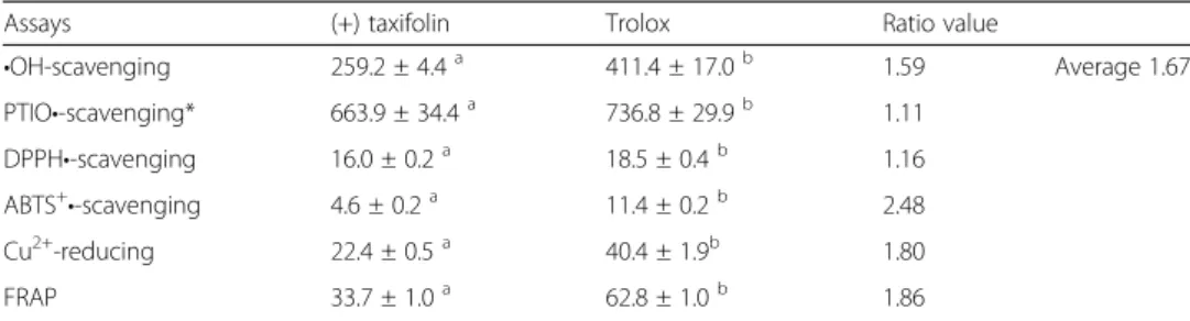
Such protective effects from •OH damage have been reported to be related to •OH scavenging [32]. In this study, (+) taxifolin was found to exhibit•OH-scavenging ability in a dose-dependent manner (Additional file 1: Figure S2). The IC50 Trolox/IC50 (+)
taxifo-linvalue (1.67; Table 1) suggests that (+) taxifolin is a better•OH scavenger than trolox,
which is a standard antioxidant.
•OH scavenging comprises two pathways: direct and indirect. The direct antioxidant pathway directly scavenges the•OH free radical that has been generated via the Fenton reaction. However, •OH is a very transient species so it is impossible to verify whether
•OH is directly scavenged. Therefore, we used a stable oxygen-centered radical, PTIO•, for the investigation. As seen in Additional file 1: Figure S3A, (+) taxifolin scavenged the PTIO•radical at various pH values in a dose-dependent manner.
Correspondingly, the IC50values varied with various pH values: 2.6 ± 0.5, 1.6 ± 0.2,
0.7 ± 0.03, 0.6 ± 0.04, 0.5 ± 0.04 and 0.4 ± 0.02 mM respectively for pH 5.0, 6.0, 7.0, 7.4, 8.0 and 9.0 (Additional file 1: Table S1). This indicates the involvement of the dir-ect antioxidant pathway in•OH scavenging by (+) taxifolin. When the IC50values were
plotted against pH values, a first-order decay curve was observed (Additional file 1: Figure S3B), suggesting that a high level of H+ (low pH value) considerably sup-pressed the PTIO•-scavenging ability of (+) taxifolin. Thus, the radical-scavenging ability of (+) taxifolin is hypothesized to be involved in H+ transfer, consistent with the cyclic voltammetry-based evidence [33].
It has been documented that at a pH≤5.0, PTIO• can be scavenged via electron transfer (ET) [33]. Our assay suggests that (+) taxifolin may also scavenge PTIO• at pH 5.0, indicating the involvement of ET in its antioxidant action. This is further sup-ported by its ABTS+•-scavenging, Cu2+-reducing and Fe3+-reducing (i.e., FRAP) abilities (Additional file 1: Figures S4-S6). ABTS+•-scavenging is considered to be an ET-based pathway [34]. The ABTS+•-scavenging ability of (+) taxifolin indicates the involvement of ET in the antioxidant process. Furthermore, (+) taxifolin increased the relative Cu2
+
-reducing and FRAP-reducing abilities in a concentration-dependent manner. The FRAP (at pH 3.6) and Cu2+-reducing activities have been demonstrated to be an ET re-action [35]. It should be noted that the Fe3+-reducing potential of flavonoids may also reduce Fe3+into Fe2+to cause pro-antioxidation [36]. It remains unknown whether the pro-antioxidation is linked to (+) taxifolin cytotoxicity to oocytes at higher concentra-tion [28].
In this study, (+) taxifolin efficiently scavenged the DPPH· radical (Additional file 1: Figure S7). DPPH· scavenging is regarded as a hydrogen atom transfer-based multi-pathway [32]. Successful DPPH· scavenging by (+) taxifolin indicated that hydrogen atom transfer may occur in its direct antioxidative process. Moreover, it was recently reported that these direct antioxidative pathways are not exclusive but are rather com-petitive based on various reaction conditions [34].
Table 1The IC50values of (+) taxifolin and trolox in various assays (μM)
Assays (+) taxifolin Trolox Ratio value
•OH-scavenging 259.2 ± 4.4a 411.4 ± 17.0b 1.59 Average 1.67
PTIO•-scavenging* 663.9 ± 34.4a 736.8 ± 29.9b 1.11
DPPH•-scavenging 16.0 ± 0.2a 18.5 ± 0.4b 1.16
ABTS+•-scavenging 4.6 ± 0.2a 11.4 ± 0.2b 2.48
Cu2+-reducing 22.4 ± 0.5a 40.4 ± 1.9b 1.80
FRAP 33.7 ± 1.0a 62.8 ± 1.0b 1.86
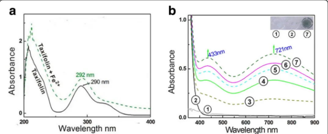
Because Fe2+ can catalyze the Fenton reaction, where H2O2yields •OH radicals, an
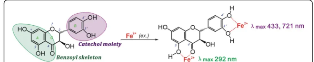
attenuation of Fe2+ levels via a binding reaction is considered an indirect antioxidant mechanism to scavenge •OH radicals [37]. In the indirect antioxidant assay, (+) taxifo-lin bound to Fe2+ to yield a green solution and two Vis absorbance peaks: λmax =
433 nm (ε=5.2 × 10²L mol−1cm−1) and λmax= 721 nm (ε= 5.1 × 10²L mol−1cm−1).
In the UV spectra, Fe2+ binding enhanced the peak strength around 290 nm (Fig. 3). These results strongly indicate a binding reaction between Fe2+ and (+) taxifolin and that Fe2+binding may act as one indirect pathway in the antioxidative process of (+) taxifolin.
As reported previously [36], adjacent keto or hydroxyl groups are potential targets of Fe2+ binding, while isolated keto-group (or hydroxyl-group) cannot bind iron. Never-theless, the 3, 4-hydroxyl-keto moiety cannot give a planar conformation (Fig. 1b), and can barely form the planar five-membered Fe2+-complex. As a dihydroflavonol, (+) taxi-folin contains only two Fe2+-binding sites: the 3′, 4′-catechol moiety and the 4, 5-hydroxyl-keto moiety (Fig. 4) [38].
Despite several reports on the metal-binding of flavonoids [38–40] and descriptions of Na+ interacting with flavonoids [41], studies focusing on UV-Vis spectral analyses (especially peak assignment) are lacking. To confirm the assignment of the UV-Vis peaks in Fig. 3, we investigated the Fe2+-binding of catechol and dihydromyricetin (ref-erence compounds), because in (+) taxifolin and dihydromyricetin, the possible π–π conjugation is blocked by a single 2, 3 carbon–carbon bond, and theB ring andA/C
fused rings are independent of each other. Thus, the whole (+) taxifolin molecule can be divided into two spectroscopic systems: the benzoyl skeleton and the catechol moi-ety (Additional file 1: Figure S8). Catechol contains a similar chemical structure to the
Bring of (+) taxifolin, while dihydromyricetin bears a similar chemical structure to the
A/Cfused rings (benzoyl skeleton) of (+) taxifolin.
Catechol–Fe2+ gave two similar absorbance peaks (at approximately λmax 433 and
721 nm) in the Vis spectra to those of (+) taxifolin-Fe2+ and yielded a green solution (Additional file 1: Figure S9). By contrast, the dihydromyricetin molecule bearing a
pyrogallol moiety in the B ring presented a strong absorbance peak at λmax 589 nm
[42], and 4’-O-methyltaxifolin without catechol moiety only gave a UV absorbance peak at λmax289 nm (Additional file 1: Figure S10). Thus, it can be deduced that the peaks
in the Vis spectra of the (+) taxifolin–Fe2+complex are from the Fe2+-binding reaction with catechol in theBring.
With respect to the UV spectra, an enhanced strength of the UV peaks was observed in the Fe2+-binding reaction with (+) taxifolin (Fig. 3a), similar to the dihydromyrice-tin–Fe2+complex and 4’-O-methyltaxifolin–Fe2+complex (Additional file 1: Figures S8 & S11). Dihydromyricetin and 4’-O-methyltaxifolin share a similar benzoyl skeleton with (+) taxifolin. Thus, the enhancement of peak around 290 nm can be attributed to the Fe2+-binding reaction of the 4-hydroxyl-5-keto moiety. This assumption is further supported by the different colors between the (+) taxifolin–Fe2+complex and the 4’-O -methyltaxifolin–Fe2+complex.
Conclusion
As an effective •OH-scavenger, (+) taxifolin can protect bmMSCs from •OH-induced damage. Its •OH-scavenging action consists of direct and indirect antioxidant effects. The direct antioxidation occurs via multiple pathways, including ET, PCET and HAT. The indirect antioxidation involved Fe2+ binding. Upon binding to Fe2+, the 3′,4′ -cat-echol moiety in theBring gives rise to two peaks (λmax433 nm and 721 nm), and the
4-hydroxyl-5-keto of the benzoyl skeleton causes an enhanced peak intensity around 290 nm.
Additional file
Additional file 1: Figure S1.The CCK-8 assay for normal bmMSCs exposed to (+) taxifolin.Figure S2.
Dose–response curves for (+) taxifolin•OH-scavenging assay based on DNA.Figure S3.Dose–response curves for (+) taxifolin in PTIO•radical-scavenging assay and its IC50values at various pH values.Figure S4. Dose–response curves for (+) taxifolin in the ABTS+•radical-scavenging assay.Figure S5.Dose–response curves for (+) taxifolin in the Cu2+-reducing assay.Figure S6.Dose–response curves for (+) taxifolin in the FRAP assay.Figure S7.Dose–response curves for (+) taxifolin in the DPPH•-radical-scavenging assay.Figure S8.The UV-visible spectra for the 4’-O-methyltaxifolin–Fe2+complex.Figure S9.The UV absorption bands of flavonoid.
Figure S10.The UV-Vis spectra and solution colors for (+) taxifolin–Fe2+and catechol–Fe2+.Figure S11.The UV-Vis spectra for (+) taxifolin–Fe2+and dihydromyricetin–Fe2+.Table S1.The IC
50values listed in different units. (DOCX 789 kb)
Abbreviations
ABTS:2, 2′-azino-bis (3-ethylbenzo-thiazoline-6-sulfonic acid); bmMSCs: bone marrow-derived mesenchymal stem cells; CCK-8: Cell counting kit-8; DMEM: Dulbecco’s modified Eagle’s medium; ET: Electron transfer; FBS: Fetal bovine serum; Ferrozine: 3-(2-pyridyl)-5,6-bis (4-phenylsulfonicacid)-1,2,4-triazine; FRAP: Ferric ion-reducing antioxidant power; HAT: Hydrogen atom transfer; MTT: Methyl thiazolyl tetrazolium; Neocuproine: 2, 9-dimethyl-1, 10-phenanthroline; PCET: Proton-coupled electron transfer; PTIO•: 2-phenyl-4,4,5, 5-tetramethylimidazoline-1-oxyl-3-oxide; ROS: Reactive oxygen species; TPTZ: 2,4,6-tripyridyl triazine; Trolox: (±)-6-hydroxyl-2,5,7,8-tetramethlychromane-2-carboxylic acid
Acknowledgements
None.
Funding
This research was supported by the National Natural Science Foundation of China (81,503,593, 81,573,558), the Natural Science Foundation of Guangdong Province (2017A030312009, 2016A030313649), and the Guangdong Science and Technology Project (2017A050506043).
Availability of data and materials
Data are all contained within the article.
Authors’contributions
XL, GW, and DC conceived and designed the experiments; HX and ZF performed the cellular experiments and drew the Figures; LL, CL, XO, and LY conducted the DPPH•-scavenging assay and ABTS+•-scavenging assay; QJ, YX, and YL analyzed the data; XL wrote the paper. All authors read and approved the final manuscript.
Ethics approval
The institution Animal Ethics Committee in Guangzhou University of Chinese Medicine (Guangzhou, China) approved the protocols used in this study.
Consent for publication
Not applicable.
Competing interests
The authors declare that they have no competing interests.
Publisher’s Note
Springer Nature remains neutral with regard to jurisdictional claims in published maps and institutional affiliations.
Author details
1
School of Chinese Herbal Medicine, Guangzhou University of Chinese Medicine, Waihuang East Road No. 232, Guangzhou Higher Education Mega Center, Guangzhou 510006, China.2Innovative Research & Development Laboratory of TCM, Guangzhou University of Chinese Medicine, Guangzhou 510006, China.3School of Basic Medical Science, Guangzhou University of Chinese Medicine, Guangzhou 510006, China.4The Research Center of Integrative Medicine, Guangzhou University of Chinese Medicine, Guangzhou 510006, China.
Received: 15 August 2017 Accepted: 15 December 2017
References
1. Alves H, Mentink A, Le B, Van Blitterswijk CA, De Boer J. Effect of antioxidant supplementation on the total yield, oxidative stress levels, and multipotency of bone marrow-derived human mesenchymal stromal cells. Tissue Eng Part A. 2013;19:928–37.
2. Ryan A. Denu, Peiman H. Effects of oxidative stress on mesenchymal stem cell biology. Oxidative Med Cell Longev. 2016;1:1–9.
3. Fang YZ, Zheng RL. Theory and application of free radical biology. Science Press, Beijing, China. 2002; pp. 541–594. 4. Mülek M, Seefried L, Genest F, Högger P. Distribution of constituents and metabolites of maritime pine bark
extract (Pycnogenol®) into serum, blood cells, and synovial fluid of patients with severe osteoarthritis: a randomized controlled trial. Nutrients. 2017;9(5):443.
5. Stafford HA, Lester HH. Flavan-3-ol biosynthesis: the conversion of (+)-dihydromyricetin to its flavan-3,4-diol (leucodelphinidin) and to (+)-gallocatechin by reductases extracted from tissue cultures of ginkgo biloba and pseudotsugamenziesii. Plant Physiol. 1985;78(4):791–4.
6. Dong Y, Ma XF, RJ W, Yang MY, Shou D, Li HY. Simultaneous determination of taxifolin rutin inPinusmassoniana lamb. By high performance liquid chromatography with diode array detection. Chin arch of Tradit. Chin Med. 2012;30(11):2517–9.
7. Fan TB, Liu HY, Tang Q, Liu SM. Determination the content of dihydroquercetin in pine needles by HPLC. China Pharm. 2009;12(8):1046–8.
8. Zhang WP, Liu W, JH F, Chai J, Liu WC, Zhen YN. Structural identification and quantitative analysis of taxifolin in Larix olgensisHenry Var. Koreana Nakai. J Food Sci. 2013;34(16):293–6.
9. Bayomy NA, Abdelaziz EZ, Said MA, Badawi MS, El-Bakary RH. Effect of pycnogenol and spirulina on vancomycin-induced renal cortical oxidative stress, apoptosis, and autophagy in adult male albino rat. Can J Physiol Pharmacol. 2016;94(8):838–48.
10. Vladimirov YA, Proskurnina EV, Demin EM, Matveeva NS, Lubitskiy OB, Novikov AA, Izmailov DY, Osipov AN, Tikhonov VP, Kagan VE. Dihydroquercetin (taxifolin) and other flavonoids as inhibitors of free radical formation at key stages of apoptosis. Biochemistry (Mosc). 2009;74:301–7.
11. Maksimovich NY, Dremza IK, Troian EI, Maksimovich YN, BorodinskiĭAN. The correcting effects of dihydroquercetin in cerebral ischemia-reperfusion injury. Biomed Khim. 2014;60(6):643–50.
13. Zhang ZR, Al Zaharna M, Wong MM, Chiu SK, Cheung HY. Taxifolin enhances and rographolide-induced mitotic arrest and apoptosis in human prostate cancer cells via spindle assembly checkpoint activation. PLoS One. 2013; 8(1):54577.
14. Dok-Go H, Lee KH, Kim HJ, Lee EH, Lee J, Song YS, Lee YH, Jin C, Lee YS, Cho J. Neuroprotective effects of antioxidative flavonoids, quercetin, (+)-dihydroquercetin and quercetin 3-methyl ether, isolated from Opuntiaficus-indica var. saboten. Brain Res. 2003;965(1–2):130–6.
15. Chen DF, Li X, Xu Z, Liu X, SH D, Li H, Zhou JH, Zeng HP, Hua ZC. Hexadecanoic acid from Buzhong Yiqi decoction induces proliferation of bone marrow mesenchymal stem cells. J Med Food. 2010;13:967–70. 16. Li XC, Hu Q, Jiang S, Li F, Lin J, Han L, Hong Y, Lu W, Gao Y, Chen D.Flos Chrysanthemi indiciprotects against
hydroxyl-induced damages to DNA and MSCs via antioxidant mechanism: a chemistry study. J Saudi Chem Soc. 2015;19:454–60.
17. Wang T, Zeng G, Li X, Zeng H. Vitro studies on the antioxidant and protective effect of 2-substituted-8-hydroxyquinoline derivatives against H2O2-induced oxidative stress in BMSCs. Chem Biol Drug Des. 2010;75(2): 214–22.
18. Li XC, Mai W, Wang L, Han W. A hydroxyl-scavenging assay based on DNA damage in vitro. Anal Biochem. 2013; 438:29–31.
19. Li XC. 2-Phenyl-4,4,5,5-tetramethylimidazoline-1-oxyl 3-oxide (PTIO•) radical-scavenging: a new and simple antioxidant assay in vitro. J Agric Food Chem. 2017;65:6288–97.
20. Anna F, Dae-Ok K, Chung SJ, Sung I. Koo, Ock KC. Comparison of ABTS/DPPH assays to measure antioxidant capacity in popular antioxidant-rich US foods. J Food Compos Anal. 2011;24:1043–8.
21. Li X, Chen D, Mai Y, Wen B, Wang X. Concordance between antioxidant activities in vitro and chemical components of radix Astragali (Huangqi). Nat Prod Res. 2012;26:1050–3.
22. Li XC, Han W, Mai W. Antioxidant activity and mechanism of tetrahydroamentoflavone in vitro. Nat Prod Commun. 2013;8:787–9.
23. Benzie IF, Strain JJ. The ferric reducing ability of plasma (FRAP) as a measure of“antioxidant power”: the FRAP assay. Anal Biochem. 1996;239:70–6.
24. Kim A, Nam YJ, Lee CS. Taxifolin reduces the cholesterol oxidation product-induced neuronal apoptosis by suppressing the Akt and NF-κB activation-mediated cell death. Brain Res Bull. 2017;134:63–71.
25. Macáková K, Mladěnka P, Filipský T,Říha M, JahodářL, Trejtnar F, Bovicelli P, Proietti Silvestri I, Hrdina R, Saso L. Iron reduction potentiates hydroxyl radical formation only in flavonols. Food Chem. 2012;135(4):2584–92. 26. Moridani MY, Pourahmad J, Bui H, Siraki A, O’Brien PJ. Dietary flavonoid iron complexes as cytoprotective
superoxide radical scavengers. Free Radic Biol Med. 2003;34(2):243–53.
27. Rajnochová Svobodová A, Ryšavá A, Psotová M, Kosina P, Zálešák B, Ulrichová J, Vostálová J. The phototoxic potential of the flavonoids, taxifolin and quercetin. Photochem Photobiol. 2017;93(5):1240–7.
28. Kang JT, Moon JH, Choi JY, Park SJ, Kim SJ, Saadeldin IM, Lee BC. Effect of antioxidant flavonoids (quercetin and taxifolin) on in vitro maturation of porcine oocytes. Asian-Australas J Anim Sci. 2016;29(3):352–8.
29. Wang T, Li X, Wu J, Huang Y, Wei G, Chen D. Mechanistic chemistry of extraordinary capacity of salvianolic acid B on oxidatively-damaged mesenchymal stem cells. J Chin Chem Soc. 2016;63(11):924–9.
30. Wang G, Li X, Zeng H. Synthesis antioxidation activity of (E)-9-p-tolyl-3- [2-(8-hydroxy-quinol-2-yl)vinyl]-carbazole and (E)-9-(p-Anisyl)-3-[2-(8-hydroxy-quinol-2-yl)vinyl]-carbazole and their induction proliferation of mesenchymal stem cells. Act Chimica Sinica. 2009;67(9):974–82.
31. Li X, Wei G, Wang X, Liu DH, Deng RD, Li H, Zhou JH, Li YW, Zeng HP, Chen DF. Targeting of the sonic hedgehog pathway by Atractylenolides promotes Chondrogenic differentiation of mesenchymal stem cells. Biol Pharm Bull. 2012;35:1328–35.
32. Lin J, Li X, Chen L, Lu W, Chen X, Han L, Chen D. Protective effect against hydroxyl radical-induced DNA damage and antioxidant mechanism of [6]-gingerol: a chemical study. B Korean. Chem Soc. 2014;35(6):1633–8. 33. Goldstein S, Russo A, Samuni A. Reactions of PTIO•and carboxy-PTIO•with•NO,•NO2, and O2. J Biol Chem. 2003;
278:50949–55.
34. Apak R, Özyürek M, Güçlü K, Çapanoğlu E. Antioxidant activity/capacity measurement. 2. Hydrogen atom transfer (HAT)-based, mixed-mode (electron transfer (ET)/HAT), and lipid peroxidation assays. J Agric Food Chem. 2016;64: 1028–45.
35. Gülçinİ. Antioxidant activity of food constituents: an overview. Arch Toxicol. 2012;86:345–91.
36. Sugihara N, Arakawa T, Ohnishi M, Furuno K. Anti- and pro-oxidative effects of flavonoids on metal-induced lipid hydroperoxide-dependent lipid peroxidation in cultured hepatocytes loaded withα-linolenic acid. Free Radic Biol Med. 1999;27(11–12):1313–23.
37. Jiang Q, Li X, Tian Y, Lin Q, Xie H, Lu W, Chi Y, Chen D. Lyophilized aqueous extracts ofMori FructusandMori Ramulusprotect mesenchymal stem cells from•OH–treated damage: bioassay and antioxidant mechanism. BMC Complem Altern M. 2017;17:242.
38. Mladěnka P, Macáková K, Filipský T, Zatloukalová L, JahodářL, Bovicelli P, Silvestri IP, Hrdina R, Saso L. Vitro analysis of iron binding activity of flavonoids. J Inorg Biochem. 2011;105:693–701.
39. Mira L, Fernandez MT, Santos M, Rocha R, Florêncio MH, Jennings KR. Interactions of flavonoids with iron and copper ions: a mechanism for their antioxidant activity. Free Radic Res. 2002;36:1199–208.
40. Li X, Gao Y, Li F, Liang A, Xu Z, Bai Y, Mai W, Han L, Chen D. Maclurin protects against hydroxyl radical-induced damages to mesenchymal stem cells: antioxidant evaluation and mechanistic insight. Chem Biol Interact. 2014; 219:221–8.
41. Xiao CH. Chemistry of Chinese Materia Medica. 3rd ed. Shanghai, China: Shanghai Scientific & Technical Publishers; 1997. p. 289–99.