Evaluation of a Culture-Dependent Algorithm and a Molecular
Algorithm for Identification of
Shigella
spp.,
Escherichia coli
,
and Enteroinvasive
E. coli
Maaike J. C. van den Beld,a,bRichard F. de Boer,cFrans A. G. Reubsaet,aJohn W. A. Rossen,bKai Zhou,b,dSjoerd Kuiling,a
Alexander W. Friedrich,bMirjam A. M. D. Kooistra-Smidb,c
aInfectious Disease Research, Diagnostics and laboratory Surveillance, Centre for Infectious Disease Control, National Institute for Public Health and the Environment, Bilthoven, The Netherlands
bMedical Microbiology, University of Groningen, University Medical Center Groningen, Groningen, The Netherlands
cDepartment of Medical Microbiology, Certe, Groningen, The Netherlands
dCollaborative Innovation Center for Diagnosis and Treatment of Infectious Diseases, State Key Laboratory for Diagnosis and Treatment of Infectious Disease, The First Affiliated Hospital, School of Medicine, Zhejiang University Hangzhou, Hangzhou, China
ABSTRACT Identification ofShigellaspp., Escherichia coli, and enteroinvasiveE. coli
(EIEC) is challenging because of their close relatedness. Distinction is vital, as infec-tions with Shigella spp. are under surveillance of health authorities, in contrast to EIEC infections. In this study, a culture-dependent identification algorithm and a mo-lecular identification algorithm were evaluated. Discrepancies between the two algo-rithms and original identification were assessed using whole-genome sequencing (WGS). After discrepancy analysis with the molecular algorithm, 100% of the evalu-ated isolates were identified in concordance with the original identification. How-ever, the resolution for certain serotypes was lower than that of previously described methods and lower than that of the culture-dependent algorithm. Although the res-olution of the culture-dependent algorithm is high, 100% of noninvasiveE. coli, Shi-gella sonnei, andShigella dysenteriae, 93% of Shigella boydii and EIEC, and 85% of Shigella flexneri isolates were identified in concordance with the original identifica-tion. Discrepancy analysis using WGS was able to confirm one of the used algo-rithms in four discrepant results. However, it failed to clarify three other discrepant results, as it added yet another identification. Both proposed algorithms performed well for the identification of Shigella spp. and EIEC isolates and are applicable in low-resource settings, in contrast to previously described methods that require WGS for daily diagnostics. Evaluation of the algorithms showed that both algorithms are capable of identifying Shigellaspecies and EIEC isolates. The molecular algorithm is more applicable in clinical diagnostics for fast and accurate screening, while the culture-dependent algorithm is more suitable for reference laboratories to identify Shigellaspp. and EIEC up to the serotype level.
KEYWORDS EIEC,Escherichia coli,Shigella, whole-genome sequencing,
enteroinvasiveE. coli, identification, molecular methods, phenotypic methods
I
n 1898, Kiyoshi Shiga first described Shigella dysenteriae as the etiologic agent of dysentery (1). Nowadays, the genusShigellacomprises four species based on antigenic properties, Shigella dysenteriae, Shigella flexneri, Shigella boydii, and Shigella sonnei. All species cause symptoms varying from mild diarrheal episodes to dysentery (2).The relatedness ofShigellaspp. with Escherichia colihas always been recognized (3–6). In addition, in the 1940s, anE. colipathotype was described that has the same
Received26 March 2018Returned for modification4 May 2018Accepted11 July 2018
Accepted manuscript posted online18 July 2018
Citationvan den Beld MJC, de Boer RF, Reubsaet FAG, Rossen JWA, Zhou K, Kuiling S, Friedrich AW, Kooistra-Smid MAMD. 2018. Evaluation of a culture-dependent algorithm and a molecular algorithm for identification of
Shigellaspp.,Escherichia coli, and
enteroinvasiveE. coli. J Clin Microbiol
56:e00510-18.https://doi.org/10.1128/JCM
.00510-18.
EditorAlexander Mellmann, University Hospital Münster
Copyright© 2018 van den Beld et al. This is an open-access article distributed under the terms
of theCreative Commons Attribution 4.0
International license.
Address correspondence to Maaike J. C. van den Beld, [email protected].
crossm
on May 16, 2020 by guest
http://jcm.asm.org/
invasive mechanism as Shigellaspecies. This pathotype was named enteroinvasiveE. coli(EIEC) and is more related to Shigellaspp. than noninvasiveE. coli (7). EIEC and Shigellaspp. possess the same virulence genes, which are located on the chromosome and carried by a large invasion plasmid (pINV) (8).
The close relatedness ofShigellaspp. andE. colichallenges identification if they are encountered in laboratories. Nowadays, an initial molecular screening of fecal samples is often used for the detection ofShigellaspp., in which theipaHgene is a frequently used target (9–11). This is a multicopy virulence gene present on both the chromosome and pINV of Shigella spp. and EIEC strains and not present in commensal or other pathotypes ofE. coli (12). Consequently, the ipaHgene can distinguishShigellaspp. from all pathotypes ofE. coli, except for EIEC. After this initial screening, most labora-tories perform culture to select Shigella and EIEC isolates for differentiation and antibiotic resistance profiling. Species identification of a selected isolate is traditionally based on phenotypical key characteristics, including motility, lysine decarboxylase, and the ability to produce both gas and indole, which are negative forShigellaspp. and usually positive forE. coli(13, 14). Unfortunately, EIEC isolates can either be positive or negative for these features (15).
In many countries, it is obligatory to notify health authorities if a laboratory confirms a case of shigellosis. In contrast, infections with EIEC are not notifiable. Therefore, a diagnostic algorithm able to distinguish Shigella spp. from E. coli, including EIEC, is required.
In the last decade, multiple molecular identification methods forShigellaspp. andE. coli, including EIEC, were reported (5, 6, 8, 16–19). One of these methods is based on the presence of theuidAandlacYgenes (16, 19). However, this method appeared to be not as accurate as expected (6). Alternatively, a few research groups used whole-genome sequencing (WGS) for the distinction ofShigellaspp. fromE. coli(5, 6, 17, 18). Although some methods based on WGS analysis showed effectiveness, the described identification markers are phylogenetic clade specific rather than species specific (5, 6, 8). In another study, identification markers were identified by a BLAST search of coding regions of genomes of the different species (17). Consequently, these identification markers were species specific instead of clade specific; however, they were validated using only one EIEC isolate (17). Pettengill et al. (6) used a k-mer-based approach to distinguish betweenShigellaspp. andE. coli; however, some EIEC isolates were incor-rectly identified asShigellaspp. by this approach (18). In conclusion, differentiation of Shigellaspp. andE. coli, and ofShigellaand EIEC in particular, is a challenge.
Despite it being proven before thatShigellaspp. and EIEC are related and that EIEC is a diverse pathotype (5, 6, 8, 18), distinction is necessary for infectious disease control measures, as in many countries, shigellosis is a notifiable disease, in contrast to infections with EIEC. In this study, a culture-dependent identification algorithm was developed, based on previously described molecular, phenotypical, and serological features of Shigella spp. and EIEC. In addition, this algorithm was compared to a recently developed molecular identification algorithm (R. F. de Boer, M. J. C. van den Beld, W. de Boer, M. C. Scholts, K. W. van Huisstede-Vlaanderen, A. Ott, and A. M. D. Kooistra-Smid, unpublished data) for the identification ofShigellaspp.,E. coli, and EIEC.
MATERIALS AND METHODS
Isolates and original identification.The selection of isolates was based onShigellaserotype orE.
coliO type and is listed in Table 1. For selection, the original identification was a guide. This original identification was established with different methods at different institutes spanning the last 50 to 60 years. Most documentation about the methods used is lost. Therefore, except for the purchased isolates, the original identification cannot be considered the gold standard, and only concordance or discordance with the results obtained by the here-described algorithms can be examined.
Culture-dependent algorithm.The culture-dependent algorithm was designed to facilitate
identi-fication and serotyping ofShigellaspp. or EIEC from pure cultures up to the serotype level. It was based on the positivity of theipaHgene and then subsequent profiling of earlier described phenotypical and serological features.
The isolates were cultured overnight at 37°C on Columbia sheep blood agar (CSA; bioTRADING, Mijdrecht, The Netherlands). Lysates were prepared by boiling strains in TE buffer (10 mM Tris-1 mM EDTA [pH 8.0]; Sigma-Aldrich, Zwijndrecht, The Netherlands) for 30 min. A PCR to detect theipaHgene
on May 16, 2020 by guest
http://jcm.asm.org/
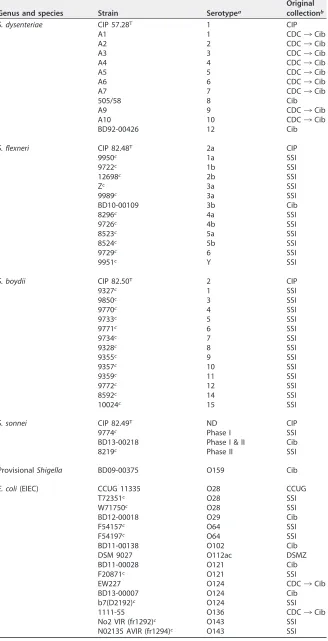
TABLE 1Original identification and original collection of the isolates used in this study
Genus and species Strain Serotypea
Original collectionb
S. dysenteriae CIP 57.28T 1 CIP
A1 1 CDC¡Cib
A2 2 CDC¡Cib
A3 3 CDC¡Cib
A4 4 CDC¡Cib
A5 5 CDC¡Cib
A6 6 CDC¡Cib
A7 7 CDC¡Cib
505/58 8 Cib
A9 9 CDC¡Cib
A10 10 CDC¡Cib
BD92-00426 12 Cib
S. flexneri CIP 82.48T 2a CIP
9950c 1a SSI
9722c 1b SSI
12698c 2b SSI
Zc 3a SSI
9989c 3a SSI
BD10-00109 3b Cib
8296c 4a SSI
9726c 4b SSI
8523c 5a SSI
8524c 5b SSI
9729c 6 SSI
9951c Y SSI
S. boydii CIP 82.50T 2 CIP
9327c 1 SSI
9850c 3 SSI
9770c 4 SSI
9733c 5 SSI
9771c 6 SSI
9734c 7 SSI
9328c 8 SSI
9355c 9 SSI
9357c 10 SSI
9359c 11 SSI
9772c 12 SSI
8592c 14 SSI
10024c 15 SSI
S. sonnei CIP 82.49T ND CIP
9774c Phase I SSI
BD13-00218 Phase I & II Cib
8219c Phase II SSI
ProvisionalShigella BD09-00375 O159 Cib
E. coli(EIEC) CCUG 11335 O28 CCUG
T72351c O28 SSI
W71750c O28 SSI
BD12-00018 O29 Cib
F54157c O64 SSI
F54197c O64 SSI
BD11-00138 O102 Cib
DSM 9027 O112ac DSMZ
BD11-00028 O121 Cib
F20871c O121 SSI
EW227 O124 CDC¡Cib
BD13-00007 O124 Cib
b7(D2192)c O124 SSI
1111-55 O136 CDC¡Cib
No2 VIR (fr1292)c O143 SSI
N02135 AVIR (fr1294)c O143 SSI
(Continued on next page)
on May 16, 2020 by guest
http://jcm.asm.org/
was performed using a Biometra TProfessional standard gradient thermocycler (Westburg, Leusden, The Netherlands), with the following program: 95°C for 3 min, followed by 35 cycles consisting of 95°C for 1 min, 57°C for 1 min, and 72°C for 1 min, and an elongation for 7 min at 72°C. As a mastermix, illustra PuReTaq Ready-To-Go PCR beads (GE Healthcare Life Sciences, Eindhoven, The Netherlands) were used, supplemented with the following primers designed for amplification of a conservative part of theipaH gene present in all differentipaHalleles (20): forward primer, 5=-TGG AAA AAC TCA GTG CCT C-3=; and reverse primer, 5=-CCA GTC CGT AAA TTC ATT CTC-3=. As an internal control for presence of bacterial DNA, a conservative part of the bacterial 16S rRNA gene was amplified with the following primers: forward primer, 5=-AGA GTT TGA TCM TGG YTC AG-3=; and reverse primer, 5=-CTT TAC GCC CAR TRA WTC CG-3=. All primers were used in a final concentration of 0.2 pmol/l.
The ipaH-positive isolates were subjected to the following phenotypic tests: oxidase, catalase, motility at 22°C and 37°C, growth on MacConkey agar and Salmonella Shigella agar (SS agar), gas from D-glucose, ornithine decarboxylase (ODC), indole, esculin hydrolysis,ortho-nitrophenyl--galactoside (ONPG), and fermentation of D-glucose, lactose, D-sucrose, D-xylose, D-mannitol, dulcitol, salicin, D-raffinose, andD-glycerol in Andrade peptone water (21), lysine decarboxylase (LDC [22]), and arginine dihydrolase (ADH [23]).
Next to the phenotypical tests, classicalShigellaserotyping was performed with all availableShigella antisera obtained from Denka Seiken Co., Ltd. (Tokyo, Japan), complemented withS. flexneriMASF IV-1, MASF IV-2, MASF 1c, and MASF B from Reagensia AB (Solna, Sweden). If slide agglutination was negative for all polyvalent antisera or an inconclusive serotype was obtained, a suspension of the isolate was boiled for 1 h, after which slide agglutination was performed again.
ClassicalE. coliO serotyping was manually performed with antisera forE. coliO1 until O187, prepared as previously described (24, 25) or purchased from Statens Serum Institut (Copenhagen, Denmark). O-antigen suspensions were prepared by boiling an overnight broth culture for 1 h to inactivate the K antigen. These prepared antigens, diluted (optical density at 600 nm [OD600], 0.44) with formalinized
(0.5%) phosphate-buffered saline (PBS), were stained with gentian violet (0.005%) and tested against the 187 O antisera in microtiter plate agglutination tests. After overnight incubation at 37°C, plates were examined against a light background, and positive reactions were titrated. O-type reactions with titers ofⱖ2,500, and reactions with titers until two steps lower than the reaction of the homologous standard were considered positive.
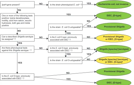
With the results of the above-described molecular, biochemical, and serological tests, an identifica-tion algorithm was applied as shown in Fig. 1, based on a previously described key (Fig. 2 in reference 26). A result was considered inconclusive if a distinction between aShigellaspecies and EIEC could not be made and the serotypes are not described as related.
Molecular algorithm. The molecular algorithm was designed to screen fecal samples for the
[image:4.585.41.375.82.278.2]presence ofShigellaspp./EIEC quickly and accurately. However, in this study, only pure cultures were examined; thus, only the molecular part of the algorithm that follows bacterial isolation was applied (de Boer et al., unpublished data).
TABLE 1(Continued)
Genus and species Strain Serotypea
Original collectionb
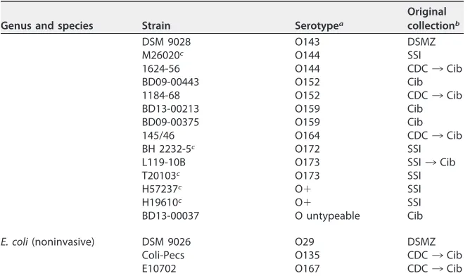
DSM 9028 O143 DSMZ
M26020c O144 SSI
1624-56 O144 CDC¡Cib
BD09-00443 O152 Cib
1184-68 O152 CDC¡Cib
BD13-00213 O159 Cib
BD09-00375 O159 Cib
145/46 O164 CDC¡Cib
BH 2232-5c O172 SSI
L119-10B O173 SSI¡Cib
T20103c O173 SSI
H57237c O⫹ SSI
H19610c O⫹ SSI
BD13-00037 O untypeable Cib
E. coli(noninvasive) DSM 9026 O29 DSMZ
Coli-Pecs O135 CDC¡Cib
E10702 O167 CDC¡Cib
aShigellaserotype in case ofShigellaspp. orE. coliO type in case ofE. colior provisionalShigella. ND, not determined.
bCIP, Collection de l’Institut Pasteur, Paris, France; CDC, Centers for Disease Control and Prevention, Atlanta, GA, USA; Cib, Centre for Infectious Disease Control, Bilthoven, The Netherlands; CDC/SSI¡Cib, historical isolates donated to Cib by the CDC or SSI, respectively, for antiserum preparation and validation from 1950s to 1980s; SSI, Statens Serum Institut, Copenhagen, Denmark; CCUG, Culture Collection, University of Göteborg, Sweden; DSMZ, Leibniz-Institut DSMZ-Deutsche Sammlung von Mikroorganismen und Zellkulturen GmbH, Braunschweig, Germany.
cProvided by F. Scheutz, SSI.
on May 16, 2020 by guest
http://jcm.asm.org/
Briefly, lysates were prepared as described above. A real-time PCR to target theipaHand thewzx genes of S. sonneiphase I,S. flexneriserotype 1-5,S. flexneri 6, andS. dysenteriaeserotype 1 was performed on a ABI 7500 sequence detection system (Applied Biosystems, Nieuwerkerk aan den IJssel, The Netherlands), as described previously (11). Each 25-l reaction mixture consisted of 5l template DNA, 1⫻Fast Advanced TaqMan Universal PCR master mix (Applied Biosystems), and 2.5g bovine serum albumin (Roche Diagnostics Netherlands B.V., Almere, The Netherlands). The primers and probes used for detection were designed based on the sequence ofwzxgenes, as described previously (27, 28). Reactions were performed under the following conditions: 50°C for 2 min, 95°C for 20 s, followed by 40 cycles of 95°C for 3 s, and 60°C for 32 s. With the result of theipaHgene PCR, a distinction between Shigella/EIEC and noninvasiveE. coliwas made. Positivity of awzxgene, in an expected ratio with a threshold cycle (CT) value of theipaHgene according to copy number (20), leads to the corresponding
serotype. If the ipaH gene had aCTvalue below 35 but all tested wzxgenes were negative, the
identification is inconclusive and was interpreted as EIEC,S. boydii,S. sonneiphase II, orS. dysenteriae serotype 2-15.
Discrepancy analysis using whole-genome sequencing.WGS analysis was performed on seven
isolates to solve discrepancies between the here-proposed algorithms and original identification (Tables 2 and 3). Isolates were cultured overnight at 37°C on CSA. For each isolate, an equivalent to 5l of colonies was suspended in 300l MicroBead solution, and DNA was extracted with the UltraClean microbial DNA isolation kit (Mo Bio Laboratories, Carlsbad, CA, USA). The DNA library was prepared with the Nextera XT version 2 index kit (Illumina, San Diego, CA, USA). Subsequently, the library was sequenced on a MiSeq sequencer (Illumina, Inc.), using a MiSeq reagent kit version 3 generating 300-bp paired-end reads.
Quality control, quality trimming, and de novo assembly was performed using CLC Genomics Workbench, version 9.1.1 (Qiagen, Aarhus, Denmark). A quality limit of 0.01 was used in trimming, and a word size of 29 and a minimum contig length of 1,000 bp were used inde novoassembly. Other parameters were set as default.
E. coliO types were predicted using SerotypeFinder (Center for Genomic Epidemiology, Lyngby, Denmark). To predict the serotype ofShigella, trimmed reads of the isolates were mapped against references of theS. flexneriO-antigen genes (29) and the O-antigen gene clusters ofS. dysenteriae,S. boydii, andS. sonnei(28). To our knowledge,S. dysenteriaeserotypes 14 and 15 are rare, and the sequence of their O antigens is not known; therefore, these serotypes were not evaluatedin silico. ThetnaCABgene cluster andrrlBgene were used as references for indole production from tryptophan and themtlA,mtlD,
Is the E. coli O-type previously associated with EIEC d?
YES
FIG 1Culture-dependent algorithm. Green, definitive identification; yellow, inconclusive identification; a, Strockbine et al. (32); b,
manufacturer’s protocol forShigellaantisera set 1, as per Denka Seiken, Sun et al. (41, 43), and Carlin et al. (44); c, Bopp et al. (13); d, O28ac, O29, O42, O96, O112ac, O115, O121, O124, O135, O136, O143, O144, O152, O159, O164, O167, O173, and O untypeable; e, if Shigellaserotype has a known relation toE. coliO type, identification isShigella[species] [serotype]; see Ewing (31), Cheasty and Rowe
(45), Liu et al. (39), and Perepelov et al. (42).
on May 16, 2020 by guest
http://jcm.asm.org/
[image:5.585.41.471.83.356.2]andmtlRgenes as references for the fermentation ofD-mannitol. All genes and gene clusters were retrieved from NCBI (see Table S2 in the supplemental material). If reads mapped with one or more mutations, the functionality of the encoded proteins was assessed using ExPASy (Swiss Institute of Bioinformatics [SIB] [30]) and BLASTp (NCBI, Bethesda, MD, USA).
The de novo assemblies were imported in SeqSphere⫹ version 3.5.1 (Ridom GmbH, Münster,
Germany), including reference genomes retrieved from NCBI, to assess the homologies of the discrepant strains with the references. A comparison of the sequences was made using theE. colicore-genome multilocus sequence typing (cgMLST) genotyping scheme, which is based on the EnteroBaseEscherichia/ ShigellacgMLST version 1 scheme (https://enterobase.warwick.ac.uk/species/index/ecoli). The resulting comparison table was imported in BioNumerics, version 7.6.3 (Applied Maths, NV), and a neighbor joining tree was inferred using 200⫻bootstrap resampling. The tree with the highest resampling support was calculated. The accession numbers of all sequences are depicted in Fig. 2.
Accession number(s).The sequences of discrepant isolates were submitted to the European
Nucleotide Archive (ENA, EMBL-EBI, Cambridge, United Kingdom) as study no. PRJEB24877with accession numbersERR2287281(isolate 12698),ERR2287282(isolate 505/58),ERR2287283(isolate 9355),
ERR2300644 (isolate F54157), ERR2300645 (isolate F54197), ERR2300646 (isolate H57237), and
ERR2300647(isolate Z) (https://www.ebi.ac.uk/ena).
RESULTS
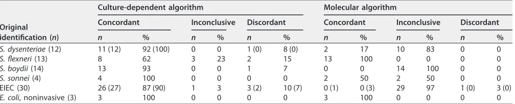
Culture-dependent algorithm. With the culture-dependent algorithm, an incon-clusive result was obtained for four isolates (Table 2). For these isolates, a distinction between EIEC and eitherS. flexneri,S. boydii, orS. dysenteriaewas impossible, and the Shigella O type has no known relationship to theE. coliO type. Only S. sonneiand noninvasiveE. coliisolates were completely concordant with the original identification, including the inconclusive results. The obtained percentages of concordance were 92%, 85%, 93%, and 90% forS. dysenteriae,S. flexneri,S. boydii, and EIEC isolates, respectively (Table 2).
Molecular algorithm.For 55 isolates (72%), only theipaHgene was detected and none of the assessedwzxgenes detected using the molecular algorithm. These isolates were binned in the rest group, meaning they can be either EIEC,S. sonneiphase II,S. boydii, orS. dysenteriaeserotypes other than 1. All isolates except for one EIEC strain (97%) were identified in concordance with the original identification or had an incon-clusive result, of which one of the results was in concordance with original identifica-tion (Table 2). One isolate had a discordant identificaidentifica-tion, although the result of the molecular algorithm was in concordance with the culture-dependent algorithm (strain H57237, Table 3).
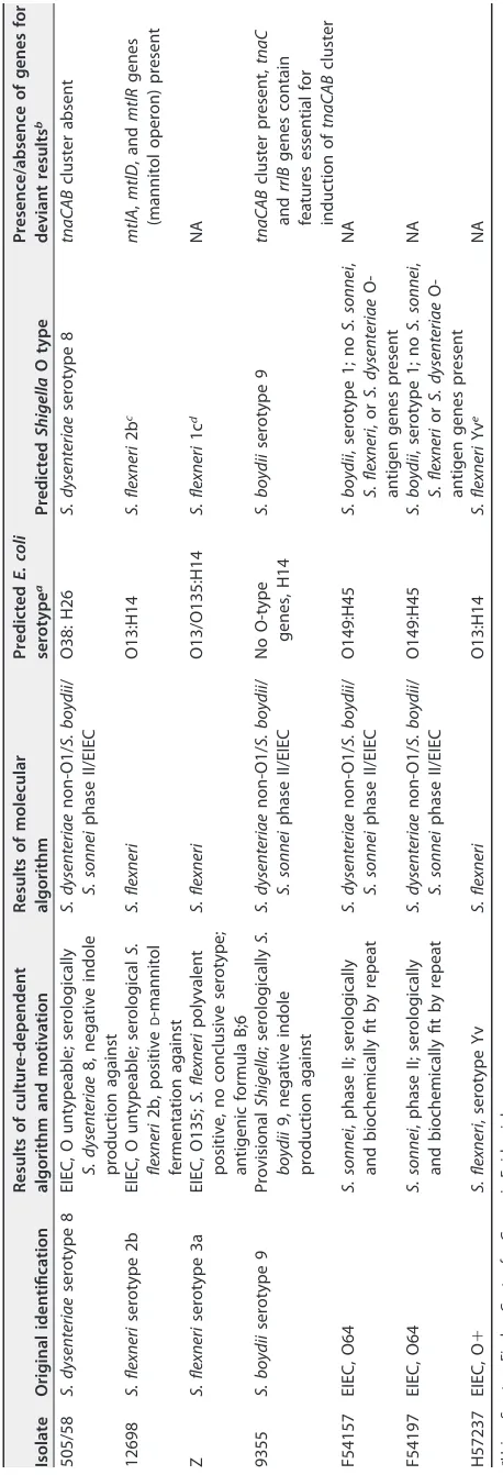
Discrepancy analysis of discordant results. Seven isolates showed discordant results with the original identification using the culture-dependent algorithm (Table 2), and a discrepancy analysis using WGS was carried out (Table 3). The predictedE. coli andShigellaserotypes and the presence of genes that encode for specific features are displayed in Table 3, as well as the results of the two tested algorithms (Table 3). The clustering of the discrepant isolates with reference isolates is shown in the cgMLST analysis (Fig. 2).
[image:6.585.42.546.83.185.2]In the discrepancy analysis of isolate 505/58, WGS data confirmed the serotype as determined at original identification and with the culture-dependent algorithm, as the predicted serotypes areE. coliO38 andS. dysenteriaeserotype 8, which are related to
TABLE 2Results of identification with culture-dependent and molecular algorithm compared to original identificationa
Original identification (n)
Culture-dependent algorithm Molecular algorithm
Concordant Inconclusive Discordant Concordant Inconclusive Discordant
n % n % n % n % n % n %
S. dysenteriae(12) 11 (12) 92 (100) 0 0 1 (0) 8 (0) 2 17 10 83 0 0
S. flexneri(13) 8 62 3 23 2 15 13 100 0 0 0 0
S. boydii(14) 13 93 0 0 1 7 0 0 14 100 0 0
S. sonnei(4) 4 100 0 0 0 0 2 50 2 50 0 0
EIEC (30) 26 (27) 87 (90) 1 3 3 (2) 10 (7) 0 (1) 0 (3) 29 97 1 (0) 3 (0)
E. coli, noninvasive (3) 3 100 0 0 0 0 3 100 0 0 0 0
aConcordant or discordant refers to comparison with the original identification (Table 1). For inconclusive identification, the original identification is in concordance with one of the results. Values in parentheses are the results after discrepancy analysis.
on May 16, 2020 by guest
http://jcm.asm.org/
TABLE 3 Discrepancy analysis of isolates with discordant results based on the culture dependent algorithm Isolate Original identification Results of culture-dependent algorithm and motivation Results of molecular algorithm Predicted E. coli serotype a Predicted Shigella O type Presence/absence of genes for deviant results b 505/58 S. dysenteriae serotype 8 EIEC, O untypeable; serologically S. dysenteriae 8, negative indole production against S. dysenteriae non-O1/ S. boydii / S. sonnei phase II/EIEC O38: H26 S. dysenteriae serotype 8 tnaCAB cluster absent 12698 S. flexneri serotype 2b EIEC, O untypeable; serological S. flexneri 2b, positive D -mannitol fermentation against S. flexneri O13:H14 S. flexneri 2b c mtlA , mtlD , and mtlR genes (mannitol operon) present Z S. flexneri serotype 3a EIEC, O135; S. flexneri polyvalent positive, no conclusive serotype; antigenic formula B;6 S. flexneri O13/O135:H14 S. flexneri 1c d NA 9355 S. boydii serotype 9 Provisional Shigella ; serologically S. boydii 9, negative indole production against S. dysenteriae non-O1/ S. boydii / S. sonnei phase II/EIEC No O-type genes, H14 S. boydii serotype 9 tnaCAB cluster present, tnaC and rrlB genes contain features essential for induction of tnaCAB cluster F54157 EIEC, O64 S. sonnei , phase II; serologically and biochemically fit by repeat S. dysenteriae non-O1/ S. boydii / S. sonnei phase II/EIEC O149:H45 S. boydii , serotype 1; no S. sonnei , S. flexneri ,o r S. dysenteriae O-antigen genes present NA F54197 EIEC, O64 S. sonnei , phase II; serologically and biochemically fit by repeat S. dysenteriae non-O1/ S. boydii / S. sonnei phase II/EIEC O149:H45 S. boydii , serotype 1; no S. sonnei , S. flexneri or S. dysenteriae O-antigen genes present NA H57237 EIEC, O ⫹ S. flexneri , serotype Yv S. flexneri O13:H14 S. flexneri Yv e NA aUsing SerotypeFinder, Center for Genomic Epidemiology. bNA, not applicable. cwzx 1-5 , gtrII , and gtrX present. dwzx 1-5 , gtrI , gtrIc , and oac present. ewzx 1-5 and lpt-O present.
on May 16, 2020 by guest
http://jcm.asm.org/
[image:7.585.76.305.71.742.2]FIG 2Neighbor-joining tree for core-genome MLST, with distance based onE. colicgMLST (EnteroBase) scheme, 2,513 columns, 200⫻bootstrapped. Accession numbers are from GenBank or EMBL; Black, isolates 505/58, 12698, Z, 9355, F54157, F54197, and H57237; light blue,S. dysenteriae; red,S. flexneri; green,S. boydii; pink,S. sonnei; blue, EIEC; gray, other pathotypes ofE. coli.
on May 16, 2020 by guest
http://jcm.asm.org/
[image:8.585.42.546.70.662.2]each other (31). However, because indole was negative, while all strains described in the literature fromS. dysenteriaeserotype 8 are capable of producing indole (13, 31), and because theE. coliO antigen is not typeable phenotypically (32), isolate 505/58 was identified as EIEC O-untypeable using the culture-dependent algorithm. WGS data confirmed that the tnaCABcluster, which contains the functional genes for the pro-duction of indole from tryptophan (33), is absent in 505/58. cgMLST showed that isolate 505/58 clustered with an EIEC reference genome and not with other S. dysenteriae reference genomes in this analysis (Fig. 2). The molecular algorithm placed 505/58 in the rest group, which is in concordance with the original identification, as well as with the culture-dependent algorithm (Table 3). The clustering combined with the absence of thetnaCABcluster indicates that 505/58 was originally misidentified asS. dysenteriae or that it has lost thetnaCABcluster over time.
With isolate 12698, WGS data confirmed the serotype as determined at the original identification and with the culture-dependent algorithm to beS. flexneriserotype 2b. The molecular algorithm confirmed these results, as it detected the presence of the wzx1-5 gene (Table 3). However, using the culture-dependent algorithm, 12698 was
repeatedlyD-mannitol positive, while all describedS. flexneri serotype 2b isolates are
D-mannitol negative (13, 31). BecauseD-mannitol was positive and theE. coliO type is
untypeable (32), 12698 was identified as EIEC O untypeable using the culture-dependent algorithm. The WGS data confirmed the D-mannitol-positive result, as it detected the mtlAandmtlDgenes and its regulatormtlR(34). However, despite the positive result of D-mannitol fermentation, isolate 12698 clustered with S. flexneri reference isolates using cgMLST (Fig. 2), supporting the original identification, as well as the classical andin silicoserotyping to designate isolate 12698S. flexneriserotype 2b. Discrepancy analysis using WGS for isolate Z added an additional identification instead of confirming one of the other results. Isolate Z was originally identified asS. flexneri3a, while with the culture-dependent algorithm, the isolate fit phenotypically to S. flexneri3a but had a serologically inconclusive serotype with antigenic formula B;6. Because theShigellaantigenic formula was inconclusive and theE. coliO type was O135 (14), isolate Z was identified as EIEC O135 with the culture-dependent algorithm. WGS analysis detected the presence of the followingS. flexnerigenes and clusters in isolate Z:wzx1-5,oac,gtrI,andgtr1C, resulting inS. flexneriserotype 1c (Table 3). Although the
completely conservedgtrIandgtrIcclusters are present, including thegtrA andgtrB genes (35, 36), with classicalShigellaserotyping, agglutination with type I and MASF 1c antisera was absent. In the cgMLST analysis, isolate Z clustered withS. flexnerireference isolates (Fig. 2). The molecular algorithm identified isolate Z asS. flexneri; however, this algorithm is not able to distinguish different serotypes (Table 3). To summarize, classical andin silico serotyping, cgMLST analysis, and the result of the molecular algorithm confirmed the original identification of isolate Z asS. flexneribut with discordances in its serotype.
In the discrepancy analysis of isolate 9355, WGS data confirmed the serotype as determined at the original identification and with the culture-dependent algorithm to be S. boydii serotype 9. However, because indole is negative, while this should be positive forS. boydiiserotype 9 (13, 31), and theE. coliO type is O132, which has never been associated with EIEC, isolate 9355 was provisionally identified asShigellausing the culture-dependent algorithm. The molecular algorithm placed 9355 in the rest group, which is in concordance with original identification as well as with the culture-dependent algorithm (Table 3). The WGS data suggest that the wholetnaCABcluster is present in isolate 9355 and contains the indole production genestnaA,tnaB, andtnaC (33), which all encode functional proteins. Furthermore, all necessary features for the induction oftnaAandtnaBgenes are present in thetnaCandrrlBgenes (37, 38). The mechanism that hinders the production of indole could not be determined by assessing the presence or absence of functional genes and features and is a subject for further investigation. Isolate 9355 clustered withS. dysenteriaegenomes in the cgMLST anal-ysis. As clustering of S. boydii with S. dysenteriae was described before (5), cgMLST
on May 16, 2020 by guest
http://jcm.asm.org/
supports the original identification and the classical andin silicoserotype to designate isolate 9355S. boydiiserotype 9.
For isolates F54157 and F54197, discrepancy analysis using WGS added an addi-tional identification instead of confirming one of the other results. They were originally identified as EIEC O64 and asS. sonnei phase II in the culture-dependent algorithm; however, they were predicted to beE. coliO149 andS. boydiiserotype 1 with WGS data (Table 3), which were described as identical antigens (31, 39). Agglutination with S. sonneiphase II antiserum in the culture-dependent algorithm could be explained by linkage between enterobacterial common antigen, which is a surface antigen present inEnterobacteriaceae, andS. sonneiphase II core oligosaccharide (40). With the molec-ular algorithm, isolates F54157 and F54197 were binned in the rest group, which is in concordance with the original identification, with the culture-dependent algorithm and with WGS data. Evaluation of theS. boydiiserotype 1 O-antigen cluster in the WGS data in more detail showed intactwzxandwzygenes but major deletions in thermlBgene for both isolates, explaining the lack of expression of the S. boydiiserotype 1/E. coli O149 phenotype (39). In the cgMLST analysis, strains F54157 and F54197 clustered with S. dysenteriae and S. boydii strains. Overall, the discrepancy analysis based on WGS showed that isolates F54157 and F54197 were originally misidentified as EIEC with O type O64 and misidentified with the culture-dependent algorithm asS. sonneiphase II. Isolate H57237 was originally identified as EIEC; however, both algorithms used in this study identified this isolate as S. flexneri. The serotype of H57237 is Yv, as determined by the culture-dependent algorithm and confirmed by the WGS analysis (Table 3). Serotype Yv has only recently been described (41), and probably, the original identification of this isolate predates the discovery of this novel serotype.
The discrepancy analysis showed that isolates H57237, F54157, F54197, and 505/58 might be misidentified during the original identification (Table 3 and Fig. 2). The results of the comparison of the molecular and culture-dependent algorithms with the original identification were corrected for these findings and are displayed in parentheses in Table 2.
DISCUSSION
After discrepancy analysis, the identification ofS. dysenteriae,S. sonnei, and nonin-vasiveE. coliisolates with the culture-dependent algorithm was 100% in concordance with the original identification, including the inconclusive results. For S. flexneri, S. boydii, and EIEC isolates, the concordance was 85%, 93%, and 93%, respectively.
With the molecular algorithm, 100% of the isolates were identified in concordance with the original identification after discrepancy analysis (Table 3). However, its reso-lution for certain serotypes is low, as it does not allow specific detection of EIEC, S. boydii,S. sonneiphase II, andS. dysenteriaeserotype 2-15. Another limitation is that cross-reactivity ofShigellaandE. coliO antigens is described. The primers from theS. dysenteriae wzxgene are likely to amplify theE. coliO-antigen clusters O1, O120, and O148 (31, 39), and the primers from theS. flexneri wzx1-5gene will probably amplify the
E. coliO-antigen clusters O1, O13, O16, O19, O62, O69, O73, O135, and O147 (31, 42). Of all theseE. coliO types, only O135 is described as an EIEC-associated O type; none of the otherE. coliO types are likely to possess theipaHgene and are therefore not considered to beShigellaspp. or EIEC in the molecular algorithm. Nevertheless, EIEC with O type O135 cannot be separated fromS. flexneri. However, this is overcome in a diagnostic setting by targeted culture from the fecal samples prompted by the results of the molecular part of the algorithm. If an isolate is selected, it is identified based on a few phenotypical key features and agglutination withShigellaand EIEC polyvalent antisera. If no isolate is selected, the physician will receive a report thatShigellaspp. or EIEC is detected but without specifications about species or serotype.
One of the strengths of this study is the discrepancy analysis with WGS. This analysis is able to confirm one of the determined identities of isolates 505/58, 12698, 9355, and H57237. In contrast to those isolates, for isolates Z, F54157, and F54197, the
on May 16, 2020 by guest
http://jcm.asm.org/
ancy analysis with WGS added an extra identification result, therefore complicating the identification further instead of clarifying it.
Isolates 12698, 9355, and 505/58 were serological congruent using all identification methods, including WGS, but had one phenotypical test in discordance with their serotype (Table 3), resulting in a different identification by the culture-dependent algorithm. Phenotypical properties of a serotype are described by testing multiple isolates of the same serotype. There is not necessarily a causal connection between the serotype and the results of phenotypic tests, and phenotypic variability increases with the number of tested isolates. If the culture-dependent algorithm was applied less stringently and one phenotypical test against it was allowed, the above-described isolates were correctly identified. However, disregarding phenotypic test results should be considered carefully, because some phenotypic traits are set as defining for genus or species, for instance, the absence of LDC or D-mannitol fermentation, which are genus specific forShigellaor set as species specific forS. dysenteriae,respectively. The results of these species specific phenotypic tests should not be disregarded.
A limitation of this study is that only a few isolates of every species were used, and it is desirable to test more isolates with the proposed algorithms in the future. However, rare serotypes were difficult to obtain, and one can debate to omit these rare serotypes for test evaluation, because they are not frequently encountered in clinical diagnostics. The here-described culture-dependent algorithm outperforms the previously de-scribed method based on the detection of theuidAgene and thelacYgene (16) that only correctly identifiedin silico100% ofS. sonnei, 92% ofS. flexneri, 86% ofS. boydii, 80% of S. dysenteriae, 77% of noninvasive E. coli, and 62% of EIEC isolates (6). In addition, thelacYgene approach is able to distinguish organisms to the genus level (16); therefore, its resolution is lower than that of the culture-dependent algorithm described in this study.
The previously described k-mer-based method outperforms the here-described culture-dependent algorithm for the identification ofShigellaspecies, because it iden-tified 100% of all Shigella species isolates in concordance with biochemical and serological profiling. In contrast, for identification of EIEC isolates, the proposed culture-dependent algorithm is superior, identifying 93% of EIEC isolates according to original identification, against 81.5% of EIEC isolates with the k-mer based approach (18). Furthermore, for the k-mer-based method, sequencing of whole genomes and subse-quent bioinformatics analysis are required, making it less applicable in low-resource settings, where Shigella spp. are encountered frequently. Moreover, to match the resolution of the culture-dependent algorithm, extra analyses should be added to the k-mer-based method in order to determine thein silicoserotype.
This study shows again that species differentiation of Shigellaspp. and E. coliis challenging, as other studies have concluded before (5, 6, 18). With some isolates, differentiation is impossible, as evidenced by the percentage of isolates (5%) for which identification is inconclusive with the culture-dependent algorithm. Using the molec-ular algorithm, 71% of the isolates resulted in an inconclusive identification; however, this algorithm was not designed for use in the distinction between EIEC,S. boydii,S. sonneiphase II, andS. dysenteriaeserotype 2-15. Nevertheless, the molecular algorithm would be sufficient for use in a developed country, because a recent study in The Netherlands (R. F. de Boer, unpublished data) showed that in 80% ofipaHgene-positive fecal samples,S. sonneiorS. flexneriis present. For use in other regions, the concept of the molecular algorithm can be adjusted to their particular needs; targets ofwzxgenes ofS. dysenteriaeandS. boydiican be added or the whole procedure can be redefined to a conventional PCR platform if real-time platforms are unavailable.
In conclusion, although not perfect, the proposed algorithms are capable of iden-tifying mostShigellasp. and EIEC isolates. The molecular algorithm is fast and accurate and is suitable for daily application in diagnostic laboratories, as it can be performed with standard PCR equipment; however, its resolution for certain serotypes is low. The culture-dependent algorithm is more time-consuming, and many phenotypical tests and antisera are required, yet the resolution is high for all serotypes. If a desirable
on May 16, 2020 by guest
http://jcm.asm.org/
complete identification cannot be obtained with the molecular algorithm, the culture-dependent algorithm can be applied by a reference laboratory to obtain a higher resolution.
Despite the genetic relationship ofShigella spp. and EIEC, causing difficulties for identification, differentiation is still necessary for epidemiological and surveillance purposes because of current guidelines for infectious disease control. One can specu-late if guidelines need to be adjusted, but evidence for guideline optimization with regard to infections with EIEC is currently lacking. In the future, the impact of infections with EIEC on individual patients and on public health should be further investigated to assess if it is justified that surveillance measures and control guidelines for infections with EIEC are different from those of shigellosis.
SUPPLEMENTAL MATERIAL
Supplemental material for this article may be found athttps://doi.org/10.1128/JCM .00510-18.
SUPPLEMENTAL FILE 1,PDF file, 0.1 MB.
ACKNOWLEDGMENTS
We thank Flemming Scheutz from the Statens Serum Institut (Copenhagen, Den-mark) for providing strains.
This research received no specific grant from any funding agency in the public, commercial, or not-for-profit sectors.
REFERENCES
1. Lampel KA, Formal SB, Maurelli AT. 2018. A brief history ofShigella. EcoSal Plus 8:3.https://doi.org/10.1128/ecosalplus.ESP-0006-2017. 2. Hale TL. 1991. Genetic basis of virulence in Shigella species. Microbiol
Rev 55:206 –24.
3. Brenner DJ, Fanning GR, Steigerwalt AG, Orskov I, Orskov F. 1972. Polynucleotide sequence relatedness among three groups of patho-genic Escherichia coli strains. Infect Immun 6:308 –15.
4. Pupo GM, Lan R, Reeves PR. 2000. Multiple independent origins of Shigella clones of Escherichia coli and convergent evolution of many of their characteristics. Proc Natl Acad Sci U S A 97:10567–72.https://doi
.org/10.1073/pnas.180094797.
5. Sahl JW, Morris CR, Emberger J, Fraser CM, Ochieng JB, Juma J, Fields B, Breiman RF, Gilmour M, Nataro JP, Rasko DA. 2015. Defining the phy-logenomics of Shigella species: a pathway to diagnostics. J Clin Micro-biol 53:951– 60.https://doi.org/10.1128/JCM.03527-14.
6. Pettengill EA, Pettengill JB, Binet R. 2015. Phylogenetic analyses of Shigella and enteroinvasive Escherichia coli for the identification of molecular epidemiological markers: whole-genome comparative analy-sis does not support distinct genera designation. Front Microbiol 6:1573.
https://doi.org/10.3389/fmicb.2015.01573.
7. Lan R, Alles MC, Donohoe K, Martinez MB, Reeves PR. 2004. Molecular evolutionary relationships of enteroinvasiveEscherichia coliand Shi-gellaspp. Infect Immun 72:5080 – 8.https://doi.org/10.1128/IAI.72.9
.5080-5088.2004.
8. Hazen TH, Leonard SR, Lampel KA, Lacher DW, Maurelli AT, Rasko DA. 2016. Investigating the relatedness of enteroinvasive Escherichia coli to other E. coli and Shigella isolates by using comparative genomics. Infect Immun 84:2362–71.https://doi.org/10.1128/IAI.00350-16.
9. van den Beld MJ, Friedrich AW, van Zanten E, Reubsaet FA, Kooistra-Smid MA, Rossen JW, participating Medical Microbiological Laboratories. 2016. Multicenter evaluation of molecular and culture-dependent diag-nostics for Shigella species and entero-invasive Escherichia coli in the Netherlands. J Microbiol Methods 131:10 –15.https://doi.org/10.1016/j
.mimet.2016.09.023.
10. Van Lint P, De Witte E, Ursi JP, Van Herendael B, Van Schaeren J. 2016. A screening algorithm for diagnosing bacterial gastroenteritis by real-time PCR in combination with guided culture. Diagn Microbiol Infect Dis 85:255–9.https://doi.org/10.1016/j.diagmicrobio.2016.03.017.
11. de Boer RF, Ott A, Kesztyus B, Kooistra-Smid AM. 2010. Improved detec-tion of five major gastrointestinal pathogens by use of a molecular
screening approach. J Clin Microbiol 48:4140 – 6. https://doi.org/10
.1128/JCM.01124-10.
12. Venkatesan MM, Buysse JM, Kopecko DJ. 1989. Use ofShigella flexneri ipaCandipaHgene sequences for the general identification ofShigella spp. and enteroinvasiveEscherichia coli. J Clin Microbiol 27:2687–91. 13. Bopp CA, Brenner FW, Fields PI, Wells JG, Strockbine NA. 2003.
Esche-richia, Shigella and Salmonella, p 654 – 671.In Murray PR, Baron EJ, Jorgensen JH, Pfaller MA, Yolken RH (ed), Manual of clinical microbiol-ogy, 8th ed, vol 1. ASM Press, Washington, DC.
14. Scheutz F, Strockbine NA. 2005. Genus I. Escherichia Castellani and Chalmers 1919, 9417, p 607– 624.InGarrity GM (ed), Bergey’s manual of systematic bacteriology, 2nd ed, vol 2. The Proteobacteria. Springer Science, New York, NY.
15. Silva RM, Toledo MR, Trabulsi LR. 1980. Biochemical and cultural char-acteristics of invasive Escherichia coli. J Clin Microbiol 11:441– 4. 16. Pavlovic M, Luze A, Konrad R, Berger A, Sing A, Busch U, Huber I. 2011.
Development of a duplex real-time PCR for differentiation between E. coli and Shigella spp. J Appl Microbiol 110:1245–51.https://doi.org/10
.1111/j.1365-2672.2011.04973.x.
17. Kim HJ, Ryu JO, Song JY, Kim HY. 2017. Multiplex polymerase chain reaction for identification of shigellae and four Shigella species using novel genetic markers screened by comparative genomics. Foodborne Pathog Dis 14:400 – 406.https://doi.org/10.1089/fpd.2016.2221. 18. Chattaway MA, Schaefer U, Tewolde R, Dallman TJ, Jenkins C. 2017.
Identification of Escherichia coli and Shigella species from whole-genome sequences. J Clin Microbiol 55:616 – 623. https://doi.org/10
.1128/JCM.01790-16.
19. Lobersli I, Wester AL, Kristiansen A, Brandal LT. 2016. Molecular differ-entiation of Shigella spp. from enteroinvasive E. coli. Eur J Microbiol Immunol (Bp) 6:197–205.https://doi.org/10.1556/1886.2016.00004. 20. Buysse JM, Hartman AB, Strockbine N, Venkatesan M. 1995. Genetic
polymorphism of theipaHmulticopy antigen gene inShigellaspps. and enteroinvasiveEscherichia coli. Microb Pathog 19:335– 49. https://doi
.org/10.1016/S0882-4010(96)80005-2.
21. Barrow GI, Feltham RKA. 1993. Cowan and Steel’s manual for the iden-tification of medical bacteria, 3rd ed. Cambridge University Press, Cam-bridge, United Kingdom.
22. Difco Laboratories. 1984. Difco manual. Difco Laboratories, Inc., De-troit, MI.
23. Lenette EH. 1985. Manual of clinical microbiology, 4th ed. American Society for Microbiology, Washington, DC.
on May 16, 2020 by guest
http://jcm.asm.org/
24. Ewing WH. 1986. The genusEscherichia, p 93–134.InEwing WH (ed), Edwards and Ewing’s identification of Enterobacteriaceae, 4th ed. Elsevier Science Publishing Co. Inc., New York, NY.
25. Guinée PA, Agterberg CM, Jansen WH. 1972. Escherichia coli O antigen typing by means of a mechanized microtechnique. Appl Microbiol 24: 127–31.
26. van den Beld MJ, Reubsaet FA. 2012. Differentiation betweenShigella, enteroinvasiveEscherichia coli(EIEC) and noninvasiveEscherichia coli. Eur J Clin Microbiol Infect Dis 31:899 –904.https://doi.org/10.1007/s10096
-011-1395-7.
27. Houng HS, Sethabutr O, Echeverria P. 1997. A simple polymerase chain reaction technique to detect and differentiate Shigella and enteroinva-sive Escherichia coli in human feces. Diagn Microbiol Infect Dis 28:19 –25.
https://doi.org/10.1016/S0732-8893(97)89154-7.
28. Li Y, Cao B, Liu B, Liu D, Gao Q, Peng X, Wu J, Bastin DA, Feng L, Wang L. 2009. Molecular detection of all 34 distinct O-antigen forms of Shigella. J Med Microbiol 58:69 – 81. https://doi.org/10.1099/jmm.0
.000794-0.
29. Sun Q, Lan R, Wang Y, Zhao A, Zhang S, Wang J, Wang Y, Xia S, Jin D, Cui Z, Zhao H, Li Z, Ye C, Zhang S, Jing H, Xu J. 2011. Development of a multiplex PCR assay targeting O-antigen modification genes for molec-ular serotyping of Shigella flexneri. J Clin Microbiol 49:3766 –70.https://
doi.org/10.1128/JCM.01259-11.
30. Artimo P, Jonnalagedda M, Arnold K, Baratin D, Csardi G, de Castro E, Duvaud S, Flegel V, Fortier A, Gasteiger E, Grosdidier A, Hernandez C, Ioannidis V, Kuznetsov D, Liechti R, Moretti S, Mostaguir K, Redaschi N, Rossier G, Xenarios I, Stockinger H. 2012. ExPASy: SIB bioinformatics resource portal. Nucleic Acids Res 40:W597–W603. https://doi.org/10
.1093/nar/gks400.
31. Ewing WH. 1986. The genusShigella, p 135–172.InEwing WH (ed), Edwards and Ewing’s identification of Enterobacteriaceae, 4th ed. Elsevier Science Publishing Co., Inc., New York, NY.
32. Strockbine NA, Bopp CA, Fields PI, Kaper JB, Nataro JP. 2015.Escherichia, ShigellaandSalmonella, p 685–713.InJorgensen JH, Pfaller MA, Carroll KC, Funke G, Landry ML, Richter SS, Warnock DW (ed), Manual of clinical microbiology, 11th ed, vol 1. ASM Press, Washington, DC.
33. Li G, Young KD. 2015. A new suite of tnaA mutants suggests that Escherichia colitryptophanase is regulated by intracellular sequestration and by occlusion of its active site. BMC Microbiol 15:14.https://doi.org/
10.1186/s12866-015-0346-3.
34. Moss GP. 2017. Nomenclature Committee of the International Union of Biochemistry and Molecular Biology (NC-IUBMB).http://www.sbcs.qmul
.ac.uk/iubmb/. Accessed 13 December 2017.
35. Stagg RM, Tang SS, Carlin NI, Talukder KA, Cam PD, Verma NK. 2009. A novel glucosyltransferase involved in O-antigen modification of Shigella flexneri serotype 1c. J Bacteriol 191:6612–7.https://doi.org/10.1128/JB
.00628-09.
36. Tang SS, Carlin NI, Talukder KA, Cam PD, Verma NK. 2016.Shigella flexneri serotype 1c derived from serotype 1a by acquisition ofgtrICgene cluster via a bacteriophage. BMC Microbiol 16:127. https://doi.org/10.1186/
s12866-016-0746-z.
37. Yanofsky C. 2007. RNA-based regulation of genes of tryptophan synthe-sis and degradation, in bacteria. RNA 13:1141–54. https://doi.org/10
.1261/rna.620507.
38. Cruz-Vera LR, Rajagopal S, Squires C, Yanofsky C. 2005. Features of ribosome-peptidyl-tRNA interactions essential for tryptophan induction oftnaoperon expression. Mol Cell 19:333– 43.https://doi.org/10.1016/j
.molcel.2005.06.013.
39. Liu B, Knirel YA, Feng L, Perepelov AV, Senchenkova SN, Wang Q, Reeves PR, Wang L. 2008. Structure and genetics of Shigella O antigens. FEMS Microbiol Rev 32:627–53. https://doi.org/10.1111/j
.1574-6976.2008.00114.x.
40. Gozdziewicz TK, Lugowski C, Lukasiewicz J. 2014. First evidence for a covalent linkage between enterobacterial common antigen and lipo-polysaccharide in Shigella sonnei phase II ECALPS. J Biol Chem 289: 2745–54.https://doi.org/10.1074/jbc.M113.512749.
41. Sun Q, Lan R, Wang J, Xia S, Wang Y, Wang Y, Jin D, Yu B, Knirel YA, Xu J. 2013. Identification and characterization of a novel Shigella flexneri serotype Yv in China. PLoS One 8:e70238. https://doi.org/10.1371/
journal.pone.0070238.
42. Perepelov AV, Shekht ME, Liu B, Shevelev SD, Ledov VA, Senchenkova SN, L’Vov VL, Shashkov AS, Feng L, Aparin PG, Wang L, Knirel YA. 2012. Shigella flexneri O-antigens revisited: final elucidation of the O-acetylation profiles and a survey of the O-antigen structure diversity. FEMS Immunol Med Microbiol 66:201–10.https://doi.org/10.1111/j.1574
-695X.2012.01000.x.
43. Sun Q, Lan R, Wang Y, Wang J, Luo X, Zhang S, Li P, Wang Y, Ye C, Jing H, Xu J. 2011. Genesis of a novelShigella flexneriserotype by sequential infection of serotype-converting bacteriophages SfX and SfI. BMC Mi-crobiol 11:269.https://doi.org/10.1186/1471-2180-11-269.
44. Carlin NI, Rahman M, Sack DA, Zaman A, Kay B, Lindberg AA. 1989. Use of monoclonal antibodies to typeShigella flexneriin Bangladesh. J Clin Microbiol 27:1163–1166.
45. Cheasty T, Rowe B. 1983. Antigenic relationships between enteroinva-siveEscherichia coliantigens O28ac, O112ac, O124, O136, O143, O144, O152, and O164 andShigellaO antigens. J Clin Microbiol 17:681– 684.