Oncotarget, December, Vol.4, No 12
Dietary protein restriction inhibits tumor growth in human
xenograft models of prostate and breast cancer
Luigi Fontana1,2,3,*, Remi M. Adelaiye4,5,*, Antonella L. Rastelli1, Kiersten Marie Miles4, Eric Ciamporcero4,6, Valter D. Longo7, Holly Nguyen8, Robert Vessella8, and Roberto Pili4,5
1 Department of Medicine, Washington University in St. Louis, MO, USA; 2 Department of Medicine, Salerno University Medical School, Salerno, Italy; 3 CEINGE Biotecnologie Avanzate, Napoli, Italy;
4 Genitourinary Program, Roswell Park Cancer Institute, Buffalo NY;
5 Department of Cancer Pathology and Prevention, Roswell Park Cancer Institute Division, University at Buffalo, NY; 6 Medicine and Experimental Oncology, University of Turin, Turin Italy;
7 Longevity Institute School of Gerontology and Department of Biological Sciences, University of Southern California, Los Angeles, CA, USA;
8 Department of Urology, University of Washington Medical Center, USA * These authors contributed equally to this research
Correspondence to: Luigi Fontana, email: LFontana@dom.wustl.edu Correspondence to: Roberto Pili, email: Roberto.Pili@RoswellPark.org Keywords: protein restriction, mTOR, prostate and breast cancer
Received: November 8, 2013 Accepted: November 21, 2013 Published: November 23, 2013
This is an open-access article distributed under the terms of the Creative Commons Attribution License, which permits unrestricted use, distribution, and reproduction in any medium, provided the original author and source are credited.
ABSTRACT
Purpose: Data from epidemiological and experimental studies suggest that dietary protein intake may play a role in inhibiting prostate and breast cancer by modulating the IGF/AKT/mTOR pathway. In this study we investigated the effects of diets with different protein content or quality on prostate and breast cancer.
Experimental Design: To test our hypothesis we assessed the inhibitory effect of protein diet restriction on prostate and breast cancer growth, serum PSA and IGF-1 concentrations, mTOR activity and epigenetic markers, by using human xenograft cancer models.
Results: Our results showed a 70% inhibition of tumor growth in the castrate-resistant LuCaP23.1 prostate cancer model and a 56% inhibition in the WHIM16 breast cancer model fed with a 7% protein diet when compared to an isocaloric 21% protein diet. Inhibition of tumor growth correlated, in the LuCaP23.1 model, with decreased serum PSA and IGF-1 levels, down-regulation of mTORC1 activity, decreased cell proliferation as indicated by Ki67 staining, and reduction in epigenetic markers of prostate cancer progression, including the histone methyltransferase EZH2 and the
associated histone mark H3K27me3. In addition, we observed that modifications of
dietary protein quality, independently of protein quantity, decreased tumor growth. A diet containing 20% plant protein inhibited tumor weight by 37% as compared to a 20% animal dairy protein diet.
Conclusions: Our findings suggest that a reduction in dietary protein intake is
highly effective in inhibiting tumor growth in human xenograft prostate and breast cancer models, possibly through the inhibition of the IGF/AKT/mTOR pathway and
INTRODUCTION
Prostate (PCa) and breast (BC) cancers are the most commonly diagnosed cancer in men and women living in Western countries [1]. Studies of populations migrating from low- to high-risk areas have shown a steep rise in PCa and BC rate [2,3]. In addition, in the last three decades the age-standardized PCa and BC incidence and mortality rate has increased dramatically in Japan and Singapore, two developed countries previously considered having a very low prevalence rate [4,5]. These studies strongly suggest that environmental factors play a key role in PCa and BC pathogenesis. It has been hypothesized that this increased prevalence of PCa and BC is partially due to the radical dietary shifts from traditional to Western diet patterns [2,6], which are characterized by high intakes of
animal protein and fats, and refined carbohydrates.
Data from epidemiological and experimental studies indicate that protein intake is one of the most important dietary regulators of circulating levels of IGF-1, a powerful growth factor, which activates the Akt/mTOR pathway [7,8]. High circulating levels of IGF-1 are associated with increased risk of PCa and BC [9-11], Moreover, multiple lines of evidence have shown that activation of the PI3K/ AKT/mTOR pathway, through insulin/IGF-1 stimulation and/or high levels of essential amino acids, play a crucial role in maintaining the malignant phenotype, and its inhibition antagonizes growth and motility of a range of cancer cells in mouse models [12-17].
In this study, we assessed whether a reduction of
protein intake or modifications in aminoacid composition
of isocaloric diets could inhibit PCa growth by using the LuCaP23.1 androgen-sensitive and castrate-resistant patient-derived xenograft model. LuCaP23.1 represents a relevant model for studying therapeutic interventions in a preclinical setting because it retains major clinical hallmarks of human PCa, including heterogeneous growth,
prostate specific antigen (PSA) production,
androgen-responsiveness, and resistance to castration [18]. In addition, we assessed whether or not protein intake could also inhibit BC growth by using the breast cancer cell line WHIM16. Finally, we investigated whether these dietary manipulations could modulate IGF-1 production, mTOR activity, cell proliferation, and key epigenetic markers of PCa progression, such as the methyltransferase EZH2 and associated histone mark H3K27me3 [19,20].
RESULTS
Protein restriction inhibits tumor growth in human prostate and breast cancer models.
To test the hypothesis whether a isocaloric decrease in dietary protein intake inhibits tumor growth in a human
animal model of PCa and BC, we first designed and
tested murine diets containing the lowest concentrations of protein that did not result in weight loss or health impairment. These studies showed that an “ad libitum” fed diet providing 7% calories from protein provided the lowest protein level compatible with health and weight
maintenance (data not shown). In our first experiment
(pre-implantation study), we acclimatized 4-6 week old male SCID mice to either the 21% or 7% protein diet for 4 weeks, prior to surgical castration and subcutaneous implantation of LuCaP23.1-CR tumors. As shown in
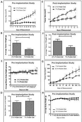
figure 1A, LuCap23.1-CR xenograft growth was strikingly
reduced in the 7% than in the 20% protein diet group, resulting in a 70% (p< 0.001, 95% CI= 55.98 to 139.7) reduced tumor size at 5 weeks post tumor implantation. Consistently, average tumor weight at the end of the experiment was 81% (p<0.0009, 95% CI =0.3814-1.243) lower in the 7% protein than in the 20% protein diet group (Fig. 1B). In a second experiment (post-implantation study), protein restriction was initiated in castrated mice 4 weeks after tumor establishment (~50 mm2). As shown in figure 1E, also in this setting the 7% protein
diet markedly inhibited tumor growth and resulted in a ~50% (p<0.0275, 95% CI = 0.04232-0.5910) reduction in tumor weight (Fig.1F). Throughout the 4-month study,
there was no significant difference in mean body weights
between the 7% and the 21% protein diet groups (Fig. 1C). Interestingly, despite the higher carbohydrate content of
the 7% protein diet, there was no significant difference
in serum glucose concentration between the two groups (Fig. 1D).
To test the hypothesis whether an isocaloric decrease in dietary protein intake inhibits tumor growth in a human animal model of BC as well, we acclimatized 4-6 week old female NOD-SCID mice to either the 21% or 7% protein diet for 4 weeks, prior to subcutaneous implantation of
WHIM16 tumor cells. As shown in figure 1G, WHIM16 xenograft growth was significantly reduced in the 7%
as compared to the 20% protein diet group, resulting in a 56% (p< 0.0232, 95% CI= -153.9 to -12.86) reduced
tumor size. No significant difference in mean body
weights between the 7% and the 21% protein diet groups was observed (Fig. 1H).
Protein restriction reduces serum IGF-1 concentration and attenuates mTOR activity in a human castrate-resistant, prostate cancer model.
To determine whether dietary protein intake affects circulating IGF-1 levels in castrated tumor bearing mice, serum IGF-1 concentration were measured in the two
groups. Serum IGF-1 concentration was significantly lower
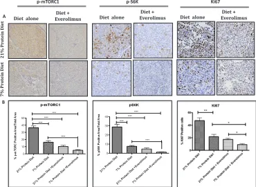
determining whether or not a reduction in protein intake down-regulates the mTOR signaling pathway in the LuCaP23.1-CR model, and whether or not the diet-induced inhibitory effect on tumor growth was dependent on mTOR inhibition. To test this hypothesis, we assessed the effects on tumor growth and the levels of phosphorylated mTOR and p70 S6K in mice treated with diet alone versus diet in combination with everolimus treatment. As shown
in figure 2B and C, everolimus treatment was as effective
as protein restriction in inhibiting tumor growth and had greater effect in combination with protein restriction as compared to lower protein diet alone. Tumor weights from the animals treated with everolimus alone or the 7%
protein diet alone were similar (Fig. 2D). However, tumor
weights were significantly smaller in animals treated with
everolimus fed the 7% protein diet than in those treated with everolimus fed the 20% protein diet, suggesting that protein restriction and mTOR inhibition have additive inhibitory effects on PCa development. Endpoint PSA
measurements confirmed the inhibitory effect of protein
restriction alone and everolimus in both the 21% and 7% protein diet groups (Fig. 2E). Immunohistochemistry
staining showed a significant decrease in
[image:3.612.167.454.223.652.2]phospho-mTORC1 levels following protein restriction (21% protein diet =37% +/- 2.09 vs. 7% protein diet=19% +/- 2.03, p<0.0001, 95% CI = 14.51- 26.19), which was even more
Figure 1: Low protein diet attenuates the growth of prostate and breast cancer in the castrate-resistant LuCaP23.1 model and in the WHIM16 model, respectively. Tumor sizes were assessed two times a week by caliper measurements. (A, B) LuCaP23.1-CR and (G) WHIM16 growth curve of tumors already exposed to low protein diet (pre-implantation studies) and endpoint tumor weights. (E, F) LuCap23.1-CR growth curve of tumors exposed to low protein diet after tumors were implanted and established (post-implantation studies), and endpoint tumor weights. (C) LuCaP23.1-CR and (H) WHIM16 mouse body weights. (D) Measurements of serum glucose in LuCaP23.1-CR bearing mice. Results are expressed as the mean +/- SE, n= 7-10; * p<0.05.
A Pre-implantation Study
B
D
E Post-implantation Study
C
Post-implantation Study Pre-implantation Study
Pre-implantation Study
Pre-implantation Study
F
Days of Measurement
Tu
mo
r Si
ze (
mm
2)
7 % Protein Diet 21 % Protein Diet
Days on Diet 21 % Protein Diet
7 % Protein Diet Pre-implantation Study
Pre-implantation Study G
H
* *
Figure 2: Low protein diet decreases IGF-1 serum levels and inhibit LuCaP23.1-CR growth in combination with everolimus. (A) Measurements of serum IGF-1 in LuCaP23.1-CR bearing animals fed with either 21% or 7% protein diet. (B, C) Mice were acclimatized for four weeks to either 21% or 7% protein diet and after LuCaP23.1-CR xenograft implantation were treated with everolimus (2 mg/kg PO, daily X5 times/week). (D, E) Endpoint PSA and tumor weights were collected. Results are expressed as the mean +/- SE, n=10.
A
B
C
D
E
Figure 3: Low protein diet decreases mTOR and proliferation activity in the LuCaP23.1-CR model. (A) At the end of the “pre-implantation” experiment, tumor samples were collected and processed. Paraffin embedded tissue specimens were stained for p-mTOR, p-S6 ribosomal protein and proliferation marker Ki67. (B) Quantification of staining. Results are based on four fields per tissue and are expressed as the mean +/- SE. **p<0.01 and ***p<0.001.
21%
Pro
te
in
D
ie
t
7%
P
ro
te
in
Die
t
p-mTORC1
Diet alone Diet + Everolimus Diet alone Diet + Everolimus
Ki67 p-S6K
Diet + Everolimus Diet alone
[image:4.612.122.492.404.674.2]Figure 4: Epigenetic alterations associated with low protein diet in the LuCaP23.1-CR model. (A) At the end of the “pre-implantation” experiment, tumor samples were collected and processed. Paraffin embedded tissue specimens were stained for the histone methyltransferase EZH2 and the associated histone mark H3K27me3. (B) Quantification of staining. Results are based on four randomly selected fields and are expressed as the mean + SE. **p<0.01, ***p<0.001
Figure 5: Effect of the different sources of protein diet on tumor growth and potential molecular mechanisms.
[image:5.612.60.289.400.639.2]Endpoint LuCaP23.1-CR tumor weights of mice fed with either 21% protein diet (which consist of both plant and animal protein sources), 20% or 10% protein diet (plant based) or 20% or 10% protein diet (animal based). Results are presented as a mean +/- SE, n= 7-8 per group. *p<0.05, ns= not statistically significant.
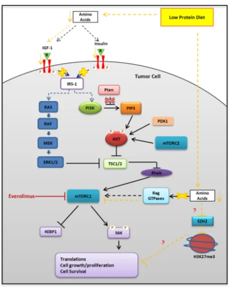
Figure 6: Representative schema of the potential molecular mechanisms responsible for the biological effects of protein restriction.
B
21%
P
ro
te
in D
ie
t
7%
P
ro
te
in D
ie
t
21%
P
ro
te
in D
ie
t
+ E
ver
ol
imu
s
7%
P
ro
te
in D
ie
t
+ E
ver
ol
imu
s
H3K27me3 EZH2
A B
[image:5.612.325.554.401.687.2]pronounced when protein restriction was combined with everolimus (Fig. 3A and B). We also observed a similar
significant decrease in ribosomal protein TOR activity
as indicated by the down-regulation of p70 S6K (21% protein diet =29%+/- 2.74 vs. 7% protein diet = 8%+/- 0.84, p<0.0001, 95% CI= 15.4- 27.34) in mice fed the 7% protein diet, which is potentiated by everolimus treatment. Interestingly, associated with the reduced mTOR activity we observed a decreased number of proliferating cells as indicated by the Ki67 staining (21% protein diet = 47% +/- 4.5 vs. 7% protein diet = 22% +/- 3.4, p<0.0041, 95% CI= 11.55- 39.10) (Fig. 3A and B). Protein diet restriction induced a similar inhibition of mTOR pathway and Ki67 expression in another patient-derived xenograft model of PCa (supplementary Fig. S1).
Protein restriction inhibits tumor growth in a human androgen-sensitive prostate cancer model.
To determine whether the androgen status could affect the inhibitory effects of protein restriction on PCa growth, we acclimatized the mice to either 21% or 7% protein diet and after four weeks we inoculated LuCaP23.1-AS tumors orthotopically in the prostate of non-castrated SCID mice. The 7% protein diet had a dramatic inhibitory effect on the growth of AS tumors implanted orthotopically in the prostate of intact animals (supplementary Fig. S2). Interestingly, the effects of protein restriction on average tumor weight was identical in the animal fed with a 7% diet alone and in those fed a 21% protein diet treated with everolimus.
Protein restriction induces epigenetic modifications.
Because in our study the tumor inhibitory effects of protein restriction were partially independent of mTOR,
we assessed whether epigenetic modifications, that are
known to be associated with PCa progression, were
modified by protein restriction in the LuCaP23.1-CR
model. We found that the mice fed with a 7% protein diet
had significantly lower expression of EZH2 (21% protein
diet = 97.3% +/- 4.4 vs. 7% protein diet = 55.1% +/- 3.46, p<0.0001, 95% CI= 32.8- 51.65) and H3K27me3 (21% protein diet =82.3% +/- 4.4 vs. 7% protein diet= 54.9% +/- 3.8, p<0.0051, 95% CI= 11.83- 43.02) (Fig. 4A, B and C). Similar changes in EZH2 and H3K27me3 were also observed in mice treated with everolimus.
Plant protein diets inhibit tumor growth independently of protein content.
To investigate whether modifications in protein quality (i.e. aminoacid composition) are beneficial in
delaying PCa growth, mice were placed on either regular (20%) or a reduced (10%) protein diet from animal or vegetable sources for 4-weeks prior to surgically castration and subcutaneous implantation of LuCaP23.1-CR xenografts. At the end of the experiment (6 weeks),
mice were sacrificed and tumor weights were collected. As shown in figure 5, a diet containing 20% plant protein
decreased tumor weight by 37% as compared to a 20% animal dairy protein diet (p<0.045, 95% CI= -1.484 to -0.01818). The inhibitory effect on tumor growth exerted by the 20% and 10% plant protein diets was similar. Interestingly, there was no additive effect of switching from animal to plant proteins when dietary protein content was 10%, suggesting that a threshold exists below which the amino acid composition is less important than the protein content of the diet.
DISCUSSION
In this study we examined the effects of isocaloric
modifications in dietary protein quantity or quality on
tumor growth in the human xenograft LuCaP23.1 (CR and AS), LuCaP35V (CR) and WHIM16 BC models. Our
findings indicate that dietary protein reduction results in a
marked inhibition of PCa and BC growth. This inhibitory effect was associated with a reduction in serum PSA and IGF-1 levels, and a down-regulation of intratumor mTOR activity in the LuCaP23.1 LuCaP35V models. Protein restriction was also associated with modulation
of specific histone markers suggesting epigenetic
modulation. Interestingly, the source of proteins appears to be important, as tumor inhibition was also achieved by changes in the quality (i.e. aminoacid composition of vegetable versus dairy) of protein food.
Accumulating scientific data indicate that
perturbations in the IGF/PI3K/Akt/mTOR pathway play a key role in the pathogenesis of PCa and BC [10-18,24]. A linear relationship exists between circulating IGF-1 levels and the risk of developing PCa and BC [9,10,24], and the PI3K/Akt/mTOR signaling pathways is upregulated in 30-50% of PCa [12,13]. Data from epidemiological and human experimental studies suggest that dietary protein or essential amino acid restriction is more powerful than calorie or fat restriction in lowering the circulating levels of IGF-1, which in turn inhibits the PI3K/AKT/mTOR pathway [7,8,21,23,24]. Consistently, our data show that an isocaloric reduction of protein
intake significantly reduces serum IGF-1 concentrations,
restriction. This inhibition in PCa growth induced by protein restriction appears to be greater than the effect induced by fat or carbohydrate restriction previously reported by other groups [25-27], and similar to the inhibitory effect induced by calorie restriction [28]. We also observed an additive effect of protein restriction and pharmacological mTOR inhibition (i.e. everolimus treatment) on mTOR activity, cell proliferation and PCa growth, suggesting that protein restriction works through multiple pathways and molecular targets. Interestingly, LuCaP23.1 is a PTEN expressing tumor, but with intrinsic mTOR pathway activation. Ongoing studies with PCa
derived from genetically modified mice will determine the
importance of mTOR activity in the sensitivity to protein restriction.
At least 15-17% (i.e. ~1.3-1.5 g/kg/body weight) of the total calories consumed by US and Northern European citizens comes from dietary protein, which is two-fold higher than what is recommended by the USDA (i.e. recommended daily allowance, 0.85 g/kg/body weight or 10% protein from calories) and of the quantity consumed (9% calorie from protein) by the Okinawan centenarians who had one of the lowest rate of PCa and BC in the world [29-32]. Moreover, the great majority of proteins in the typical Western diet come from meat, egg and dairy food sources, which are very rich in essential amino acids. Elevated levels of essential amino acids are able to fully activate, independently of IGF-1-insulin signaling, the TORC1 complex, which regulates cell growth, protein synthesis, and autophagy [12]. Our data suggest that feeding mice isocaloric and isoproteic diets with lower concentrations of some essential aminoacids (i.e. isoleucine, lysine, tyrosine, threonine, tryptophan, valine and methionine) reduces PCa growth, though at a lesser extent than protein restriction. Our experimental
findings support the notion that the high intake of animal
(dairy) proteins in Western diets may play a role in PCa development and progression, whereas more traditional diets rich in proteins from cereals and legumes might partially inhibit PCa growth [33].
Epigenetics is a potential mechanistic link between diet, energy metabolism, and gene expression modulation [34,35]. Highly accessible chromatin or open chromatin is typically observed at active regulatory regions like enhancers and promoters. Closed chromatin, on the other hand, is observed at silenced regions like hyper-methylated promoters of repressed genes. Diet and energy metabolism affect epigenetic enzymes that regulate histone marks [34-36]. Enhancer of zeste homolog 2 (EZH2), a subunit of Polycomb repressive complex 2, inhibits gene expression via its histone methyltransferase activity. A recent report has shown that the oncogenic function of EZH2 in cells of CR prostate cancer is independent of its role as a transcriptional repressor and involves its the ability to act as a coactivator for critical transcription factors including the androgen receptor [16]. Interestingly, in our
study we observed that protein restriction decreased the expression of EZH2 and its associated histone repressive marker H3K37me3. This observation suggests that protein restriction is responsible for selective epigenetic changes
that may drive modulation specific pathways involved
in PCa growth and survival (Fig. 6). Additional studies
are needed to shed light on the specificity of the histone
markers and associated gene expression modulation following protein restriction.
The results from our studies may have a rapid translation into the clinic. We envision that patients with
recurrent PCa and BC could be offered diet modifications
involving protein dietary content at different stages of their disease with appropriate monitoring. The absence of a differential effect in the AS and CR LuCaP23.1 models suggest that the androgen status does not affect the response of PCa to protein restriction. Furthermore, intratumor androgen receptor expression was not inhibited in the 7% protein diet group (data not shown). An ongoing clinical study in patients with newly diagnosed PCa will provide important information on the biological changes following this dietary intervention before prostatectomy.
In summary, a high-protein diet promoted the growth of human LuCaP23.1 and WHIM16 tumors in mice, and
a reduction in protein intake resulted in a significant
inhibition of tumor growth even when the diet was started after the formation of measurable tumors, possibly through inhibition of the IGF/Akt/mTOR pathway and
epigenetic modifications. Moreover, our findings indicate
that plant proteins partially inhibit prostate cancer growth independently of caloric or proteic intake, suggesting that protein quality plays a key role in the progression of prostate and breast cancer. Clinical studies in patients
with prostate and breast cancer are warranted to confirm the impact of dietary modifications in protein content and
quality on tumor progression.
MATERIALS AND METHODS
Androgen-sensitive and castrate-resistant LuCaP23.1 xenograft models of human prostate cancer.
under the skin or orthotopically into the dorsal lobe of the prostate in both intact (LuCaP23.1-AS) or surgically-castrated (LuCaP23.1-CR) mice. Similar approach was used for LuCaP35V-CR. All mice were operated under
sedation with oxygen, isoflurane and buprenorphine.
Mice were randomly grouped and placed on either 21% or 7% protein diets prior to tumor implantation (n=16-20 per group) or after tumor implantation (n=7 per group) or placed on diets from different sources of protein (n= 7-8 per group).
WHIM16 xenograft model of human breast cancer.
[image:8.612.66.553.67.576.2]Six-weeks old, female NOD.Cg-Prkdcscid Il2rgtm1Wjl/SzJ mice were purchased from the Jackson Labs. Mice were housed and maintained in a sterile and pathogen free facility. WHIM16 tumor cell line was generated from a patient with ER positive/PR negative/ HER2 negative breast cancer (Washington University).
Table 1: Composition and ingredients of experimental diets
21% mix protein
7% mix protein
20% plant protein
10% plant protein
20% dairy protein
10% dairy protein
Diet composition Diet composition Diet composition Total energy value
(kcal/g) 3.6 3.6 Total energy value (kcal/g) 3.7 3.7 Total energy value (kcal/g) 3.7 3.7 Carbohydrate (%Kcal) 58.9 73.0 Carbohydrate (%Kcal) 62.4 73.5 Carbohydrate (%Kcal) 63.6 74.0 Fat (%kcal) 20.1 20.2 Fat (%kcal) 17.6 16.6 Fat (%kcal) 16.4 16.0 Protein (%Kcal) 20.9 6.8 Protein (%Kcal) 20.0 10.0 Protein (%Kcal) 20.0 10.0 Leucine (g/kg) 25.4 8.8 Leucine (g/kg) 21.2 10.6 Leucine (g/kg) 18.7 9.4 Isoleucine (g/kg) 7.8 2.7 Isoleucine (g/kg) 7.9 4.0 Isoleucine (g/kg) 10.1 5.0 Lysine (g/kg) 16.3 4.0 Lysine (g/kg) 3.9 1.9 Lysine (g/kg) 15.7 7.9 Methionine (g/kg) 6.7 1.9 Methionine (g/kg) 4.1 2.1 Methionine (g/kg) 4.9 2.4 Cysteine (g/kg) 7.2 3 Cysteine (g/kg) 3.0 1.5 Cysteine (g/kg) 2.0 1.0 Arginine (g/kg) 6.3 2.9 Arginine (g/kg) 7.8 3.9 Arginine (g/kg) 6.7 3.3 Phenylalanine (g/kg) 6.6 2.4 Phenylalanine (g/kg) 10.4 5.2 Phenylalanine (g/kg) 8.8 4.4 Tyrosine (g/kg) 6.9 2.4 Tyrosine (g/kg) 5.0 2.5 Tyrosine (g/kg) 9.2 4.6 Histidine (g/kg) 3.4 1.4 Histidine (g/kg) 4.2 2.1 Histidine (g/kg) 4.9 2.4 Threonine (g/kg) 9.7 3.3 Threonine (g/kg) 5.5 2.7 Threonine (g/kg) 8.4 4.2 Tryptophan (g/kg) 3.4 1.0 Tryptophan (g/kg) 1.6 0.8 Tryptophan (g/kg) 2.5 1.2 Valine (g/kg) 8.4 3.2 Valine (g/kg) 8.7 4.3 Valine (g/kg) 11.8 5.9
Formula (g/kg) Formula (g/kg) Formula (g/kg)
Corn 430 430 Wheat gluten 110 55 Casein 170 85 Lactalbumin 177 35 Corn gluten (60%) 136 68 Lactalbumin 44 22 DL-Methionine 2.0 0.4 Isolated soy protein 22 11
Corn starch 149 287.4 Corn starch 325.4 459 Corn starch 380.1 486.3 Maltodextrin 100 100 Maltodextrin Sucrose 100150 100150 Maltodextrin Sucrose 100150 100150 Corn oil 29 32 Corn oil 32 32 Corn oil 32 32 Olive oil 29 32 Olive oil 32 32 Olive oil 32 32 Cellulose 30 30 Cellulose 50 50 Cellulose 50 50 * Mineral Mix,
AIN-93G-MX 35 35 ** Mineral Mix, w/o Ca & P 13.4 13.4 **Mineral Mix, w/o Ca & P 13.4 13.4 Calcium phosphate 8 8 Calcium phosphate Calcium carbonate 127.0 12.66.8 Calcium phosphate Calcium carbonate 810.3 10.88.3 ***Vitamin Mix,
Tekland 10 10 ***Vitamin Mix, Teklad 10 10 ***Vitamin Mix, Teklad 10 10
This tumor cell line carries also a PI3K mutation with activation of the AKT/mTOR pathway (manuscript submitted). Five million WHIM16 cells were implanted subcutaneously under the skin. All mice were operated
under sedation with oxygen, isoflurane and buprenorphine.
Mice were randomly grouped and placed on either 21% or 7% protein diets prior to tumor implantation (n=10 per group).
Feeding protocol and drug treatment.
The six experimental diets were prepared and sterilized by irradiation by Harlan Laboratories (Madison, WI). A summary of the composition and ingredients of each diet are shown in table 1. Animals were allowed free access to food in cage and autoclaved water supply via auto-watering system. Male mice that were randomized into 21% protein diet and 7% protein diet were further randomized within each group to either receive treatment with everolimus (10 mg/kg, 5 days on/2 days off) or vehicle.
Tumor assessment.
For tumors implanted subcutaneously, tumor sizes and body weights were recorded twice and once a week, respectively. Tumor weights were measured by using a weighing scale at the end of the experiments. Tumor sizes were assessed by caliper measurements of two diameters of the tumor (longest length x shortest length = mm2).
Blood and prostate tissue collection, and IGF-1 measurement.
At the end of the experiment, blood was drawn from all mice by cardiac bleed. Serum was separated, and aliquots were either used to assess levels of PSA and glucose in the serum or stored in -800C for further analysis.
Serum insulin growth factor 1 (IGF-1) concentration was measured in duplicates (n=8 per diet group) using
a mouse specific IGF-1 ELISA kit (Abcam, Cambridge,
MA, USA). Tumor tissues were excised, weighed and either stored in -800C or fixed in 10% buffered formalin
for histopathology.
Immunohistochemistry.
Tissue specimens were fixed for 24-hr, paraffin embedded and sectioned (4μm). Sections were de-paraffinized and rehydrated through graded alcohol
washes. Antigen unmasking was achieved by boiling slides in sodium citrate buffer (pH=6.0). Sections were further incubated in hydrogen peroxide to reduce
endogenous activity. To examine the expressions of our proteins of interests, tissue section were blocked with 2.5% horse serum (Vector Laboratories) and incubated overnight in primary antibodies against p-mTOR (1:400, Cell Signaling), p-S6K (1:200, Cell Signaling), Ki67 (1:50, Thermo Fisher), EZH2 (1:50, Cell Signaling), and H3k27me3 (1:200 Cell Signaling). Following primary incubation, tissue sections were incubated in horseradish-conjugated anti-rabbit or anti-mouse antibody according to manufacturer’s protocol (Vector Laboratories) followed by enzymatic development in diaminobenzidine (DAB) and counter stained in hemotoxyline. Section were dehydrated
and mounted with cytoseal 60 (Thermo Scientific). Stained sections were analyzed under bright field using
the Zeiss Axio microscope. The number of positive cells was determined in a blinded fashion by analyzing four
random 20x fields per tissue and quantified using Image
J software.
Statistical analysis.
Quantitative measures were compared between
groups (normal diet versus low protein or modified diets,
with or without treatment) using two-tailed Student’s t-test calculated by Graph-Pad software. The P values < 0.05
were considered to be statistically significant. The data are
presented as mean ± SE.
ACKNOWLEDGMENTS
Financial Disclosures:
None of the authors had conflicts of interest.
Funding/Support:
This research was supported in part by the National Cancer Institute, National Institutes of Health (P30CA016056) (RP), Glenn Foundation for Medical Research (LF), and the Bakewell Foundation (LF). LuCaP23.1-AS, LuCaP23.1-CR and LuCaP35V-CR were generated with support from the Richard M Lucas Foundation and the Prostate Cancer Foundation (RV).
REFERENCES
1. Ferlay J, Shin HR, Bray F, Forman D, Mathers C, Parkin DM. Estimates of worldwide burden of cancer in 2008: GLOBOCAN 2008. Int J Cancer. 2010;127(12):2893-917 2.
3. Shimizu H, Ross RK, Bernstein L, Yatani R, Henderson BE, Mack TM. Cancers of the prostate and breast among Japanese and white immigrants in Los Angeles County. Br J Cancer. 1991;63(6):963-6.
4. Jemal A, Center MM, DeSantis C, Ward EM. Global patterns of cancer incidence and mortality rates and trends. Cancer Epidemiol Biomarkers Prev. 2010;19(8):1893-907. 5. The Research Group for Population-based Cancer
Registration in Japan. Cancer Incidence and Incidence Rates in Japan in 1998: Estimates Based on Data from 12 Population-based Cancer Registries. Jpn J Clin Oncol 2003; 33: 241–5.
6. Chan JM, Gann PH, Giovannucci EL. Role of diet in prostate cancer development and progression. J Clin Oncol 2005;23:8152– 60.
7. Giovannucci E, Pollak M, Liu Y, Platz EA, Majeed N, Rimm EB, Willett WC. Nutritional predictors of insulin-like growth factor-1 and their relationships to cancer in men. Cancer Epidemiol Biomarkers Prev. 2003;12(2):84-9. 8. Fontana L, Weiss EP, Villareal DT, Klein S, Holloszy JO.
Long-term effects of calorie or protein restriction on serum IGF-1 and IGFBP-3 concentration in humans. Aging Cell. 2008;7(5):681-7.
9. Yu H, Rohan T. Role of the insulin-like growth factor family in cancer development and progression. J Natl Cancer Inst. 2000;92(18):1472-89.
10. Chan JM, Stampfer MJ, Giovannucci E, Gann PH, Ma J, Wilkinson P, Hennekens CH, Pollak M. Plasma insulin-like growth factor-I and prostate cancer risk: a prospective study. Science. 1998;279(5350):563-6.
11. Endogenous Hormones and Breast Cancer Collaborative Group, Key TJ, Appleby PN, Reeves GK, Roddam AW. Insulin-like growth factor 1 (IGF1), IGF binding protein 3 (IGFBP3), and breast cancer risk: pooled individual data analysis of 17 prospective studies. Lancet Oncol. 2010;11(6):530-42.
12. Morgan TM, Koreckij TD, Corey E. Targeted therapy for advanced prostate cancer: inhibition of the PI3K/Akt/ mTOR pathway. Curr Cancer Drug Targets. 2009;9(2):237-49.
13. Dai B, Kong YY, Ye DW, Ma CG, Zhou X, Yao XD. Activation of the mammalian target of rapamycin signaling pathway in prostate cancer and its association with patient clinicopathological characteristics. BJU Int. 2009;104(7):1009-16.
14. Ma CX, Crowder RJ, Ellis MJ. Importance of PI3-kinase pathway in response/resistance to aromatase inhibitors. Steroids. 2011;76(8):750-2.
15. Efeyan A, Zoncu R, Sabatini DM. Amino acids and mTORC1: from lysosomes to disease. Trends Mol Med. 2012;18(9):524-33.
16. Sachdev D and Yee D. Disrupting insulin-like growth factor signaling as a potential cancer therapy. Mol Cancer Ther 2007; 6(1): 1-12.
17. Pollak MN. Insulin-like growth factors and neoplasia. Norvartis Found Symp 2004;262: 84-98.
18. Corey E. and Vessella RL. Xenograft models of human prostate cancer. In: in Prostate Cancer: Biology, Genetics and the New Therapeutics, 2nd Edition; Ed. Leland Chung, William Isaacs, Jonathan Simons; Humana Press, Totowa, NJ, 2007: 3 – 32.
19. Xu K, Wu ZJ, Groner AC, et al. EZH2 oncogenic activity in castration-resistant prostate cancer cells is Polycomb-independent. Science. 2012;338(6113):1465-9
20. Pellakuru LG, Iwata T, Gurel B, et al. Global levels of H3K27me3 track with differentiation in vivo and are deregulated by MYC in prostate cancer. Am J Pathol. 2012;181(2):560-9.
21. Norat T, Dossus L, Rinaldi S, Overvad K, Grønbaek H, et al. Diet, serum insulin-like growth factor-I and IGF-binding protein-3 in European women. Eur J Clin Nutr. 2007;61(1):91-8.
22. Nardella C, Carracedo A, Salmena L, Pandolfi PP. Faithfull modeling of PTEN loss driven diseases in the mouse. Curr Top Microbiol Immunol. 2010;347:135-68.
23. Underwood LE, Thissen JP, Lemozy S, Ketelslegers JM, Clemmons DR. Hormonal and nutritional regulation of IGF-I and its binding proteins. Horm Res.1994;42(4-5):145-51.
24. Clemmons DR, Seek MM, Underwood LE. Supplemental essential amino acids augment the somatomedin-C/insulin-like growth factor I response to refeeding after fasting. Metabolism. 1985;34(4):391-5.
25. Wang Y, Corr JG, Thaler HT, Tao Y, Fair WR, Heston WD. Decreased growth of established human prostate LNCaP tumors in nude mice fed a low-fat diet. J Natl Cancer Inst. 1995;87(19):1456-62.
26. Lloyd JC, Antonelli JA, Phillips TE, et al. Effect of isocaloric low fat diet on prostate cancer xenograft progression in a hormone deprivation model. J Urol. 2010;183(4):1619-24.
27. Freedland SJ, Mavropoulos J, Wang A, Darshan M, et al. Carbohydrate restriction, prostate cancer growth, and the insulin-like growth factor axis. Prostate. 2008;68(1):11-9. 28. Blando J, Moore T, Hursting S, et al Dietary energy
balance modulates prostate cancer progression in Hi-Myc mice. Cancer Prev Res (Phila). 2011;4(12):2002-14. 29. Moshfegh A, Goldman J, Cleveland L (2005) What we eat
in America, NHANES 2001-2002: Usual nutrient intakes from food compared to Dietary Reference Intakes. http:// www.ars.usda.gov/SP2UserFiles/Place/12355000/pdf/ usualintaketables2001-02.pdf. Accessed Mar 23, 2013. 30. Rand WM, Pellett PL, Young VR (2003) Meta-analysis of
nitrogen balance studies for estimating protein requirements in healthy adults. Am J Clin Nutr 2003;77(1): 109-27. 31. U.S. Department of the Office of the Civil Administrator
http://www.archives.gov/research/guide-fed-records/groups/260.html
32. Willcox, B. J., Willcox, D. C., Todoriki, H., et al. Caloric restriction, the traditional Okinawan diet, and healthy aging: the diet of the world’s longest-lived people and its potential impact on morbidity and life span. Annals of the New York Academy of Sciences 2007;1114: 434-455.
33. Gao X, LaValley MP, Tucker KL. Prospective studies of dairy product and calcium intakes and prostate cancer risk: a meta-analysis. J Natl Cancer Inst. 2005;97(23):1768-77. 34. Zheng S, Rollet M, Yang K, Pan YX. A gestational
low-protein diet represses p21WAF1/Cip1 expression in the mammary gland of offspring rats through promoter histone modifications. Br J Nutr. 2012;108(6): 98-1007.
35. Cong R, Jia Y, Li R, et al. Maternal low-protein diet causes epigenetic deregulation of HMGCR and CYP7α1 in the liver of weaning piglets. J Nutr Biochem. 2012 Dec;23(12):1647-54.