5
X
October 2017
Classification of Breast cancer Using
Local Binary Pattern and Gabor Filter
Abhijeet V. Sansare1, Sanjay kumar. R. Kinge2
1
M.E. Student, Dept. Of Electronics and Telecommunication, MIT College Of Engineering, Pune, India
2
Assistant Professor, Dept. Of Electronics and Telecommunication, MIT College Of Engineering, Pune India 2
Abstract: The proposed paper presents a novel approach for classification of breast cancer. Digital mammograms have become the most effective techniques for the detection of breast cancer. The goal of this research is to increase the diagnostic accuracy of image processing and machine learning techniques for optimum classification between malignant and benign abnormalities in digital mammograms by reducing the number of misclassified cancers. The Local Binary Pattern method and Gabor Filter is used to extract the texture features of the digital mammographic images. The respective texture features are given to Support Vector Machine classifier which successfully classifies benign cases, malignant cases and normal breast cases with accuracy 96.72%, 84% and 81.90% respectively.
Keywords : breast cancer detection, CAD, LBP, Gabor filter, SVM, etc.
I. INTRODUCTION
The aim of this research is the development of a reliable tool for the detection of breast cancer using digital mammography images. Digital mammography is a generally new strategy for the early recognition of breast cancer. It depends on aggregated density of tissues, that is, to identify shadows. This is the reason with respect to why digital mammography has been considered as a productive instrument for the identification of masses and MCCs. The real issue related with mammogram screening programs is the expansive level of missed cancer. Studies demonstrate that amid screening, radiologists neglect to recognize 15 precents of bosom diseases that are obvious in review contemplate. Besides, when insignificant signs are considered, appraisals of missed cases increment to 50 precents, because of mistakes of observation. Without considering recorded eye movements, search and detection mistakes are those that happen when radiologists don't report the presence of an unmistakable tumor and interpretation errors as those that happen when the tumor is accounted for however not considered significant.
With a specific end goal to distinguish if a breast tissue is cancerous, a biopsy is generally performed. Amid a biopsy the suspicious breast tissue is expelled from the patient for an indicative examination. The breast tissue is evacuated by a surgical extraction and is analysed by the pathologist. A variation from the normal once identified in a breast tissue, can be delegated either benign (not cancerous) or malignant (cancerous). Computer Aided Detection methods are composed image processing and machine learning techniques. Mammogram processing usually utilizes image processing strategies with the end goal of removal of artefacts and labels, eliminating digitization noise and improving mammograms for ideal review, mostly for discovery of mass lesions and MCCs. Many investigations give confirm that radiologists perform better on computer enhanced images to change mammograms such that they can be printed or inspected on a screen ideally. Machine learning systems are regularly connected in computer Aided Detection scheme for pattern classification. In classification process, feature extracted from the Region of Interest (ROI) are utilized for the training and testing (approving) a learning machine. A classifier prepared on known irregularities (mass lesions and MCCs) consolidates the selected features and utilizes confidence measures to show if the tested specimen is malignant or benign.
II.PROPOSEDSYSTEM
A. Image database
Digital Mammographic images for proposed system are acquired from Mammographic Image Analysis Society (MIAS) Minimammographic Database. The MIAS database is composed of 322 cases from normal, benign and malignant cases [12]. For experimental analysis 158 images are extracted from the database. Digital
B. Image Preprocessing
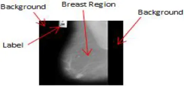
Fig 1. Detail Mammogram Image
[image:3.612.245.368.244.349.2]As shown in fig.1, after removing the noise or labels from the input image, it necessary to extract region of interest i.e. breast region. As shown in fig.1, the input image is composed of the background and breast region. This background part gets eliminated in preprocessing [4].
Fig.2. Breast region with Pectorial muscle
Fig.2. show extracted breast region containing pectorial muscle. The pectorial muscle present in the image can bias the final result of classification. Because the pectorial region has same intensity level as cancerous region, so it is necessary to remove the pectorial region from the image. As shown in fig.2, the pectorial muscle touches the border of the image. It is possible to remove the pectorial muscle part touching the border line.
C. Feature extraction
Feature extraction is a collection of significant information form given data. Feature plays a very important role in the area of image processing. Before extracting features, various image preprocessing techniques like binary image, thresholding, resizing, and normalization etc [2], [6], [11] are applied on the input image. After that, feature extraction techniques are applied to get features that will be useful in classifying and recognition of images.
Texture is an important characteristic for the analysis of various types of the images including natural scenes, remotely sensed data and biomedical modalities. It is believed that the texture plays an important role in the visual system for recognition and interpretation of data. Local binary pattern [4], [7], [9], [10] and Gabor filter is used in proposed system to extract the texture features from the processed image.
1) Local Binary Pattern: Local binary pattern (LBP) is an image descriptor whose purpose is to compute binary patterns for the local structure of an image. Local binary pattern (LBP) is a straightforward yet exceptionally effective surface administrator which labels the pixels of an image by thresholding the neighbourhood of every pixel and considers the outcome as a binary number. Because of its discriminative power and computational erffortlessness, LBP texture operator has turned into a prevalent approach in different applications. The most essential property of LBP texture descriptor in genuine applicaions is its robustness to monotonic gray scale changes created, for instance, by intensity varieties.
Fig.3. LBP operator
Given a pixel in the image, an LBP code is computed by comparing it with its neighbours,
Where gc is the gray value of the central pixel, g p is the value of its neighbours, P is the total number of involved neighbours, and R
is the radius of the neighbourhood. Suppose the coordinate of gp is (0,0), then the coordinates of (R cos(2л/P), R sin(2л/P)). The
gray values of neighbour that are not in the image grids can be estimated by interpolation. Suppose the image is of size I*J. After the LBP pattern of each pixel is identified, a histogram is built to represent the texture image
[image:4.612.125.497.315.725.2]2) Gabor Filter: The gabor filter is a linear filter used for texture analysis, which means that it is basically analyses whether there are any specific frequency content in the image in specific direction in a localized region around the point or region of analysis. Frequency and orientation representation of Gabor filter are similar to those of the human visual system, and then they have been found to be particularly appropriate for texture representation and discrimination. A Gabor filter is obtained by modulating a sinusoid with a Gaussian. For the case of one dimensional (1D) signals, a 1D sinusoid is modulated with a Gaussian. This filter will therefore respond to some frequency, but only in a localized part of the signal. The Gabor filter can be expressed as:
It has been shown that σ, the standard deviation of the Gaussian kernel depends upon the spatial frequency to measured, i.e. .
D. Classification
In classification stage, SVM classifier is trained with the extracted features i.e. Gabor filter and LBP features. The Gabor features and LBP features are saved in a ‘.mat’ file in MatLab. These extracted features are first given to SVM classifier for training. After completion of training of SVM classifier, extracted feature of testing image can be used to classify given input image as normal, benign or malignant. Support Vector Machine (SVM) originally separates the binary classes (k=2) with a maximized margin criterion. However, real world problems often require the discrimination for more than two categories.
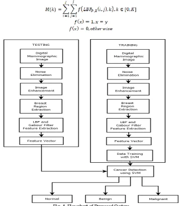
Fig.4 shows the flow diagram of proposed system explained above. As shown, initially features of all images (i.e. 158 images) present in the database are extracted and all features are labeled with respective cases i.e. benign, malignant, and normal. This extracted features and labels are used to train the support vector machine. Once the SVM is training is completed, it can classify the given image as benign, normal, and malignant.
III.EXPERIMENTALRESULTS
This section presents the experimental result of proposed CAD system. The proposed algorithm has been evaluated on MIAS database. The MIAS database contains 322 normal, benign and malignant cases from which 158 cases are extracted for evaluation purpose.
Fig.5 shows original mammographic images which are converted to '.png' image format from '.pgm'. Fig.5 (a) shows different class from benign, malignant and normal class. Fig.6 shows results of input image (Digital Mammographic Images from MIAS Database) preprocessing i.e. noise removal, contrast enhancement and breast region extraction. Fig.7 shows pectorial muscle removed from the given input image. Presence of pectorial muscle in preprocessed image can bias the final result, so it is necessary to remove the pectorial muscle from the image. As shown in fig.7, proposed system successfully removes all part of pectorial muscle present in the input image.
Fig.5 Digital Mammographic Images from MIAS Database
Fig.7 Result Image Segementation Pectorial Muscle Removed
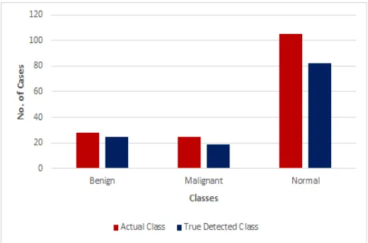
Fig.8 shows the plot of Actual class versus True detected class. The result shown in fig.6.4. are obtained using 'rbf' as the 'kernel_function' of support vector machine classifier. Actual class is the number of benign, normal and malignant classes given in MIAS databse. True detected class is the result of the proposed system classified as benign, normal and malignant classes. The database extracted for given system contains 28 benign cases, 27 malignant cases and 103 normal cases.
Fig. 8. Comparative Analysis of Classification using SVM with ‘Kernel_function’ as ‘rbf’
[image:6.612.175.435.260.432.2]As The proposed system classifies 25 benign cases, 19 malignant cases and 82 normal cases correctly from the given actual class cases by using 'rbf' as a 'kernel_function' of SVM. The proposed system detects and classifies benign, normal and malignant classes with 89.28%, 79.61% and 70.37% accuracy respectively as shown in fig.9.
Fig. 9: Accuracy of proposed system using SVM with ‘Kernel function’ as ‘rbf’
[image:6.612.175.437.505.691.2]Fig. 10. Comparative Analysis of Classification using SVM with ‘Kernel_function’ as ‘quadratic’
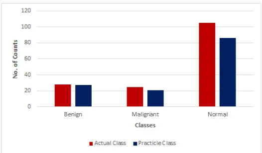
By using 'quadratic' as a 'kernel_function' of SVM classifier, the proposed system detects and classifies benign, normal, malignant classes with accuracy 96.72%, 81.90%and 84% respectively as shown in fig.11.
Fig. 11: Accuracy of proposed system using SVM with ‘Kernel function’ as ‘quadratic’
IV. CONCLUSIONS
The objectives of the proposed study were to develop CAD system for breast cancer detection at an early stage. The proposed system detects the benign, malignant and normal cases successfully. For proposed system, 158 images were extracted from MIAS database for experimentation. Local Binary Pattern method is used with Gabor Filter to increase the accuracy of detection and classification. The SVM with ‘kernel_function’ as ‘rbf’ has comparatively less accuracy than SVM with
‘kernel_function’ as ‘quadratic’. The proposed system with ‘quadratic’ as ‘kernel_function’ of SVM classifies benign cases with accuracy 96.72%, malignant cases with accuracy 84% and normal cases with accuracy 81.90%.
REFERENCES
[1] American Cancer Society, Breast Cancer: Facts and Figures,
[2] R. Guzman-Cabrera, J.R. Guzaman-Supulveda, M. Torres-Cisneros, D.A. May-Arrioja, J. Ruiz-Pinales, O.G. Ibarra-Manzano, G. Avina-Cervantes, A.
Donzalez Parada, “Digital image processing technique for breast cancer detection”, ,Int J Thermophys(2013)34:1519-1531
[3] Jawad Nagi, Sameem Abdul Kareem Farukh Nagi, Syed Khaleel Ahmed, “Automated breast profile segmentation for ROI detection using digital
mammograms”, 2010 IEE EMBS Conference on biomedical Engineering and Sciences (IECBES 2010), Malaysia, pp.87-92.
[4] Pavel Kral, Ladislav Lenc, “LBP features for breast cancer detection”, ICIP2016, pp. 2643-2647.
[5] Leonardo de Oliveira Martins, Geraldo Braz Junior, Aristifanes Correa Silva, Anselmo Cardoso de Paiva, and Marcelo Gattass, “Detection of masses in digital
mammogram using K-means and support vector machine”, Electronic Letters on Computer Vision and Image Analysis8(2):39-50, 2009.
[6] Anuj Kumar Singh, Bhupendra Gupta, “A novel approach for breast cancer detection and segmentation in a mammogram”, Eleventh International
Multi-Conference on Information Processing-2015(IMCIP-2015), Procedia Computer Science 54(2015), pp. 676-682.
[7] S. Naresh, S. Vani Kumari, “Breast Cancer Detection using Local Binary Patterns”, International Journal Of Computer Applications (0975-8887), Volume 123-
No.16, August 2015, pp. 6-9.
[8] Arnau Oliver, Joan Marti, Robert Marti, Anna Bosch, Jordi Freixenet, “A new Approach to the classification of mammographic masses and normal breast
[9] Eanes Torres Pereira, Sidney Pimentel Eleuterio, Joao Marques de Carvalho, “Local Binary Patterns Applied to Breast Cancer Classification in Mammographies”, RITA Volume Numero, pp. 1-15.
[10] Arnau Oliver, Xavier Llado, “False Positive Reduction in Mammographic Mass Detection Using Local Binary Patterns”, MICCAI2007, Part I, LNCS 4791,
pp.286-293, 2007.
[11] Spandana Paramkusham, K. M. M. Rao, B. V. V. S. N. Prabhakar Rao, “Early Stage Detection of Breast Cancer using Novel Image Processing Technique,
Matlab and Labview Implementation”,15th International Conference on Advance Computing Technologies (ICACT) 2013, pp. 1-5.
[12] Mammographic image analysis socity (MIAS) MiniMammographic Database, http://peipa.essex.ac.uk/info/mias.html
[13] V. Shiv Naga Prasad, Justin Domke, “Gabor filter visualization”.