ISSN Online: 1937-688X ISSN Print: 1937-6871
Evaluating the Combined Optimization of
Oxygenation and Ventilation in a Patient
Simulator
J. Kretschmer, C. Bibiano, P. Stehle, K. Möller
Institute of Technical Medicine, Furtwangen University, Villingen-Schwenningen, Germany
Abstract
The use of mathematical models can aid in optimizing therapy settings in ventilated patients to achieve certain therapy goals. Especially when multiple goals have to be met, the use of individualized models can be of great help. The presented work shows the potential of using models of respiratory mechanics and gas exchange to optimize minute ventilation and oxygen supply to achieve a defined oxygenation and carbon dioxide removal in a patient while guaranteeing lung protective ventilation. The ven-tilator settings are optimized using respiratory mechanics models to compute a res-piration rate and tidal volume that keeps the maximum airway pressure below the critical limit of 30 cm H2O while ensuring a sufficient expiration. A three-parameter
gas exchange model is then used to optimize both minute ventilation and oxygen supply to achieve defined arterial partial pressures of oxygen and carbon dioxide in the patient. The presented approach was tested using a JAVA based patient simulator that uses various model combinations to compute patient reactions to changes in the ventilator settings. The simulated patient reaction to the optimized ventilator settings showed good agreement with the desired goals.
Keywords
Physiological Model, Model Based Optimization, Decision Support, Patient Simulator
1. Introduction
Mechanical ventilation is a life-saving intervention, routinely used in intensive care. It provides breathing support in critically ill patients that are not able to maintain suffi-cient oxygenation. However, if the ventilator settings are not properly adapted to the
How to cite this paper: Kretschmer, J., Bibiano, C., Stehle, P. and Möller, K. (2016) Evaluating the Combined Optimization of Oxygenation and Ventilation in a Patient Simulator. J. Biomedical Science and
Engi-neering, 9, 90-98.
http://dx.doi.org/10.4236/jbise.2016.910B012
individual patient physiology, it can cause injuries to the lung tissue through barotrau-ma or collaps of alveoli [1]. Optimizing ventilator settings in patients with critically poor lung function poses a trade-off between multiple conflicting goals, such as apply-ing high airway pressures and tidal volumes to ensure sufficient oxygenation versus us-ing low airways pressures to protect healthy lung tissue [2]. Mathematical models of the human physiology can be adapted to the individual physiological properties of a patient and thus can be used to predict reactions of that patient to changes in the ventilator set-tings. Those predictions can aid in providing decision-making support by using opti-mization algorithms to calculate ventilator settings that lead to achieving the goals de-fined by the clinician [3]. Model based decision support in ventilated patients should consider the effect of air volume on the air pressure in the lung, but should also con-sider other physiological processes that are influenced by the ventilation. The most critical goals to achieve in a ventilated patient are a sufficient minute volume with low air pressures, avoiding intrinsic PEEP (positive end-expiratory pressure) by setting an expiration time that allows the patient to exhale the air before starting with the next inspiration phase and to apply enough oxygen to secure a sufficient oxygenation in the blood. Thus, gas exchange in the patient has to be taken into account when calculating the optimal ventilator settings. The following example should therefore demonstrate how to exploit information from different mathematical models to optimize ventilator settings individually for a patient. The goal was to calculate the necessary minute vo-lume (MV) and inspiratory oxygen fraction (FiO2) to achieve a desired partial pressure
of oxygen and carbon dioxide in arterial blood (PaO2, PaCO2) while keeping the
maxi-mum airway pressure below a critical limit and avoiding the build-up of intrinsic PEEP through a sufficient expiration time. The presented approach is evaluated using a pa-tient simulator that has been presented previously [4]. It incorporates models of respi-ratory mechanics, gas exchange and cardiovascular dynamics to simulate mechanically ventilated patients with various diseases.
2. Methods
2.1. Respiratory Mechanics Models
[6]. The models available for the presented optimization approach are a model of first order (FOM) [5], a viscoelastic model (VEM) [5], a recruitment model (PRM) [7] and a recruitment model with viscoelastic elements (PRVEM) [8]. Input to the models is air flow, output is air volume and airway pressure.
2.2. Gas Exchange Model
The presented optimization approach uses a three-parameter gas exchange model that allows simulating different ventilation to perfusion ratios (V/Q) to predict the effect of oxygen supply and minute volume on PaO2 and PaCO2 [9]. The model consists of two
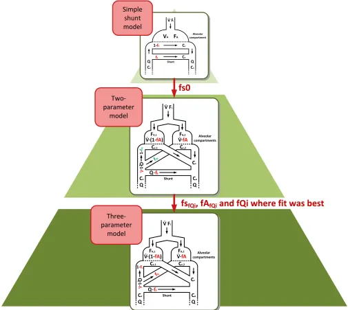
alveolar compartments, each of which are provided with a fraction of the inspired air. Parameter fA defines the fraction of air that reaches one of the compartments while the other compartment receives the rest. Venous blood coming from the body is partly shunted away and mixed with the oxygenated blood coming from the capillaries. The non-shunted blood is then distributed among the two alveolar compartments. Parame-ter fs defines the amount of shunted blood, parameParame-ter fQ defines the amount of blood reaching the alveolar compartment that receives the fraction of air defined by fA. In-puts to the model are the measured end-tidal oxygen and carbon-dioxide gas fractions (FetO2, FetCO2) and blood gas parameters such as Hemoglobin concentration, body
temperature, base excess and pH. The outputs of the model then are the arterial partial pressures of oxygen and carbon dioxide (PaO2, PaCO2). To improve the quality of
pa-rameter identification, a hierarchical approach has been used here as well. Figure 1 shows a schematic overview of the identification of the three-parameter gas exchange model. An initial estimate of the shunt is calculated using a simple shunt-model that consists of only one alveolar compartment [10]. This estimate is then used as an initial guess in the identification of a two-parameter model that is derived from the three- pa-rameter model. In the two-papa-rameter model, fQ is fixed to a certain value (fQi = 0.1, 0.2, 0.3, ∙∙∙, 0.9). Parameters fs and fA of the two parameter model are then identified for the specific fQi. The combination of fs, fA and fQi, where the parameter identifica-tion received the best fit to the data are finally used as initial estimates for the identifi-cation of the three-parameter model. The simple shunt model requires one measured PaO2 and PaCO2 together with the FiO2 that was supplied, while the two- and the three-
parameter models require four measurements of PaO2 and PaCO2 and different levels
of FiO2.
2.3. Optimization Algorithm
To achieve a specific PaO2 and PaCO2 in the patient, both minute ventilation (MV) and
supplied oxygen (FiO2) need to be optimized. Simultaneously, the minute ventilation
needs to be calculated with regards to the underlying respiratory mechanics so that the peak airway pressure (Ppeak) does not lead to additional lung injury and the expiration
time is enough to avoid intrinsic PEEP. A study by the ARDS network has shown that ventilation with a peak pressure of 30 cmH2O instead of 50 cm H2O leads to a reduction
Figure 1. Hierarchical order of the gas exchange models. The simple shunt model is used to calculate an initial shunt estimate, the two-parameter model is used to calculate appropriate initial guesses for the identification of the three-parameter model by testing differ-ent blood distributions (fQi) and the fitting shunt fs and alveolar distribution fA to it. The combination of fs, fA and fQi that leads to the model reproducing recorded data best is then used.
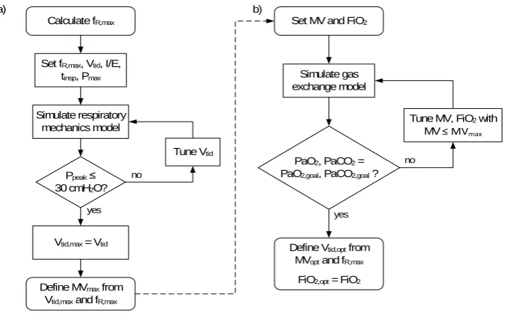
expiration time should be at least 3 times the expiratory time constant (τE) [3]. The
op-timization thus starts with finding the maximum respiratory frequency that still allows a complete expiration by using
exp, min E 3
t =τ ⋅ (1)
in, total, max exp, min
t =t ⋅I E (2)
(
)
R , max 60 in, total, max exp, min
f = t +t (3)
Two-parameter model Simple shunt model Three-parameter model
fs0
fs
fQi, fA
fQiand fQi where fit was best
V� Fi
VA FA compartmentAlveolar
Shunt
fS
1-fS Cc
Cv
Q Q
Ca
Cv
V� Fi
Alveolar compartments Shunt fS 1-f S Cc,1 Q Cc,2
V� fA V� (1-fA)
1-f Qi fQi Q Q• • • • Q Cc Ca Cv FA,2 FA,1
V� Fi
Alveolar compartments Shunt fS 1-f S Cc,1 Q Cc,2
1-fQ
fQ Q Q• • Q Cc Ca Cv
V� fA
V� (1-• fA) •
FA,2
[image:4.595.44.550.71.522.2]where texp,min is the minimum expiration time necessary for a complete expiration,
tin,total,max is the derived maximum inspiration time using a given inspiration to
expira-tion ratio (I/E) and fR,max is the resulting maximum respiration rate. I/E is given as a
goal by the user, while τE is calculated using an exponential fit of the recorded
expira-tion phase. The maximum tidal volume (Vtid,max) is then found by tuning tidal volume
(Vtid) so that the forward simulation of the respiratory mechanics model results in a
peak pressure of 30 cm H2O. Subsequently, both MV and FiO2 are tuned to achieve the
PaO2 and PaCO2 goals in the forward simulation of the gas exchange model using
MVmax derived from the previously calculated Vtid,max and fR,max as a boundary condition.
The optimal tidal volume (Vtid,opt) then is the quotient of MVopt and fR,max:
All models and algorithms were programmed in MATLAB (R2015a, The Mathworks, Natick, MA, USA). Parameter identification and tuning of Vtid, MV and FiO2 was done
using a Nelder-Mead Simplex Search method, realized in MATLAB as fminsearch function. Figure 2 gives an overview of the optimization algorithm.
2.4. Data
[image:5.595.197.552.430.650.2]The proposed algorithm was tested using a patient simulator that has been published previously. The simulator allows calculating real time reaction of a ventilated patient to changes in the ventilator settings. It combines models of respiratory mechanics, gas exchange and cardiovascular dynamics to achieve a global simulation of physiological interactions caused by applying mechanical ventilation. It allows simulating various model combinations and different parameter settings. Four different model combina-tions have been tested with the proposed optimization algorithm (Permutacombina-tions of the
Figure 2. Optimization algorithm to tune MV and FiO2 to achieve specific PaO2 and PaCO2 while
protecting lung tissue from exceedingly high airway pressures. (a) Algorithm to calculate fR,max
and Vtid,max; (b) Algorithm to calculate optimal MV and FiO2.
Calculate fR,max
Ppeak ≤ 30 cmH2O? Set fR,max, Vtid, I/E,
tinsp, Pmax
Simulate respiratory mechanics model
Tune Vtid
Define MVmax from Vtid,max and fR,max
Set MV and FiO2
Vtid,max = Vtid
Simulate gas exchange model
PaO2, PaCO2 = PaO2,goal, PaCO2,goal ?
Tune MV, FiO2 with
MV ≤ MVmax
Define Vtid,opt from MVopt and fR,max
FiO2,opt = FiO2 no
yes yes
no
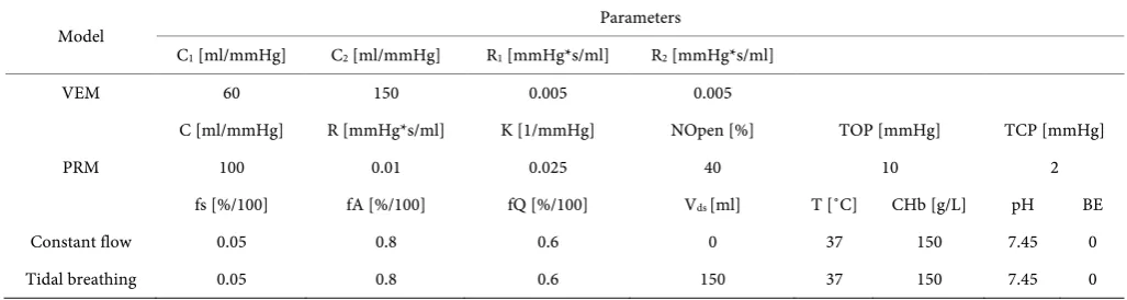
viscoelastic and the recruitment model with a constant flow and a tidal breathing model of gas exchange). Table 1 shows the relevant parameters that have been used in the models. To identify the respiratory mechanics models, flow, volume and airway pres-sure (Paw) were recorded, for the gas exchange models FetO2, FetCO2, PaO2 and PaCO2
was recorded at four different levels of FiO2 (30%, 50%, 70% and 90%). For a realistic
scenario, the recorded data for Paw, flow, volume, FetO2, FetCO2, PaO2 and PaCO2 were
superimposed with white noise with an amplitude of ±3%.
The viscoelastic model (VEM) and the recruitment model (PRM) used to simulate the respiratory mechanics reaction coincide with the models described in 2.2. To simu-late gas exchange reactions, models need to comprise body gas exchange, therefore the models used to create the patient data differed from the model used for optimizing MV and FiO2. The models used for creating the patient data and subsequent evaluation of
the optimized ventilator settings are based on the model presented by Chiari et al. [11] but were extended to comprise two alveolar compartments [9] [12]. One of the models assumes constant air flow into the lungs, the other comprises a dead space compart-ment to simulate distinct inspiration and expiration phases [12].
3. Results
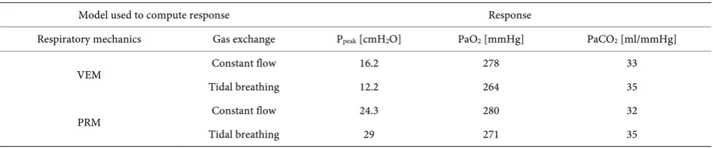
[image:6.595.40.554.566.703.2]Table 2 shows the results of the parameter identification, Table 3 shows the response to optimized ventilator settings as computed in the patient simulator. Parameter identi-fication of the respiratory mechanics model resulted in a maximum deviation of the identified parameters from their true value of 40% and a minimum deviation of 0% when the VEM was used to create the patient data, and a maximum deviation of 19% with a minimum deviation of 8.9% when the PRM was used to simulate the patient. The identification of the gas exchange model resulted in a maximum deviation of 32% for fs and fQ when using the tidal breathing model for simulating the patient and a maximum deviation of 12% for fs and fQ when using the constant breathing model in the patient simulator. Identified values for parameter fA did show no match (>50% deviation) with the value used to create the data.
Table 1. Parameters used in the respiratory mechanics and gas exchange models. C—Compliance, R—Resistance, K—Overdistention factor, NOpen—Number of recruited alveoli at the beginning of simulation, TOP/TCP—Threshold opening and closing pressure at which alveoli open and close, Vds = Dead space volume.
Model Parameters
C1 [ml/mmHg] C2 [ml/mmHg] R1 [mmHg*s/ml] R2 [mmHg*s/ml]
VEM 60 150 0.005 0.005
C [ml/mmHg] R [mmHg*s/ml] K [1/mmHg] NOpen [%] TOP [mmHg] TCP [mmHg]
PRM 100 0.01 0.025 40 10 2
fs [%/100] fA [%/100] fQ [%/100] Vds [ml] T [˚C] CHb [g/L] pH BE
Constant flow 0.05 0.8 0.6 0 37 150 7.45 0
Table 2. Parameter identification results.
Model used to
create patient data Parameters
Respiratory
mechanics Gas exchange C
1
[ml/mmHg] C
2
[ml/mmHg] R
1
[mmHg*s/ml] R
2
[mmHg*s/ml] [%/100] fs [%/100] fA [%/100] fQ
VEM Constant flow 55.9 185.7 0.007 0.005 0.045 0.104 0.467
Tidal breathing 0.034 0.108 0.528
C [ml/mmHg]
R [mmHg*s/ml]
K [1/mmHg]
NOpen [%]
TOP [mmHg]
fs [%/100]
fA [%/100]
fQ [%/100]
PRM Constant flow 108.9 0.009 0.028 34 11.9 0.050 0.120 0.550
Tidal breathing 0.036 0.116 0.481
Table 3. Patient response to the optimized ventilator settings as computed in the simulator.
Model used to compute response Response
Respiratory mechanics Gas exchange Ppeak [cmH2O] PaO2 [mmHg] PaCO2 [ml/mmHg]
VEM Constant flow 16.2 278 33
Tidal breathing 12.2 264 35
PRM Constant flow 24.3 280 32
Tidal breathing 29 271 35
The resulting patient responses show a maximum airway pressure below 30 cm H2O
in all tested combinations, a maximum deviation of 20% and a minimum deviation of 5.6% in the PaO2 response and a maximum deviation of 8.6% and a minimum
devia-tion of 0% in the PaCO2 response.
4. Discussion
Despite its regular use, individually optimizing mechanical ventilation in patients on the ICU is a challenging task. Multiple conflicting goals need to be weighed against each other while the patient response can predicted on a limited basis only. Using ma-thematical models helps to not only predict those responses and compute optimal ven-tilator settings to achieve certain therapy goals but also allow a more detailed insight into the patient’s physiology through the identified model parameters. The presented work aims at showing the potential of using such models, especially when multiple goals need to be achieved.
[image:7.595.43.554.283.389.2]parameters correctly. Instead, multiple maneuvers should be applied to reveal the un-derlying parameter values. Still, the peak pressures resulting from the optimized set-tings were all below the critical limit of 30 cm H2O. The identification of shunt and
blood distribution in the gas exchange models mostly showed a good agreement with the values used in the simulator, while the identification of the air distribution seems to have failed. However, the models used to create the data and to compute the patient response differed from the model that was used to calculate the optimal ventilator set-tings, thus the results of parameter identification should not be used a quality measure for the proposed algorithm. Here, the desired PaCO2 was achieved with only a small
deviation while the resulting PaO2 did not deviate more than 20% from the desired
val-ue. The reason for the deviation seems to be that model used to optimize the ventilator settings shows a stronger decrease in oxygenation when the minute ventilation is re-duced than the models used in the simulator.
The presented evaluation of the optimization algorithm is purely simulation based and thus can only be used to show the potential of such an approach, a subsequent evaluation with real patient data is therefore planned for the future.
5. Conclusion
The use of mathematical models can aid in optimizing therapy settings in ventilated pa-tients to achieve certain therapy goals. Especially when multiple goals have to be met, the use of individualized models can be of great help. The presented work shows the potential of using models of respiratory mechanics and gas exchange to optimize minute ventilation and oxygen supply to achieve a defined oxygenation and carbon dioxide removal in a patient while guaranteeing lung protective ventilation.
Acknowledgements
This work was partially supported by the EU (eTime, Grant FP7-PIRSES 318943) and the German Ministry of Education and Research (TREFFER, Grant 01PL11008).
References
[1] Slutsky, A.S. and Tremblay, L. (1998) Multiple System Organ Failure—Is Mechanical Ven-tilation a Contributing Factor. Am J Respir Crit Care Med, 157, 1721-1725.
http://dx.doi.org/10.1164/ajrccm.157.6.9709092
[2] The Acute Respiratory Distress Syndrome Network (2000) Ventilation with Lower Tidal Volumes as Compared with Traditional Tidal Volumes for Acute Lung Injury and the Acute Respiratory Distress Syndrome. N Engl J Med, 342, 1301-1308.
http://dx.doi.org/10.1056/NEJM200005043421801
[3] Schranz, C., Becher, T., Schadler, D., Weiler, N. and Möller, K. (2014) Model-Based Setting of Inspiratory Pressure and Respiratory Rate in Pressure-Controlled Ventilation. Physiol Meas, 35, 383-397. http://dx.doi.org/10.1088/0967-3334/35/3/383
[4] Kretschmer, J., Lehmann, T., Redmond, D., Stehle, P. and Möller, K. (2016) A Modular Pa-tient Simulator for Evaluation of Decision Support Algorithms in Mechanically Ventilated Patients. In: E. Kyriacou, et al., Eds., XIV Mediterranean Conference on Medical and
Cy-prus, Cham, 697-702. http://dx.doi.org/10.1007/978-3-319-32703-7_134
[5] Schranz, C., Knobel, C., Kretschmer, J., Zhao, Z. and Moller, K. (2011) Hierarchical Para-meter Identification in Models of Respiratory Mechanics. IEEE Trans Biomed Eng, 58, 3234-3241. http://dx.doi.org/10.1109/TBME.2011.2166398
[6] Kretschmer, J., Riedlinger, A. and Moeller, K. (2015) Evaluation of an Algorithm to Choose between Competing Models of Respiratory Mechanics. Current Directions in Biomedical
Engineering, 1, 428-432. http://dx.doi.org/10.1515/cdbme-2015-0103
[7] Schranz, C., Docherty, P.D., Chiew, Y.S., Chase, J.G. and Möller, K. (2012) Structural Iden-tifiability and Practical Applicability of an Alveolar Recruitment Model for ARDS Patients.
IEEE Trans Biomed Eng., 59, 3396-3404. http://dx.doi.org/10.1109/TBME.2012.2216526
[8] Schranz, C., Kretschmer, J. and Moller, K. (2013) Hierarchical Individualization of a Re-cruitment Model with a Viscoelastic Component for ARDS Patients. Conf Proc IEEE Eng
Med Biol Soc, 2013, 5220-5223.
[9] Karbing, D.S., Kjaergaard, S., Andreassen, S., Espersen, K. and Rees, S.E. (2011) Minimal Model Quantification of Pulmonary Gas Exchange in Intensive Care Patients. Med Eng Phys, 33, 240-248. http://dx.doi.org/10.1016/j.medengphy.2010.10.007
[10] Kretschmer, J., Becher, T., Riedlinger, A., Schadler, D., Weiler, N. and Moller, K. (2013) A Simple Gas Exchange Model Predicting Arterial Oxygen Content for Various FiO2 Levels.
Conf Proc IEEE Eng Med Biol Soc, 2013, 465-468.
[11] Chiari, L., Avanzolini, G. and Ursino, M. (1997) A Comprehensive Simulator of the Human Respiratory System: Validation with Experimental and Simulated Data. Ann Biomed Eng, 25, 985-999. http://dx.doi.org/10.1007/BF02684134
[12] Kretschmer, J. (2013) Komplexe Modellsysteme zur Automatisierung mechanischer Beatmung. PhD Thesis, Faculty of Medicine Carl Gustav Carus, Technical University Dresden, Dresden.
Submit or recommend next manuscript to SCIRP and we will provide best service for you:
Accepting pre-submission inquiries through Email, Facebook, LinkedIn, Twitter, etc. A wide selection of journals (inclusive of 9 subjects, more than 200 journals)
Providing 24-hour high-quality service User-friendly online submission system Fair and swift peer-review system
Efficient typesetting and proofreading procedure
Display of the result of downloads and visits, as well as the number of cited articles Maximum dissemination of your research work
Submit your manuscript at: http://papersubmission.scirp.org/