JOURNAL OF MEDICAL
CASE REPORTS
Revision of a nonunited subtrochanteric femoral fracture
around a failed intramedullary nail with the use of RIA
products, BMP-7 and hydroxyapatite: a case report
Tzioupis
et al
.
C A S E R E P O R T
Open Access
Revision of a nonunited subtrochanteric femoral
fracture around a failed intramedullary nail with
the use of RIA products, BMP-7 and
hydroxyapatite: a case report
Christopher Tzioupis
1, Pavlos Panteliadis
1, Zakareya Gamie
1, Eleftherios Tsiridis
1,2*Abstract
Introduction:Femoral subtrochanteric fractures are commonly treated using intramedullary devices. Failure of the implant and subsequent nonunion is still an issue, however, and limited evidence exists regarding the most appropriate treatment.
Case presentation:We report the case of an 80-year-old Caucasian woman with a subtrochanteric fracture originally treated using a trochanteric gamma nail which failed, resulting in a nonunion and fracture of its proximal end. The nonunion was revised with the removal of the broken trochanteric gamma nail, application of a condylar blade plate, ipsilateral Reamer/Irrigator/Aspirator autografting, recombinant human bone morphogenetic protein-7 and injectable hydroxyapatite cement. The fracture united fully at ten months following revision surgery, with no signs of femoral head avascular necrosis at 18-month follow-up.
Conclusion:The essential requirements for success when revising a nonunited fracture are to provide anatomical reduction, mechanical stability, bone defect augmentation and biological stimulation to achieve healing. Current advances in molecular biology, such as recombinant human bone morphogenetic protein-7, and biotechnology such as the Reamer/Irrigator/Aspirator system and hydroxyapatite injectable cement can improve patient outcomes over the use of our traditional revision techniques.
Introduction
Most fractures of the subtrochanteric region of the femur heal when treated using contemporary methods of internal fixation [1]. Improved understanding of the biomechanics of this region has shifted treatment toward the use of intramedullary devices (IMD) as the shorter-levered arm on the proximal fixation results in greater load sharing and less bending movement across the fracture and implant [2,3], reducing the rate of implant failure [2,4]. The overall incidence of failure for any type of fixation and subsequent nonunion of subtro-chanteric fractures varies from 7% to 20% [5]. Complica-tions occur mainly in patients with poor bone quality,
unfavorable fracture patterns and suboptimal positioning of the fixation implant [1,5]. IMD complications include femoral shaft fracture below the tip of the IMD, collapse of the fracture and cutting out of the femoral neck screw, for which reoperation is required [6]. For extra-medullary devices such as the sliding hip screw or the dynamic condylar screw, failure often occurs following screw cutout [2,3].
There is limited evidence regarding the most appro-priate method of treating a nonunion of a subtrochan-teric fracture [1,3]. Debridement of fibrous tissue, correction of varus malalignment, autografting and frac-ture compression are essential to achieve union [5]. It has been reported that subtrochanteric nonunions trea-ted with the condylar blade plate (CBP) are associatrea-ted with good healing rates [1,5]. Autograft harvesting from the iliac crest, however, is related to comorbidities [7], increasing the need for autograft substitution. The * Correspondence: etsiridis@doctors.org.uk
1Academic Department of Trauma and Orthopaedics, School of Medicine,
University of Leeds, Leeds General Infirmary, Leeds Teaching Hospitals NHS Trust, Clarendon Wing A, Great George Street, Leeds, LS1 3EX, UK Full list of author information is available at the end of the article
Reamer/Irrigator/Aspirator (RIA) system (Synthes North America, Inc., West Chester, PA, USA) is a recently developed device used to perform corticocancellous intramedullary autografts containing human mesenchy-mal stem cells (hMSCs) to stimulate bone healing [8]. In addition, recombinant human bone morphogenetic protein-7 (rhBMP-7) has been introduced with success for the treatment of nonunions [9]. Biocompatible mate-rials such as hydroxyapatite (HA) have also been tested in combination with rhBMP-7 in vivoto induce osteo-genic differentiation of hMSCs [10]. We report the case of a patient with a subtrochanteric fracture originally treated using a trochanteric gamma nail (TGN) (Gamma 3 IM nailing system; Stryker Biotech, Hopkin-ton, MA, USA) which failed and resulted in a nonunion and fracture of the proximal end of the TGN device. The nonunion was revised with the removal of the broken TGN, application of a CBP, ipsilateral RIA auto-grafting, and use of BMP-7 and HA injectable cement, with success and healing achieved at 10 months follow-ing revision surgery.
Case presentation
An 80-year-old Caucasian woman sustained a right sub-trochanteric femoral fracture following a domestic fall, classified according to the AO Foundation (AO)/Ortho-paedic Trauma Association (OTA) fracture classification system as 31-A3.3 (Figure 1). The fracture was reduced and stabilized with a TGN (Figure 2). The patient had an uncomplicated recovery and was discharged to home. After three months, the patient reported pain on ambu-lation, and radiographs failed to demonstrate sufficient
callus formation. Subsequent radiographs obtained at four and six months revealed delayed union; therefore, the nail was dynamized by removing the two distal lock-ing screws to promote union. At 10 months followlock-ing revision surgery, the patient’s pain had increased, mak-ing her unable to bear weight, and at that time a further radiograph revealed failure of the TGN with fracture of the proximal end of the nail, nonunion of the fracture site and varus deformity of the proximal femur (Figures 3 and 4). A computed tomographic scan confirmed the diagnosis of nonunion (Figure 5), and revision surgery was planned to remove the failed TGN and to stabilize the fracture with an extramedullary device and graft.
[image:3.595.307.539.88.301.2]The patient was placed in a lateral decubitus position without traction on a radiolucent table. Four hundred milligrams of teicoplanin were administered preopera-tively according to the standard antibiotic prophylaxis protocol for revision trauma surgery at our institution. The old incision was incorporated and extended distally into a straight lateral approach to the femur with the fracture site fully exposed. The broken TGN was removed through the fracture site, and the fibrous non-union tissue was taken out until bleeding bone was exposed (Figure 6). Care was taken to protect the vascu-lar supply to the fracture site by minimal muscle strip-ping. Six tissue samples were sent for microbiological testing to exclude infection according to revision surgery protocol. The fracture was then aligned over an intrame-dullary guidewire for reaming. The RIA reamers were used to ream and irrigate the endosteal bone-implant interface, and thereafter intramedullary corticocancellous
[image:3.595.58.290.493.682.2]Figure 1Anteroposterior radiograph of the pelvis demonstrating a right subtrochanteric femoral fracture classified as 31-A3.3 under the AO Foundation (AO)/Orthopaedic Trauma Association (OTA) fracture classification system.
Figure 2Anteroposterior radiograph demonstrating reduction and stabilization of the fracture with a trochanteric gamma nail (TGN).
Tzioupiset al.Journal of Medical Case Reports2011,5:87 http://www.jmedicalcasereports.com/content/5/1/87
reaming autograft was collected following the standard RIA protocol (Figure 7). Reduction forceps were then used to accurately reduce the fracture in the desired ana-tomical position, and guidewires were placed to deter-mine the direction and starting point for the CBP insertion. A 90° CBP was inserted, restoring the proper shaft-neck hip angle compared to the contralateral site (Figures 8 and 9). Prior to CBP insertion, the femoral neck was filled with injectable HA cement (BoneSource BVF; Stryker Biotech) to fill the void created by the removal of the proximal TGN screw and augment its mechanical strength. The RIA autograft was mixed with the rhBMP-7 implant (Stryker Biotech) and added onto the fracture site.
Postoperatively, the patient was administered low-molecular-weight heparin prophylaxis for six weeks. Par-tial weight bearing was commenced from the second postoperative week onward as dictated by the patient’s tolerance of pain. Clinical and radiographic follow-up was arranged at 6 weeks and 3, 6, 12 and 18 months. The fracture united fully at 10 months following revi-sion surgery, with no sign of femoral head avascular necrosis at the 18-month follow-up examination. The patient achieved a full range of hip movement, scoring 80 on the Charnely D’Aubigne Postel scale [11].
Discussion
[image:4.595.58.292.87.370.2]There has been controversy in the literature regarding the best type of implant for the fixation of subtrochan-teric femoral fractures [2]. Both intramedullary and extramedullary devices have been advocated for the man-agement of subtrochanteric fractures [3]. Less favorable results and implant failure occur in patients with osteo-porotic bone, complex fracture patterns, suboptimal
Figure 3Anteroposterior radiograph demonstrating failure of the TGN with fracture of the proximal end of the nail, nonunion of the fracture site and varus deformity of the
proximal femur. Figure 4Lateral radiograph demonstrating failure of the TGN.
[image:4.595.307.538.89.404.2] [image:4.595.307.539.555.700.2]implant positioning, shaft medialization and varus malre-duction, for which revision fixation may be recom-mended [1,2,5,12]. The biomechanical advantages of IMD are often diminished by suboptimal fracture reduc-tion and false entry point prior to nail inserreduc-tion [5]. The incidence of neck screw cutout and fracture below the nail was found to be 4% and 3.2%, respectively, for the TGN nail in a comparison study with the proximal femoral nail (PFN) [13]. The PFN was associated with varus malreduction in 7.2% of patients and screw migra-tion resulting in fracture collapse in 8% of patients; how-ever, with a lower incidence of shaft fractures and neck screw cut-out incidence, compared to TGN [13]. In a prospective study comparing the success rate of TGNs, PFNs and dynamic hip screws for unstable trochanteric fractures, the TGN group had four failures in 40 patients attributed to screw cutout and nonunion, which was greater than the number of failures in the other groups studied [6].
[image:5.595.306.540.87.476.2]In a recent systematic review, pooled analysis of level I studies suggested a nonsignificant lower risk of failure in the IMD group compared with extramedullary devices and no difference in the rate of nonunion [2]. Modes of failure included femoral fracture in the IMD group and screw cutout in the extramedullary device group. Another frequent mode of failure in the dynamic condy-lar screw (DCS) implant group was fracture of the plate through the proximal screw hole due to inadequate restoration of the medial calcar and fatigue loading of the DCS implant [2]. It is therefore important to restore the medial column to prevent cyclical loading of the plate on the tension side of the femur and potentially implant failure. This study also highlighted a lack of agreement regarding the definition of a subtrochanteric
Figure 6TGN with fracture of the proximal end of the nail.
[image:5.595.58.295.89.239.2]Figure 7Reamer/Irrigator/Aspirator aspirate.
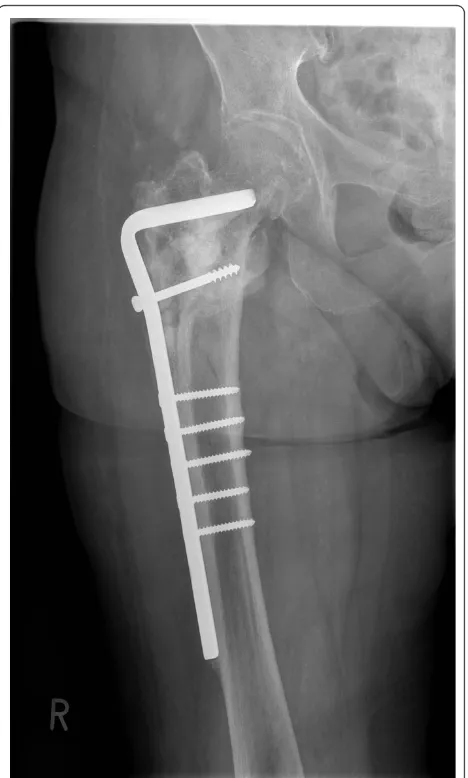
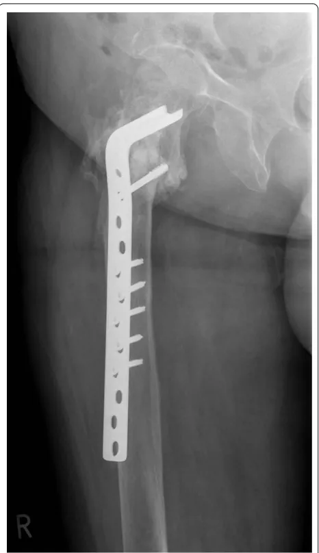
Figure 8Anteroposterior radiograph demonstrating the 90° condylar blade plate (CBP) restoring the proper shaft-neck hip angle and union of the fracture site at 10 months following revision surgery with no signs of avascular necrosis of the femoral head.
Tzioupiset al.Journal of Medical Case Reports2011,5:87 http://www.jmedicalcasereports.com/content/5/1/87
[image:5.595.58.290.517.712.2]fracture. It has been defined as a fracture occurring at the level of the lesser trochanter or approximately 5 cm below it [2]. Classification systems have also included intertrochanteric fractures with distal extension into the subtrochanteric region, such as reverse obliquity inter-trochanteric fractures [2]. However, the AO/OTA classi-fication system has classified these types of fractures separately under 31-A3, and they have been included in other studies and the current case report because of the rare occurrence of a pure subtrochanteric fracture.
The revision of a nonunited subtrochanteric fracture is challenging because of the varus deformity of the proximal fragment, bone loss and comminution, and occasionally by the failed previous implant [1,5]. Currently, there is no strong evidence to support the use of either IMD or
extramedullary devices in the revision of a failed subtro-chanteric nonunion [1]. However, the CBP has been advo-cated for fractures with a very short proximal fragment and large deformities or defects in the region of the piri-formis fossa and greater trochanter entry site [1]. The CBP is able to target the area below the femoral head that is unlikely to be compromised by the previous fixation.
In our present case, we elected to revise the failed TGN device with an extramedullary CBP to provide anatomical reduction and fracture site compression, as bone loss and the proximity of the fracture to the femoral neck would not have allowed the insertion of a revision nail to achieve these successfully [1,5]. Previous reports in the literature have confirmed the limited capacity of an IMD to correct the alignment and com-press subtrochanteric nonunions to healing in a surgical revision, which are advantages that a CBP can offer [5,14]. This added advantage was protected by augment-ing the bone biology. The combination of RIA autograft, BMP-7 and HA cement was used because of the patient’s bone loss and to restore the medial column to prevent cyclical loading of the plate on the tension side of the femur and potentially implant failure.
The gold standard for enhancing bone healing in non-united fractures is an autologous bone graft [7]; however, this procedure has been associated with donor site mor-bidity and limited availability [7]. The RIA system was developed originally as a simultaneous reaming and aspiration system to reduce intramedullary pressure, heat generation and possibly fat embolism [15]. In addition, it has been recently reported that RIA aspirate contains hMSCs [16], which are known to differentiate toward the osteogenic lineage under the appropriate stimuli [10,17].
Removing the TGN and proximal screw, as well as the fibrous tissue, from the nonunion site left a significant bone defect to be filled in our surgical revision case. Using intramedullary RIA reamings and BMP-7 was considered appropriate, as RIA reamings were available through the fracture site, avoiding the potential hazards of iliac crest harvesting. Furthermore, BMP-7 has pre-viously been used with success in randomized human nonunion studies [9] and in experimental healing of metaphyseal bone defects [18]. In addition, the injected HA cement provided temporary mechanical support to the subchondral zone of the femoral head after removal of the proximal TGN screw, as the CBP blade did not reach this zone [19].
Conclusion
[image:6.595.58.291.87.491.2]To the best of our knowledge, this is the first case study to report the successful combination of RIA autograft, BMP-7 and HA cement for the treatment of an estab-lished subtrochanteric nonunion. The essential require-ments for success when revising a nonunited fracture
are to provide anatomical reduction, mechanical stabi-lity, bone defect augmentation and biological stimulation to achieve healing. Current advances in molecular biol-ogy, such as rhBMP-7, and biotechnolbiol-ogy, such as the RIA system and HA injectable cement, can improve the outcomes of patients over the use of our traditional sur-gical revision techniques.
Consent
Written informed consent was obtained from the patient for publication of this case report and accompanying images. A copy of the written consent is available for review by the Editor-in-Chief of this journal.
Abbreviations
CBP: condylar blade plate; HA: hydroxyapatite; hMSCs: human mesenchymal stem cells; IMD: intramedullary device; rhBMP-7: recombinant human bone morphogenetic protein-7; RIA: Reamer/Irrigator/Aspirator; TGN: trochanteric gamma nail.
Author details
1Academic Department of Trauma and Orthopaedics, School of Medicine,
University of Leeds, Leeds General Infirmary, Leeds Teaching Hospitals NHS Trust, Clarendon Wing A, Great George Street, Leeds, LS1 3EX, UK.
2
Academic Orthopaedic Unit, Faculty of Medicine, Aristotle University of Thessaloniki 541 24, Greece.
Authors’contributions
CT reviewed the literature and was involved in manuscript preparation and editing. PP reviewed the literature, wrote a first draft of the manuscript and was involved in manuscript preparation and editing. ZG reviewed the literature and was involved in manuscript preparation, editing and submission. ET carried out the surgical procedure and was involved with the conception of the report, reviewed the literature, corrected and finalised the manuscript. All authors read and approved the final manuscript.
Competing interests
The authors declare that they have no competing interests.
Received: 1 July 2010 Accepted: 1 March 2011 Published: 1 March 2011
References
1. Haidukewych GJ:Nonunion of fractures of the subtrochanteric region of the femur.Tech Orthop2008,23:131-136.
2. Kuzyk PR, Bhandari M, McKee MD, Russell TA, Schemitsch EH:
Intramedullary versus extramedullary fixation for subtrochanteric femur fractures.J Orthop Trauma2009,23:465-470.
3. Parker MJ, Handoll HH:Gamma and other cephalocondylic intramedullary nails versus extramedullary implants for extracapsular hip fractures in adults.Cochrane Database Syst Rev2008, ,3:CD000093.
4. Shukla S, Johnston P, Ahmad MA, Wynn-Jones H, Patel AD, Walton NP: Outcome of traumatic subtrochanteric femoral fractures fixed using cephalo-medullary nails.Injury2007,38:1286-1293.
5. De Vries JS, Kloen P, Borens O, Marti RK, Helfet DL:Treatment of subtrochanteric nonunions.Injury2006,37:203-211.
6. Papasimos S, Koutsojannis CM, Panagopoulos A, Megas P, Lambiris E:A randomised comparison of AMBI, TGN and PFN for treatment of unstable trochanteric fractures.Arch Orthop Trauma Surg2005, 125:462-468.
7. Arrington ED, Smith WJ, Chambers HG, Bucknell AL, Davino NA: Complications of iliac crest bone graft harvesting.Clin Orthop Relat Res
1996,329:300-309.
8. Porter RM, Liu F, Pilapil C, Betz OB, Vrahas MS, Harris MB, Evans CH: Osteogenic potential of reamer irrigator aspirator (RIA) aspirate collected from patients undergoing hip arthroplasty.J Orthop Res2009,27:42-49.
9. Friedlaender GE, Perry CR, Cole JD, Cook SD, Cierny G, Muschler GF, Zych GA, Calhoun JH, LaForte AJ, Yin S:Osteogenic protein-1 (bone morphogenetic protein-7) in the treatment of tibial nonunions.J Bone Joint Surg Am2001,83-A(Suppl 1):S151-S158.
10. Tsiridis E, Ali Z, Bhalla A, Heliotis M, Gurav N, Deb S, DiSilvio L:In vitro and in vivo optimization of impaction allografting by demineralization and addition of rh-OP-1.J Orthop Res2007,25:1425-1437.
11. Charnley J:The long-term results of low-friction arthroplasty of the hip performed as a primary intervention.J Bone Joint Surg Br1972,54:61-76. 12. Haidukewych GJ, Israel TA, Berry DJ:Reverse obliquity fractures of the
intertrochanteric region of the femur.J Bone Joint Surg Am2001, 83-A:643-650.
13. Herrera A, Domingo LJ, Calvo A, Martínez A, Cuenca J:A comparative study of trochanteric fractures treated with the Gamma nail or the proximal femoral nail.Int Orthop2002,26:365-369.
14. Rahme DM, Harris IA:Intramedullary nailing versus fixed angle blade plating for subtrochanteric femoral fractures: a prospective randomised controlled trial.J Orthop Surg (Hong Kong)2007,15:278-281.
15. Giannoudis PV, Tzioupis C, Green J:Surgical techniques: how I do it? The Reamer/Irrigator/Aspirator (RIA) system.Injury2009,40:1231-1236. 16. Porter RM, Ivkovic A, Wells J, Glatt V, Harris MB, Vrahas MS, Evans C: Characterization and utilization of mesenchymal progenitor cells recovered with the Reamer-Irrigator-Aspirator [Abstract].Eur Cell Mater
2008,16(Suppl 2):19.
17. Belthur MV, Conway JD, Jindal G, Ranade A, Herzenberg JE:Bone graft harvest using a new intramedullary system.Clin Orthop Relat Res2008, 466:2973-2980.
18. Tsiridis E, Morgan EF, Bancroft JM, Song M, Kain M, Gerstenfeld L, Einhorn TA, Bouxsein ML, Tornetta P:Effects of OP-1 and PTH in a new experimental model for the study of metaphyseal bone healing.J Orthop Res2007,25:1193-1203.
19. Tsiridis E, Ali Z, Bhalla A, Gamie Z, Heliotis M, Gurav N, Deb S, DiSilvio L:In vitro proliferation and differentiation of human mesenchymal stem cells on hydroxyapatite versus human demineralised bone matrix with and without osteogenic protein-1.Expert Opin Biol Ther2009,9:9-19. doi:10.1186/1752-1947-5-87
Cite this article as:Tzioupiset al.:Revision of a nonunited subtrochanteric femoral fracture around a failed intramedullary nail with the use of RIA products, BMP-7 and hydroxyapatite: a case report.
Journal of Medical Case Reports20115:87.
Submit your next manuscript to BioMed Central and take full advantage of:
• Convenient online submission
• Thorough peer review
• No space constraints or color figure charges
• Immediate publication on acceptance
• Inclusion in PubMed, CAS, Scopus and Google Scholar
• Research which is freely available for redistribution
Submit your manuscript at www.biomedcentral.com/submit
Tzioupiset al.Journal of Medical Case Reports2011,5:87 http://www.jmedicalcasereports.com/content/5/1/87