ScholarWorks @ Georgia State University
ScholarWorks @ Georgia State University
Biology Theses Department of Biology
Fall 12-10-2012
Is TGF-
β
playing a role in ectopic neuromuscular junction
Is TGF- playing a role in ectopic neuromuscular junction
formation in the nematode Caenorhabditis elegans?
formation in the nematode Caenorhabditis elegans?
Abir A. Rahman
Follow this and additional works at: https://scholarworks.gsu.edu/biology_theses
Recommended Citation Recommended Citation
Rahman, Abir A., "Is TGF-β playing a role in ectopic neuromuscular junction formation in the nematode Caenorhabditis elegans?." Thesis, Georgia State University, 2012.
https://scholarworks.gsu.edu/biology_theses/40
This Thesis is brought to you for free and open access by the Department of Biology at ScholarWorks @ Georgia State University. It has been accepted for inclusion in Biology Theses by an authorized administrator of
IS TGF-β PLAYING A ROLE IN ECTOPIC NEUROMUSCULAR JUNCTION FORMATION IN THE NEMATODE
CAENORHABDITIS ELEGANS?
by
ABIR ASHFAKUR RAHMAN
Under the Direction of Walter W. Walthall
ABSTRACT
The neuromuscular junction (nmj) is a commonly studied synapse, used often to investigate reciprocal
signaling between a motor neuron and the appropriate target muscle. In Caenorhabditis
elegans, ectopic nmjs can be created by eliminating selected embryonic muscle cells that act as
guideposts for the migration of post-embryonic muscles. The ectopic muscles are required to induce
sprouting from DD motor neurons, indicating the presence of a muscle derived signaling molecule that
interacts with the neurons. A TGF-β homolog, unc-129, is reported to be transiently expressed in the
dorsal body wall muscles. The timing of the expression of TGF-β coincides with the time that the DD
motor neurons respecify their synapses. In this study, we show that TGF-β is expressed by the ectopic
muscle and that in unc-129 mutant animals, the ectopic muscle is unable to induce sprouting from the
DD motor neurons. Therefore, we conclude that TGF-β is necessary for ectopic nmj formation in
C.elegans.
IS TGF-β PLAYING A ROLE IN ECTOPIC NEUROMUSCULAR JUNCTION FORMATION IN THE NEMATODE
CAENORHABDITIS ELEGANS?
by
ABIR ASHFAKUR RAHMAN
A Thesis Submitted in Partial Fulfillment of the Requirements for the Degree of
Master of Science
in the College of Arts and Sciences
Georgia State University
Copyright by Abir Ashfakur Rahman
IS TGF-β PLAYING A ROLE IN ECTOPIC NEUROMUSCULAR JUNCTION FORMATION IN THE NEMATODE
CAENORHABDITIS ELEGANS?
by
ABIR ASHFAKUR RAHMAN
Committee Chair: Walter W. Walthall
Committee: Casonya Johnson
Vincent Rehder
Electronic Version Approved:
Office of Graduate Studies
College of Arts and Sciences
Georgia State University
DEDICATION
To Sharmin Islam, who decided to take up a career in Molecular Biology because I told her that it was a
really cool field. You have always been and will continue to be one of my biggest inspirations. And to
ACKNOWLEDGEMENTS
I would like to thank my professor Dr. Bill Walthall for being such a great teacher and mentor. With you,
I learned how to use genetics and molecular biology to understand the nervous system. I learned that
molecular biology, in essence, is a tool that can be used to approach any biological phenomenon. I
appreciate the patience that you have shown with my slow yet promising research. I would also like to
thank my committee members Dr. Casonya Johnson and Dr. Vincent Rehder, for their time and valuable
input on my project as well as my thinking as a research scientist.
I would like to extend my gratitude to the Department of Biology, the Neuroscience Institute, and all the
faculty and staff therein for giving me this opportunity and for nurturing the scientist in me. Thanks to
LaTesha Warren, for making my life as a graduate student so much nicer. Special thanks go to my
colleagues in the lab: Richard, Manali, Vaishali, Shelley, David, Farmaan, Kelsey, Jennifer, Kachael and
Adrian for their support, suggestions and ideas. I cannot thank my cousin Tamanna Rahman and her
husband Atiqur Rahman Bhuiyan enough, for being with me through thick and thin, all the way. And I do
not have the words to thank Ismat Ara. If she didn’t show up in July 2012, I doubt I would have ever
finished my thesis.
Most importantly, I would like to thank my parents and my little sister, Tasnuva Rahman, who have
showered me with so much love and support that I simply could not have come this far without. I hope I
TABLE OF CONTENTS
ACKNOWLEDGEMENTS ... v
LIST OF TABLES... vii
LIST OF FIGURES ...viii
1. INTRODUCTION ... 1
2. MATERIALS AND METHODS ... 7
3. RESULTS ... 9
4. DISCUSSION ... 17
REFERENCES ... 23
LIST OF TABLES
Table 1 – Genotypes of the animals used 7
LIST OF FIGURES
Figure 1 – The direction of information flow in DD and VD motor neurons 3
Figure 2 - The lineage and a Nomarski image of the Cap and the Cpp cells 4
Figure 3 – Ectopic muscle generated when Cap/Cpp are ablated 4
Figure 4 – Schematic explanation of ectopic muscle formation 5
Figure 5 – Neuron and muscle morphology in an unablated pflp-13::gfp animal 10
Figure 6 – Ectopic muscle innervated by DD motor neurons. 11
Figure 7 – Neuron and muscle morphology in pflp11::gfp animals 12
Figure 8 – The dorsal body wall muscles and the DA and DB motor neurons expressing TGF-β 13
Figure 9 – The ectopic muscle is expressing TGF-β 14
Figure 10 – Aberrant neurite growth in unc-129 mutant animals 16
Figure A1 – Neuron morphology in unc-40 mutant animal 27
1. INTRODUCTION
The assembly of functional neural networks requires interactions that allow recognition among
the participating cells. The developing neuron first sends processes that follow guidance cues on
appropriate substrates to find appropriate target and then initiates synaptogenesis, a complex
developmental process that leads to the formation of the synapse, which consists of a pre- and
postsynaptic element. Both process guidance and synaptogenesis require the exchange of various
signaling molecules. Molecules secreted from the target cells are collectively known as retrograde
signaling molecules; whereas the signaling molecules released from the presynaptic neuron are known
as orthograde signaling molecules (Fitzsimonds and Poo 1998). Reciprocal signaling has been shown to
be a very important mechanism by which the events of synaptogenesis are coordinated (Sanyal, Kim and
Ramaswami 2004).
The neuromuscular junction presents an intriguing location to study these phenomena. This is a
synapse where the presynaptic and the postsynaptic elements arise from two distinct lineages. The
ectodermally derived motor neuron must innervate appropriate target muscles derived from the
mesodermal germ layer. In vertebrates, a single muscle cell is initially innervated by multiple axons.
However, in the adult animal, the individual muscle cells are innervated by single motor axons. Synapses
made by all but one motor axon are eliminated in an activity-dependant manner (Redfern 1970;
Lichtman 1977). This high degree of specificity of pairing between the neuronal presynaptic elements
and the post-synaptic elements on the muscle is achieved through reciprocal signaling.
Evidence for retrograde signaling at neuromuscular junctions is seen in Xenopus (Dai and Peng
1993) and in Helisoma (Zoran, Funte, Kater and Haydon 1993) cell cultures as well as in Drosophila
melanogaster (Aberle et al. 2002; Keshishian and Kim 2004; Marques et al. 2002) and Caenorhabditis
elegans (Hedgecock, Culotti and Hall 1990; Colavita et al. 1998; Macneil et al. 2009). Work on C.elegans
ease of performing genetic manipulations. The nematodehas a relatively short reproductive span, which
reduces the time required to generate and maintain true breeding mutant animals. In addition, the
animal is transparent and it is possible to fluorescence-label entire neurons or the presynaptic termini of
the neurons of interest (Chalfie et al. 1994).
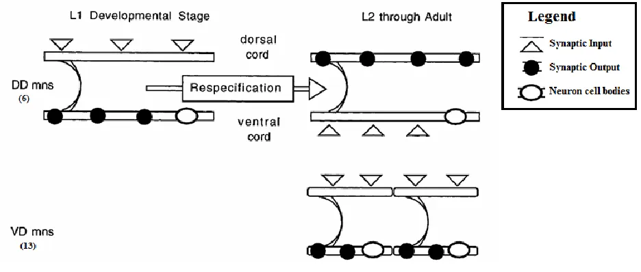
The nervous system of C. elegans consists of 302 neurons, out of which there are 19 GABA-ergic
motor neurons that are designated the D motor neurons. There are 6 dorsal D neurons (DD) that initially
innervate ventral body wall muscles, when the animal hatches, and undergo synaptic respecification at
around the first larval molt, innervating dorsal body wall muscles thereafter. This change of polarity
occurs without any morphological change in the neurons themselves (White et al 1978; Walthall
1990).The remaining 13 D motor neurons are designated ventral D (VD) neurons because they innervate
the ventral muscles (Figure 1). They are postembryonic and are born around the same time as the DD
synaptic respecification event. The DD motor neurons are seeking new muscle targets during the
respecification and, presumably, are receptive at this time point, to retrograde signaling molecules from
Figure 1 – The direction of information flow in DD and VD motor neurons
(Top) DD motor neurons innervate ventral muscle when the animal is born, but undergo a respecification event and innervate dorsal muscle in the later stages. (Bottom) VD motor neurons are born after the animal hatches from the egg, around the first larval molt, and innervate ventral muscle. (Figure adopted from Plunkett, Simmons and Walthall 1996)
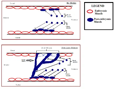
There are 95 body wall muscles in C.elegans, organized into four contiguous strands running
along the length of the animal (Figure 3A). Out of these 95 muscle cells, 81 are embryonic and are
present when the animal hatches, and the remaining 14 are born at around the first larval molt. Since
the complete lineage of the 959 somatic cells in the adult animal has been mapped out (Sulston et al.
1983), it is possible to trace each muscle cell and each neuron to their precursors. The 14 postembryonic
muscle cells and some other cells are descendants of a myoblast called the M cell (born from MSapa in
figure 2). A subset of the 81 embryonic body wall muscle cells are derived from the myoblasts Cap and
Cpp (figure 2). This lineage map is invariant and it is, therefore, possible to eliminate specific cells from
the adult animal, by performing laser ablation of the appropriate precursors in embryos. Such ablation
of the Cap and Cpp cells results in the absence of the descendant body wall muscles from the animal
(Plunkett, Simmons and Walthall 1996). This, in turn, creates a gap in the dorsal body wall muscle
Figure 2 – The lineage and a Nomarski image of the Cap and the Cpp cells
(A) Partial lineage of C.elegans showing the body wall muscle precursors Cap, Cpp and MSapa. Anterior daughters are shown to the left and posterior to the right (Adopted from Plunkett, Simmons and Walthall 1996). (B) Nomarski image of a C.elegans egg approximately 100 minutes post fertilization. The Cap and the Cpp have just been born.
Figure 3 – Ectopic muscle generated when Cap/Cpp are ablated
[image:14.612.73.329.395.663.2]As the animal develops, ectopic strands are set up by the postembryonic muscle cells (Figure 3)
presumably because the embryonic muscles act as guideposts that are now absent due to the ablation.
Figure 4 shows a schematic description of the process. These ectopic muscles are then innervated by
processes from the DD motor neurons that have sprouted from the dorsal nerve cord (Plunkett,
Simmons and Walthall 1996). Ablation of the MSapa (post-embryonic muscle precursor, figure 2) along
with Cap and Cpp leads to a gap in the dorsal muscle strands as well as absence of all ectopic muscle. In
such a case, the neurite sprouting from the DD neurons was no longer seen (Plunkett, Simmons and
Walthall 1996). We hypothesize that the ectopic muscles are secreting a signaling molecule that might
[image:15.612.83.482.319.622.2]be responsible for inducing these innervations.
Figure 4 – Schematic explanation of ectopic muscle formation
The C.elegans TGF-β homolog, unc-129, is secreted by the dorsal body wall muscles (Colavita et
al. 1998). Its expression in the dorsal body wall muscles is transient such that at L1, all dorsal body wall
muscles are expressing unc-129 but by around the L1/L2 molt, only a subset of the muscles is expressing
unc-129, gradually reaching the point when, in the adult animal, none of the dorsal muscles are
expressing unc-129. In this study, the ectopic muscles were seen to express unc-129 at around the L1/L2
molt. We suggest that this TGF-β is functioning as a retrograde signaling molecule, inducing innervation
of the ectopic muscles when the DD motor neurons are seeking new targets, and is then no longer
expressed once the synaptogenesis is complete.
According to Macneil et al. (2009), UNC-129 interacts with two non-canonical receptors, UNC-5
and UNC-40, to mediate pioneer axon guidance via the UNC-6/Netrin pathway. UNC-129 is thought to
switch the signal transduction mechanism of the developing neuron from 5 alone signaling to
UNC-5+UNC-40 signaling, which in turn, increases the sensitivity of the growth cone to UNC-6. This becomes
important in cases where UNC-6 acts as a repulsive signal and neural processes extend away from
regions of high concentrations of UNC-6, such as the case of DD motor neuron axon migration. As the
growth cone migrates from UNC-6 rich regions to UNC-6 deficient regions, an increase in sensitivity
takes place in the growth cone, in order to compensate for the decrease in 6 concentration.
UNC-129 is graded opposite to UNC-6 and is thought to promote this compensatory increase in sensitivity
(Colavita and Culotti 1998; Macneil et al. 2009). In this paper, we show that the ectopic muscle,
generated by the Cap/Cpp ablation process, expresses unc-129. The mechanism described by Macneil et
al. (2009) might therefore be the mechanism for ectopic muscle innervation via retrograde signaling
mediated by the UNC-129, UNC-5 and UNC-40. We, therefore, tested a hypothesis that unc-129 is
2. MATERIALS AND METHODS
Strain maintenance: All strains of C. elegans were grown and maintained at 22°C on NGM agar plates as
described by Brenner (1974). Mating plates were set with 6 males and 2 L4/adult hermaphrodites per
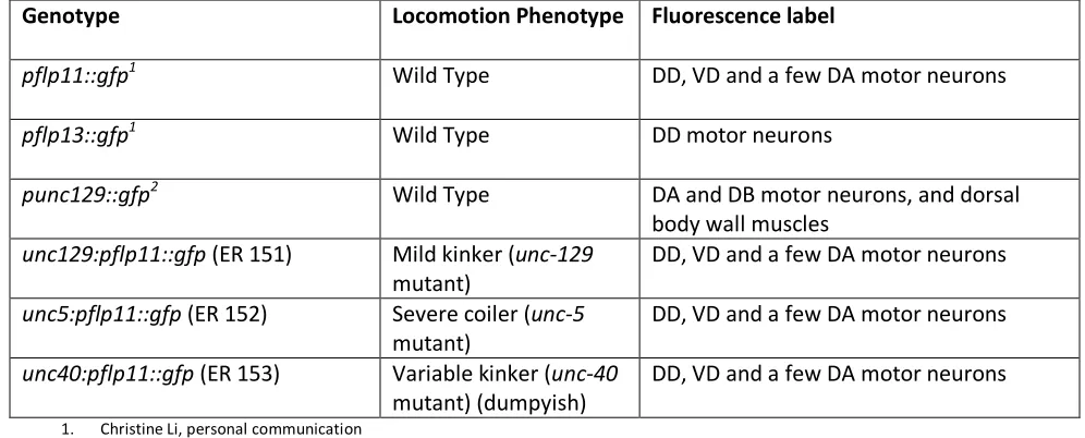
plate. Table 1 shows a list of the genotypes of the different strains used and the respective tissues that
[image:17.612.62.560.253.454.2]expressed GFP. The N2 Bristol strain was used as wild type for crossing purposes.
Table 1 - Genotypes of the animals used.
Transgenic animals expressing GFP under neuron-specific promoters were used to visualize the respective neurons.
Genotype Locomotion Phenotype Fluorescence label
pflp11::gfp1 Wild Type DD, VD and a few DA motor neurons
pflp13::gfp1 Wild Type DD motor neurons
punc129::gfp2 Wild Type DA and DB motor neurons, and dorsal
body wall muscles
unc129:pflp11::gfp (ER 151) Mild kinker (unc-129
mutant)
DD, VD and a few DA motor neurons
unc5:pflp11::gfp (ER 152) Severe coiler (unc-5
mutant)
DD, VD and a few DA motor neurons
unc40:pflp11::gfp (ER 153) Variable kinker (unc-40
mutant) (dumpyish)
DD, VD and a few DA motor neurons
1. Christine Li, personal communication 2. J. G. Culloti, personal communication
Genetic crossing: Hermaphrodites from transgenic strains expressing the desired GFP transcriptional
reporter were first crossed with N2 male animals to generate heterozygous males. These heterozygous
males were then mated with homozygous hermaphrodites containing the desired mutation (recessive).
The progeny were screened for heterozygous, GFP-positive hermaphrodites, with wild-type locomotion.
Approximately 6 of these hermaphrodites were then placed on culture plates together and allowed to
reproduce by self-fertilization. The progeny of these six worms were screened for individuals that were
homozygous for the mutation, as indicated by the locomotion defective phenotype, and expressed the
Laser Ablations: Healthy, gravid hermaphrodites were cut open to obtain eggs. The eggs were then
mounted on 0.1% agarose gel pads and visually monitored at 1000x magnification until the embryonic
precursors to the dorsal body wall muscles, Cap and Cpp, were born, around 100 minutes
post-fertilization (Sulston et al. 1983). These cells were then ablated using a nitrogen laser. They were shot 3
times for 10 seconds each at the firing rate of approximately 4 shots per second, under the 100x
objective lens set at a numerical aperture setting of 1.6. Eggs were then recovered in a culture plate at
22°C. Ablations were performed in animals from each of the genotype groups from table 1. Successfully
ablated animals were then identified, fixed and stained with RITC-Phalloidin by a procedure modified
from Plunkett, Simmons and Walthall (1996), and analyzed using deconvolution microscopy (Deltavision
Olympus IX70 microscope). The images thus obtained were then color coded green for GFP and red for
RITC-phalloidin. Images were taken only of the posterior third of the animal because the Cap/Cpp
ablation only affects that part of the animal. The rest of the animal remains mostly unaffected.
Scoring criteria: Ectopic muscle was defined as any muscle strand outside the four muscle quadrants and
that did not form part of the gut of the animal. Ectopic neurite branches were counted only if they
appeared to terminate in close proximity to an ectopic muscle strand. Otherwise, they were considered
3. RESULTS
To determine whether the findings of Plunkett, Simmons and Walthall (1996), could be
reproduced in embryos carrying transgenic GFP reporters we used the promoter of flp-13 (pflp13::gfp).
This GFP construct is expressed in the DD motor neurons (Kim and Li 2004). In unablated animals, this
allowed observation of the ventrally located cell bodies of the neurons, ventral postsynaptic processes,
commissures projecting to the dorsal nerve cord where they branched. Four body wall muscle strands
(shown in red) flanked the two nerve cords (Figure 5). Images were always taken of the posterior third
of the animal because the Cap/Cpp descendants are normally located in this region of the animal and
the ablation procedure only affects this part of the animal. The rest of the animal remains mostly
unaffected.
In ablated animals, the tail was severely deformed because the missing muscles normally act as
girdles and help maintain the shape of the worm. Ectopic muscle was observed in the ablated regions as
described by Plunkett, Simmons and Walthall (1996). Any muscle strand observed outside the four
contiguous body wall muscle strands, and not contributing to the gut, was considered ectopic. Ectopic
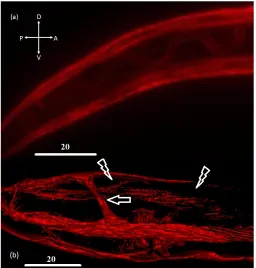
muscle was observed in 8 of preps out of 10 (Table 2). In addition, ectopic GFP-positive branches
emanating from the DD motor neurons extended from the dorsal nerve cord to the ectopic muscle
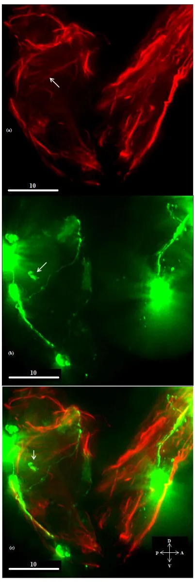
strands as shown figure 6 (shown with white arrow). This was consistent with the observations made by
Plunkett, Simmons and Walthall (1996) that the ectopic muscle is able to attract innervation.
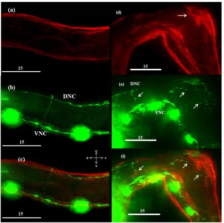
This finding was further confirmed by performing ablations in pflp11::gfp transgenic animals.
The animals in this background looked similar to the pflp-13 animals, with GFP being present in the DD,
VD and DA motor neurons (Kim and Li 2004). In the unablated animals, GFP was observed in the cell
bodies and the processes of these neurons, including those forming the dorsal and ventral nerve cords.
muscles were also seen after ablations performed in these animals, along with ectopic neural processes
[image:20.612.75.448.149.658.2]that innervated the muscles (Figure 7, d-f).
Figure 5 - Neuron and muscle morphology in an unablated pflp-13::gfp animal.
Figure 6 – Ectopic muscle innervated by DD motor neurons.
Figure 7 – Neuron and muscle morphology in a pflp11::gfp animal
Body wall muscles are shown in red while DD and VD motor neurons are shown in green. (a-c) Unablated specimen. (d-f) Ablated specimen. The white arrow in (d) shows an ectopic muscle strand. The arrows in (e) and (f) indicate associated neurite sprouting.
Table 2 - The number of animals with ectopic nmjs in the tested genetic backgrounds
Animal Total number of specimens
with ectopic muscle
Total number of specimens that displayed ectopic neurite sprouting associated with the ectopic muscle
Wild Type 10 8
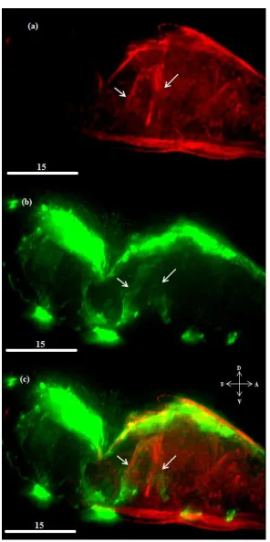
[image:22.612.65.548.621.697.2]Next, we wanted to test if TGF-β was the signal from the ectopic muscles that was necessary in
order to induce sprouting from the DD motor neurons. First, ablations were performed in animals
expressing the punc-129::gfp transgene (ER80). This construct allowed the visualization of the cells that
expressed unc-129, which are the DA and DB motor neurons and dorsal body wall muscles (Colavita and
Culotti 1998). This was to see if the ectopic muscle strands were, indeed, expressing TGF-β. If TGF-β is
indeed the suspected signal, then it should be expressed in the ectopic muscles at the time of DD
respecification. In the unablated animals, expression of unc-129 in the dorsal body wall muscles is
transient such that at around the time of the DD motor neuron respecification, only a subset of the
muscle cells expresses unc-129 (Figure 8). We hypothesized that this subset consisted primarily of
[image:23.612.70.548.366.439.2]post-embryonic muscle cells that are seeking neuronal innervation.
Figure 8 – The dorsal body wall muscles and the DA and DB motor neurons expressing TGF-β
Unablated ER80 animal. Body wall muscles are shown in red and cells expressing unc-129 are shown in green. Muscle strands expressing unc-129 are shown using white pointers.
In the ablated ER80 animals, the ectopic muscle fluoresced green (n=3), indicating that unc-129
is expressed by the ectopic muscle cells (Figure 9). Thus ectopic muscles express TGF-β coincident with
Figure 9 – The ectopic muscle is expressing TGF-β
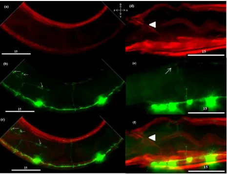
To determine whether TGF-β is necessary for the induction of ectopic neurites, ablations were
then performed in unc-129 mutant embryos (ev554) carrying the pflp11::gfp transgene (pflp11::gfp;
unc-129(ev554)). If TGF-β is necessary, then mutant animals should not display ectopic neural processes
associated with ectopic muscles in ablated animals. The unablated animals in this background often
displayed axon guidance defects. The dorsal nerve cord was often absent or formed laterally below the
two dorsal strands. Colavita et al (1998) reported that mutations in unc-129 show axon guidance defects
similar to unc-6, unc-5 and unc-40 mutations, which includes commissures that misguided and fail to
reach the dorsal nerve cord. Therefore, aberrant commissures were seen in the ablated as well as
unablated animals. Figure 10 shows the morphology of the neurons in an unablated specimen (top) and
an ablated specimen (bottom). As suspected, the single commissure visible in the ablated specimen is
not close to the ectopic muscle strand (shown by the white arrow), and is therefore not considered to
have formed an ectopic nmj. Out of the 9 animals that displayed ectopic muscle strands, none displayed
ectopic muscle-associated neurite sprouting (Table 2). Therefore, we concluded that TGF-β, was, in fact,
Figure 10 – Aberrant neurite growth in unc-129 mutant animals.
4. DISCUSSION
Synapses play a key role in regulating the information flow in nervous systems. It is, therefore,
important for pre-synaptic elements to locate appropriate post-synaptic partners. The neuromuscular
junction (nmj) is an interesting synapse. Given the ectodermal developmental origin of neurons and
mesodermal origin of muscles, the involved cells face a challenge to locate each other and establish a
functional connection. This is achieved through various signaling mechanisms (Fitzimonds and Poo 1998;
Sanyal, Kim and Ramaswami 2004), many of which have already been characterized in vertebrates.
Despite all that is known about nmjs, many more pathways remain to be discovered in this complex
process. In this research, C.elegans neuromuscular junctions were used to study the genes important for
neuron-muscle communication and subsequent nmj development. Specifically, we used a technique
where selected embryonic myoblasts that act as guideposts for the postembryonic muscles, are
laser-ablated. This, then, resulted in the postembryonic muscle setting up ectopic strands that induced
neurite sprouting from DD motor neurons.
The developmental time at which the ectopic (postembryonic) muscle strands are formed
coincides with the respecification of the DD motor neurons. Plunkett, Simmons and Walthall (1996)
found that these ectopic muscles are able to induce sprouting from the DD motor neurons. They have
shown that the presence of ectopic muscle leads to neurite sprouting from the dorsal nerve cord, and
when the same ectopic muscle (postembryonic) precursors were laser-ablated along with the embryonic
myoblasts, the sprouting from the DD motor neurons was not observed. This indicates that the ectopic
muscle is sufficient for ectopic neurite sprouting. We have shown that unc-129 (TGF-β) is expressed by
these ectopic muscles. We have also shown that in the absence of TGF-β, the ectopic muscles are no
longer innervated by DD motor neurons. Therefore, we conclude that unc-129 (TGF-β), from the
The underlying mechanism for the formation of ectopic nmjs is tied closely to the series of
events presumably taking place in the DD motor neurons during the synaptic respecification event. The
DD motor neurons respecify their synapses in the four hours surrounding the first larval molt (White et
al. 1978; Hallam and Jin 1998). At some point before this time, the DD respecification process is most
likely initiated by some form of an intrinsic trigger(s). Very little is known about this intrinsic trigger, but
lin-14 is a heterochronic gene which has been implicated in controlling the time of DD respecification
(Hallam and Jin 1998).
Following the initiation of the respecification event, the DD motor neurons begin eliminating
synapses from the ventral side of the animal and forming synapses on the dorsal side. Park et al. (2011)
have identified cyy-1 (cyclin-box containing protein) and cdk-5 (cyclin-dependant kinase -5) to be playing
a part in the process. Specifically, cyy-1 is important for synapse removal and cdk-5 is shown to be
important for new synapse formation. Park et al. (2011) have also shown that materials from previous
ventral pre-synaptic termini are recycled to construct the new dorsal pre-synaptic termini. This indicates
a potentially critical role for cytoskeletal factors and motors, such as kinesin and dynein, because this
recycling of pre-synaptic material is an important characteristic of the DD respecification event. The
recycling of presynaptic materials is reduced in unc-104/kinesin3 mutants. Park et al. (2011) concluded
that CDK-5 facilitates UNC-104-mediated intracellular transport of presynaptic material. CDK-5, along
with a regulatory subunit p39, has also been implicated in actin modifications and microtubule
formation in mice (Humbert, Dhavan and Tsai 2000). Coincident with these cytoskeletal changes,
presumable changes at the new dorsal pre-synaptic termini allow the neuron to be receptive to new
synaptic partners, and this probably leads to ectopic neurite branching if the appropriate
post-synaptic muscle are present in ectopic locations, instead of the dorsal body wall muscle strands.
Various key genes and pathways have been identified in the process of synaptic partner
the respecification event, the dorsal process of the DD motor neurons probably undergoes changes in
the receptor profile, possibly by enrichment of UNC-5 and UNC-40 receptor proteins (see below), at
their pre-synaptic (dorsal) termini so as to become sensitive to TGF-β, which then reveals the location of
the appropriate post-synaptic muscles. In the case where the appropriate post-synaptic target muscles
(post-embryonic) are formed in ectopic locations, due to ablation of the embryonic muscle precursors,
for example, sprouting of neurites that are seeking new targets, takes place.
A possible mechanism by which TGF-β induces such sprouting was first described by Colavita et
al (1998). They showed that TGF-β is expressed transiently in dorsal body wall muscles and plays a role
in pioneer axon guidance. They have shown that TGF-β acts via the netrin pathway (Colavita et al 1998;
Macneil et al. 2009). The netrin receptors characterized therein are UNC-5 and UNC-40. src-1 codes for
a kinase that has also been implicated in mediating unc-5 signaling (Lee, Li and Guan 2005). Together,
these genes might be part of a gene network that normally promotes ventral-to-dorsal migration of
axons earlier in development and is underlying the formation of ectopic nmjs, upon the ablation
procedure.
This mechanism entails that neural processes that migrate from ventral to dorsal respond
initially to unc-6/netrin gradients via unc-5 only mediated signaling. During their migration, they switch
to unc-5+unc-40 mediated signaling, which increases the sensitivity of the growth cones to lower
concentrations of unc-6/netrin (Macneil et al 2009). This is consistent with observed netrin signaling
patterns in Drosophila (Keleman and Dickson 2001). UNC-129 is reported to mediate this switch in
signaling. This is probably achieved by blocking UNC-5 signaling, as is indicated by binding assays where
UNC-129 was shown to be able to physically bind UNC-5. A similar interaction was also observed
between BMP7 (vertebrate TGF-β homolog) and RCM (vertebrate unc-5 homolog). Presumably, it is a
consequence of this increased sensitivity to unc-6/netrin induced repulsion that UNC-129 can attract
In the absence of target muscle (embryonic and postembryonic) that would normally flank the
dorsal nerve cord, the DD motor neurons send out ectopic neural processes, presumably in search of
target muscle at other locations. This could be an intrinsic mechanism that is normally suppressed by
the intact muscle flanking the dorsal nerve cord. The suppression may be mediated by a retrograde
signal from the muscle. Aberrant sprouting has been shown to occur AVB neurons if synaptic activity is
blocked (Loria et al. 2003; Hobert, Tessmer and Ruvkun 1999; Zhao and Nonet 2000), indicating that this
process is very dynamic and synaptic activity is necessary for maintenance of a previously established
synapse. In the absence of post-synaptic muscle (embryonic), the sprouting from the DD motor neurons
may be due to the result of the activation of a similar genetic program.
Following sprouting, neurites migrate towards the ectopic muscle. This is caused by further
changes in cytoskeletal structure, cell surface membrane structure, and interactions with the
extracellular matrix and any secreted factors. A comparable process is that of axon regeneration (Chen
et al. 2011). Following a laser axotomy procedure, some but not all C.elegans neurons regrow their
axons. Among those that do regenerate are the PLM sensory neurons and motor neurons. Chen et al.
(2011) identified several mutants that displayed defects and enhancements in this regeneration ability.
The list of mutations included genes for cytoskeletal factors and extracellular matrix (ECM) proteins.
Although most mutations reduced regeneration abilities, mutations in the ECM protein coding genes
often caused enhancements in regeneration. This suggested that interplay, between the ECM proteins
and cytoskeletal factors, was important for axon guidance. Secreted guidance molecules such as TGF-β
probably interact with ECM proteins such as agrin, and ephrins that have been shown to be important in
regeneration as well as axon guidance in vertebrate model systems.
In addition, parts of the Wnt signaling pathway and the slit-robo pathway have been identified
by Chen et al. (2011). This suggests that regeneration is somehow reactivating the axon guidance
several genes to be involved in regeneration that are not known to be associated with axon guidance or
neuronal development. For example, genes involved in endocytosis of synaptic vesicles and in the
biosynthesis of GABA were involved. This led to the conclusion that regeneration is more complex than
was initially thought to be. Nevertheless, it can be speculated that at least part of the regeneration
process comprises reactivation of earlier developmental programs in the neuron. This is consistent with
a previous description of the laser axotomy procedure, where the proximal end of the severed axon was
seen to reform the growth cone and migrate to the appropriate post-synaptic partner (Yanik et al.
2004). This, therefore, raises the possibility that the non-canonical TGF-β pathway identified by Macneil
et al. (2009) works in parallel with the regeneration pathways identified by Chen et al. (2011) and to a
certain extent, the regeneration process might be reactivating an axon guidance program already in
place, presumably suppressed by factors in a functional synapse.
Nevertheless, our results as well as those of Chen et al. (2011) indicate that DD motor neurons
have more developmental potential during the respecification than previously characterized. The
presence of ectopic muscles as well as surgery of the axon can induce neurite sprouting, followed by
guidance to appropriate post-synaptic targets. In the case of ectopic nmj formation, TGF-β and the
netrin pathway are presumably instrumental in promoting the sprouting of ectopic processes, possibly
by the mechanism described by Macneil et al (2009).
We have demonstrated that TGF-β is necessary for ectopic nmj formation and that it is
expressed by the appropriate post-synaptic cell. The findings of Colavita et al. (1998) were that motor
axons are misguided in the absence of TGF-β. In this research, we were able to change the location of
the TGF-β expressing cell and, in a way, demonstrated that it is sufficient to induce ectopic neurite
sprouting. The downstream factors of this process still need to be elucidated. The receptors have been
proposed by Macneil et al. (2009) as unc-5 and unc-40. However, it remains to be seen whether there is
TGF-β, and the intracellular factors such as cytoskeletal elements etc., that underlie the migration of the
neurite. These are to be identified as well. TGF-β may also have an effect on synaptogenesis, which can
be characterized if expression of TGF-β could be temporally manipulated. It is important in this case to
make sure that axon guidance is completed and that only the effect on synaptogenesis is tested. Finally,
all this information can be put together as a set of signaling cascades that operate to promote the
unusual phenomenon of ectopic nmj formation. With this information, it will then be possible to
formulate models of nmj formation in other organisms.
TGF-β pathways are highly conserved across species. There is evidence that TGF-β plays a role in
axon guidance in Drosophila melanogaster. The development of the Drosophila neuromuscular junction
involves the TGF-β homolog, Gbb (Glass-bottom boat) and the receptor wit (Wishful Thinking). The
TGF-β homologs in vertebrates, such as the BMPs (Bone Morphogenic Proteins), have also been implicated in
axon guidance (Sanyal et al. 2004). The findings of this research have considerable implications in our
understanding of the development of nmjs in general. It would be interesting to see if the role played by
TGF-β in nmj formation in more complex organisms is similar to that in C.elegans. However, the pathway
described by Macneil et al (2009), is a non-canonical one. Characterization of this pathway will present
us with an interesting example of how a ligand from one particular signaling pathway, can play a
pleiotropic role in a second, considerably different pathway. The neuromuscular junction has been
studied for a long time and there appears to be numerous signaling mechanisms at play to ensure
proper formation the nmj. A better understanding of the nmj formation will generate insight into the
complex signals that allow neural circuits to be formed in general. This, in turn, will shed light on how
REFERENCES
1. Aberle H., Haghighi A.P., Fetter R.D., McCabe B.D., Maghalhaes T.R. and Goodman C.S. (2002).
Wishful thinking encodes a BMP type II receptor that regulates synaptic growth in Drosophila.
Neuron. 33(4), 545-558.
2. Brenner, S. (1974). The genetics of Caenorhabditis elegans. Genetics 77, 71-94.
3. Chalfie M., Tu Y., Euskirchen G., Ward W.W. and Prasher D. C. (1994) Green fluorescent protein
as a marker for gene expression. Science 263 (5148), 802-805
4. Chen L., Wang Z., Ghosh-Roy A., Hubert T., Yan D., O’Rourke S., Bowerman B., Wu Z., Jin Y. and
Chisholm A. D. (2011) Axon regeneration pathways identified by systematic genetic screening in
C. elegans. Neuron 71, 1043–1057.
5. Colavita, A., Krishna, S., Zheng, H., Padgett, R. W., and Culotti, J. G. (1998) Pioneer Axon
Guidance by UNC-129, a C.elegans TGF-β. Science 281, 706-709.
6. Dai Z. and Peng H.B. (1993) Elevation in presynaptic Ca2+ level accompanying initial nerve-muscle
contact in tissue culture. Neuron. 10, 827-837.
7. Fitzsimonds, R.M., and Poo, M.M. (1998). Retrograde signaling in the development and
modification of synapses. Physiol. Rev. 78, 143–170.
8. Georgi, L. L., Albert, P. S., and Riddle, D. L. (1990) daf-1, a C. elegans gene controlling dauer larva
development, encodes a novel receptor protein kinase. Cell 61, 635-645.
9. Hallam S.J. and Jin Y. (1998). Lin-14 regulates the timing of synaptic remodeling in
Caenorhabditis elegans. Nature. 395, 78-82.
10. Hedgecock E.M., Culotti J.G. and Halls D.H. (1990). The unc-5, unc-6, and unc-40 Genes Guide
Circumferential Migrations of Pioneer Axons and Mesodermal Cells on the Epidermis in C.
11. Hobert O., Tessmer K. and Ruvkun G. (1999) The Caenorhabditis elegans lim-6 LIM homeobox
gene regulates neurite outgrowth and function of particular GABAergic neurons. Development
126, 1547-1562.
12. Humbert S., Dhavan R. and Tsai L. (2000). P39 activates cdk5 in neurons, and is associated with
the actin cytoskeleton. Journal of Cell Science. 113, 975-983.
13. Keleman K., Ribeiro C. and Dickson B.J. (2005) Comm function in commissural axon guidance:
cell-autonomous sorting of Robo in vivo. Nature Neuroscience 8, 156-163.
14. Keshishian H. and Kim Y.M. (2004). Orchestrating development and function: retrograde BMP
signaling in the Drosophila nervous system. Trends Neurosci. 27, 143–147.
15. Kim K. and Li C. (2004). Expression and regulation of an FMRFamide-related neuropeptide gene
family in Caenorhabditis elegans. J Comp. Neurol. 475, 540-550.
16. Lee J., Li W. and Guan K. (2005) SRC-1 mediates UNC-5 signaling in Caenorhabditis elegans.
Molecular and Cellular Biology. 25, 6485-6495.
17. Lichtman J.W. (1977). The reorganization of synaptic connexions in the rat submandibular
ganglion during post-natal development. J Physiol. 273, 155-177.
18. Loria P.M., Duke A., Rand J.B. and Hobert O. (2003) Two Neuronal, Nuclear-Localized RNA
Binding Proteins Involved in Synaptic Transmission. Current Biology 13, 1317–1323.
19. Macneil, L. T., Hardy, H. W., Pawson, T., Wrana, J. L., and Culotti, J. G. (2009) UNC-129 regulates
the balance between UNC-40 dependent and independent UNC-5 signaling pathways. Nature
Neuroscience. 12, 150-155.
20. Marques G., Bao H., Haerry T.E., Shimell M.J., Duchek P., Zhang, B. and O’Connor M.B. (2002).
The Drosophila BMP Type II Receptor Wishful Thinking Regulates Neuromuscular Synapse
21. McCabe B.D., Marques G., Haghighi P., Fetter R.D., Crotty M.L., Haerry T. E., Goodman C.S. and
O’Connor M.B. (2003). The BMP Homolog Gbb Provides a Retrograde Signal that Regulates
Synaptic Growth at the Drosophila Neuromuscular Junction. Neuron 39, 241-254
22. Park M., Watanabe., Poon V.Y.N., Ou C., Jorgensen E.M. and Shen K. (2011). CYY-1/Cyclin Y and
CDK-5 differentially regulate synapse elimination and formation for rewiring neural circuits.
Neuron. 70, 742-757.
23. Plunkett, J. A., Simmons, R. B., and Walthall, W. W. (1996) Dynamic Interactions between Nerve
and Muscle in Caenorhabditis elegans. Dev. Biol. 175, 154-165.
24. Redfern P.A. (1970) Neuromuscular transmission in new-born rats. J Physiol. 209(3), 701-709.
25. Sanyal, S., Kim, S. M., and Ramaswami, M. (2004) Retrograde Regulation in the CNS:
Neuron-Specific Interpretations of TGF-β Signaling. Neuron 41, 845-848.
26. Shen K. and Scheiffele P. (2010) Genetics and Cell Biology of Building Specific Synaptic
Connectivity. Annu Rev Neurosci. 33:473-507.
27. Sulston, J. E. and Horvitz, H. R. (1977). Post-embryonic lineages of the nematode Caenorhabditis
elegans. Dev. Biol. 56, 110-156.
28. Sulston J.E., Scheirenberg E., White J.G. and Thomas J.N. (1983). The embryonic cell lineage of
the nematode Caenorhabditis elegans. Developmental Biology. 100, 64-119.
29. Walthall, W.W. (1990). Metamorphic- like changes in the nervous system of the nematode.
Developmental neurobiology 21, 1085-1091.
30. White, J. G., Southgate, E., Thomson, J. N., and Brenner, S. (1983). Factors that determine
connectivity in the nervous system of Caenorhabditis elegans. Cold Spring Harbor Symp. Quant.
Biol. 48, 633-640.
31. White J. G., Albertson D. G., and Anness M. A. R., (1978) Connectivity changes in a class of
32. Yanik M.H., Cinar H., Cinar H. N., Chisholm A. D., Jin Y. and Ben-Yakar A. (2004) Functional
regeneration after laser axotomy. Nature. 432, 822.
33. Zhao H. and Nonet M.L. (2000). A retrograde signal is involved in activity-dependent remodeling
at a C.elegans neuromuscular junction. Development 127, 1253-1266.
34. Zoran M.J., Funte L.R., Kater S.B. and Haydon P.G. (1993). Neuron-muscle contact changes
APPENDIX: INCOMPLETE DATA SETS
Additional ablations were performed in unc-5 and unc-40 mutants carrying the pflp11::gfp
transgene. Both of these mutants, without any ablations performed, displayed severe axon guidance
defects. Similar to unc-129 mutants, these mutants often lacked a dorsal nerve cord. Unablated unc-40
mutants displayed a higher frequency of animals with neuronal processes that failed to complete the
ventral-to-dorsal migration, when compared to unc-129 animals (Figure A1). This mutant was more
similar to the unc-129 mutant because the misguided neurites were able to reach the midline, before
getting disrupted, whereas unc-5 mutants displayed misguided axons that got disrupted at the beginning
of the migration to the ventral side. Neuronal processes were often seen to stay restricted to the ventral
[image:37.612.72.543.350.528.2]side in the unc-5 mutant animals (Figure A2).
Figure A1 – Neuron morphology in unc-40 mutant animal.
Figure A2 – Neuron morphology in unc-5 mutant animals.
(a) Unablated unc-5 mutant animal. The arrow shows a misguided commissure. The dorsal nerve cord is absent and all but one neural processes are restricted to the ventral side of the animal. (b-d) Ablated unc-5 mutant animal. The pointer marks the position of a probable ectopic muscle. There are no neural processes seen to be innervating it. The dorsal nerve cord is absent in this animal as well. Body wall muscles and gut muscles are depicted in red and DD, VD and DA motor neurons are shown in green (pflp11::gfp).
In addition, the unc-5 and unc-40 mutant animals displayed an increased fragility to the ablation
procedure. Most of the eggs recovered from the ablations performed did not hatch. Those that did
hatch were prone to early deaths. For this reason, only two ablated specimens were successfully ablated
and analyzed in the unc-40 mutant background (Figure A1) and one ablated specimen was analyzed in
the unc-5 background (Figure A2).
Although ectopic muscles were observed in these animals, there were no neural processes
associated with them. Some aberrant commissures were seen in the unc-40 mutant animals, but they
occurred in regions where there was no ectopic muscle present. In addition, dorsal nerve cords were
often absent. At this point, the results of the ablations in the unc-5 and unc-40 mutants remain
inconclusive. Further experiments are to be done if necessity is to be established. However, our data