0095-1137/10/$12.00 doi:10.1128/JCM.00568-10
Copyright © 2010, American Society for Microbiology. All Rights Reserved.
Evaluation of a Commercial Microarray System for Detection of
SHV-, TEM-, CTX-M-, and KPC-Type

-Lactamase Genes
in Gram-Negative Isolates
䌤
†
Andrea Endimiani,
1,2Andrea M. Hujer,
1,2Kristine M. Hujer,
1,2Julian A. Gatta,
1Andrew C. Schriver,
1Michael R. Jacobs,
3Louis B. Rice,
1and Robert A. Bonomo
1,2,4,5*
Research Service, Louis Stokes Cleveland Department of Veterans Affairs Medical Center, Cleveland, Ohio1; Department of
Medicine, Case Western Reserve University School of Medicine, Cleveland, Ohio2; Department of Pathology,
University Hospitals Case Medical Center, Case Western Reserve University, Cleveland, Ohio3;
Department of Pharmacology, Case Western Reserve University School of Medicine, Cleveland, Ohio4; and Department of Molecular Biology and Microbiology,
Case Western Reserve University School of Medicine, Cleveland, Ohio5
Received 16 March 2010/Returned for modification 5 May 2010/Accepted 14 May 2010
We evaluated the ability of a commercial microarray system (Check KPC/ESBL; Check-Points Health BV)
to detect clinically important class A-lactamase genes. A total of 106 Gram-negative strains were tested. The
following sensitivity and specificity results were recorded, respectively: forblaSHV, 98.8% and 100%; forblaTEM,
100% and 96.4%; and forblaCTX-MandblaKPC, 100% and 100%.
The spread of class A or group 2be extended-spectrum
-lactamases (ESBLs) represents an emerging public-health concern (1, 17). Among the organisms of the Enterobacteria-ceaefamily (e.g.,Klebsiella pneumoniaeandEscherichia coli), the most frequently detected and clinically important ESBLs belong to the TEM, SHV, and CTX-M families (17). While TEM- and SHV-type ESBLs arise via substitutions in strate-gically positioned amino acids (e.g., Gly238 and Arg164) from the natural narrow-spectrum TEM-1, TEM-2, or SHV-1 -lac-tamase genes, all currently identified CTX-M enzymes dem-onstrate an ESBL phenotype (7, 14).
The ability to rapidly identify narrow-spectrum-lactamases (e.g., SHV-11 and TEM-1) and ESBLs (e.g., SHV-5 and SHV-12 or TEM-10) has important clinical implications. Usu-ally, Enterobacteriaceae species producing narrow-spectrum enzymes are resistant to penicillins and narrow-spectrum ceph-alosporins, whereas those producing ESBLs manifest resis-tance to extended-spectrum oxyimino-cephalosporins and aztreonam (14). Since resistance to quinolones and aminogly-cosides is frequently observed among ESBL producers, car-bapenems represent one of the therapeutic options of last resort for life-threatening infections due to these organisms (6, 14).
In some geographic areas, the spread of carbapenemases belonging to class A (e.g., KPCs), class B (e.g., VIMs and IMPs), and class D (e.g., OXA-48) has significantly compro-mised the clinical use of carbapenems, consigning clinicians to the use of “last-line” antimicrobials such as colistin (2, 19). In particular, the KPC -lactamases (primarily KPC-2 and
KPC-3) are the serine carbapenemases that are most wide-spread in the United States, and strains producing these en-zymes are responsible for numerous outbreaks with high mor-tality rates (3, 9, 13). Although nine KPC-type-lactamases have been described, their susceptibility profiles are similar, rendering the differentiation of these variants less clinically relevant (13, 22).
Prompt and appropriate antibiotic treatment for infections due to ESBLs- and/or KPC-producingEnterobacteriaceaemay positively affect the final outcome for infected patients (6, 13). Unfortunately, standard and confirmatory phenotypic tests may fail to identify ESBL- and, more frequently, KPC-produc-ing organisms. For the latter group, the use of the modified Hodge test delays the final report by an additional 24 h (11, 12, 21). Therefore, a rapid and reliable method is needed to per-form a quick and accurate analysis of the most importantbla
genes possessed by clinical isolates.
Microarray technologies are promising genotyping systems that possess a high multiplexing capacity and can be used for detecting different -lactamase genes that are present in a single strain (8, 10, 23). This ability can assist clinicians in directing antimicrobial therapy. In the present work, we eval-uated the ability of Check KPC/ESBL (Check-Points Health BV, Wageningen, Netherlands), the first rapid, commercially available, microarray-based diagnostic test system for detec-tion and identificadetec-tion of bla genes belonging to the TEM, SHV, CTX-M, and KPC types. This system can detect single nucleotide polymorphisms found in the most important TEM-and SHV-type ESBLs (www.lahey.org/studies), including sin-gle mutations corresponding to amino acid positions Val84Ile, Glu104Lys, Arg164Ser/His/Cys, and Gly238Ser in TEMs and Gly238Ser/Ala and/or Glu240Lys in SHVs (7).
A total of 102 Enterobacteriaceae and four Acinetobacter baumanniiisolates possessing differentbla genes were tested (Table 1; see also Table S1 in the supplemental material). The majority of strains (n⫽61) had previously been characterized
* Corresponding author. Mailing address: Infectious Diseases Sec-tion, Department of Veterans Affairs Medical Center, 10701 East Blvd., Cleveland, OH 44106. Phone: (216) 791-3800, ext. 4399. Fax: (216) 231-3482. E-mail: [email protected].
† Supplemental material for this article may be found at http://jcm .asm.org/.
䌤Published ahead of print on 26 May 2010.
2618
on May 16, 2020 by guest
http://jcm.asm.org/
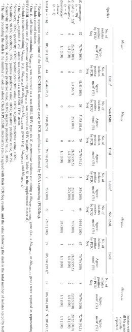
TABLE 1. Performance of the Check KPC/ESBL microarray assay in identification of  -lactamase genes a Species bla KPC bla SHV bla TEM bla CTX-M Isolates with all bla genes correctly reported No. of isolates positive by PCR/ Seq Agree-ment i (%) ESBL d Non-ESBL Total ESBL d Non-ESBL Total No. of isolates positive by PCR/ Seq Agree-ment i (%) Agree-ment i (%) No. of isolates positive by PCR/ Seq Agree-ment i (%) No. of isolates positive by PCR/ Seq c Agree-ment i (%) No. of isolates positive by PCR/ Seq Agree-ment i (%) No. of isolates positive by PCR/ Seq Agree-ment i (%) No. of isolates positive by PCR/ Seq e Agree-ment i (%) No. of isolates positive by PCR/ Seq Agree-ment i (%) K. pneumoniae ( n ⫽ 79) b 52 79/79 (100) 41 41/41 (100) 38 31/38 (81.6) 79 72/79 (91.1) 3 3/3 (100) 64 64/64 (100) 67 79/79 (100) 8 79/79 (100) 72/79 (91.1) E. coli ( n ⫽ 22) b 4 22/22 (100) 3 2/3 (66.7) 2 2/2 (100) 5 21/22 (95.5) 2 2/2 (100) 7 7/7 (100) 9 21/22 (95.5) 9 22/22 (100) 20/22 (90.9) A. baumannii ( n ⫽ 4) 0 4/4 (100) 0 0 0 4/4 (100) 2 2/2 (100) 2 4/4 (100) 2 4/4 (100) 4/4 (100) Enterobacter cloacae ( n ⫽ 1) 1 1/1 (100) 0 0 0 1/1 (100) 0 1 1/1 (100) 1 1/1 (100) 0 1/1 (100) 1/1 (100) Total ( n ⫽ 106) 57 106/106 (100) f 44 43/44 (97.7) 40 33/40 (82.5) 84 98/106 (92.5) g 7 7/7 (100) 72 72/72 (100) 79 105/106 (99.1) h 19 106/106 (100) f 97/106 (91.5) a Results represent comparisons of the Check KPC/ESBL microarray assay to PCR amplification followed by DNA sequencing (PCR/Seq). b Includes control isolates. c One E. coli isolate containing bla SHV-38 was reported as a non-ESBL SHV type; six K. pneumoniae isolates containing a bla SHV-non-ESBL gene (i.e., a bla SHV-1 or bla SHV-11 gene) were reported as representing SHV-ESBL strains; one K. pneumoniae isolate containing bla SHV-11 was reported as SHV negative (see Table S1 in the supplemental material). d Includes isolates containing bla ESBL (e.g., bla SHV-12 )o r bla ESBL plus bla non-ESBL genes (e.g., bla SHV-12 and bla SHV-11 ). e One bla TEM -negative E. coli isolate was reported as a non-ESBL TEM strain. f Sensitivity, 100%; specificity, 100%; positive predictive value, 100%; negative predictive value, 100%. g Sensitivity, 98.8%; specificity, 100%; positive predictive value, 100%; negative predictive value, 95.7%. h Sensitivity, 100%; specificity, 96.4%; positive predictive value, 98.8%; negative predictive value, 100%. i The value preceding the slash is the number of isolates whose Check KPC/ESBL microarray results agreed with their PCR/Seq results, and the value follo wing the slash is the total number of isolates tested by both assays.
on May 16, 2020 by guest
http://jcm.asm.org/
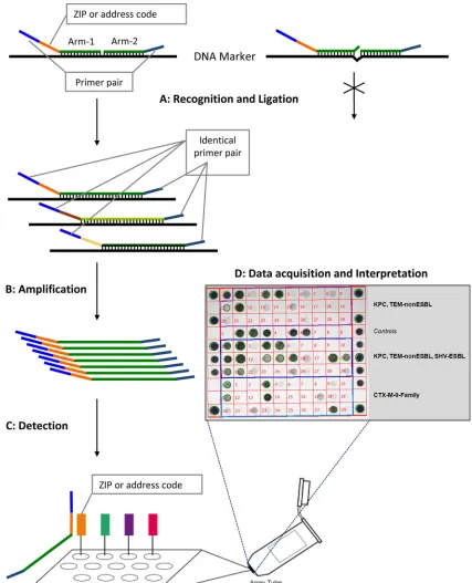
[image:2.585.159.419.69.723.2]FIG. 1. Schematic flowchart representing the different steps used by the Check-Points KPC/ESBL platform to recognize specificblagenes. (Step A) Target DNA recognition and ligation (thermocycling conditions, 95°C for 3 min, 24 cycles of 95°C for 30 s and 65°C for 5 min, and 98°C for 2 min [total, 2.75 h]). Each target-specific probe consists of two oligonucleotide probe arms that are used to detect single nucleotide polymorphisms (SNPs). These two probe arms are connected by the ligase, generating a single probe molecule only when they perfectly match the target sequence. Only connected probe arms produce the labeled amplification products detected in step C. Every target-specific probe is equipped with the same consensus primer pair necessary for step B and a unique “ZIP code” necessary for step C. (Step B) PCR amplification of the target DNA sequences (thermocycling conditions, 95°C for 10 min, 35 cycles of 95°C for 5 s, 55°C for 30 s, and 72°C for 30 s, and 98°C for 2 min [total, 1.5 h]). Using a common primer pair, target ligated sequence templates labeled with specific “ZIP codes” are multiplied. (Step C) Detection (requiring approximately 1 to 2 h of processing, depending on the number of samples). Amplification products are targeted to specific addresses on the microarray. This targeting is dependent on the specific “ZIP code.” (Step D) Immediate acquisition of images by scanning of the microarray using the array tube reader and immediate interpretation of the acquired pictures by the use of dedicated software.
on May 16, 2020 by guest
http://jcm.asm.org/
(3–5, 15), whereas theblagenes of the remaining isolates were characterized by PCR amplification, standard DNA sequenc-ing, and analytical isoelectric focusing (aIEF) as previously described (4). In this collection, isolates possessed an average of three differentblagenes (range, one to five; see Table S1 in the supplemental material). The collection also included K. pneumoniaeATCC 700603, which produces the SHV-18 ESBL (20), and sixE. coliDH10B control strains in which singlebla
genes are carried in different plasmid vectors (see Table S1 in the supplemental material).
Genomic DNA of strains was extracted from overnight colonies grown on blood agar (BBL, Sparks, MD) by the use of a DNeasy blood and tissue kit (Qiagen Sciences, Ger-mantown, MD). Microarray assays were performed according to the instructions of the manufacturer (Check-Points Health BV). Briefly, templates of the targetbla DNA sequences are generated during the ligation step. These templates are then amplified, and the products are hybridized in specific array tubes. Tubes are then inserted in the array tube reader upon completion of the detection reaction, and images are acquired and interpreted with software supplied by the manufacturer (Fig. 1). For 50 isolates, the complete procedure (i.e., from genomic DNA extraction to results) can be performed in ap-proximately 8 h.
Overall, the Check KPC/ESBL system correctly identified representatives of the fourblagene families tested, including differentiation between non-ESBL and ESBL genes, in 97 of 106 isolates (91.5%). Specificities of 100% were recorded for the blaKPC, blaSHV, and blaCTX-M genes, whereas one false
positive was reported forblaTEMgenes (specificity of 96.4%). The system detected allblaKPC-,blaTEM-, andblaCTX-M
-possess-ing isolates, includ-possess-ing differentiation of ESBL from non-ESBL
blaTEM-containing strains (Table 1). Notably, all blaCTX-M
genes detected were classified into the appropriate family group (i.e., group I, CTX-M-1-like; group II, CTX-M-2-like; group III, CTX-M-8-like; group IV, CTX-M-9-like; group V, CTX-M-25/CTX-M-26) according to the classification method of Pitout et al. (16) (see Table S1 in the supplemental mate-rial).
Detection and recognition of the blaSHV genes showed 92.5% agreement, with sensitivity and specificity of 98.8% and 100%, respectively (Table 1). Only 1 in 44 blaESBL-positive
strains (i.e., blaSHV-38-positive strains) was not identified (97.7% agreement). SHV-38 is a very rare chromosomal ESBL enzyme (group 2be) that was found in a single clinical isolate. It possesses a unique amino acid substitution (i.e., Ala146Val) and is capable of conferring resistance to ceftazidime and imi-penem (18). The amino acid at position 146 is not included in those analyzed by the Check KPC/ESBL system.
Six strains with non-ESBL blaSHVgenes were misclassified as ESBLs (Table 2). Notably, three of these wereblaSHV-11 -positive K. pneumoniae isolates (non-ESBL), which showed
-lactamase bands at pIs of 7.6 and 8.2 by aIEF and double spikes at positions 238 and/or 240 in the DNA sequencing traces of theblaSHVgene. This pattern is consistent with the possible production of an SHV-ESBL (along with the non-ESBL SHV-11) that was not detected with a cloning and DNA sequencing method that we previously employed (4). There-fore, blaSHV-positive total agreement and the overall agree-ment (i.e., allblagenes correctly reported) would improve by 2.8% if these three strains were classified as ESBL producers (Table 1).
[image:4.585.42.541.79.299.2]The data presented above also support the previous obser-vation that standard DNA sequencing of PCR amplification products fails to accurately detect more than oneblagene of a given family (4). In particular, manyK. pneumoniae isolates
TABLE 2. Details of discrepancies between conventional PCR plus DNA sequencing results and Check KPC/ESBL genotyping results
Strain Source or reference
Gene identified by PCR and DNA sequencing
Gene identified by Check KPC/ESBL
genotyping Comment(s)
E. coliDH10B This study blaSHV-38 SHV-non-ESBL Rare chromosomal genotype found
in a singleK. pneumoniaeisolate (18); mutation conferring ESBL phenotype not assayed with current microarray primers
K. pneumoniaeVA-361a 4 bla
KPC-2,blaTEM-1,blaSHV-11 KPC, TEM-non-ESBL, SHV-ESBL Possible production of an
SHV-ESBL (e.g., SHV-5/SHV-12)
K. pneumoniaeVA-388 4 blaKPC-3,blaTEM-1,blaSHV-11 KPC, TEM-non-ESBL SHV genes were not detected
K. pneumoniaeVA-392a 4 bla
KPC-3,blaTEM-1,blaSHV-11 KPC, TEM-non-ESBL, SHV-ESBL Possible production of an
SHV-ESBL (e.g., SHV-5/SHV-12)
K. pneumoniaeVA-412a 4 bla
KPC-2,blaTEM-1,blaSHV-11 KPC, TEM-non-ESBL, SHV-ESBL Possible production of an
SHV-ESBL (e.g., SHV-5/SHV-12)
K. pneumoniaeVA-414 4 blaKPC-3,blaTEM-1,blaSHV-11 KPC, TEM-non-ESBL, SHV-ESBL SHV-11 is not an ESBL
K. pneumoniae111b 15 bla
TEM-10-like,blaSHV-1-like TEM-ESBL, SHV-ESBL SHV-1 is not an ESBL
K. pneumoniae438b 15 bla
TEM-2-like,blaSHV-1-like TEM-non-ESBL, SHV-ESBL SHV-1 is not an ESBL
E. coli25 This study blaCTX-M-9-like CTX-M-(IV),
cTEM-non-ESBL bla
TEMgenes were not detected by
PCR analysis (including using internal primers); aIEF showed only one-lactamase band at a pI of 6.7, possibly related to the CTX-M enzyme expression
a
Analytical isoelectric focusing (aIEF) revealed-lactamase bands with pIs of 7.6 and 8.2 and initial standard DNA sequencing ofblaSHVshowed double spikes in
amino acid position 238 and/or 240 (4). b
Partial DNA sequencing (i.e., from amino acid 35 to 274 for TEMs and from 8 to 249 for SHVs). TheblaSHVandblaTEMsequences did not show substitutions
conferring an ESBL phenotype. c
Number in parentheses following CTX-M designation indicates the family group for CTX-M-type ESBLs (16).
on May 16, 2020 by guest
http://jcm.asm.org/
possessing bothblaSHV-11(non-ESBL) andblaSHV-12(ESBL) genes were initially identified incorrectly asblaSHV-11-positive
isolates only with standard DNA sequence analysis (4). In contrast, the microarray can accurately identify the blaESBL
gene (e.g., blaSHV-12) regardless of the coexistence of addi-tionalblanon-ESBLgenes (e.g.,blaSHV-1and/orblaSHV-11) (see
Table S1 in the supplemental material).
In conclusion, the results of the present work show that the microarray Check KPC/ESBL system is a highly accurate tool for detection of the clinically important -lactamase genes found among contemporary Gram-negative organisms. Due to its rapid performance, this platform could be used in epidemi-ological or infection control studies in which large collections of isolates need to be characterized. Furthermore, the use of Check KPC/ESBL in clinical practice may lead to more appro-priate use of antimicrobial agents, reduction of costs, and im-proved patient outcomes. More-extensive evaluations (e.g., us-ing clinical isolates possessus-ing bla genes not tested in this study) are needed to establish the full potential of this meth-odology for detecting different resistance genes.
This work was supported in part by the Veterans Affairs Merit Review Program (R.A.B.), the National Institutes of Health (grant RO3-AI081036 to R.A.B.), and the Geriatric Research Education and Clinical Center (grant VISN 10 to R.A.B.).
We thank Sarah Drawz for the critical revision of the manuscript and Francesco Luzzaro, Antonio Q. Toniolo, John Quale, David L. Paterson, Gerri S. Hall, and Stephen G. Jenkins for providing clinical isolates. We also thank Check-Points for the technical support and for providing the material necessary for the study.
REFERENCES
1.Bush, K., and G. A. Jacoby.2010. An updated functional classification of
-lactamases. Antimicrob. Agents Chemother.54:969–976.
2.Cornaglia, G., and G. M. Rossolini.2010. The emerging threat of acquired
carbapenemases in Gram-negative bacteria. Clin. Microbiol. Infect.16:99– 101.
3.Endimiani, A., J. M. Depasquale, S. Forero, F. Perez, A. M. Hujer, D.
Roberts-Pollack, P. D. Fiorella, N. Pickens, B. Kitchel, A. E. Casiano-Colon,
F. C. Tenover, and R. A. Bonomo.2009. Emergence ofblaKPC-containing
Klebsiella pneumoniaein a long-term acute care hospital: a new challenge to our healthcare system. J. Antimicrob. Chemother.64:1102–1110.
4.Endimiani, A., A. M. Hujer, F. Perez, C. R. Bethel, K. M. Hujer, J. Kroeger,
M. Oethinger, D. L. Paterson, M. D. Adams, M. R. Jacobs, D. J. Diekema,
G. S. Hall, S. G. Jenkins, L. B. Rice, F. C. Tenover, and R. A. Bonomo.2009.
Characterization of blaKPC-containingKlebsiella pneumoniae isolates
de-tected in different institutions in the eastern USA. J. Antimicrob. Che-mother.63:427–437.
5.Endimiani, A., F. Luzzaro, R. Migliavacca, E. Mantengoli, A. M. Hujer,
K. M. Hujer, L. Pagani, R. A. Bonomo, G. M. Rossolini, and A. Toniolo.
2007. Spread in an Italian hospital of a clonalAcinetobacter baumanniistrain producing the TEM-92 extended-spectrum-lactamase. Antimicrob. Agents Chemother.51:2211–2214.
6.Endimiani, A., and D. L. Paterson.2007. Optimizing therapy for infections
caused by enterobacteriaceae producing extended-spectrum-lactamases. Semin. Respir. Crit. Care Med.28:646–655.
7.Gniadkowski, M. 2008. Evolution of extended-spectrum-lactamases by
mutation. Clin. Microbiol. Infect.14(Suppl.)1:11–32.
8.Grimm, V., S. Ezaki, M. Susa, C. Knabbe, R. D. Schmid, and T. T.
Bach-mann.2004. Use of DNA microarrays for rapid genotyping of TEM
-lac-tamases that confer resistance. J. Clin. Microbiol.42:3766–3774.
9.Kitchel, B., J. K. Rasheed, J. B. Patel, A. Srinivasan, S. Navon-Venezia, Y.
Carmeli, A. Brolund, and C. G. Giske.2009. Molecular epidemiology of
KPC-producingKlebsiella pneumoniaeisolates in the United States: clonal expansion of multilocus sequence type 258. Antimicrob. Agents Chemother.
53:3365–3370.
10.Leinberger, D. M., V. Grimm, M. Rubtsova, J. Weile, K. Schroppel, T. A.
Wichelhaus, C. Knabbe, R. D. Schmid, and T. T. Bachmann.2010.
Inte-grated detection of extended-spectrum--lactam resistance by DNA mi-croarray-based genotyping of TEM, SHV, and CTX-M genes. J. Clin. Mi-crobiol.48:460–471.
11.Luzzaro, F., G. Gesu, A. Endimiani, G. Ortisi, S. Malandrin, L. Pagani, and
G. M. Rossolini.2006. Performance in detection and reporting-lactam
resistance phenotypes inEnterobacteriaceae: a nationwide proficiency study in Italian laboratories. Diagn. Microbiol. Infect. Dis.55:311–318.
12.Miriagou, V., G. Cornaglia, M. Edelstein, I. Galani, C. G. Giske, M.
Gniad-kowski, E. Malamou-Lada, L. Martinez-Martinez, F. Navarro, P. Nord-mann, L. Peixe, S. Pournaras, G. M. Rossolini, A. Tsakris, A. Vatopoulos,
and R. Canton.2010. Acquired carbapenemases in Gram-negative bacterial
pathogens: detection and surveillance issues. Clin. Microbiol. Infect.16:112– 122.
13.Nordmann, P., G. Cuzon, and T. Naas.2009. The real threat ofKlebsiella
pneumoniaecarbapenemase-producing bacteria. Lancet Infect. Dis.9:228– 236.
14.Paterson, D. L., and R. A. Bonomo.2005. Extended-spectrum-lactamases:
a clinical update. Clin. Microbiol. Rev.18:657–686.
15.Paterson, D. L., K. M. Hujer, A. M. Hujer, B. Yeiser, M. D. Bonomo, L. B.
Rice, and R. A. Bonomo.2003. Extended-spectrum-lactamases inKlebsiella
pneumoniae bloodstream isolates from seven countries: dominance and widespread prevalence of SHV- and CTX-M-type-lactamases. Antimicrob. Agents Chemother.47:3554–3560.
16.Pitout, J. D., A. Hossain, and N. D. Hanson.2004. Phenotypic and molecular
detection of CTX-M--lactamases produced byEscherichia coliand Kleb-siellaspp. J. Clin. Microbiol.42:5715–5721.
17.Pitout, J. D., and K. B. Laupland.2008. Extended-spectrum
-lactamase-producingEnterobacteriaceae: an emerging public-health concern. Lancet Infect. Dis.8:159–166.
18.Poirel, L., C. Heritier, I. Podglajen, W. Sougakoff, L. Gutmann, and P.
Nordmann.2003. Emergence inKlebsiella pneumoniaeof a
chromosome-encoded SHV-lactamase that compromises the efficacy of imipenem. An-timicrob. Agents Chemother.47:755–758.
19.Queenan, A. M., and K. Bush.2007. Carbapenemases: the versatile
-lacta-mases. Clin. Microbiol. Rev.20:440–458.
20.Rasheed, J. K., G. J. Anderson, H. Yigit, A. M. Queenan, A.
Domenech-Sanchez, J. M. Swenson, J. W. Biddle, M. J. Ferraro, G. A. Jacoby, and F. C.
Tenover.2000. Characterization of the extended-spectrum-lactamase
ref-erence strain,Klebsiella pneumoniaeK6 (ATCC 700603), which produces the novel enzyme SHV-18. Antimicrob. Agents Chemother.44:2382–2388.
21.Tenover, F. C., M. J. Mohammed, T. S. Gorton, and Z. F. Dembek.1999.
Detection and reporting of organisms producing extended-spectrum -lac-tamases: survey of laboratories in Connecticut. J. Clin. Microbiol.37:4065– 4070.
22.Wolter, D. J., P. M. Kurpiel, N. Woodford, M. F. Palepou, R. V. Goering, and
N. D. Hanson.2009. Phenotypic and enzymatic comparative analysis of the
novel KPC variant KPC-5 and its evolutionary variants, KPC-2 and KPC-4. Antimicrob. Agents Chemother.53:557–562.
23.Zhu, L. X., Z. W. Zhang, D. Liang, D. Jiang, C. Wang, N. Du, Q. Zhang, K.
Mitchelson, and J. Cheng.2007. Multiplex asymmetric PCR-based
oligonu-cleotide microarray for detection of drug resistance genes containing single mutations inEnterobacteriaceae. Antimicrob. Agents Chemother.51:3707– 3713.