Copyright © 2002, American Society for Microbiology. All Rights Reserved.
Genome Scale Comparison of
Mycobacterium avium
subsp.
paratuberculosis
with
Mycobacterium avium
subsp.
avium
Reveals
Potential Diagnostic Sequences
John P. Bannantine,
1* Emily Baechler,
2,3Qing Zhang,
2LingLing Li,
2and Vivek Kapur
2 National Animal Disease Center, Agricultural Research Service, U.S. Department of Agriculture, Ames, Iowa,1and BiomedicalGenomics Center2and Department of Medicine,3University of Minnesota, Minneapolis, Minnesota
Received 16 November 2001/Returned for modification 6 January 2002/Accepted 27 January 2002
The genetic similarity betweenMycobacterium aviumsubsp.paratuberculosisand other mycobacterial species has confounded the development of M. avium subsp.paratuberculosis-specific diagnostic reagents. Random shotgun sequencing of the M. avium subsp. paratuberculosis genome in our laboratories has shown >98% sequence identity withMycobacterium aviumsubsp.aviumin some regions. However, an in silico comparison of the largest annotatedM. aviumsubsp.paratuberculosiscontigs, totaling 2,658,271 bp, with the unfinishedM. aviumsubsp.aviumgenome has revealed 27 predictedM. aviumsubsp.paratuberculosiscoding sequences that do not align withM. aviumsubsp.aviumsequences. BLASTP analysis of the 27 predicted coding sequences (genes) shows that 24 do not match sequences in public sequence databases, such as GenBank. These novel sequences were examined by PCR amplification with genomic DNA from eight mycobacterial species and ten independent isolates ofM. avium subsp. paratuberculosis. From these analyses, 21 genes were found to be present in allM. aviumsubsp.paratuberculosisisolates and absent from all other mycobacterial species tested. One region of theM. avium subsp. paratuberculosis genome contains a cluster of eight genes, arranged in tandem, that is absent in other mycobacterial species. This region spans 4.4 kb and is separated from other predicted coding regions by 1,408 bp upstream and 1,092 bp downstream. The gene upstream of this eight-gene cluster has strong similarity to mycobacteriophage integrase sequences. The GC content of this 4.4-kb region is 66%, which is similar to the rest of the genome, indicating that this region was not horizontally acquired recently. Southern hybridization analysis confirmed that this gene cluster is present only inM. aviumsubsp. paratuberculosis. Collectively, these studies suggest that a genomics approach will help in identifying novelM. aviumsubsp.paratuberculosisgenes as candidate diagnostic sequences.
Paratuberculosis, or Johne’s disease, is a granulomatous en-teritis of ruminant animals that may be prevalent in approxi-mately 35% of United States dairy herds (7, 24). Diarrhea, reduced feed intake, weight loss, and eventual death charac-terize this intestinal disorder in cattle. Based upon prevalence figures and information from animal producers, economic losses for the dairy industry exceed 200 million dollars annually
(18).Mycobacterium aviumsubsp.paratuberculosisis the
etio-logic agent of this economically significant disease. This veter-inary pathogen has also been implicated as the etiologic agent of Crohn’s disease (15), leading researchers to speculate on a potential pathogenic role for this organism in humans.
The control of Johne’s disease is severely hampered by in-adequate diagnostic tools (26). The prolonged incubation time and presence of subclinical cases permit infected animals to shed large amounts of bacilli in their feces before detection
(21). Culture ofM. avium subsp.paratuberculosisfrom feces
has been the most reliable method for identifying infected animals; however, the slow growth of this organism results in a minimum of 6 weeks before culture data are available.
Re-search on the pathogenesis and immunology of M. avium
subsp.paratuberculosisinfections of cattle will allow the design
of better diagnostic and control procedures. New approaches that yield improved diagnostic tests will enable early detection and removal of subclinically infected animals. This will effec-tively reduce the incidence of Johne’s disease in beef and dairy herds.
With the availability of over 60 published microbial ge-nomes, some of which are in the same genus or even species, the age of comparative genomics has arrived. This approach is
particularly useful in the genusMycobacteriumdue to the
num-ber of sequenced species. M. tuberculosis and M. leprae
ge-nomes have been published (5, 6), and projects are under way for M. bovis, M. avium subsp. avium, M. smegmatis, and M. aviumsubsp.paratuberculosis(3). Comparative mycobacterial genomic approaches have been used to identify small-scale
genomic deletions among M. tuberculosis isolates (12).
Fur-thermore, large genome rearrangements (2) as well as deleted
regions (14) were identified in studies comparing theM. bovis
BCG vaccine strain withM. tuberculosis. Genome-wide
com-parisons in this genus will lead to an increased understanding of the genes required for pathogenicity as well as highlighting the sequences that make each species distinct.
Our laboratories have been actively engaged in sequencing
the genome ofM. avium subsp. paratuberculosisin order to
reveal diagnostic sequences and/or antigens as well as to better understand the pathogenesis of Johne’s disease. The strong
nucleotide identity between M. avium subsp. avium and M.
aviumsubsp.paratuberculosis(11, 22) has prevented the
devel-* Corresponding author. Mailing address: National Animal Disease Center, ARS-USDA, 2300 North Dayton Ave., Ames, IA 50010. Phone: (515) 663-7340. Fax: (515) 663-7458. E-mail: jbannant@nadc.ars.usda .gov.
1303
on May 15, 2020 by guest
http://jcm.asm.org/
opment ofM. aviumsubsp.paratuberculosis-specific DNA se-quences or antigens. To date, the only routinely used
diagnos-tic sequence is that of the insertion element IS900, which is
present in multiple copies in theM. aviumsubsp.
paratubercu-losisgenome (9). In this study, we performed a partial genome comparison between the largest annotated contiguous DNA
fragments (contigs) of M. avium subsp. paratuberculosisand
the genetically similar M. avium subsp. avium genome.
Se-quences present inM. aviumsubsp.paratuberculosisbut notM.
avium subsp. avium were further analyzed by PCR with genomic DNA from several mycobacterial species. From these
analyses, 21 uniqueM. aviumsubsp.paratuberculosispredicted
coding sequences were identified. These unique sequences may be used to develop improved diagnostic reagents.
MATERIALS AND METHODS
Mycobacterial strains.Mycobacteria used in this study are listed in Table 1. All mycobacteria were cultured in Middlebrook 7H9 medium with 0.05% Tween 80 and oleic acid-albumin-dextrose-complex (Becton Dickinson Microbiology, Sparks, Md.). Cultures containingM. aviumsubsp.paratuberculosisisolates were supplemented with 2 mg of ferric mycobactin J (Allied Monitor Inc., Fayette, Mo.) per liter. All growth flasks were incubated at 37°C without shaking.
Annotation ofM. aviumsubsp.paratuberculosiscontigs greater than 10 kb.The sequencing and assembly strategies used here will be described elsewhere. For these studies, we chose assembledM. aviumsubsp.paratuberculosiscontig frag-ments greater than 10 kb. Predicted coding sequences (genes) were identified with ARTEMIS software (http://www.sanger.ac.uk/Software/) and TB-parse, a program used to identify coding sequences in theM. tuberculosisgenome (5).
The results were compared and verified manually in ARTEMIS. A putative ribosome binding site was also evaluated for each coding sequence. The presence of an AG-rich sequence approximately 30 bp upstream of the start codon was scored as a putative ribosome binding site sequence. Similarities were identified by BLASTP analysis by using GenBank and a local database constructed by the Computational Biology Center at the University of Minnesota (http://www.cbc .umn.edu).
Sequence analysis.Sequence alignments ofM. aviumsubsp.paratuberculosis
andM. aviumsubsp.aviumwere compared and visualized with ACT software (http://www.sanger.ac.uk/Software/).M. aviumsubsp.aviumis being sequenced by The Institute for Genomic Research (TIGR; http://www.tigr.org/cgi-bin /BlastSearch/blast.cgi?organism⫽m_avium). Sequence alignments used to pro-duce illustrations were made with AssemblyLIGN software (Accelrys, Princeton, N.J.).
DNA hybridization.Genomic DNA was extracted from several species of mycobacteria by a method modified from that described by Whipple et al. (25). One liter of Middlebrook 7H9-cultured mycobacteria was incubated at 37°C until an optical density at 540 between 0.50 and 0.56 was attained.D-Cycloserine was added to the medium at a final concentration of 0.5 mg/ml and incubated for an additional 24 h. Mycobacteria were harvested by centrifugation at 9,950⫻gfor 15 min, and the pellet was resuspended in 11 ml of Qiagen buffer B1 containing 1 mg of Qiagen RNase A per ml. Lipase was added (450,000 U; catalog no. L4384; Sigma, St. Louis, Mo.) to digest mycobacterial cell wall lipids. Following a 2-h incubation at 37°C, 20 mg of lysozyme was added, and incubation pro-ceeded for an additional 3 h at 37°C. Qiagen proteinase K (500l; 20 mg/ml) was added and incubated for 1.5 h at 37°C. Qiagen buffer B2 (4 ml) was added, and the slurry was mixed and incubated 16 h at 50°C. The remaining cellular debris was removed by centrifugation at 12,100⫻gfor 20 min. The supernatant was poured over a preequilibrated Qiagen 500/G genomic tip. The loaded column was washed and processed according to the instructions of the manufacturer.
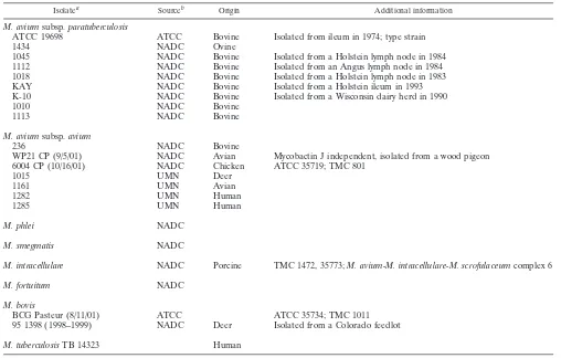
[image:2.587.40.548.82.406.2]PstI-restricted DNA fragments were separated on a 1% agarose gel. DNA-containing gels were depurinated, denatured, and neutralized as described by TABLE 1. Mycobacterial strains used in this study
Isolatea Sourceb Origin Additional information
M. aviumsubsp.paratuberculosis
ATCC 19698 ATCC Bovine Isolated from ileum in 1974; type strain
1434 NADC Ovine
1045 NADC Bovine Isolated from a Holstein lymph node in 1984
1112 NADC Bovine Isolated from an Angus lymph node in 1984
1018 NADC Bovine Isolated from a Holstein lymph node in 1983
KAY NADC Bovine Isolated from a Holstein ileum in 1993
K-10 NADC Bovine Isolated from a Wisconsin dairy herd in 1990
1010 NADC Bovine
1113 NADC Bovine
M. aviumsubsp.avium
236 NADC Bovine
WP21 CP (9/5/01) NADC Avian Mycobactin J independent, isolated from a wood pigeon
6004 CP (10/16/01) NADC Chicken ATCC 35719; TMC 801
1015 UMN Deer
1161 UMN Avian
1282 UMN Human
1285 UMN Human
M. phlei NADC
M. smegmatis NADC
M. intracellulare NADC Porcine TMC 1472, 35773;M. avium-M. intracellulare-M. scrofulaceumcomplex 6
M. fortuitum NADC
M. bovis
BCG Pasteur (8/11/01) ATCC ATCC 35734; TMC 1011
95 1398 (1998–1999) NADC Deer Isolated from a Colorado feedlot
M. tuberculosisTB 14323 Human
aDates of isolation (month/day/year) are in parentheses.
bATCC, American Type Culture Collection; NADC, National Animal Disease Center; UMN, University of Minnesota.
on May 15, 2020 by guest
http://jcm.asm.org/
Sambrook et al. (19). DNA was transferred by capillary action (20) to BrightStar-Plus membranes (Ambion, Austin, Tex.), and probes were labeled with [␣-32P]dCTP (ICN, Costa Mesa, Calif.) by random priming. Hybridization was performed in an Autoblot hybridization oven (Bellco Biotechnology, Vineland, N.J.) at 45°C for 16 h in ExpressHyb hybridization solution (Clontech, Palo Alto, Calif.). Probed blots were washed sequentially with increasing stringency solu-tions as described previously (20). Detection was by autoradiography using Bio-Max MR film (Kodak, Rochester, N.Y.).
PCR amplification.Primers listed in Table 2 were designed fromM. avium
subsp. paratuberculosis-specific sequences to amplify mycobacterial genomic DNA. Amplification recipes containing nucleotides, buffer, primers, template, and DNA polymerase were standard except for the addition of 5% dimethyl sulfoxide (Sigma). Amplification conditions included a 5-min denaturation step at 94°C and 35 cycles of 45 s at 94°C, 1 min at 55°C, and 2 min at 72°C. High-fidelity Pwo polymerase (Boehringer Ingelheim Pharmaceutical Inc., Ridgefield, Conn.) was used in amplifications to generate probes used in South-ern hybridization experiments. All other amplifications usedTaqDNA polymer-ase (Roche Molecular Biochemicals, Indianapolis, Ind.). Primers used to amplify the no. 7 sequence for a probe in Southern hybridizations were 5⬘-ATCAGGC TGACGGGATTGCCC-3⬘and 5⬘-TCAACGAGTGCACGGGAACC-3⬘.
Nucleotide sequence accession numbers.The nucleotide sequences of allM. aviumsubsp.paratuberculosisgenes described in this study were deposited in the GenBank/EMBL nucleotide sequence data library under accession numbers AF445420 through AF445446.
RESULTS
Twenty-seven M. avium subsp. paratuberculosis predicted coding sequences are not present inM. avium subsp.avium.
Our laboratories are sequencing the complete genome ofM.
aviumsubsp. paratuberculosisK-10, a field isolate recovered from a cow with clinical Johne’s disease (http://www.cbc.umn .edu/ResearchProjects/AGAC/Mptb/Mptbhome.html). The
genome size is estimated to be ⬎5 Mb based on assembled
sequence data, and at the time of this analysis (July 2001), 2.65 Mb was contained in contig fragments greater than 10 kb. Contigs that are above 10 kb were annotated with ARTEMIS and represent 48% of the total genome. The average size of the
annotated contigs is 25 kb, with one contig over 70 kb. Each gene within the annotated contig set was also checked manu-ally and confirmed by TB-parse. These contigs were aligned withM. aviumsubsp.aviumsequence data generated at TIGR. TIGR has 612 contigs that total 5,867,714 bp in the 8 July 2001 data set.
M. aviumsubsp.aviumandM. aviumsubsp.paratuberculosis
display a high degree of similarity at the nucleotide level as well as local gene order conservation. An analysis of an 11-kb region surrounding the origin of replication for each of these genomes shows 98% nucleotide identity (Q. Zhang, E. Baech-ler, L. Li, J. P. Bannantine, and V. Kapur, unpublished data).
The sequence similarity between orthologs inM. aviumsubsp.
paratuberculosisand M. aviumsubsp.aviumwas greater than
that betweenM. aviumsubsp.paratuberculosisand other
my-cobacterial species. A more global comparison shows that these strong nucleotide identities are present throughout both genomes. Despite this strong genetic similarity, a total of 27
genes from the annotated M. avium subsp. paratuberculosis
contigs were identified that did not align with the unfinishedM.
avium subsp. avium genome by computerized alignments.
These uniqueM. aviumsubsp.paratuberculosissequences are
[image:3.587.46.285.81.328.2]listed in Table 3 along with some sequence characteristics. Of these, three contained weak similarity to proteins in other mycobacterial species or proteins in GenBank (Table 3). This
TABLE 2. PCR primers used in this studya
Gene Primer 1 Primer 2
10 CGGCGGATCAGCATCTAC CACCTCATCGTGGCCAGGTT 11 ACCGAACACGAGTGGAGCA CAGACTCTGACCGACGTCAT 38 GCATTTCGGCTCCCACGGTG TACGTCGGTTCGGCGCGCAT 48 CTGACACCGGCCTACGAACG CATCCTCGCCGCGCCAGCAC 49 TGCTCAGGCCAACCCGCGC TACTGGGCGGCGCACCCGAC 50 GGCATCCGCACCTTCGTCTG CAATTCGTCGATCGGGCCGA 56 ATGAACACTTCTTCCTCTCTA CATATCGCGGTGATCCTGAC 57 ATGGCCACCAACGACGACCA CGCGGCCGTCGGGCCGGCTG 93 TTGCTGCGGGAAGGTTGCC GAGAACGAGATGTGCGTCAG 134 GCGATGGTCAACGCCACCGG TACAGCCCCGTGCAGACCGG 135 GCAGGCGTTTGCGTTCTTG CGAGGTCCGAAATAGCGTAG 159 ATGCGTTTCGCCCTCCCGAC TCACGCCTTGATTTCGTCCT 217 TGGCCGAACGCGGACTGTTC TAGGAATCCGCGTCGACGAT 218 CAAGGTTCGTGACGGTATCG TGACCCCAGCAGGTATGGC 219 CATCTACTGAGCGCCGTTTG CACGCCGCCACCCCGTCCCG 228 GCAAGGTGGGCTTTGAAG TGCGTGGGAGGATAAGGC 240 TTGGCACTGGCGTTTATG ACATCGGGAACACAGGTCTC 241 ATCCTCCGGTTTGGCGGGAA ACAGAGGTCGATCGGGTCG 250 CAGTCGGCCGGCGAAACGCC CGCGGCGAAATCGAACGC 251 CACGTGCTGTCCCCATCGGC CTACGTCTTCGTGACCAAAG 252 TGACCACCGACAACCCCACG CATGAGGGCTGTCCCTCTCC 253 TTGACCGCGTTGACGGCGTT CAGCGGTCCGCGCTCTTCGC 254 TGGGCAGCCCGGTGTCCCG CACGCGCTCCTTTCAGCCTT 255 CAGTCACCCCGCGGCCGGTA TCTACTGACCCGCAGATCGAA 256 TGGCCGTCAAGGACCAGAAC CATGACCCTGCCGGCGTCCC 257 TGGCATTGGATCGCGTCGGA TCAAACCCGGCGAGTTCTTC
aPrimers are listed 5⬘to 3⬘.
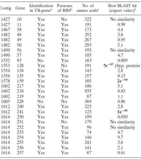
TABLE 3. M. aviumsubsp.paratuberculosispredicted coding sequences not present inM. aviumsubsp.avium,
as determined in silico
Contig Gene Identificationin TB-parsea Presenceof RBSb amino acidsNo. of c Best BLAST hit(expect value)d
1427 10 Yes No 322 No similarity
1427 11 Yes Yes 191 0.99
1467 38 Yes Yes 173 4.4
1482 48 Yes Yes 352 3.8
1482 49 Yes Yes 267 0.95
1482 50 Yes Yes 293 5.1
1490 56 Yes Yes 193 No similarity
1490 57 Yes Yes 103 4.7
1532 93 No Yes 183 0.009
1553 128 Yes No 191 7eⴚ07(hyp. protein)
1556 134 Yes Yes 143 2.8
1556 135 Yes Yes 157 0.13
1578 159 Yes Yes 185 2eⴚ04
1602 217 Yes Yes 106 5.1
1602 218 Yes Yes 835 0.83
1602 219 Yes Yes 87 1.1
1605 228 No No 369 0.86
1612 240 Yes Yes 223 2.8
1612 241 Yes Yes 123 1eⴚ05
1614 250 Yes Yes 199 0.030
1614 251 Yes No 179 No similarity 1614 252 Yes Yes 96 No similarity
1614 253 Yes Yes 74 4.7
1614 254 Yes Yes 146 9.7
1614 255 Yes Yes 241 3.6
1614 256 Yes Yes 141 2.1
1614 257 Yes Yes 87 0.61
aTB-parse is the gene prediction program used to annotate theM. tuberculosis
genome (6). “Yes,” TB-parse identified the listedM. paratuberculosisgene; “No,” TB-parse did not recognize it as a coding sequence.
bPresence or absence of a consensus ribosome binding site (RBS). cNumber of amino acids encoded by the predicted coding sequence. dExpect value of the best match in GenBank. The three predicted coding
sequences that contain the highest similarity to a sequence in Genbank are in bold. hyp. protein, hypothetical protein in GenBank.
on May 15, 2020 by guest
http://jcm.asm.org/
[image:3.587.302.540.397.665.2]leaves 24 genes with no significant similarity to any known
proteins. Since only approximately half of theM. aviumsubsp.
paratuberculosisgenome was used in these analyses, a complete
genome analysis may reveal an estimated 50 uniqueM. avium
subsp.paratuberculosisgenes.
Some M. avium subsp.paratuberculosissequences that did
not align withM. aviumsubsp.avium,either in silico or
exper-imentally, contain similarity to other mycobacterial species. One such sequence, designated no. 7, was tested by PCR and
Southern hybridization with two M. aviumsubsp.avium
iso-lates and twoM. aviumsubsp.paratuberculosisstrains (Fig. 1).
An amplified PCR fragment was produced only withM. avium
subsp. paratuberculosis genomic DNA as the template (Fig.
1A). Likewise, DNA hybridization on Southern blots detected onlyM. aviumsubsp.paratuberculosissequences, notM. avium
subsp.avium(Fig. 1B). However, BLASTP analysis of the no.
7 sequence revealed strong similarity to hypothetical proteins
in theM. tuberculosisgenome. Therefore, caution must be used
in determining whether a sequence is truly unique toM. avium
subsp.paratuberculosis. More comprehensive experiments
us-ing additional mycobacterial species are necessary before such conclusions can be made.
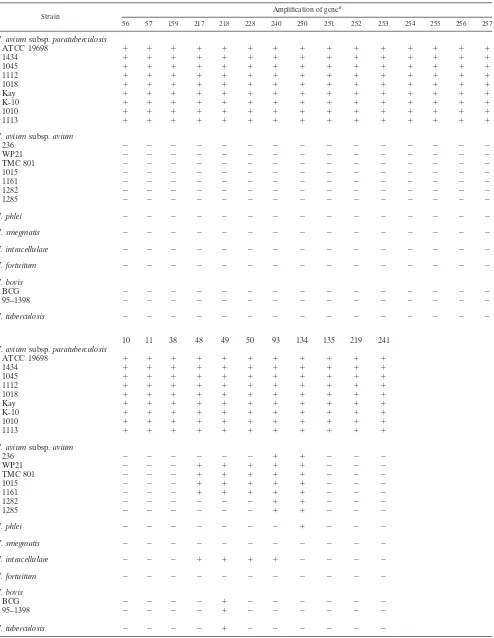
PCR analysis.PCR amplification was performed on several mycobacterial species, strains, and isolates to experimentally determine the specificity for 26 of the 27 sequences (Table 4). Gene 128 was not included in these analyses because it had the lowest expect value (highest similarity to a sequence in Gen-Bank) of the 27 sequences by BLASTP analysis (Table 3).
These data show that primers designed from all 26M. avium
subsp.paratuberculosisK-10 genes could produce an amplified
product in all 10 M. aviumsubsp.paratuberculosisstrains or
isolates tested. In addition, despite an absence of any homol-ogous sequences in public databases, PCR products of the correct size were obtained for five genes by using templates from other mycobacterial species. Following this analysis, a
core group of 21 genes that are present only inM. aviumsubsp.
paratuberculosisremained (Table 4).
Sequence analysis of anM. avium subsp.paratuberculosis -specific eight-gene cluster.Table 3 lists eight genes present on contig fragment 1614. These eight genes are arranged in tan-dem, span a total of 4.4 kb at the end of the 1614 contig (Fig.
2), and are present only in M. aviumsubsp.paratuberculosis
(Table 4). Located 1,408 bp upstream of gene 250 is an inte-grase gene that contains similarity to other mycobacteriophage integrases. As larger contiguous fragments were assembled
from the gap closure phase of theM. aviumsubsp.
paratuber-culosis genome project, a search to define the ends of the
4.4-kb sequence not present in M. avium subsp. avium was
performed. This 4.4-kb segment containing genes 250 to 257, herein termed no. 481, is located at the end of the 46-kb contig 1614 and it was found to align with the 94-kb contig 1398 present in a more recent contig assembly data set (Fig. 2). The no. 481 sequence aligned near the center of the 94-kb contig essentially at 35 to 45 kb. A trimmed portion of the 1398 contig is shown in the alignment in Fig 2. The results of this analysis further extended the region of no. 481 sequence to 9.4 kb, none
of which aligns with theM. avium subsp.aviumsequence in
silico.
A TBLASTX analysis was performed on the 9.4-kb sequence (designated contig 1398-trimmed in Fig. 2). The results of these analyses revealed that, while no sequences aligned with
M. aviumsubsp.avium, the ends of contig 1398-trimmed align
with sequences inM. tuberculosis(Table 5). The open reading
frames designated by a question mark in Table 5 are present on contig 1398, which has not yet been annotated. This again leaves a core sequence of eight open reading frames,
compris-ing the no. 481 sequence, that are present only inM. avium
subsp.paratuberculosis. This core sequence is flanked by 1,408
bp of noncoding sequence downstream and 1092-bp of non-coding sequence upstream (Fig. 2). Therefore, this novel core sequence is well separated from other predicted open reading frames.
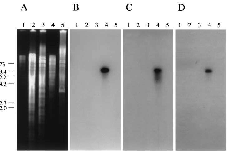
Southern hybridization analysis shows that the no. 481 se-quence is specific toM. aviumsubsp.paratuberculosis.To
con-firm experimentally that no. 481 is present only inM. avium
subsp.paratuberculosis, three arbitrarily chosen genes of the
no. 481 sequence (251, 253, and 255) were radiolabeled and used as probes in DNA hybridization with several
mycobacte-rial species, includingM. fortuitum,M. bovis,M. intracellulare,
M. aviumsubsp.avium, andM. aviumsubsp.paratuberculosis FIG. 1. PCR and Southern hybridization analysis of a unique DNA
fragment show that it is conserved among twoM. aviumsubsp. para-tuberculosisisolates tested but not present inM. aviumsubsp.avium genomic DNA. (A) PCR amplification of specific products represent-ing unique fragment no. 7 from genomic DNA of two representative strains of M. aviumsubsp. paratuberculosis and two representative isolates ofM. aviumsubsp.avium. Lanes: 1, 100-bp DNA size stan-dards; 2,M. avium subsp.paratuberculosisK-10; 3,M. aviumsubsp. aviumTMC801; 4,M. aviumsubsp.paratuberculosisATCC 19698; 5, M. aviumsubsp. aviumdeer isolate. (B) Southern hybridization of PstI-restricted genomic DNA (2g each) from these same isolates. A DNA fragment amplified with primers designed from the no. 7 se-quence was labeled and used as the probe. The data show the presence of this fragment inM. aviumsubsp.paratuberculosisbut notM. avium subsp.avium. The same blot, when stripped and reprobed with the gene encoding the 65-kDa heat shock protein, revealed bands in all four lanes with equal intensities, ruling out the possibility of false-negative hybridizations for the lanes containingM. aviumsubsp.avium genomic DNA (not shown). The lanes in panel B are identical to those in panel A except that lane 1 contains-HindIII size standards.
on May 15, 2020 by guest
http://jcm.asm.org/
TABLE 4. PCR analysis ofM. paratuberculosispredicted coding sequences
Strain Amplification of gene
a
56 57 159 217 218 228 240 250 251 252 253 254 255 256 257
M. aviumsubsp.paratuberculosis
ATCC 19698 ⫹ ⫹ ⫹ ⫹ ⫹ ⫹ ⫹ ⫹ ⫹ ⫹ ⫹ ⫹ ⫹ ⫹ ⫹
1434 ⫹ ⫹ ⫹ ⫹ ⫹ ⫹ ⫹ ⫹ ⫹ ⫹ ⫹ ⫹ ⫹ ⫹ ⫹
1045 ⫹ ⫹ ⫹ ⫹ ⫹ ⫹ ⫹ ⫹ ⫹ ⫹ ⫹ ⫹ ⫹ ⫹ ⫹
1112 ⫹ ⫹ ⫹ ⫹ ⫹ ⫹ ⫹ ⫹ ⫹ ⫹ ⫹ ⫹ ⫹ ⫹ ⫹
1018 ⫹ ⫹ ⫹ ⫹ ⫹ ⫹ ⫹ ⫹ ⫹ ⫹ ⫹ ⫹ ⫹ ⫹ ⫹
Kay ⫹ ⫹ ⫹ ⫹ ⫹ ⫹ ⫹ ⫹ ⫹ ⫹ ⫹ ⫹ ⫹ ⫹ ⫹
K-10 ⫹ ⫹ ⫹ ⫹ ⫹ ⫹ ⫹ ⫹ ⫹ ⫹ ⫹ ⫹ ⫹ ⫹ ⫹
1010 ⫹ ⫹ ⫹ ⫹ ⫹ ⫹ ⫹ ⫹ ⫹ ⫹ ⫹ ⫹ ⫹ ⫹ ⫹
1113 ⫹ ⫹ ⫹ ⫹ ⫹ ⫹ ⫹ ⫹ ⫹ ⫹ ⫹ ⫹ ⫹ ⫹ ⫹
M. aviumsubsp.avium
236 ⫺ ⫺ ⫺ ⫺ ⫺ ⫺ ⫺ ⫺ ⫺ ⫺ ⫺ ⫺ ⫺ ⫺ ⫺
WP21 ⫺ ⫺ ⫺ ⫺ ⫺ ⫺ ⫺ ⫺ ⫺ ⫺ ⫺ ⫺ ⫺ ⫺ ⫺
TMC 801 ⫺ ⫺ ⫺ ⫺ ⫺ ⫺ ⫺ ⫺ ⫺ ⫺ ⫺ ⫺ ⫺ ⫺ ⫺
1015 ⫺ ⫺ ⫺ ⫺ ⫺ ⫺ ⫺ ⫺ ⫺ ⫺ ⫺ ⫺ ⫺ ⫺ ⫺
1161 ⫺ ⫺ ⫺ ⫺ ⫺ ⫺ ⫺ ⫺ ⫺ ⫺ ⫺ ⫺ ⫺ ⫺ ⫺
1282 ⫺ ⫺ ⫺ ⫺ ⫺ ⫺ ⫺ ⫺ ⫺ ⫺ ⫺ ⫺ ⫺ ⫺ ⫺
1285 ⫺ ⫺ ⫺ ⫺ ⫺ ⫺ ⫺ ⫺ ⫺ ⫺ ⫺ ⫺ ⫺ ⫺ ⫺
M. phlei ⫺ ⫺ ⫺ ⫺ ⫺ ⫺ ⫺ ⫺ ⫺ ⫺ ⫺ ⫺ ⫺ ⫺ ⫺
M. smegmatis ⫺ ⫺ ⫺ ⫺ ⫺ ⫺ ⫺ ⫺ ⫺ ⫺ ⫺ ⫺ ⫺ ⫺ ⫺
M. intracellulare ⫺ ⫺ ⫺ ⫺ ⫺ ⫺ ⫺ ⫺ ⫺ ⫺ ⫺ ⫺ ⫺ ⫺ ⫺
M. fortuitum ⫺ ⫺ ⫺ ⫺ ⫺ ⫺ ⫺ ⫺ ⫺ ⫺ ⫺ ⫺ ⫺ ⫺ ⫺
M. bovis
BCG ⫺ ⫺ ⫺ ⫺ ⫺ ⫺ ⫺ ⫺ ⫺ ⫺ ⫺ ⫺ ⫺ ⫺ ⫺
95–1398 ⫺ ⫺ ⫺ ⫺ ⫺ ⫺ ⫺ ⫺ ⫺ ⫺ ⫺ ⫺ ⫺ ⫺ ⫺
M. tuberculosis ⫺ ⫺ ⫺ ⫺ ⫺ ⫺ ⫺ ⫺ ⫺ ⫺ ⫺ ⫺ ⫺ ⫺ ⫺
10 11 38 48 49 50 93 134 135 219 241
M. aviumsubsp.paratuberculosis
ATCC 19698 ⫹ ⫹ ⫹ ⫹ ⫹ ⫹ ⫹ ⫹ ⫹ ⫹ ⫹
1434 ⫹ ⫹ ⫹ ⫹ ⫹ ⫹ ⫹ ⫹ ⫹ ⫹ ⫹
1045 ⫹ ⫹ ⫹ ⫹ ⫹ ⫹ ⫹ ⫹ ⫹ ⫹ ⫹
1112 ⫹ ⫹ ⫹ ⫹ ⫹ ⫹ ⫹ ⫹ ⫹ ⫹ ⫹
1018 ⫹ ⫹ ⫹ ⫹ ⫹ ⫹ ⫹ ⫹ ⫹ ⫹ ⫹
Kay ⫹ ⫹ ⫹ ⫹ ⫹ ⫹ ⫹ ⫹ ⫹ ⫹ ⫹
K-10 ⫹ ⫹ ⫹ ⫹ ⫹ ⫹ ⫹ ⫹ ⫹ ⫹ ⫹
1010 ⫹ ⫹ ⫹ ⫹ ⫹ ⫹ ⫹ ⫹ ⫹ ⫹ ⫹
1113 ⫹ ⫹ ⫹ ⫹ ⫹ ⫹ ⫹ ⫹ ⫹ ⫹ ⫹
M. aviumsubsp.avium
236 ⫺ ⫺ ⫺ ⫺ ⫺ ⫺ ⫹ ⫹ ⫺ ⫺ ⫺
WP21 ⫺ ⫺ ⫺ ⫹ ⫹ ⫹ ⫹ ⫹ ⫺ ⫺ ⫺
TMC 801 ⫺ ⫺ ⫺ ⫹ ⫹ ⫹ ⫹ ⫹ ⫺ ⫺ ⫺
1015 ⫺ ⫺ ⫺ ⫹ ⫹ ⫹ ⫹ ⫹ ⫺ ⫺ ⫺
1161 ⫺ ⫺ ⫺ ⫹ ⫹ ⫹ ⫹ ⫹ ⫺ ⫺ ⫺
1282 ⫺ ⫺ ⫺ ⫺ ⫺ ⫺ ⫹ ⫹ ⫺ ⫺ ⫺
1285 ⫺ ⫺ ⫺ ⫺ ⫺ ⫺ ⫹ ⫹ ⫺ ⫺ ⫺
M. phlei ⫺ ⫺ ⫺ ⫺ ⫺ ⫺ ⫺ ⫹ ⫺ ⫺ ⫺
M. smegmatis ⫺ ⫺ ⫺ ⫺ ⫺ ⫺ ⫺ ⫺ ⫺ ⫺ ⫺
M. intracellulare ⫺ ⫺ ⫺ ⫹ ⫹ ⫹ ⫹ ⫺ ⫺ ⫺ ⫺
M. fortuitum ⫺ ⫺ ⫺ ⫺ ⫺ ⫺ ⫺ ⫺ ⫺ ⫺ ⫺
M. bovis
BCG ⫺ ⫺ ⫺ ⫺ ⫹ ⫺ ⫺ ⫺ ⫺ ⫺ ⫺
95–1398 ⫺ ⫺ ⫺ ⫺ ⫹ ⫺ ⫺ ⫺ ⫺ ⫺ ⫺
M. tuberculosis ⫺ ⫺ ⫺ ⫺ ⫹ ⫺ ⫺ ⫺ ⫺ ⫺ ⫺
a⫹, an amplification product of the correct size was detected by ethidium bromide staining;⫺, no amplification product was detected.
on May 15, 2020 by guest
http://jcm.asm.org/
(Fig. 3). Only an M. aviumsubsp. paratuberculosisfragment greater than 9.5 kb was detected by each of the three gene probes.
DISCUSSION
A major research effort in the study of M. avium subsp.
paratuberculosishas been directed at unraveling the complex-ities surrounding diagnosis of infected animals. However, no
DNA sequence besides the IS900element has been routinely
used to detect the presence ofM. aviumsubsp.paratuberculosis
(9, 16, 17). IS900is a repeated sequence in M. aviumsubsp.
paratuberculosis, present in 14 copies in strain K10 (V. Kapur, L. Li, Q. Zhang, and J. P. Bannantine, unpublished data). The results of this study reveal an initial list of 27 sequences that
are likely specific toM. aviumsubsp.paratuberculosis, as
de-termined by an in silico comparison with M. avium subsp.
avium. Subsequent analysis by PCR amplification has trimmed
this list down to 21M. aviumsubsp.paratuberculosis-specific
sequences. Nearly one half of the genome was analyzed; there-fore, the list reported here will likely expand when the genome sequence is completed. These novel sequences provide inves-tigators with a list of potential diagnostic candidate sequences that can be applied in a multiplex PCR format to better diag-nose Johne’s disease in cattle.
A surprising finding revealed by this comparative genomic
approach was the presence ofM. aviumsubsp.paratuberculosis
sequences that contain similarity to M. tuberculosis but are
absent inM. aviumsubsp.avium. This was observed for
por-tions of contig 1398-trimmed and the no. 7 sequence, which is
not listed in Table 3. BecauseM. aviumsubsp.aviumis most
closely related to M. avium subsp.paratuberculosis, it is the
genome of choice for initial screening of novelM. aviumsubsp.
paratuberculosissequences. However, each sequence must be subsequently evaluated for specificity experimentally with a
complete panel ofMycobacteriumsp. DNA before specificity is
concluded.
One of the annotated contigs (1614) contained 8 of the 27
M. aviumsubsp.paratuberculosissequences not present inM. aviumsubsp.avium. This region of eight genes (no. 481 se-quence) was examined further, as it seems likely to be cotrans-cribed from one or a small number of promoters, although this was not experimentally shown. The function of this novel gene
cluster is not known, although it is possible that the no. 481 sequence may represent a cryptic prophage or prophage rem-nant. The presence of an upstream coding sequence with strong identity to mycobacteriophage integrases supports this hypothesis. The discovery of a novel mycobacteriophage that is selectively present in certain mycobacterial species is not
un-precedented. The prophage phiRv1 is present inM.
tuberculo-sisand some strains ofM. bovisbut is missing from allM. bovis
BCG genomes (1, 2, 14). Conversely, other mycobacterio-phages do not have the genomic structure reported for the no. 481 sequence. For example, the genomes of D29 and L5 my-cobacteriophages are much larger at 50 kb, with the integrase present near the middle of the bacteriophage genome next to
anattP attachment site (8). Furthermore, the integrase gene is
separated from the rest of the no. 481 sequence by 1.4 kb, and further upstream of the putative integrase gene on the 1614 contig, another 2.3 kb of sequence separates the next predicted coding sequence. This situation is different from the high cod-ing density seen in bacteriophages (8). Finally, sequenccod-ing of theM. tuberculosisgenome shows that there are several seg-ments containing phage-related genes (3). One of these ap-pears to be small and contains part of a phage-like integrase gene and a putative excisionase gene but no other functions that are obviously phage related (5).
Horizontally transferred DNA segments that may
corre-TABLE 5. Tera-BLASTX data of nucleic acid database using predicted coding sequences in contig 1398-trimmed
ORFa Positionb No. of
amino acids Best BLAST hit e value
? 491–742 83 M. tuberculosisRv2517c 2e-20 ? 739–1257 172 M. tuberculosisRv2516c 9e-55 257 2349–2627 c 92 No similarity
256 2624–3049 c 141 No similarity 255 3051–3776 c 241 No similarity 254 3782–4222 c 146 No similarity 253 4222–4446 c 74 No similarity 252 4789–5079 c 96 No similarity 251 5570–6109 c 179 No similarity 250 6164–6763 c 199 No similarity
249 8171–9151 c 326 Integrase (M. tuberculosis
CDC1551) 9e-77
[image:6.587.57.532.75.199.2]aORF, open reading frame. ?, ORF has not yet been annotated. bc, coding sequence is on the complementary DNA strand.
FIG. 2. Sequence alignment schematic showing positions of predicted coding sequences relative to assembled contig fragments. Alignments of contig 1614 and a trimmed fragment of the 94-kb contig 1398 are shown along with each predicted coding sequence listed in Table 5. Coding sequences labeled by start and stop coordinates are present on a contig that has not yet been annotated. Note that the core region of genes 250 to 257 is well separated from neighboring coding regions. The integrase gene upstream of gene 250 is also designated gene 249.
on May 15, 2020 by guest
http://jcm.asm.org/
[image:6.587.302.540.576.711.2]spond to pathogenicity islands can often be identified by dif-ferences in their GC content (10, 13). The 9.0-kb GS element inM. aviumsubsp.paratuberculosis(4, 23), for example, has an average GC content of 57.1% (4), significantly lower than the
69.31% average for theM. avium subsp.paratuberculosis
ge-nome. Although the source of this low-GC island was never identified, the element is bounded by short inverted repeats, further suggesting its acquisition by horizontal transfer. The fact that the no. 481 sequence is adjacent to a putative inte-grase may suggest that the no. 481 sequence is part of a hor-izontally acquired element. However, the GC content of the no. 481 region (66%) is similar to that of the rest of the genome (69.31%), which argues against a recent horizontally transferred element from a species with different GC content. InM. tuberculosis,the average GC content is 65.6%, although some areas show dramatic differences in GC content. Regions that were unusually GC rich or poor were found to correspond to the novel PE-PGRS gene family (5) or to genes encoding polyketide synthases or transmembrane proteins (5).
One of the primary goals set when our laboratories
under-took the sequencing of the M. aviumsubsp.paratuberculosis
genome was to identify novel sequences with potential diag-nostic utility. This communication represents our initial efforts to achieve this goal. Heterologous expression of these genes is in progress. The resulting purified proteins will then be used in studies to determine if they are recognized by sera from cattle with Johne’s disease. These findings may have significant ap-plication in the development of new diagnostic tests to identify cattle infected with Johne’s disease.
ACKNOWLEDGMENTS
We thank individuals at TIGR for their work on sequencing theM. aviumsubsp.aviumgenome. We thank Chad Reinke and Janis Hansen for technical assistance.
This work was funded by USDA-NRI grant 00-02215 to V.K. and J.P.B. Portions of this work were also supported by the Agricultural Research Service.
REFERENCES
1.Behr, M. A., M. A. Wilson, W. P. Gill, H. Salamon, G. K. Schoolnik, S. Rane, and P. M. Small.1999. Comparative genomics of BCG vaccines by whole-genome DNA microarray. Science284:1520–1523.
2.Brosch, R., S. V. Gordon, C. Buchrieser, A. S. Pym, T. Garnier, and S. T. Cole. 2000. Comparative genomics uncovers large tandem chromosomal duplications inMycobacterium bovisBCG Pasteur. Yeast17:111–123. 3.Brosch, R., A. S. Pym, S. V. Gordon, and S. T. Cole.2001. The evolution of
mycobacterial pathogenicity: clues from comparative genomics. Trends Mi-crobiol.9:452–458.
4.Bull, T. J., J. M. Sheridan, H. Martin, N. Sumar, M. Tizard, and J. Hermon-Taylor.2000. Further studies on the GS element. A novel mycobacterial insertion sequence (IS1612), inserted into an acetylase gene (mpa) in My-cobacterium aviumsubsp.silvaticumbut not inMycobacterium aviumsubsp.
paratuberculosis. Vet. Microbiol.77:453–463.
5.Cole, S. T., R. Brosch, J. Parkhill, T. Garnier, C. Churcher, D. Harris, S. V. Gordon, K. Eiglmeier, S. Gas, C. E. Barry III, F. Tekaia, K. Badcock, D. Basham, D. Brown, T. Chillingworth, R. Connor, R. Davies, K. Devlin, T. Feltwell, S. Gentles, N. Hamlin, S. Holroyd, T. Hornsby, K. Jagels, B. G. Barrell, et al.1998. Deciphering the biology of Mycobacterium tuberculosis from the complete genome sequence. Nature393:537–544.
6.Cole, S. T., K. Eiglmeier, J. Parkhill, K. D. James, N. R. Thomson, P. R. Wheeler, N. Honore, T. Garnier, C. Churcher, D. Harris, K. Mungall, D. Basham, D. Brown, T. Chillingworth, R. Connor, R. M. Davies, K. Devlin, S. Duthoy, T. Feltwell, A. Fraser, N. Hamlin, S. Holroyd, T. Hornsby, K. Jagels, C. Lacroix, J. Maclean, S. Moule, L. Murphy, K. Oliver, M. A. Quail, M. A. Rajandream, K. M. Rutherford, S. Rutter, K. Seeger, S. Simon, M. Sim-monds, J. Skelton, R. Squares, S. Squares, K. Stevens, K. Taylor, S. White-head, J. R. Woodward, and B. G. Barrell.2001. Massive gene decay in the leprosy bacillus. Nature409:1007–1011.
[image:7.587.111.475.69.312.2]7.Collins, M. T., D. C. Sockett, W. J. Goodger, T. A. Conrad, C. B. Thomas,
FIG. 3. DNA hybridization of various mycobacterial species shows that genes 251, 253, and 255 are present only in M. avium subsp. paratuberculosis. The 1% agarose gel in panel A containingPstI-restricted DNA fragments was transferred to a nylon membrane and sequentially probed with genes 255 (B), 253 (C), and 251 (D). The nylon filter was stripped between hybridizations. AnM. aviumsubsp.paratuberculosisDNA fragment greater than 9.4 kb was detected in each hybridization.-HindIII size standards are indicated on the left. Lanes: 1,M. fortuitum; 2,M. bovisBCG; 3,M. intracellulare; 4,M. aviumsubsp.paratuberculosisK-10; 5,M. aviumsubsp.aviumTMC801.
on May 15, 2020 by guest
http://jcm.asm.org/
and D. J. Carr.1994. Herd prevalence and geographic distribution of, and risk factors for, bovine paratuberculosis in Wisconsin. J. Am. Vet. Med. Assoc.204:636–641.
8.Ford, M. E., G. J. Sarkis, A. E. Belanger, R. W. Hendrix, and G. F. Hatfull. 1998. Genome structure of mycobacteriophage D29: implications for phage evolution. J. Mol. Biol.279:143–164.
9.Green, E. P., M. L. Tizard, M. T. Moss, J. Thompson, D. J. Winterbourne, J. J. McFadden, and J. Hermon-Taylor.1989. Sequence and characteristics of IS900, an insertion element identified in a human Crohn’s disease isolate ofMycobacterium paratuberculosis. Nucleic Acids Res.17:9063–9073. 10.Hurtado, A., and F. Rodriguez-Valera.1999. Accessory DNA in the genomes
of representatives of theEscherichia colireference collection. J. Bacteriol. 181:2548–2554.
11.Ji, Y. E., M. J. Colston, and R. A. Cox.1994. Nucleotide sequence and secondary structures of precursor 16S rRNA of slow-growing mycobacteria. Microbiology140:123–132.
12.Kato-Maeda, M., J. T. Rhee, T. R. Gingeras, H. Salamon, J. Drenkow, N. Smittipat, and P. M. Small.2001. Comparing genomes within the species
Mycobacterium tuberculosis. Genome Res.11:547–554.
13.Lio, P., and M. Vannucci.2000. Finding pathogenicity islands and gene transfer events in genome data. Bioinformatics16:932–940.
14.Mahairas, G. G., P. J. Sabo, M. J. Hickey, D. C. Singh, and C. K. Stover. 1996. Molecular analysis of genetic differences betweenMycobacterium bovis
BCG and virulentM. bovis. J. Bacteriol.178:1274–1282.
15.McFadden, J. J., P. D. Butcher, R. Chiodini, and J. Hermon-Taylor.1987. Crohn’s disease-isolated mycobacteria are identical toMycobacterium para-tuberculosis, as determined by DNA probes that distinguish between myco-bacterial species. J. Clin. Microbiol.25:796–801.
16.Millar, D., J. Ford, J. Sanderson, S. Withey, M. Tizard, T. Doran, and J. Hermon-Taylor.1996. IS900 PCR to detectMycobacterium
paratubercu-losisin retail supplies of whole pasteurized cows’ milk in England and Wales. Appl. Environ. Microbiol.62:3446–3452.
17.Moss, M. T., E. P. Green, M. L. Tizard, Z. P. Malik, and J. Hermon-Taylor. 1991. Specific detection ofMycobacterium paratuberculosisby DNA hybridi-sation with a fragment of the insertion element IS900. Gut32:395–398. 18.Ott, S. L., S. J. Wells, and B. A. Wagner.1999. Herd-level economic losses
associated with Johne’s disease on US dairy operations. Prev. Vet. Med. 40:179–192.
19.Sambrook, J., E. F. Fritsch, and T. Maniatis.1989. Molecular cloning: a laboratory manual, 2nd ed. Cold Spring Harbor Laboratory Press, Cold Spring Harbor, N.Y.
20.Southern, E. M.1975. Detection of specific sequences among DNA frag-ments separated by gel electrophoresis. J. Mol. Biol.98:503–517. 21.Stabel, J. R.1998. Johne’s disease: a hidden threat. J. Dairy Sci.81:283–288. 22.Stahl, D. A., and J. W. Urbance.1990. The division between fast- and slow-growing species corresponds to natural relationships among the myco-bacteria. J. Bacteriol.172:116–124.
23.Tizard, M., T. Bull, D. Millar, T. Doran, H. Martin, N. Sumar, J. Ford, and J. Hermon-Taylor.1998. A low G⫹C content genetic island in Mycobacte-rium avium subsp.paratuberculosisandM. avium subsp.silvaticum with homologous genes inMycobacterium tuberculosis. Microbiology144:3413– 3423.
24.Wells, S. J., S. L. Ott, and A. H. Seitzinger.1998. Key health issues for dairy cattle–-new and old. J. Dairy Sci.81:3029–3035.
25.Whipple, D. L., R. B. Le Febvre, R. E. Andrews, Jr., and A. B. Thiermann. 1987. Isolation and analysis of restriction endonuclease digestive patterns of chromosomal DNA fromMycobacterium paratuberculosisand other Myco-bacteriumspecies. J. Clin. Microbiol.25:1511–1515.
26.Whitlock, R. H., and C. Buergelt.1996. Preclinical and clinical manifesta-tions of paratuberculosis (including pathology). Vet. Clin. N. Am. Food Anim. Pract.12:345–356.