www.impactjournals.com/oncotarget/
Oncotarget, 2016, Vol. 7, (No. 49), pp: 80820-80841
TRAIL induces pro-apoptotic crosstalk between the TRAIL-receptor
signaling pathway and TrkAIII in SH-SY5Y cells, unveiling a
potential therapeutic “Achilles heel” for the TrkAIII oncoprotein in
neuroblastoma
Luciana Gneo
1,*, Pierdomenico Ruggeri
1,*, Lucia Cappabianca
1, Antonietta Rosella
Farina
1, Natalia Di Ianni
1, Andrew Reay Mackay
11Department of Applied Clinical and Biotechnological Sciences, University of L'Aquila, L’Aquila 67100, Italy *These authors contributed equally to this work
Correspondence to: Andrew Reay Mackay, email: andrewreay.mackay@univaq.it
Keywords:TrkAIII oncoprotein, TRAIL, apoptosis, neuroblastoma, SH-SY5Y
Received: October 07, 2016 Accepted: October 28, 2016 Published: November 04, 2016
ABSTRACT
TrkAIII expression in neuroblastoma (NB) associates with advanced stage disease, worse prognosis, post therapeutic relapse, and in NB models TrkAIII exhibits oncogenic activity and promotes chemotherapeutic-resistance. Here, we report a potential therapeutic “Achilles heel” for the TrkAIII oncoprotein in a SH-SY5Y NB model that is characterised by one-way TRAIL-induced, pro-apoptotic crosstalk between the TRAIL receptor signaling pathway and TrkAIII that results in the delayed induction of apoptosis. In TrkAIII SH-SY5Y cells, blocked in the intrinsic apoptosis pathway by elevated constitutive Bcl-2, Bcl-xL and Mcl-1 expression, TRAIL induced delayed caspase-dependent apoptosis via the extrinsic pathway and completely abrogated tumourigenic capacity in vitro. This effect was initiated by TRAIL-induced SHP-dependent c-Src activation, the induction of TrkAIII/SHP-1/c-Src complexing leading to SHP-mediated TrkAIII de-phosphorylation, subsequent induction of complexing between de-phosphorylated TrkAIII and cFLIP associated with a time-dependent increase the caspase-8 to cFLIP ratio at activated death receptors, resulting in delayed caspase cleavage and caspase-dependent apoptosis. We also confirm rate-limiting roles for c-FLIP and Mcl-1 in regulating the sensitivity of TrkAIII SH-SY5Y cells to TRAIL-induced apoptosis via the extrinsic and intrinsic pathways, respectively. Our study unveils a novel mechanism for the TRAIL-induced apoptosis of TrkAIII expressing NB cells that depends upon SHP/Src-mediated crosstalk between the TRAIL-receptor signaling pathway and TrkAIII, and supports a novel potential pro-apoptotic therapeutic use for TRAIL in TrkAIII expressing NB.
INTRODUCTION
The developmental and stress-regulated alternative
TrkAIII splice variant of the neurotrophin receptor
tropomyosin related kinase A (TrkA) is expressed by
advanced stage human neuroblastomas (NB) and NB cell
lines, associates with metastatic disease, worse prognosis
and post-therapeutic disease-relapse in high TrkA
expressing unfavourable tumours and exhibits oncogenic
activity in NB models [1–5]. It is characterised by exon 6–7
skipping and is expressed as an immature N-glycosylated
100 kDa protein that is devoid of the extracellular D4
Ig- like domain and several N-glycosylation sites that
prevent spontaneous receptor activation and facilitate
cell surface expression. In contrast to fully-spliced TrkA
receptors, TrkAIII is miss-localised to intracellular
membranes within which it undergoes ligand-independent
activation that promotes self-perpetuating re-cycling
between the endoplasmic reticulum (ER) and ERGIC
and recruitment to the centrosome. The result is chronic
oncogenic signaling through IP3K/Akt but not Ras/
MAPK, a pro-survival ER-stress response characterised
by ATF6 activation and increased Grp78/Bip expression, a
more angiogenic and stem cell-like phenotype, centrosome
amplification and genetic instability, increased resistance
to ROS and chemotherapeutic agent-induced death and
enhanced primary and metastatic tumour growth in NB
models [1, 2, 6–12]. Although small molecular TrkA
inhibitors and PNA inhibitors of TrkAIII expression do
not directly induce the death of TrkAIII expressing NB
cells, they reduce TrkAIII oncogenic activity and sensitize
TrkAIII expressing NB cells to chemotherapeutic and
cytotoxic agents [1, 12].
In pursuit of novel ways to kill NB cells, the
pro-apoptotic TNFα family cytokine Apo2L/TRAIL has been
identified as a promising chemotherapeutic candidate that
acts selectively on tumour but not non-transformed cells
[13–18]. TRAIL induces apoptosis by ligating functional
DR4 and/or DR5 TRAIL receptors that recruit FADD and
caspase 8 (or 10) to form the death-receptor complex,
DISC, resulting in cleavage-dependent activation of
caspase-8 (or –10), initiating apoptosis through the extrinsic
pathway in type I tumour cells or via cBID to tBID
cleavage through the intrinsic mitochondrial pathway in
type 2 tumour cells [14, 19, 20]. In both tumour cell types,
TRAIL-induced apoptosis is regulated by the equilibrium
between functional (DR4 and DR5) and decoy receptors
(DcR1, DcR2 and OPG), caspase 3, 8, 9 and 10 expression
levels and the equilibrium between caspase 8 and its
inhibitory analogues cFLIP
Sand cFLIP
L. In type I but
not type 2 tumour cells, TRAIL-induced apoptosis is also
regulated by the equilibrium between pro-apoptotic BH-3
only and anti-apoptotic B-cell lymphoma-2 (Bcl-2) proteins
that regulate mitochondrial outer membrane permeability
[14, 21–26]. Therapeutic TRAIL use in NB, however, has
been hampered by reports of TRAIL-resistance [27–40],
making the characterisation and circumvention of the
mechanisms responsible fundamental for future therapeutic
use of TRAIL in NB [41–48]. Within this context, we
recently reported that NGF sensitizes TRAIL-resistant TrkA
expressing NB cells to TRAIL-induced apoptosis, unveiling
a novel immunological pro-apoptotic dimension to NGF/
TrkA interaction and potential therapeutic use for NGF plus
TRAIL in TrkA expressing NB [49, 50].
In the present study, we report a potential
therapeutic “Achilles heel” for TrkAIII in a human
SH-SY5Y NB model. We show that TRAIL induces one way,
pro-apoptotic crosstalk between the TRAIL receptor
signaling pathway and TrkAIII, resulting in the induction
of delayed apoptosis through the extrinsic pathway and
the complete abrogation of tumourigenic activity
in vitro
.
This TRAIL-induced pro-apoptotic cross talk is mediated
by SHP protein tyrosine phosphatase(s) and the
non-receptor tyrosine kinase c-Src, depends upon TrkAIII
de-phosphorylation, and TrkAIII binding and sequester
of cFLIP, which increases the caspase-8 to cFLIP ratio
at activated death receptors, inducing delayed
caspase-dependent apoptosis.
RESULTS
TrkAIII sensitizes SH-SY5Y cells to
TRAIL-induced apoptosis
In
2-dimensional cultures, TRAIL at concentrations
> 50 ng/ml induced significant cell death of two
independent TrkAIII expressing SH-SY5Y cell lines
(clones 1 and 2) but not two independent empty pcDNA3.1
vector (pcDNA) transfected SH-SY5Y cell lines (clones 1
and 2) or non-transfected (NT) parental SH-SY5Y cells
(Figure 1A and 1B, data shown for TRAIL concentration
of 200 ng/ml only). TRAIL-induced cell death was
confirmed by Acridine Orange/ethidium bromide cell
death assay (Figure 1A) and caspase-dependent apoptosis
was confirmed by the complete inhibition of
TRAIL-induced death by z-VAD-fmk pan caspase and
z-IETD-fmk caspase 8 inhibitors (see Figure 8). Maximal apoptosis
occurred at TRAIL concentrations > 100 ng/ml, and at
200 ng/ml TRAIL induced a mean (±SD) of 77.2 ± 26.2%
(
P
= 0.0002,
n = 6) death in TrkAIII SH-SY5Y clone 1
and 59.3 ± 28.4% (p =
0.0026,
n =
6) death in TrkAIII
SH-SY5Y clone 2, measured at 16 hours (Figure 1B).
In substrate-independent tumourigenesis assays
in vitro, TRAIL (200 ng/ml) completely abrogated
tumourigenic growth of TrkAIII SH-SY5Y clone 1 and
2 but did not reduce the tumourigenic growth of either
pcDNA SH-SY5Y clone 1 and clone 2 or NT SH-SY5Y
cells over 14 days (Figure 2A–2C, data displayed for NT
SY5Y, pcDNA SY5Y clone 1 and TrkAIII
SH-SY5Y clone 1, only). SiRNA knockdown of Mcl-1 in
NT-SH-SY5Y or pcDNA SH-SY5Y cells did not reduce
tumorigenic activity
in vitro in the presence of TRAIL
(200 ng/ml) over 14 days (Figure 2A) nor sensitize to
NT-SH-SY5Y or pcDNA SH-SY5Y cells to
TRAIL-induced apoptosis (not shown). The clear difference in
NT-SH-SY5Y, pcDNA SH-SY5Y and TrkAIII SH-SY5Y
tumourigenic activity
in vitro in the presence of TRAIL
(200 ng/ml) is demonstrated in Figure 2C, at higher
magnification. TrkAIII SY5Y clone 1 and pcDNA
SH-SY5Y clone 1 were selected for further study.
Together, these data show that TrkAIII sensitizes
SH-SY5Y cells to TRAIL-induced apoptosis, resulting in
the abrogation of tumorigenic activity
in vitro
.
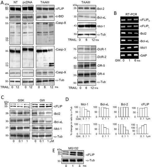
NT, pcDNA and TrkAIII SH-SY5Y cells express
all components required for TRAIL-induced
apoptosis
Western blot and RT-PCR comparisons of NT
SH-SY5Y, pcDNA SH-SY5Y and TrkAIII SH-SY5Y cells
revealed similar levels of functional (DR4 and DR5) and
decoy (DcR1 and DcR2) TRAIL receptor mRNA and
protein expression (Figure 3A and 3B), with cell surface
DR4 and DR5 expression detected in TrkAIII SH-SY5Y
adding to our previous report of cell surface DR4 and DR5
expression in NT SH-SY5Y and pcDNA SH-SY5Y cells
[49]. NT, pcDNA and TrkAIII SH-SY5Y cell lines also
exhibited similar levels of constitutive caspase-3, caspase-8,
caspase-9 mRNA and protein expression and cBID protein
expression (Figure 3A and 3B). In contrast to NT and
pcDNA SH-SY5Y cells, TrkAIII SH-SY5Y cells also
exhibited higher constitutive mRNA and protein expression
of the intrinsic apoptosis pathway inhibitors Bcl- 2, Bcl-xL
and Mcl-1, and exhibited lower levels of cFLIP protein but
not mRNA expression (Figure 3A and 3B).
Treatment of TrkAIII SH-SY5Y cells with TRAIL
(200 ng/ml for 0–12 hours) did not alter the levels of
DR4, DR5, DcR1, DcR2, Bcl-2a, Bcl-xL, Mcl-1 or
cFLIP protein expression but did induced delayed (post
6 hours) cleavage of caspase-3 and caspase-8, and also a
delayed reduction in c-BID protein levels, consistent with
[image:3.612.152.467.164.654.2]degradation, not detected in NT-SH-SY5Y or pcDNA
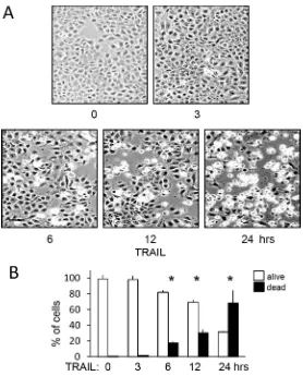
Figure 1:
TRAIL induces apoptosis of TrkAIII SH-SY5Y cells.
(A) Representative phase contrast (black and white) andFigure 2:
TRAIL abrogates the tumorigenic activity of TrkAIII SH-SY5Y cells
in vitro
.
(A) Representative photographs,demonstrating the abrogation of TrkAIII SH-SY5Y but not NT SH-SY5Y or pcDNA SH-SY5Y tumourigenic activity in vitro, in the presence
but not absence of TRAIL (200 ng/m). (B) Histograms demonstrating the mean (±SD) percentage change in tumour numbers grown from NT SH-SY5Y, pcDNA SH-SY5Y and TrkAIII SH-SY5Y cells, in the absence (100%) or presence of TRAIL (200 ng/ml). Tumour sphere numbers were evaluated in 10 ×10 magnification fields in triplicate experiments, each performed in duplicate (* = statistical significance
compared to untreated control). (C) Representative phase contrast micrographs demonstrating the appearance of tumour spheroid grown
SH- SY5Y cells (Figure 4A). Treatment of TrkAIII
SH-SY5Y cells with the TrkA inhibitor GW441756 (0.1
and 1 µM for 24 hours) [51] decreased Bcl-2 and Bcl-xL
mRNA and protein expression, increased cFLIP protein
but not mRNA expression (Figure 4B and 4C) but did not
alter Mcl-1 mRNA or protein expression (Figure 4B and
4C). In contrast, treatment of TrkAIII SH-SY5Y cells with
the PERK inhibitor GSK2656157 (0.1 and 1 µM) reduced
Mcl-1 but not Bcl-2 or Bcl-xL protein expression and did
not modulate cFLIP protein levels (Figure 4B and 4C).
The possibility that degradation at the proteasome
was responsible for reducing cFLIP protein levels
in TrkAIII SH-SY5Y cells was confirmed using the
proteasome inhibitor MG132 (10 µM), which increased
cFLIP protein levels in TrkAIII SH-SY5Y cells (Figure 4E).
Together, these data show that resistance to
induced apoptosis does not result from the lack of
TRAIL-induced apoptosis component expression or the expression
of Bcl2 family inhibitors but is associated with higher
levels of cFLIP expression. Conversely, TrkAIII SH-SY5Y
sensitivity to TRAIL-induced apoptosis is associated with
reduced cFLIP levels that result from TrkAIII activity and
cFLIP degradation at the proteasome and occurs despite
[image:5.612.92.527.207.653.2]increased Bcl2 family expression.
Figure 3: NT, pcDNA and TrkAIII SH-SY5Y cells express all of the major components required for TRAIL-induced
apoptosis.
(A) Ethidium bromide stained agarose gels demonstrating similar levels of RT-PCR products for major components involved inTRAIL-induced apoptosis generated from RNAs purified from NT-SH-SY5Y, pcDNA SH-SY5Y and TrkAIII SH-SY5Y cells. (B) Western blots demonstrating similar protein levels of major components involved in TRAIL-induced apoptosis in whole NT-SY5Y, pcDNA SH-SY5Y and TrkAIII SH-SH-SY5Y cell extracts (20 µg). (C) Representative IF micrographs demonstrating cell surface expression of functional
Figure 4: Effects of TRAIL, GW441756, GSK2656157 and MG132 upon the expression of components of
TRAIL-induced apoptosis.
(A) Western blots demonstrating the effect of TRAIL (200 ng/ml 0–12 hours) upon cFLIP, cBID, Caspase-8, Caspase-3 and Caspase-9 levels in whole cell extracts from NT SH-SY5Y (NT), pcDNA SH-SY5Y (pcDNA) and TrkAIII SH-SY5Y cells (TrkAIII) and Bcl-2, Bcl-xL, Mcl-1, DcR-1, DcR-2, DR-5 and DR-4 protein levels in whole cell extracts from TrkAIII SH-SY5Y cells (TrkAIII) (20 μg of whole cell extract loaded per sample). (B) Agarose gels demonstrating cFLIPS, cFLIPL, Bcl-2, Bcl-xL, Mcl-1 andGAPDH RT-PCR product levels generated from mRNAs purified from TrkAIII SH-SY5Y cells treated with 1 µM GW441756 (GW; 0, 1
TRAIL-induced apoptosis is delayed,
caspase-dependent and cFLIP and Mcl-1-regulated
TRAIL (200 ng/ml)-induced TrkAIII SH-SY5Y
apoptosis was not detected prior to 6 hours. At 6 hours,
TRAIL-induced a mean (±SD) of 18.3 ± 9.8% cell death
(
p =
0.0264,
n = 6), 30.4 ± 13.8% cell death at 12 hours
(
p =
0.0033,
n = 6) and 68.4 ± 23.4% cell death at 24
hours (
p <
0.0001,
n =
6) (Figure 5A and 5B).
SiRNA cFLIP knockdown in TrkAIII SH-SY5Y
cells (Figure 6A) significantly accelerated and augmented
TRAIL (200 ng/ml)-induced apoptosis to a mean (±SD)
of 90.7 ± 15.8% at 6 hours (P <
0.0001,
n =
4), compared
to 17.2 ± 8.6% in sham-transfected and 20.3 ± 18.6% in
control siRNA transfected TrkAIII SH-SY5Y counterparts
(Figure 6B and 6C). SiRNA Mcl-1 knockdown in TrkAIII
SH-SY5Y cells (Figure 6A) also significantly accelerated
and augmented TRAIL (200 ng/ml)-induced death to
88.5 ± 22.2% at 6 hours (P <
0.0001,
n =
4), compared
to both sham and control siRNA transfected counterparts
(Figure 6B and 6C). TRAIL-induced death of TrkAIII
SH-SY5Y cells exhibiting cFLIP knockdown was completely
abrogated by z-IETD-fmk caspase-8 inhibitor (10 µM)
but not by z-LEHD-fmk (10 µM) caspase-9 inhibitor
(
p <
0.0001,
n =
4), whereas both z-IETD-fmk (10 µM)
and z-LEHD-fmk (10 µM) significantly inhibited
TRAIL-induced death of TrkAIII SH-SY5Y cells exhibiting
Mcl-1 knockdown (
p <
0.0001,
n =
4 for both) (Figure 6C).
Transient FLAG-tagged cFLIP overexpression in TrkAIII
SH-SY5Y cells (Figure 7A) significantly reduced TRAIL
(200 ng/ml)-induced apoptosis at 24 hours from a mean
(± SD) of 75.3 ± 18.2% in TrkAIII SH-SY5Y cells
transfected with empty pcDNA vector to 29.2 ± 18.4%
(
P <
0.02,
n = 4) (Figure 7B and 7C). Transient Bcl-xL
overexpression (Figure 7A) did not significantly inhibit
TRAIL-induced TrkAIII SH-SY5Y apoptosis compared
to empty pcDNA vector-transfected TrkAIII SH-SY5Y
counterparts (Figure 7B and 7C).
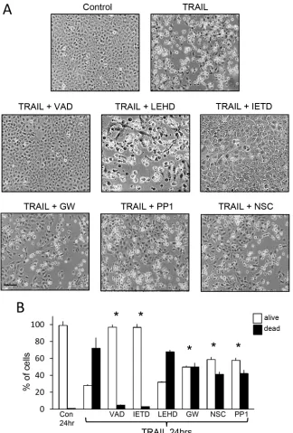
Under non-knockdown conditions, TRAIL (200
ng/ml)-induced TrkAIII SH-SY5Y apoptosis was
abrogated by z-IETD-fmk (10 µM) caspase-8 inhibitor
and by z-VAD-fmk (10 µM) pan-caspase inhibitor
but not by z-LEHD-fmk (10 µM) caspase-9 inhibitor
[image:7.612.168.446.323.668.2](Figure 8A and 8B).
Figure 5: TRAIL induces delayed and not immediate apoptosis of TrkAIII SH-SY5Y cells.
(A) Representative phase(bar = 100 μm) contrast micrographs demonstrating time-dependent TRAIL-induced (200 ng/ml) TrkAIII SH-SY5Y cell death from 0–24 hours.
Figure 6: SiRNA cFLIP and Mcl-1 knockdown accelerate TRAIL-induced apoptosis.
(A) Western blots demonstrating knockdown of cFLIP and Mcl-1 protein expression in whole cell extracts from TrkAIII SH-SY5Y cells transiently transfected with cFLIP- and Mcl-1-specific siRNAs but not control siRNAs, compared to cFLIP and Mcl-1 levels in sham-treated controls (Con).Together, these data confirm that TRAIL-induced
TrkAIII SH-SY5Y apoptosis is caspase-dependent,
delayed in association with delayed caspase activation
(see Figure 4A), is regulated by cFLIP and occurs via the
extrinsic pathway. Furthermore, the data identify Mcl-1 as
the dominant inhibitor of the intrinsic apoptosis pathway
in TrkAIII SH-SY5Y cells and show that reducing cFLIP
or Mcl-1 expression augments sensitivity to
TRAIL-induced apoptosis.
TRAIL activates c-Src, induces TrkAIII
complexing with c-Src and SHP-1 and
TRAIL-induced apoptosis involves TrkAIII, c-Src and
SHP activity
Mean (±SD) TRAIL-induced TrkAIII SH-SY5Y
death of 72.2 ± 17.3% at 24 hours was significantly
reduced to 50.1 ± 9.3% (P
= 0.0203,
n = 6) by GW441756
(1 µM) TrkA inhibitor (51); to 41.3 ± 7.2% (P
= 0.0023,
n = 6) by NSC87877 (1 µM) SHP-1/2 inhibitor (52) and
to 42.2 ± 9.8% (P
= 0.0041,
n =
6) by PP1 (1 µM) c-Src
inhibitor (53) (Figure 8A and 8B).
SHP-1 immunoprecipitated from untreated whole
TrkAIII SH-SY5Y cell extracts (500 μg) pulled down
c-Src but not TrkAIII, confirming constitutive
SHP-1/c-Src but not SHP-1/TrkAIII complexing (Figure 9A).
TRAIL (200 ng/ml for 6 hours) did not alter SHP-1/c-Src
complexing but induced SHP-1 pull-down of TrkAIII
together with c-Src (Figure 9A).
Western blots detected high constitutive c-Src Y527
phosphorylation relative to Y416 phosphorylation in
TrkAIII SH-SY5Y cells (Figure 9B). TRAIL (200 ng/ml,
[image:9.612.129.486.280.632.2]0–12 hours) reduced c-Src Y527 phosphorylation at
6 and 12 hours in association with increased c-Src
Y416 phosphorylation, consistent with c-Src activation
(Figure 9B). PP1 (1 µM) c-Src inhibitor did not alter
Figure 7: Overexpression of cFLIP but not Bcl-xL inhibits TRAIL-induced TrkAIII SH-SY5Y cell apoptosis.
(A) Western blots demonstrating expression of exogenous FLAG-cFLIP and Bcl-xL in TrkAIII SH-SY5Y transient transfectants compared to emptyeither constitutive c-Src Y527 or Y416 phosphorylation
or TRAIL-induced c-Src Y527 de-phosphorylation in
TrkAIII SH-SY5Y cells over the 12 hour time course
but did abrogate TRAIL stimulation of c-Src Y416
phosphorylation, consistent with autophosphorylation
(Figure 9B and 9C). NSC87877 (1 µM) SHP1/2
inhibitor prevented both TRAIL-induced Src Y527
de- phosphorylation and increased Y416 phosphorylation
(Figure 9B and 9C), implicating SHP in TRAIL-induced
c-Src activation via the de-phosphorylation of Y527.
In addition to inducing TrkAIII/SHP-1/c-Src
complexing, TRAIL also induced de-phosphorylation of
[image:10.612.146.470.137.616.2]TrkAIII Y674/675 within 6 hours (Figure 10A and 10B),
prevented by both PP1 and NSC87877 (Figure 10A and 10B)
Figure 8: TRAIL-induced TrkAIII SH-SY5Y apoptosis is inhibited by z-VAD-fmk, z-IETD-fmk, GW441756, PP1 and
NSC-87877 but not by z-LEHD-fmk.
(A) Representative phase contrast micrographs demonstrating inhibition of TRAIL-induced TrkAIII SH-SY5Y apoptosis following 24 hour treatment (200 ng/ml) by 10 µM z-VAD-fmk (VAD), 10 µM z-IETD-fmk (IETD) but not 10 µM z-LEHD-fmk (LEHD) and by 1 µM GW441756 (GW), 1 µM PP1 and 1 µM NSC-87877 (NSC) (bar = 100 µm). (B) Histograms displaying the mean (+SD) percentage (%) TrkAIII SH-SY5Y survival (white) or death (black) following treatment with TRAIL alone (200 ng/ml), TRAIL plus 10 µM z-VAD-fmk (VAD), TRAIL plus 10 µM z-LEHD-fmk (LEHD), TRAIL plus 10 µM z-IETD-fmk (IETD), TRAIL plus 1 µM GW441756 (GW), TRAIL plus 1 µM PP1 and TRAIL plus 1 µM NSC-87877 (NSC), in three independent AO/EBr experiments, each performed in duplicate (* = significant difference compared to TrkAIII SH-SY5Y cells treated for 24 hours with TRAILFigure 9: TRAIL induces complexing between TrkAIII, SHP-1 and c-Src and activates c-Src in TrkAIII SH-SY5Y
cells.
(A) Immunoprecipitation/Western blots demonstrating SHP-1 pull down of c-Src but not TrkAIII from untreated TrkAIII SH-SY5Y whole cell extracts (500 µg) and SHP-1 pull down of both c-Src and TrkAIII in TRAIL-treated (200 ng/ml for 12 hours) TrkAIII SH-SY5Y whole cell extracts (500 µg), plus Western blots showing input levels of SHP-1, c-Src and TrkAIII in TrkAIII SH-SH-SY5Y wholecell extracts (20 µg). (B) Western blots demonstrating TRAIL-induced (200 ng/ml for 0, 6 and 12 hours) c-Src Y527 de-phosphorylation and stimulation of c-Src Y416 phosphorylation; 1 µM PP1 inhibition of TRAIL-induced stimulation of c-Src Y416 phosphorylation but not TRAIL-induced c-Src Y527 de-phosphorylation, and 1 µM NSC-87877 (NSC) inhibition of both TRAIL-induced stimulation of c-Src Y416 phosphorylation and c-Src Y527 de-phosphorylation, compared to total c-Src levels in whole cell TrkAIII SH-SY5Y extracts
in association with significant inhibition of TRAIL-induced
apoptosis, confirming roles for both c-Src and SHP in
TRAIL-induced TrkAIII de-phosphorylation and subsequent
apoptosis. PP1 and NSC87877 did not reduce constitutive
TrkAIII Y674/675 phosphorylation in the absence of TRAIL
(data not shown). Association between TRAIL-induced
TrkAIII Y674/675 de-phosphorylation and apoptosis was
confirmed in Western blots by exclusive detection of TrkAIII
Y674/675 de-phosphorylation in apoptotic but not surviving
TrkAIII SH-SY5Y cells (see Figure 11B, lower panel).
Together, these data implicate both SHP and c-Src
in TRAIL-induced pro-apoptotic crosstalk with TrkAIII,
and implicate SHP in c-Src activation and TrkAIII
de-phosphorylation.
TRAIL promotes TrkAIII/cFLIP complexing
and alters the caspase-8 to cFLIP ratio recruited
to death receptor complexes
In co-immunoprecipitation experiments, cFLIP
immunoprecipitated from untreated whole TrkAIII SH-SY5Y
cell extracts pulled down very low levels of TrkAIII, that
were not reduced further by GW441756 (1 µM for 24 hours)
(Figure 11A). C-FLIP immunoprecipitates pulled down
non-phosphorylated TrkAIII from TRAIL-treated (200 ng/ml for
24 hours) apoptotic but not surviving TrkAIII SH-SY5Y cells
(Figure 11A and 11B). TRAIL-induced TrkAIII Y674/675
de-phosphorylation was confirmed in input samples from
[image:12.612.182.434.230.619.2]apoptotic but not surviving cells (Figure 11B, INPUT).
Figure 10: TRAIL-induced de-phosphorylation of TrkAIII Y674/675 associates with apoptosis and is prevented by PP1
and NSC-87877.
(A) Western blots demonstrating no change total and Y674/675 phosphorylated TrkAIII levels in TrkAIII SH-SY5Y cells incubated for 0, 3 and 6 hours in absence of TRAIL, compared to time-dependent TRAIL-induced (200 ng/ml) TrkAIII Y674/675 de-phosphorylation associated with constant TrkAIII levels, in TrkAIII SH-SY5Y cells incubated for 3 and 6 hours plus the inhibition of TRAIL-induced TrkAIII Y674/675 de-phosphorylation by 1 µM PP1 (TRAIL + PPI) and by 1 µM NSC-87877 (TRAIL + NSC) at bothIn ligand-precipitation/Western blots, streptavidin
precipitation of biotin-labelled TRAIL-ligated DR4
positive death receptor complexes from TrkAIII SH-SY5Y
cells treated for 1 and 6 hours with biotinylated TRAIL,
revealed an increase in the caspase 8 to cFLIP ratio from
0.66 following 1 hour TRAIL-treatment to 1.53 following
6 hour TRAIL-treatment. In contrast, the caspase-8 to
cFLIP ratio of 0.68 pcDNA SH-SY5Y cells treated for 1
hour with biotinylated TRAIL remained at the same level
in 6 hour-treated cells (Figure 12A and 12B).
Together, these data link TRAIL-induced
SHP/c-Src-mediated TrkAIII de-phosphorylation and complexing
with cFLIP to a pro-apoptotic increase in caspase-8 to
cFLIP ratio at TRAIL-activated death receptors.
TRAIL does not modulate Caspase-8 Y387
phosphorylation in TrkAIII SH-SY5Y cells
Western blots detected similar constitutive levels of
inhibitory caspase 8 Y380 phosphorylation [54], in NT
[image:13.612.57.558.189.633.2]SY5Y, pcDNA SH-SY5Y and TrkAIII SH-SY5Y cells. Neither
GW441756 (1 µM for 24 hours) nor TRAIL (200 ng/ml for
12 hours) reduced Caspase 8 Y380 phosphorylation levels in
TrkAIII SH-SY5Y cells (Figure 12C and 12D). These data
associate constitutive inhibitory caspase-8 Y380 phosphorylation
[image:14.612.120.490.134.597.2]with the TRAIL-resistant SH-SY5Y phenotype, show that
caspase-8 Y380 phosphorylation in TrkAIII SH-SY5Y cells
is not TrkAIII-dependent and confirm that caspase-8 Y380
de-phosphorylation is not involved in sensitizing TrkAIII
SH-SY5Y cells to TRAIL-induced apoptosis.
Figure 12: TRAIL induces a time-dependent increase in the caspase-8 to cFLIP ratio at ligand-activated death
receptors in TrkAIII SH-SY5Y but not pcDNA SH-SY5Y cells.
(A) Western blots demonstrating reduced cFLIP levels and increased caspase-8 levels in ligand-precipitated DR4 positive death receptor complexes from TrkAIII SH-SY5Y but not pcDNA SH-SY5Y cells treated with biotin-labelled TRAIL (500 ng/ml) for 6 hours compared to 1 hour. Input levels of cFLIP, Caspase 8 (Casp-8) and α-tubulin (α-Tub) in total cell extracts (20 µg) are provided (Input). (B) Histogram displaying differences in the c-FLIP to caspase-8 ratios in the adjacent Western blots, adjusted for differences in DR4 densitometric levels, in ligand-precipitated DR4 positive death receptors purified from pcDNA SH-SY5Y (white) and TrkAIII SH-SY5Y cells (grey), obtained by image J densitometry. (C) Western blots demonstrating similar levels of total and Y380 phosphorylated caspase 8 in NT SH-SY5Y, pcDNA SH SY5Y and TrkAIII SH-SY5Y whole cell extractsDISCUSSION
In the present study, we report a potential
therapeutic “Achilles heel” for the TrkAIII oncoprotein
in a SH-SY5Y NB model. In this model, TRAIL induced
delayed caspase-dependent apoptosis of SH-SY5Y cells
engineered to express TrkAIII, resulting in the complete
abrogation of tumorigenic growth capacity
in vitro
.
Consistent with the TRAIL-resistant phenotype of parental
and control-transfected SH-SY5Y cells (this study and 49),
TRAIL-induced apoptosis of TrkAIII SH-SY5Y cells
resulted from one-way SHP/c-Src-mediated pro-apoptotic
crosstalk between the TRAIL receptor signaling pathway
and TrkAIII. This results from SHP/c-Src-dependent
binding and de-phosphorylation of TrkAIII, inducing
cFLIP complexing with de-phosphorylated TrkAIII,
increasing the caspase-8 to cFLIP ratio at TRAIL-activated
death receptors, which explains the delayed induction of
caspase cleavage and apoptosis, with rate-limiting roles
confirmed for both c-FLIP and Mcl-1.
All the cell lines used in this study exhibited
constitutive expression of components required for
TRAIL-induced apoptosis (this study and 49), a
pro-apoptotic expression equilibrium between functional and
decoy TRAIL receptors and cell-surface functional DR4
and DR5 TRAIL receptor expression (this study and 49).
In contrast to some NB cell lines [55], all of our cell
lines expressed caspase 8 mRNA and protein. However,
inhibitory caspase-8 Y380 phosphorylation [54] was also
detect in all cell lines, providing an additional mechanism
for reducing SH-SY5Y sensitivity to TRAIL-induced
apoptosis (this study and 49).
TrkAIII SH-SY5Y cells expressed lower levels of
cFLIP but higher levels of Bcl-2, Bcl-xL and Mcl-1 than
either NT SH-SY5Y or pcDNA SH-SY5Y cells. Inhibitor
(GW441756 and MG132) studies indicated that lower
cFLIP protein expression by TrkAIII SH-SY5Y cells
was post-transcriptional and dependent upon enhanced
cFLIP degradation at the proteasome and TrkAIII tyrosine
kinase activity. We are currently investigating whether this
may depend upon Nedd-4 family E3 ligases, which bind
activated TrkA receptors and promote cFLIP degradation
at the proteasome [56, 57].
TRAIL-induced TrkAIII SH-SY5Y apoptosis was
caspase-dependent and was abrogated by z-VAD-fmk
pan-caspase and z-IETD-fmk caspase-8 inhibitors but
not by z-LEHD-fmk caspase-9 inhibitor [58–60] or
Bcl-xL overexpression. This characterises TrkAIII SH-SY5Y
cells as type I tumour cells, sensitive to TRAIL-induced
apoptosis through the extrinsic pathway [14, 19, 20, 25],
with the intrinsic pathway blocked by constitutive Bcl-2,
Bcl-xL and Mcl-1 expression (this study and 49).
TRAIL-induced TrkAIII SH-SY5Y apoptosis was
delayed and not immediate, was not detected prior to
6 hours and increased to a maximum post 12 hours. Delayed
apoptosis was associated with delayed cleavage of caspase
8 and delayed cleavage of the caspase 8 substrates c-BID
[61] and caspase 3 [62], confirming a delay in caspase 8
activation. Therefore, low cFLIP levels combined with
inhibitory caspase 8 Y380 phosphorylation were sufficient
to prevent immediate apoptosis, implicating additional
TRAIL-induced time-dependent pro apoptotic changes.
The central role of cFLIP in delaying
TRAIL-induced TrkAIII SH-SY5Y apoptosis was confirmed by
siRNA knockdown, which accelerated and augmented
apoptosis to > 90% within 6 hours, and by cFLIP over
expression that significantly inhibited TRAIL-induced
TrkAIII SH-SY5Y apoptosis. This suggests that
TRAIL-induced reduction in cFLIP function resulting from
pro-apoptotic crosstalk with TrkAIII may underpin the delay
in TRAIL-induced apoptosis. However, since TRAIL did
not reduce cFLIP protein expression in TrkAIII SH-SY5Y
cells any further reduction in cFLIP function would have
to be post-translational.
Inhibitor studies confirmed that TRAIL-induced
pro-apoptotic crosstalk between the TRAIL-receptor pathway
and TrkAIII involved SHP, c-Src and TrkAIII activity.
TrkA (GW441756), c-Src (PP1) and SHP1/2 (NSC-87877)
inhibitors significantly reduced TRAIL-induced TrkAIII
SH-SY5Y apoptosis, which was associated with
time-dependent changes in c-Src and TrkAIII phosphorylation.
These changes were characterised by initial
TRAIL-induced de-phosphorylation of c-Src inhibitory tyrosine
Y527 combined with increased phosphorylation of c-Src
active site tyrosine Y416, consistent with activation [63],
which was accompanied by the induction of complexing
between phosphorylated TrkAIII, SHP-1 and c-Src,
followed by TrkAIII Y674/675 de-phosphorylation,
consistent with inactivation [64], implicating SHP-1 and
c-Src in TRAIL-induced pro-apoptotic crosstalk with
TrkAIII. It remains unclear, however, whether both
SHP-1 and c-Src, which directly bind activated TrkA receptors
[65, 66], interact directly with TrkAIII under these conditions.
The SHP inhibitor NSC-87877 prevented both
TRAIL-induced Src Y527 de-phosphorylation and
increased Y416 phosphorylation. The c-Src inhibitor PP1
prevented TRAIL-induced c-Src Y416 phosphorylation but
not Y527 de-phosphorylation, and both inhibitors prevented
TRAIL-induced TrkAIII Y674/675 de-phosphorylation.
This suggest that TRAIL first activates SHP, which
de-phosphorylates c-Src Y527 within constitutive complexes
leading to c-Src activation and Y416 auto phosphorylation,
which in turn promotes interaction with phosphorylated
TrkAIII, resulting in SHP-mediated TrkAIII Y674/675
de-phosphorylation and subsequent apoptosis. This possibility
is supported by reports that SHP is recruited and activated
at death receptors [67], TRAIL activates c-Src [68], c-Src
and SHP bind activated TrkA receptors [65, 66] and SHP
de-phosphorylates TrkA Y674 and Y675 residues [66].
surviving but not apoptotic cells. TrkAIII de-phosphorylation
alone, however, would appear to be insufficient to sensitize
SH-SY5Y cells to TRAIL-induced apoptosis, since TrkAIII
inactivated by GW441756 (this study and 8) significantly
inhibited TRAIL-induced TrkAIII SH-SY5Y apoptosis. This
suggests that a more complex interaction between c-Src/
SHP and phosphorylated TrkAIII, resulting in TrkAIII
de-phosphorylation is required for a pro-apoptotic outcome.
Furthermore, a pro-apoptotic outcome may also reflect
changes in the c-Src/SHP equilibrium, since activated c-Src
promotes apoptotic resistance and TrkA phosphorylation
[65, 68], whereas activated SHP promotes death
receptor-mediated apoptosis and TrkA de-phosphorylation [66, 67],
and both exhibit reciprocal regulation [69]. However,
since inhibitors of both SHP and c-Src reduced
TRAIL-induced TrkAIII SH-SY5Y apoptosis in association with
maintenance of TrkAIII phosphorylation, both SHP and
c-Src are required for TRAIL-induced pro-apoptotic cross
talk between the TRAIL receptor pathway and TrkAIII.
In a direct comparison between TRAIL-treated
apoptotic and surviving TrkAIII SH-SY5Y cells, increased
complexing between cFLIP and de-phosphorylated
TrkAIII was detected in apoptotic but not in surviving
TrkAIII SH-SY5Y cells. This suggests that TRAIL may
induce cFLIP sequester by de-phosphorylated TrkAIII with
intracellular membranes [1, 8], reducing its recruitment
to activated cell surface TRAIL receptors, which was
confirmed by the time-dependent increase in caspase-8 to
c-FLIP ratio recruited to DR4 positive receptors in
TRAIL-treated TrkAIII SH-SY5Y but not NT-SH-SY5Y cells.
This extend our previous reports that c-FLIP complexes
with NGF-activated TrkA receptors [49, 70], sensitizing
TrkA SH-SY5Y cells to TRAIL-induced apoptosis [49] to
include a role for TrkAIII/cFLIP complexing in
TRAIL-induced TrkAIII SH-SY5Y apoptosis. cFLIP, therefore,
can interact with both phosphorylated TrkA and
non-phosphorylated TrkAIII receptors depending upon the
circumstance, which may reflect not only differences
in receptor structure but also localisation, post receptor
signaling and/or complex composition.
SiRNA Mcl-1 knockdown accelerated and augmented
TRAIL-induced TrkAIII SH-SY5Y apoptosis, characterising
Mcl-1 as a major regulator of TrkAIII SH-SY5Y sensitivity
to TRAIL-induced apoptosis. In contrast to siRNA cFLIP
knockdown, which accelerated and augmented
TRAIL-induced apoptosis through the extrinsic pathway,
z-LEHD-fmk caspase-9 inhibitor prevented accelerated and
augmented TRAIL-induced apoptosis following siRNA
Mcl-1 knockdown, confirming intrinsic apoptosis pathway
involvement [23–25]. Interestingly, Mcl-1 expression
in TrkAIII SH-SY5Y cells was inhibited by the PERK
inhibitor GSK2656157 [71] but not by the TrkA inhibitor
GW441756, whereas Bcl2 and Bcl-xL expression was
reduced by GW441756 but not GSK2656157, implicating
TrkAIII activity in Bcl-2 and Bcl-xL but not Mcl-1
expression and the PERK arm of the ER-stress response
in the expression of Mcl-1 but not Bcl-2 and Bcl-xL.
This supports reports of ER-stress regulated Mcl-1
expression [72] and the activation an ER-stress response
survival adaptation in TrkAIII SH-SY5Y cells [11, 12],
adding PERK up-regulated expression of Mcl-1 as a
novel potential “non-oncogene addiction” mechanism to
oncogenic TrkAIII repertoire.
Constitutive inhibitory caspase 8
Y380-phosphorylation [73] was detected at similar levels in all
SH-SY5Y cell lines, providing an additional mechanism
for reducing sensitivity to TRAIL-induced apoptosis.
However, neither GW441756 nor TRAIL reduced caspase
8 Y380-phosphorylation in TrkAIII SH-SY5Y cells,
indicating that caspase 8 Y380-phosphorylation does not
depend upon TrkAIII and its de-phosphorylation was not
involved in sensitizing TrkAIII SH-SY5Y cells to
TRAIL-induced apoptosis.
In conclusion, we have identified a potential
therapeutic “Achilles heel” for the TrkAIII oncoprotein,
characterised by TRAIL-induced one-way pro-apoptotic
crosstalk between the TRAIL receptor signaling pathway
and TrkAIII, in SH-SY5Y NB cells. We propose that
TRAIL-induced apoptosis initiates with TRAIL activation
of SHP, followed by SHP-mediated activation of c-Src.
This promotes complexing between phosphorylated
TrkAIII, SHP and c-Src, leading to SHP-mediated
TrkAIII de-phosphorylation and the sequester cFLIP,
which increases the caspase-8 to cFLIP ratio recruited
to activated TRAIL-receptors, explaining the delay in
caspase cleavage and apoptosis via the extrinsic pathway.
Our observations add significantly to a previous report
of pro-apoptotic cross talk between TNF-α signaling
networks and tyrosine kinase receptors [74] and provide a
novel rational for the therapeutic use of TRAIL, combined
with siRNA cFLIP and Mcl-1 inhibitors, in TrkAIII
expressing NBs. Such an approach may work best in
first-line therapy, as TRAIL promotes KRAS-driven metastasis
[75] and KRAS mutations are detected in relapsed but not
primary NBs [76–78].
MATERIALS AND METHODS
Cell lines and reagents
Non-transfected, pcDNA control transfected and
TrkAIII transfected SH-SY5Y NB cell lines have been
described previously [1]. Cells were grown in RPMI,
supplemented with appropriate antibiotics (Zeocin for
stable transfectants, penicillin and streptomycin) and 10%
foetal calf serum. GW441756 TrkA inhibitor [51],
z-VAD-fmk pan-caspase inhibitor, z-IETD-z-VAD-fmk caspase 8 inhibitor
and z-LEHD-fmk caspase 9 inhibitor; human recombinant
TRAIL and PP1 c-Src inhibitor were purchased from
Sigma-Aldrich (St. Louis Mo). NSC-87877 SHP1/2
TrkA, α-tubulin, c-FLIP, caspase-8, caspase-9, BID and
Mcl-1 were purchased from SantaCruz (SantaCruz, Ca).
Antibodies against Y674/675 phosphorylated TrkA, c-Src,
Y527 phosphorylated and Y416 phosphorylated c-Src and
Caspase-3, were from Cell Signalling (Danvers, MA).
Antibodies against DR4, DR5, DcR1 and DcR2 were
from ANASPEC (Belgium). Antibodies against Bcl2 and
Bcl-xL were from AbCam (Cambridge, UK). Hybond
C-extra nitrocellulose membranes and ECL solutions were
purchased from Amersham International (Bedford, UK).
RNA-easy RNA purification kits were purchased from
Qiagen (Hilden, Ge). Mammalian pcDNA expression
vectors for Bcl-xL and Flag-tagged c-FLIP were kindly
provided by Dr. Francesca Zazzeroni (Univ. of L’Aquila)
and have been described previously [79].
Transient Bcl-xL and cFLIP expression
Bcl-xL and flag-tagged cFLIP were overexpressed
in TrkAIII SH-SY5Y cells by transient transfection of
mammalian expression vectors bearing Bcl-xL, FLAG-tagged
cFLIP [79] or empty pcDNA. Briefly, 0.6
µg/ml of eachvector were transfected into sub-confluent cell cultures using
FUGENE transfection reagent as directed by the manufacturer
(Promega, Madison, Wi). At 6 hours transfection medium
was replaced with fresh growth medium (RPMI/10%FCS/
glutamine plus antibiotic) and the cells left for 48 hours,
at which time they were utilised for experimentation.
Overexpression of Bcl-xL and Flag-tagged cFLIP was verified
by Western blotting of total cell extracts (20
µg).SiRNA knockdown of c-FLIP and Mcl-1
C-FLIP and Mcl-1 expression was knocked down in
TrkAIII SH-SY5Y cells using a TriFECTa Dicer-Substrate
RNAi kit, employing three cFLIP-specific or three
Mcl-1-specific Dicer-Substrate siRNA duplexes, as described
by the manufacturer (Integrated DNA Technologies,
Coralville, IA). Briefly, cells were subjected to 48 hour
transient transfection with either 25 nM negative control
siRNA duplex (provided with the kit) or 25 nM of a mix
of cFLIP specific or Mcl-1 specific siRNA duplexes,
using INTERFERin
in vitro
siRNA transfection reagent,
as described by the manufacturer (Polyplus Transfection
Inc., New York, NY). Sham transfected controls received
transfection reagent alone. SiRNA knockdown of cFLIP
and Mcl-1 expression was confirmed by Western blot
comparison to α-tubulin in whole cell extracts (20μg).
Mcl-1 siRNA sets
5′-AGCCUAGUAUGUCAAUAAAGCAAAT-3′ and
5′-AUUUGCUUUUAUUGACAUACUA GGCUUA-3′;
5′-GGAACAAAUCUGAUAACUAUGCAGG-3′ and
5′- CCUGCAUAGUUAUCAGAUUUGUUCCAC-3′;
and 5′-CAAGUGCAUAGAUGUGAAUUGGUTT-3′ and
5′-AAACCAAUUCACAUCUAUGCACUUGUU-3′.
cFLIP siRNA sets
5′-UGAGUUGGAGAAACUAAAUCUGGTT-3′ and
5′-AACCAGAUUUAGUUUCUCCAAC UCAAC-3′; 5′-CG
AAGACCCUUGUGAGCUUCCCUAG-3′ and 5′-CUAG
GGAAGCUCACA AGGGUCUUGCAG-3′; and 5′-GCC
GAGGCAAGAUAAGCAAGGAGAA-3′ and 5′-UUC
UCCUUGCUUAUCUUGCCUCGGCCC-3′
Transfection efficiency was confirmed using a HPRT-S1
DS positive control and validated using a negative control
duplex (NC1) not present in the human genome, as described
by the manufacturer (Integrated DNA Technologies; www.
IDTDNA.com)
Cell extraction, immunoprecipitation and
Western blotting
Cells were extracted in lysis buffer (PBS containing
0.5% sodium deoxycholate, 1% NP40, 0.1% SDS, 1 mM
sodium orthovanadate, 1 mM PMSF, 1 μg/ml of pepstatin A
and Aprotinin) and protein concentrations were calculated
by Bradford protein concentration assay (Sigma-Aldrich).
Samples were mixed with reducing SDS-PAGE sample
buffer and subjected to reducing SDS-PAGE/Western
blotting. Briefly, proteins separated by reducing
SDS-PAGE, were trans-blotted onto Hybond C+ nitrocellulose
membranes by electrophoresis (Amersham Int. UK) and
the membranes subsequently air-dried. Non-specific protein
binding-site on membranes were blocked by incubation for
2 hours in 5% non-fat milk in TBS-T prior to incubation
with primary antibodies, at recommended dilutions, for
2–16 hours at 4°C. Membranes were then washed in TBS-T,
incubated with secondary HRP-conjugated antibodies
(Jackson ImmunoResearch Laboratories, West Grove, PA)
diluted in blocking solution and immunoreactive species
detected by chemiluminescence reaction, as directed by the
manufacturer (Amersham Int).
RNA purification and RT-PCR
RT reactions were performed on total RNAs
(1 μg), purified using RNA-easy Plus, as described by
the manufacturer (Qiagen), using the Moloney Murine
Leukemia virus RT kit, as detailed by the manufacturer
(LifeTechnologies, Inc, Paisley, UK). RT reactions were
subjected to PCR using the following primers:
GAP: 5′- AGGTCCACCACTGACAGTT-3′ (forward)
and 5′- CTGCACCACCAACTGCTT AG-3′ (reverse) (300 bp).
DcR1: 5′-GAAGAATTTGGTGCCAATGCCACTG-3′
(forward) and 5′- CTCTTGGACTTGGCTGGGAGA
TGTG-3′ (reverse) (612bp);
DcR2: 5′-CCCCCGGCAGGACGAAGTT-3′ (forward)
and 5′- CTCCTCCGCTGCTGGGGTTTT-3′ (reverse)
(418 bp);
DR5: 5′-CTGAAAGGCATCTGCTCAGGTG-3′
(forward) and 5′-CAGAGTCTGCATTACCTTCTAG-3′
(reverse) (347 bp);
c-FLIPs: 5′-GGACCTTGTGGTTGAGTTGG-3′
(forward) and 5′-ATCAGGACAATGGGCATAGG-3′
(reverse) (241 bp);
c-FLIP
L: 5′-GGCTCCCAGAGTGTGTATGG-3′
(forward) and 5′-AGCTTCTCGGTGAACTGTGC-3′
(reverse) (249 bp);
Bcl-2: 5′-GACTTCGCCGAGATGTCC-3′ (forward)
and 5′-CAAGCTCCCACCAGGGCCAAAC-3′ (reverse)
(356 bp);
Bcl-XL:
5′-GTGAATTCTGAGGCCAAGGGAAC-3′ (forward)
and 5′-GAACGGCGGCTGGGATACTTTTG-3′ (reverse)
(373 bp);
Caspase-8: 5′-TCTGGAGCATCTGCTGTCTG-3′
(forward) and 5′-CCTGCCTGGTGTCTGAAGTT-3′
(reverse) (427 bp);
Caspase-10: 5′-GGGAACGGACACACAACTCT-3′
(forward) and 5′-CTAGCTTTTGGCCCTGACTG-3′
(reverse) (293 bp);
Caspase-3:
5′-TTAATAAAGGTATCCATGGAGAACACT-3′
(forward) and 5′-TTAGTGATAAAAATAGAGTTCT
TTTGTGAG-3′ (reverse) (849 bp);
Mcl-1: 5′-AAGCC AATGGGCAGGTCT-3′ (forward)
and 5′-TGTCCAGTTTCCGAAGCAT-3′ (reverse) (121 bp);
For each primer set, PCRs were performed on
reverse transcription reactions serially diluted from 1 to
1:1000. Reactions below saturation were compared by
densitometric analysis of Jpeg images of ethidium bromide
stained gels, using ImageJ64 software [80].
Cell death assay
TRAIL-induced cell death was routinely assayed
using a modification of previously described methods
[81, 82]. Briefly, cells were washed once in Ca
2+free
PBS, detached with ice cold PBS containing 1 mM
EDTA, transferred to sterile 15 mls tubes, centrifuged
for 5 minutes at 1,000 × g at 4°C, washed with ice cold
PBS and re-pelleted by centrifugation at 1,000 × g for 5
minutes, at 4°C. Cell pellets were re-suspended in 25 μl of
PBS containing 2 μl of acridine orange/ethidium bromide
solution (100 μg/ml acradine orange and 100 μg/ml
ethidium bromide in PBS) plated onto glass slides and
examined immediately under a Zeiss “Axioplan-2”
fluorescence microscope. Representative fields were
digitally photographed under identical exposure conditions
and the number of dead cells (orange/red nuclei) and
live cells (green nuclei) counted. In addition, phase
contrast micrographs of parallel cultures were used to
confirm changes in the relative percentage of adherent
(surviving) and suspension (apoptotic) cells, following
TRAIL-treatment. TRAIL-induced TrkAIII SH-SY5Y
cell apoptosis was confirmed by its complete inhibition by
z-VAD-fmk pan caspase inhibitor.
Tumour growth in soft agar
For substrate-independent tumour growth assays
[49], 1 × 10
4cells in single-cell suspension (passed
through a gauge × 18 syringe needle) were mixed in a
33% solution of agar (BiTec; Difco) in RPMI containing
5% FCS at 37°C, with or without TRAIL (200 ng/ml) and
layered onto a solid 0.6% agarose substrate also with or
without TRAIL, prepared in the same growth medium.
Following top phase agar solidification, complete medium
was added and re-placed every 2 days. Tumour spheroid
growth was monitored over a 14-day period by phase
contrast microscopy. Tumour spheroids were counted in
10 random fields at ×10 magnification.
Isolation of TRAIL-activated death receptor
complexes
TRAIL-ligated death receptor complexes were
purified from biotin-TRAIL treated pcDNA SH-SY5Y and
TrkAIII SH-SY5Y cells by ligand affinity precipitation,
as previously described [83]. Briefly, biotinylated TRAIL
was prepared by incubating TRAIL (1 mg/ml) with
Sulfo-NHS-LC-Biotin (1 mg/ml) (Pierce) for 1 hour on ice.
The reaction was stopped by adding 1/10 volume of 1M
Tris-HCl [pH. 7.5] and unincorporated biotin removed
by buffer exchange into 150 mM NaCl, 30 mM HEPES
(pH 7.5) using PD-10 columns (Amersham Pharmacia
Biotech). For ligand affinity precipitation, cells (5 × 10
6cells per sample) were washed twice in RPMI at 37°C and
incubated with 1 mg/ml biotinylated TRAIL for 1 and 6
hours. Death receptor complex formation was stopped by
the addition of 15 volumes of ice cold PBS and cells were
then lysed in 4.5 mls of lysis buffer (30 mM TRIS-HCl
[pH. 7.5], 150 mM NaCl, 10% glycerol, 1% Triton X-100,
supplemented with complete protease inhibitor cocktail)
(Roche Diagnostics, Mannheim, GE). TRAIL receptor
protein complexes were precipitated from lysates by
co-incubation with 20 μl of Streptavidin beads (Pierce) for
3 hours at 4°C with rotation. Ligand affinity precipitates
were washed four times in lysis buffer, eluted from beads
in reducing SDS-PAGE sample buffer and subjected to
SDS-PAGE Western blotting.
Indirect immunofluorescence
Cells grown on Nunc glass chamber slides
(Sigma-Aldrich) were washed in PBS, fixed in 4%
paraformaldehyde, washed in PBS then processed for
indirect immunofluorescence (IF). Fixed, non-permeabilised
cells were incubated for 1 h in blocking solution (1% bovine
serum albumin in PBS-0.03% TX100) and then incubated for
temperature. Slides were washed three times in PBS-0.03%
TX100, incubated with secondary fluorochrome-conjugated
antibody diluted in blocking solution for 1 h at room
temperature, washed in PBS-0.03% TX100 and mounted
using VectorMount. IF images were obtained using a Zeiss
Axioplan 2 fluorescence microscope with a digital camera
and Leica M500 Image Manager software.
Statistical analysis
Data were analysed statistically by Student’s
t-test and statistical significance was associated with
probabilities of < 0.05.
ACKNOWLEDGMENTS AND FUNDING
The first two authors contributed equally to
this work. This work was supported by the Maugeri
Foundation.
CONFLICTS OF INTEREST
The authors declare no conflicts of interest.
REFERENCES
1. Tacconelli A, Farina AR, Cappabianca L, DeSantis G,
Tessitore A, Vetuschi A, Sferra R, Rucci N, Argenti B, Screpanti I, Gulino A, Mackay AR. TrkA alternative
splicing: a regulated tumor-promoting switch in human neuroblastoma. Cancer Cell. 2004; 6:347–360.
2. Ruggeri P, Farina AR, Cappabianca L, Di Ianni N,
Ragone M, Merolle S, Gulino A, Mackay AR. Neurotrophin
and Neurotrophin Receptor Involvement in Human Neuroblastoma. In: Neuroblastoma, Edited by Shimada H. Intech. 2013; doi: 10.5772/55536.
3. Schramm A, Schowe B, Fielitz K, Heilmann M, Martin M, Marschall T, Koster J, Vandesompele J, Vermeulen J, de Preter K, Koster J, Versteeg R, Noguera R, et al. Exon-level
expression analysis identify MYCN and NTRK1 as major determinants of alternative exon usage and robustly predict
neuroblastoma outcome. Br J Cancer. 2012; 107:1409–1417. 4. Simpson AM, Iyer R, Mangino JL, Minturn JE, Zhao H,
Kolla V, Garrett M. Brodeur. TrkAIII isoform expression upregulates stem cell markers and correlates with worse outcome in neuroblastomas (NBs). Proc Adv Neuroblast
Res. 2012; p 164 (POT055).
5. Luberg K, Park R, Aleksejeva E, Timmunsk T. Novel
transcripts reveal a complex structure of the human TrkA gene and imply the presence of multiple protein isoforms. BMC
Neurosci. 2015, 16; 78. doi: 10.1186/s12868–015–0215-x 6. Arevalo JC, Conde B, Hempstead BL, Chao MV,
Martin-Zanca D, Perez P. TrkA immunoglobulin-like ligand binding domains inhibit spontaneous activation of the receptor. Mol
Cell Biol. 2000; 20:5908–5916.
7. Watson FL, Porcionatto MA, Bhattacharyya A, Stiles CD,
Segal RA. TrkA glycosylation regulates receptor
localisation and activity. J Neurobiol. 1999; 39:323–336. 8. Farina AR, Cappabianca L, Ruggeri P, Gneo L,
Maccarone R, Mackay AR. Retrograde TrkAIII transport
from ERGIC to ER; a re-localisation mechanism for oncogenic activity. Oncotarget. 2015; 6:35636–35651. doi:
10.18632/oncotarget.5802.
9. Farina AR, Tacconelli A, Cappabianca L, Cea G, Panella S,
Chioda A, Romanelli A, Pedone C, Gulino A, Mackay AR. The TrkAIII splice variant targets the centrosome and promotes
genetic instability. Mol Cell Biol. 2009; 29:4812–4830. 10. Farina AR, Di Ianni N, Cappabianca L, Ruggeri PD,
Ragone M, Ianni G, Gulino A, Mackay AR. TrkAIII promotes microtubule nucleation and assembly at the
centrosome in SH-SY5Y neuroblastoma cells, contributing
to an undifferentiated anaplastic phenotype. Biomed Res
Int. 2013; doi: 10.1155/2013/740187.
11. Farina AR, Tacconelli A, Cappabianca L, Cea G,
Chioda A, Romanelli A, Pensato S, Pedone C, Gulino A, Mackay AR. The neuroblastoma tumour-suppressor TrkAI and its oncogenic alternative TrkAIII splice variant exhibit
geldanamycin-sensitive interactions with Hsp90 in human neuroblastoma cells. Oncogene. 2009; 28:4075–4094. 12. Ruggeri P, Farina AR, Di Ianni N, Cappabianca L, Ragone M,
Ianni G, Gulino A, Mackay AR. The TrkAIII oncoprotein
inhibits mitochondrial free radical ROS-induced death of
SH-SY5Y neuroblastoma cells by augmenting SOD2 expression and activity at the mitochondria, within the context of a tumor
stem cell-like phenotype. PLoS One. 2014; 15:e94568. 13. Carswell EA, Old LJ, Kassel RL, Green S, Fiore N,
Williamson B. An endotoxin-induced serum factor that causes necrosis of tumors. Proc Natl Acad Sci USA 1975; 72: 3666–3670.
14. Ashkenazi A, Dixit VM. Death receptors: signaling and modulation. Science. 1998; 281:1305–1308.
15. Nagane M, Huang HJ, Cavenee WK. The potential of TRAIL for cancer chemotherapy. Apoptosis. 2001; 6:191–197. 16. Hall MA, Cleavland JL. Clearing the TRAIL for cancer
therapy. Cancer Cell. 2007; 12:4–6.
17. Ashkenazi A, Holland P, Eckhardt SG. Ligand-based targeting of apoptosis in cancer: the potential of
recombinant human apoptosis ligand 2/Tumor necrosis
factor-related apoptosis- inducing ligand (rhApo2L/ TRAIL). J Clin Oncol. 2008; 26:3621–3630.
18. Van Dijk M, Halpin-McCormick A, Sessler T, Samali A, Szegezdi E. Resistance to TRAIL in non-transformed cells
is due to multiple redundant pathways. Cell Death Dis.
2013; 4:e702.
19. Russo M, Mupo A, Spagnuolo C, Russo GL. Exploring
death receptor pathways as selective targets in cancer
therapy. Biochem Pharmacol. 2010; 80:674–682.
21. Ashkenazi A.Targeting the extrinsic apoptotic pathway in
cancer: lessons learned and future directions. J Clin Invest. 2015; 125:487–489.
22. Xiong S, Mu T, Wang G, Jiang X. Mitochondria-mediated apoptosis in mammals. Protein Cell. 2014; 5:737–749. 23. Tait SW, Green DR. Mitochondria and cell death: outer
membrane permeabilization and beyond. Nat Rev Mol Cell
Biol. 2010; 11:621–632.
24. Cregan SP, Dawson VL, Slack RS. Role of AIF in
caspase-dependent and caspase-incaspase-dependent cell death. Oncogene.
2004; 23:2785–2796.
25. Fulda S, Debatin K-M. Extrinsic versus intrinsic apoptosis pathways in anticancer chemotherapy. Oncogene. 2006; 25:4798–4811.
26. Safa AR, Pollok KE. Targeting the anti-apoptotic protein
c-FLIP for cancer therapy. Cancers. 2011; 3:1639–1671. 27. Trivedi R, Mishra DP. Trailing TRAIL resistance: novel
targets for TRAIL sensitization in cancer cells. Frontiers in Oncol. 2015; 5:1–20.
28. Fulda S, Meyer E, Debatin K-M. Inhibition of
TRAIL-induced apoptosis by Bcl-2 overexpression. Oncogene.
2002; 21:2283–2294.
29. Yang X, Thiele CJ. Targeting the tumour necrosis
factor-related apoptosis-inducing ligand pathway in
neuroblastoma. Cancer Letters. 2003; 197:137–143. 30. Miller MA, Karacay B, Zhu X, D’Orisio MS, Sandler AD.
Caspase 8L, a novel inhibitory isoform of caspase 8 is associated with undifferentiated neuroblastoma. Apoptosis. 2006; 11:15–24. 31. Hopkins-Donaldson S, Bodmer J-L, Balmas Bourloud K,
Brognara CB, Tschopp J, Gross N. Loss of caspase-8
expression in highly malignant human neuroblastoma correlates with resistance to tumor necrosis factor-related apoptosis-inducing ligand-induced apoptosis. Cancer Res.
2000; 60:4315–4319.
32. Goldsmith KC, Hogarty MD. Targeting programmed cell death pathways with experimental therapeutics: opportunities in high-risk neuroblastoma. Cancer Letters. 2005; 228:133–141.
33. Karacay B, Sanlioglu S, Griffith TS, Sandler A, Bonthius DJ. Inhibition of the NF-κB pathway enhances TRAIL-mediated apoptosis in neuroblastoma cells. Cancer Gene Ther. 2004; 11:681–690.
34. Efron PA, Chen MK, Iyengar M, Dai W, Nagaram A,
Beierle EA. Differential response of neuroblastoma cells
to TRAIL is independent of PI3K/Akt. J Ped Surg. 2006; 41:1072–1080.
35. Poulaki V, Mitsiades N, Romero ME, Tsokos M.
Fas-mediated apoptosis in neuroblastoma cells requires mitochondrial activation and is inhibited by FLICE inhibitor proteins and bcl-2. Cancer Res. 2001; 61:4864–4872.
36. van Noesel MM, Pieters R, Voute PA, Versteeg R. The
N-myc paradox: N-myc overexpression in neuroblastomas
is associated with sensitivity as well as resistance to
apoptosis. Cancer Letters. 2003; 197:165–172.
37. Eggert A, Grotzer MA, Zuzak TJ, Wiewrodt BR, Ho R,
Ikegaki N, Brodeur GM. Resistance to tumor necrosis factor-related apoptosis-inducing ligand-induced apoptosis in neuroblastoma cells correlates with a loss of caspase-8
expression. Cancer Res. 2001; 61:1314–1319.
38. Van Noesel MM, Van Bezouw S, Salomons GS, Voute PA,
Pieters R, Baylin SB, Herman JG, Versteeg R. Tumor-specific down-regulation of the tumor necrosis
factor-related apoptosis inducing ligand decoy receptors DcR1 and DcR2 is associated with dense promoter hypermethylation.
Cancer Res. 2002; 62:2157–2161.
39. Sheard MA, Asgharzadeh S, Liu Y, Lin TY, Wu HW, Ji L, Groshen S, Lee DA, Seeger RC. Membrane-bound TRAIL supplements natural killer cell cytotoxicity against neuroblastoma cells. J Immunother. 2013; 36:319–329. 40. Ammann JU, Haag C, Kasperczyk H, Debatin KM, Fulda S.
Sensitization of neuroblastoma cells for TRAIL-induced apoptosis by NF-κB inhibition. Int J Cancer Res. 2009; 124:1301–1311.
41. Opel D, Naumann I, Schneider M, Bertele D, Debatin KM, Fulda S. Targeting aberrant PI3K/Akt activation by
PI103 restores sensitivity to TRAIL-induced apoptosis in neuroblastoma. Clin Cancer Res. 2011; 17:3233–3247. 42. Shenoy K, Wu Y, Pervaiz S. LY303511 enhances TRAIL
sensitivity of SHEP-1 neuroblastoma cells via hydrogen
peroxide-mediated mitogen-activated protein kinase activation and
up-regulation of death receptors. Cancer Res. 2009; 69:1941–1950. 43. Kim HR, Lee MW, Kim DS, Jo HY, Lee SH, Chueh HW,
Jung HL, Yoo KH, Sung KW, Koo HH. Etoposide sensitizes neuroblastoma cells expressing caspase 8 to TRAIL. Cell Biol Int Rep. 2011; 19:23–30.
44. Flahaut M, Muhlethaler-Mottet A, Auderset K, Bourloud KB,
Meier R, Popovic MB, Joseph JM, Gross N. Persistent inhibition of FLIPL expression by lentiviral small hairpin
RNA delivery restores death-receptor-induced apoptosis in
neuroblastoma cells. Apoptosis. 2006; 11:255–263. 45. Chang DW, Xing Z, Pan Y, Algeciras-Schimnich A,
Barnhart BC, Yaish-Ohad S, Peter ME, Yang X. c-FLIPL
is a dual function regulator for caspase-8 activation and
CD95-mediated apoptosis. EMBO J 2002; 21:3704–3714. 46. Tong HX, Lu CW, Zhang JH, Ma L, Zhang JH. Combination
of gamma-interferon with TRAIL and cisplatin or etoposide induces apoptosis in human neuroblastoma cell line SH-SY5Y. Chin Med Sci J 2007; 22:38–43.
47. Naumann I, Kappler R, von Schweinitz D, Debatin K-M, Fulda
S. Bortezomib primes neuroblastoma cells for TRAIL-induced
apoptosis by linking death receptor to the mitochondrial
pathway. Clin Cancer Res. 2011; 17:3204–3218.
48. Wang M-J, Liu S, Liu Y, Zheng D. Actinomycin D enhances TRAIL-induced caspase-dependent and -independent apoptosis in SH-SY5Y neuroblastoma cells. Neurosci Res. 2007; 59:40–46.


![(E) 2 {2 [4 (Trifluoromethyl)phenyl]ethenyl} 1,3,2 benzodioxaborole](data:image/gif;base64,R0lGODlhAQABAIAAAP///wAAACH5BAEAAAAALAAAAAABAAEAAAICRAEAOw==)