A p47-phox pseudogene carries the most
common mutation causing p47-phox- deficient
chronic granulomatous disease.
A Görlach, … , S J Chanock, J T Curnutte
J Clin Invest.
1997;
100(8)
:1907-1918.
https://doi.org/10.1172/JCI119721
.
The predominant genetic defect causing p47-phox-deficient chronic granulomatous disease
(A47 degrees CGD) is a GT deletion (DeltaGT) at the beginning of exon 2. No explanation
exists to account for the high incidence of this single mutation causing a rare disease in an
unrelated, racially diverse population. In each of 34 consecutive unrelated normal
individuals, both the normal and mutant DeltaGT sequences were present in genomic DNA,
suggesting that a p47-phox related sequence carrying DeltaGT exists in the normal
population. Screening of genomic bacteriophage and YAC libraries identified 13 p47-phox
bacteriophage and 19 YAC clones. The GT deletion was found in 11 bacteriophage and 15
YAC clones. Only 5 exonic and 33 intronic differences distinguished all DeltaGT clones
from all wild-type clones. The most striking differences were a 30-bp deletion in intron 1 and
a 20-bp duplication in intron 2. These results provide good evidence for the existence of at
least one highly homologous p47-phox pseudogene containing the DeltaGT mutation. The
p47-phox gene and pseudogene(s) colocalize to chromosome 7q11.23. This close linkage,
together with the presence within each gene of multiple recombination hot spots, suggests
that the predominance of the DeltaGT mutation in A47 degrees CGD is caused by
recombination events between the wild-type gene and the pseudogene(s).
Research Article
Find the latest version:
http://jci.me/119721/pdf
The Journal of Clinical Investigation
Volume 100, Number 8, October 1997, 1907–1918 http://www.jci.org
A p47-
phox
Pseudogene Carries the Most Common Mutation Causing
p47-
phox
–deficient Chronic Granulomatous Disease
Agnes Görlach,* Pauline L. Lee,* Joachim Roesler,‡ Penelope J. Hopkins,§ Barbara Christensen,i Eric D. Green,¶ Stephen J. Chanock,i and John T. Curnutte‡*
*The Scripps Research Institute, Department of Molecular & Experimental Medicine, La Jolla, California 92037; ‡Genentech Inc., Department of Immunology, S. San Francisco, California 94080; §Sequana Therapeutics Inc., La Jolla, California 92037;iNational Cancer
Institute, Pediatric Branch, Bethesda, Maryland 20892; and ¶National Center for Human Genome Research, National Institutes of Health, Bethesda, Maryland 20892
Abstract
The predominant genetic defect causing p47-phox–deficient chronic granulomatous disease (A478 CGD) is a GT dele-tion (DGT) at the beginning of exon 2. No explanation exists to account for the high incidence of this single mutation causing a rare disease in an unrelated, racially diverse pop-ulation. In each of 34 consecutive unrelated normal individ-uals, both the normal and mutant DGT sequences were present in genomic DNA, suggesting that a p47-phox re-lated sequence carryingDGT exists in the normal popula-tion. Screening of genomic bacteriophage and YAC libraries identified 13 p47-phox bacteriophage and 19 YAC clones. The GT deletion was found in 11 bacteriophage and 15 YAC clones. Only 5 exonic and 33 intronic differences dis-tinguished all DGT clones from all wild-type clones. The most striking differences were a 30-bp deletion in intron 1 and a 20-bp duplication in intron 2. These results provide good evidence for the existence of at least one highly homol-ogous p47-phox pseudogene containing theDGT mutation. The p47-phox gene and pseudogene(s) colocalize to chromo-some 7q11.23. This close linkage, together with the presence within each gene of multiple recombination hot spots, sug-gests that the predominance of theDGT mutation in A478
CGD is caused by recombination events between the wild-type gene and the pseudogene(s). (J. Clin. Invest. 1997. 100: 1907–1918.) Key words: human chromosome 7 • neutrophils • gene conversion • NADPH oxidase • respiratory burst
Introduction
The phagocyte NADPH oxidase is a complex enzyme system that plays an important role in host defense. After stimulation with opsonized microorganisms or other activating agents, the oxygen consumption of these cells increases dramatically (res-piratory burst) and they release a large amount of superoxide (1). Superoxide is then converted to more potent reactive oxy-gen species such as hydrooxy-gen peroxide, hydroxyl radical, and
hypohalous acids, which are used by phagocytes to control mi-crobial infections. The importance of this defense mechanism is made evident by a rare inherited syndrome, chronic granulo-matous disease (CGD),1 in which phagocytes fail to generate
superoxide, rendering the patients highly susceptible to life-threatening microbial infections (2).
Characteristically, the components of the NADPH oxidase are found in different cellular compartments in the resting state. Two tightly linked membrane components, p22-phox1
and gp91-phox, which form an unusual low potential b-type cy-tochrome (cycy-tochrome b558) contain the two redox centers of
the oxidase (heme and flavin) as well as the NADPH-binding site (3–5). Upon proper stimulation, the cytosolic proteins
p47-phox and p67-phox translocate to the membrane and associate with the cytochrome, thus allowing the electron transfer from NADPH to molecular oxygen (6–8). Both cytosolic compo-nents contain two SH3 (src homology region 3) domains that bind to proline-rich regions, and are probably sites of interac-tion between the NADPH oxidase components during activa-tion (9, 10). In addiactiva-tion, a low molecular weight GTP-binding protein, Rac, is required for the function of the NADPH oxi-dase (11, 12). Recently, another cytosolic factor, p40-phox, has been identified (13, 14). The importance of this new factor for the function of NADPH oxidase, however, remains to be elu-cidated.
The genes encoding many of the NADPH oxidase compo-nents have been cloned, and their chromosomal localizations have been identified (13–20). Different forms of CGD are caused by mutations in the genes that encode p47-phox,
p67-phox, p22-phox, and gp91-phox (for review see references 2, 21, and 22). Defects in the X-linked gp91-phox gene lead to the most common form of CGD. Various types of family-spe-cific mutations have been detected in gp91-phox–deficient CGD, ranging from a decreased amount of protein and super-oxide production to a total lack of protein and oxygen radical formation. Various genetic defects have also been described for the rare forms of p22-phox and p67-phox–deficient CGD. In contrast, studies from Europe, the United States, and Japan investigating mutations in the second most common form of CGD, the deficiency of p47-phox (A478), reported the same mutation in 19 alleles in ten patients (23–25). At the beginning of exon 2 of this gene, a dinucleotide deletion within a GTGT repeat was found that predicts a frameshift and a premature stop codon at amino acid 51. One A478 CGD patient has been described as a compound heterozygote carrying the GT dele-tion on one allele, and a deledele-tion of a G at bp 502 on the other
Address correspondence to John T. Curnutte, M.D., Ph.D., Depart-ment of Immunology, Mail Stop 34, Genentech, Inc., 1 DNA Way, South San Francisco, CA 94080. Phone: 8131; FAX: 650-225-8136; E-mail: curnutte.john@gene.com
Received for publication 4 October 1996 and accepted in revised form 23 June 1997.
allele (24). Considering that CGD affects about 1 in 500,000 in-dividuals (21), and only 23% of these cases are caused by A478
CGD (2), it appears surprising that more than 90% of A478
CGD patients carry the same mutation, even though they are coming from unrelated and racially diverse populations. To date no explanation for the prevalence of the GT deletion in A478 CGD has been presented. It has been speculated that the high frequency of this mutation might be related to the pres-ence of the dinucleotide repeat, which would lead to a ten-dency for the DNA strands to slip at this site, generating dele-tions during copying of this sequence by DNA polymerase (24, 25). It has also been hypothesized that the dinucleotide repeat could act as a site for unequal crossovers (23), such as in the case of one form of a-thalassaemia (hemoglobin H disease) where an AG deletion in an AG tandem repeat at the exon 1/in-tron 1 border causes a frameshift and a premature termination 25 amino acids further downstream (26).
Here we describe the structure and sequence of a previ-ously unrecognized p47-phox pseudogene that carries the GT deletion identified in the majority of A478 CGD patients. This pseudogene colocalizes with the p47-phox gene at chromo-some 7q11.23. Sequence analysis of the p47-phox gene and pseudogene showed a high degree of identity in exonic and in-tronic regions. The identification of multiple potential hot spots for recombination suggests that such events between the p47-phox wild-type gene and the pseudogene(s) might occur. It is proposed that recombination events might allow the trans-fer of the GT deletion from the pseudogene to the wild-type gene, resulting in mutant alleles. Such a mechanism might ex-plain the unusually high number of p47-phox–deficient CGD patients carrying the DGT mutation.
Methods
Identification of genomic p47-phox clones. An EMBL3A bacterio-phage library of human leukocyte DNA was screened with a full-length p47-phox cDNA probe spanning the entire coding region through the polyA tail. The cDNA probe was made by PCR with the primers ATGGGGGACACCTTCATCCGT and CACTCCAAGC-AACATTTATTG. Positive clones were plaque-purified and identi-fied by restriction digestion and Southern blot hybridization. Two out of three positive clones (L14 and L24) were characterized in detail in this analysis.
A human genomic P1 library (Genome Systems, St. Louis, MO) was screened by PCR amplification using two sets of primers. One set of primers (TTTTCCTTGTCCCTGCAGGT and GACTGGGTG-GCCTCCAGTGCTCCCT) amplified a 212-bp fragment correspond-ing to the region immediately 59 of the initiator methionine (termed STSA). Another set (AGACGCAGCGCTCTAAACCGCA and CTATAGAGCCTGGCGTCTGGA) amplified a 194-bp fragment including exon 11 and the flanking 39 region of the p47-phox gene (termed STSB). Ten p47-phox P1 clones were identified with either primer pair. Six P1 clones (P38–P43) were investigated in detail in this study.
The inserts of the phage clones span about 15 kB as concluded from restriction analysis and direct sequencing. The phage clones ex-tended z 2 kB from the start of translation. The average insert size of the P1 clones was about 80 kB.
Polymerase chain reaction. Genomic DNA was isolated from whole blood stored in EDTA using a DNA extractor (Applied Bio-systems Inc., Foster City, CA). DNA from bacteriophage and P1 clones was isolated using standard protocols. Oligonucleotide primers were synthesized with a Model 394 DNA synthesizer (Applied Bio-systems Inc.). Polymerase chain reaction was performed using a
GeneAmp 9600 thermal cycler (Perkin Elmer Corp., Norwalk, CT). Typically 500–1,000 ng of genomic DNA (derived from healthy do-nors or p47-phox–deficient CGD patients), or 50–100 ng of cloned genomic DNA was amplified. Standard PCR reactions (50 ml) contained 2 ml 10 mM dNTP mix (dGTP was substituted by 1.5 mM 7-deaza-29-dGTP and 0.5 mM dGTP) (Pharmacia LKB Biotechnology, Inc., Piscataway, NJ), 5 ml 103 PCR buffer (100 mM Tris-HCl, pH 8.3, 500 mM KCl, 1.5 mM MgCl2) (Boehringer Mannheim Biochemicals,
Indi-anapolis, IN) and 0.7 ml of each primer (100 ng/ml stock solution). DNA was denatured at 988C for 3 min and kept at 808C for the addi-tion of 0.5 ml Taq polymerase (5,000 U/ml) (Boehringer Mannheim Biochemicals). Amplification conditions were as follows: 948C for 1 min, 628C for 1 min and 728C for 2 min for 30 cycles, and a final exten-sion for 7 min at 728C. Total bacteriophage DNA was denatured at 728C for 7 min before PCR. Longer fragments (. 2,000 bp) were am-plified using the Expand Long Template PCR system (Boehringer Mannheim Biochemicals). After an initial denaturation step at 728C for 7 min, 100 ng phage DNA was added to a 50-ml PCR reaction con-taining 5 ml 103 buffer I (500 mM Tris-HCl, pH 9.2, 160 mM (NH4)2SO4, 17.5 mM MgCl2), 1.75 ml of a 10 mM solution of each
dNTP, and 1.5 ml of each primer (100 ng/ml stock solution). After an initial denaturation step at 948C for 2 min, amplification was per-formed for 10 cycles at 948C for 10 s, 608C for 30 s, and 688C for 2 min, followed by 15 cycles at 948C for 10 s, 608C for 30 s, and 688C for 2 min with a cycle elongation of 20 s/cycle. A final extension was carried out at 688C for 7 min. PCR fragments were purified using the Qiaquick PCR purification kit (QIAGEN Inc., Chatsworth, CA) and eluted with either H2O or 10 mM Tris-HCl, pH 8.5. PCR products were
ana-lyzed on agarose gels to verify proper amplification.
Sequencing. Cycle sequencing of PCR-amplified DNA was per-formed using the fmol DNA cycle sequencing system (Promega Corp., Madison, WI). The sequencing primers (100 ng) were labeled in a 10-ml reaction containing 1 ml 32P g-ATP (6,000 Ci/mmol,
Amer-sham Corp., Arlington Heights, IL), 1 ml 103 T4 polynucleotide ki-nase buffer (500 mM Tris-HCl, pH 7.5, 100 mM MgCl2, 50 mM DTT,
1 mM spermidine), and 1 ml T4 polynucleotide kinase (10 U) by incu-bation for 30 min at 378C. The reaction was stopped at 808C for 2 min. Labeled primers (1.5 ml) were mixed with 9.5 ml of PCR-amplified DNA (100–500 ng), 5 ml 53 sequencing buffer (250 mM Tris-HCl, pH 9, 10 mM MgCl2) and 1 ml sequencing grade Taq polymerase (5,000
U/ml). Aliquots of this mixture (3.5 ml) were added to 2 ml of each d/ddNTP. After an initial denaturing step at 958C for 1 min, 30 cycles were performed (958C for 20 s, 428C for 20 s, and 708C for 30 s) fol-lowed by an extension at 728C for 7 min. Stop solution (3.5 ml) pro-vided with the kit was then added to each reaction. The samples were heated at 728C for 2 min, immediately chilled on ice, and then loaded on an 8% denaturing polyacrylamide gel. Electrophoresis was per-formed at a constant power of 50 W in 13 Tris borate EDTA. The se-quencing gels were vacuum dried and autoradiographed at 2708C overnight. Sequencing was also performed using the dye primer cycle sequencing method (27) with a Model 373A DNA sequencer (Ap-plied Biosystems Inc.).
Reverse transcriptase-polymerase chain reaction (RT-PCR). To-tal RNA was isolated from lymphocytes/monocytes from healthy do-nors or from EBV-transformed B-cell lines derived from normal indi-viduals (kindly provided by Dr. Ernest Beutler, The Scripps Research Institute) using the Trizol reagent (GIBCO BRL, Gaithersburg, MD) according to the manufacturer’s instructions. RNA was dissolved in RNAse-free H2O and stored at 2708C until used. RT-PCR was
per-formed using the Superscript RT-PCR system (GIBCO BRL) ac-cording to the manufacturer’s instructions. In a 12 ml reaction 1 mg RNA and 1 ml oligo(dT) (0.5 g/ml) were heated for 10 min at 708C and immediately chilled on ice. The reaction was first preincubated for 5 min at 428C with a mixture containing 2 ml of 103 buffer (200 mM Tris-HCl, pH 8.4, 500 mM KCl), 2 ml 25 mM MgCl2, 1 ml dNTP
on ice. E. coli RNaseH (2 U) was then added and incubated for 20 min at 378C. Amplification was performed with 2 ml cDNA and ex-onic primers using the PCR conditions described above.
Restriction analysis. Restriction analysis with Bsp1407I (New En-gland Biolabs Inc., Beverly, MA) was performed directly on 1 mg of bacteriophage DNA or on 500–1000 ng amplified DNA derived from cloned or genomic DNA. Restriction digestion was carried out in a 20-ml reaction containing 1.5 ml Bsp1407I (10 U/ml), 2 ml of the buffer supplied and 2 ml of bovine serum albumin (0.1 mg/ml) at 378C over-night. Restriction digestion with DraIII (UUSSB Biologicals, Cleve-land, OH) was performed with 5 ml of amplified cDNA in a 10-ml re-action containing 3 ml DraIII (3 U/ml), 1 ml of the buffer supplied, and 1 ml of BSA (0.1 mg/ml) for 2 h at 378C. The reaction products were analyzed by electrophoresis on 3% Nusieve/1% agarose gels (FMC Bioproducts, Rockland, ME).
Ratio of pseudogene and wild-type gene copies. Five normal hu-man DNA samples (50 ng) were used to amplify three genomic frag-ments encompassing the 59 region of exon 2 using conditions as de-scribed above except that the primers were end-labeled with T4 polynucleotide kinase, and the reaction was amplified for only 25 cy-cles. The first primer pair I-1 (59 -TGCAATCCAGGACAACCG-CAA-39) and I-2RA (59-TGTAATCAGAGAATCATGA-39) ampli-fied a 512-bp wild-type fragment and a 530-bp pseudogene fragment. The amplified products were separated on a 1.5% Trevigel 500 gel, visualized, and the bands were excised and counted in a beta scintilla-tion counter. Alternatively, the amplified bands were digested with 15U of Bsp1407I which cuts only the wild-type PCR product into a 138-bp fragment and a 374-bp fragment. The second primer pair, I-1 and I-2R (59-TGGAACTCGTAGATCTCG-39), amplified a 216-bp or 218-bp fragment from the pseudogene and gene, respectively. Re-striction digestion with Bsp1407I specifically cuts the wild-type frag-ment leading to two bands of 138-bp and 80-bp. The third primer pair, I-1 and I-2RB (59-CTTCCCAAAGGGTGGAGCT-39), ampli-fied a 337- or 339-bp fragment from the pseudogene and gene, respec-tively. When digested with Bsp1407I, fragments of 138 bp and 201 bp were obtained from the wild-type amplification product.
Chromosomal localization. The chromosomal location of the p47-phox gene and related sequences were determined by amplifica-tion of DNA derived from a panel of human–hamster hybrid cell lines, each containing different human chromosomes (Corriell Insti-tute for Medical Research, Camden, NJ). Two sets of primers were used: STSB, which amplifies exon 11 and the flanking 39 region (see above), and STSC (TGCAATCCAGGACAACCGCAA and GCT-CATGCCTGTAATCAGA) which amplifies a 451-bp fragment con-taining exon 2 and flanking intronic sequences. In addition, a set of yeast artificial chromosomes (YACs) highly enriched for human chromosome 7 DNA was also screened with the primer sets STSB and STSC as described previously (28). With this method a total of 19 positive YAC clones were identified.
Database analysis, sequence assembly, and editing. Assembly and analysis of DNA and protein sequences were performed with the software package of the University of Wisconsin Genetics Computer Group (29). Sequences were compared with the program FASTA (30).
Results
Sequence analysis of exon 2 in normal individuals and A478 CGD patients. In the course of mutation analysis of our series of A478 CGD patients, we also examined genomic DNA from normal donors. Exon 2 was amplified using the primers 2LB (CTTTCTGCAATCCAGGACAA), and 2RB (ATCACCT-GGGCTAAGGTCCT), and the resulting DNA fragment was subjected to direct sequencing. Surprisingly, the (unrelated) control donors appeared to be heterozygous for the GT dele-tion frequently found in A478 CGD patients. To investigate further this unexpected presence of DGT in these normal indi-viduals, we performed sequence analysis of exon 2 in genomic
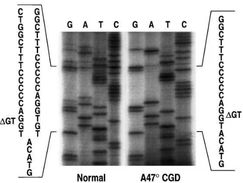
DNA from an additional 33 normals. Starting with the second GT of the GTGT tandem repeat at the beginning of exon 2, two different overlying sequences were observed in all of the normal individuals analyzed (Fig. 1). One was consistent with the published wild-type sequence containing the GTGT tan-dem repeat, while the other was identical to the sequence found in A478 CGD patients homozygous for the GT deletion. This result suggests that more than one p47-phox–related se-quence might be present in normal genomic DNA.
To further confirm this finding, restriction digestion was performed with Bsp1407I, which cuts the wild-type sequence AGGTGTACA but cannot digest the DGT sequence (AGG-TACA). No digestion was observed with amplified fragments containing exon 2 (using primers 2LB and 2RB) derived from ten A478 CGD patients homozygous for the GT deletion (Fig. 2 a). Bsp1407I digestion of the same fragment derived from a total of ten different normal subjects revealed three bands in all cases. One band was consistent with the undigested D GT-containing fragment, whereas the two smaller bands were con-sistent with the digestion products of the GTGT-containing fragment. This finding was confirmed by Bsp1407I digestion of the same amplification products derived from two genomic bacteriophage clones (to be further discussed below). One clone (L14) carried the GT deletion while the other clone (L24) carried the normal GTGT sequence. As expected, no di-gestion was observed in the fragment derived from the DGT clone L14, whereas the fragment from clone L24 carrying the GTGT sequence was completely digested into two fragments (Fig. 2 a).
Several explanations can be provided for this finding. First, all 34 normal individuals investigated here were carriers for A478 CGD. This is highly unlikely considering that the
[image:4.612.316.555.441.621.2]lence of this autosomal recessive disease is probably about 1:2,000,000 (22) with an expected carrier frequency of approxi-mately 1:700. Second, the GT deletion could have been consis-tently generated as an artifact by PCR amplification and/or the sequencing methods used. Third, a very similar gene (such as a pseudogene) carrying the GT deletion may be present in the normal population, and was therefore coamplified and cose-quenced in these experiments.
Identification and characterization of p47-phox genomic clones. To examine further the presence of the GT deletion in the normal population, two different types of human genomic libraries (EMBL3A bacteriophage and P1) were screened with
a p47-phox probe. Three bacteriophage clones and ten P1 clones were identified (Table I). Direct sequencing of exon 2 revealed that, of the three bacteriophage clones, two carried the GT deletion and one showed the normal GTGT sequence, thus giving a DGT:GTGT ratio of 2:1. In the ten P1 clones this ratio was 9:1. Since none of the clones showed both the GTGT and the DGT sequences, it appeared unlikely that the GT dele-tion was generated by PCR or sequencing artifacts. To confirm the presence of DGT by a method independent of PCR or se-quencing, however, restriction digestion of unamplified bacte-riophage clones was performed using Bsp1407I (Fig. 2 b). Whereas clone L24 contained four restriction fragments, only three fragments were observed in the clones L14 and L25, indi-cating that these two clones were lacking a restriction site com-pared to clone L24. This finding was consistent with the se-quencing data indicating that L14 and L25 carried the GT deletion, whereas L24 showed the wild-type GTGT sequence. Thus, the GT deletion can be demonstrated in normal genomic DNA clones by a method independent of PCR. As the GT de-letion predicts premature termination in the p47-phox protein, the most likely explanation for the large number of DGT clones obtained from the genomic DNA libraries is that there is at least one pseudogene. The identification of the DGT se-quence in all normal subjects investigated would also be con-sistent with the presence of a DGT containing pseudogene.
The high proportion of DGT-containing p47-phox clones identified suggested that more than one pseudogene might ex-ist in the genome. Using three radiolabeled primer pairs en-compassing the GTGT/DGT region of exon 2, quantitative PCR amplification was performed on genomic DNA from nor-mal individuals. The amplified DNA was subsequently di-gested with Bsp1407I to distinguish between the gene (GTGT) and the pseudogene (DGT). Quantitative analysis of the corre-sponding fragments suggested a ratio of pseudogene to gene of 2:1 (Table II).
[image:5.612.57.299.72.352.2]To characterize further the DGT containing p47-phox ge-nomic clones, we analyzed two of the bacteriophage clones (L14 and L24) and six of the P1 clones (P38-P43) in greater de-tail. The six clones carrying the GT deletion (L14, P38, P39, P40, P41, P43) were designated p47-phox pseudogene clones, whereas the two clones carrying the GTGT sequence (L24, P42) were considered to be wild-type clones. The size and
Figure 2. Restriction digestion with Bsp1407I. Restriction digestion was performed with Bsp1407I, which specifically cuts the wild-type sequence T/GTACA. (a) Genomic DNA from normal individuals (n5 10) and A478 CGD patients homozygous for the GT deletion (n5 10) was amplified with primers 2LB (CTTTCTGCAATCCAG-GACAA) and 2RB (ATCACCTGGGCTAAGGTCCT), and the amplification products (305 bp and 303 bp) were digested with Bsp1407I. Representative examples are shown here. The PCR prod-uct derived from a A478 patient was not digested, indicating that only sequence carrying the GT deletion was present. Bsp1407I digestion of PCR products derived from three normal individuals (N1–N3) vealed three DNA fragments: the fragments of 145 and 160 bp re-sulted from digestion of the wild-type product, and a 303-bp fragment corresponded to the uncut product. Bsp1407I digestion of the wild-type clone L24 showed complete digestion resulting in two fragments of 145 and 160 bp, whereas the fragment from the DGT clone L14 re-mained uncut. (b) Restriction digestion of unamplified bacteriophage clones L14 and L25 (pseudogene) as well as L24 (wild-type). The pseudogene clones were missing a restriction site, leaving the 12 kB fragment uncut, indicating that the GT deletion was not generated by a PCR or sequencing artifact.
Table I. Characterization of p47-phox Genomic Clones
Exon 2
Type of library No. of clones GTGT DGT
EMBL3Al 3 1 2
P1 10 1 9
YAC 19 4 15
S 32 6 26
A total of 32 p47-phox genomic clones were isolated from two normal
human genomic bacteriophage libraries (EMBL3Al and P1) as well as
a normal human YAC library by screening with either hybridization
us-ing a p47-phox cDNA probe (EMBL3Al) or with PCR amplification
using p47-phox primers (P1 and YAC). Sequence analysis of exon 2 of
[image:5.612.314.556.74.168.2]structure of the two wild-type and six pseudogene clones were determined by PCR amplification and sequence analysis using exonic primers. The wild-type clone P42 was found to be full-length containing 11 exons and 10 introns, consistent with the gene structure of the p47-phox gene (Chanock, et al., manu-script submitted for publication). The gene structures of the pseudogene clones P40, P41, and P43 were virtually identical to the wild-type clone in that they contained all 11 exons and ten introns (Fig. 3). All exon–intron borders followed the GT/ AG rule (data not shown) (31). Each of the remaining four clones (L14, L24, P38 and P39) contained sequence from exon 1 to exon 8.
Comparison of the exonic regions of the p47-phox gene and pseudogene clones. The exonic regions of the six pseudogene clones were amplified with intronic primers, sequenced, and compared to the corresponding regions of both wild-type clones as well as to genomic DNA from a series of healthy do-nors. A total of nine single base pair substitutions, in addition to the GT deletion in exon 2, were found in the exonic regions of the pseudogene clones (Table III). Single base pair changes in exons 4, 6, and 9 (nucleotide positions 269, 558, 825, and 861) segregated between wild-type and pseudogene clones; i.e., all wild-type clones differed from all pseudogene clones at these four positions. For example, the wild-type clones L24 and P42 showed a G at position 269 in concordance with the published sequence, whereas all six pseudogene clones carried an A at this position (Table III). This G→A substitution
pre-dicts an Arg90→His exchange. At position 558, the pseudo-gene clones showed a G compared to an A in the wild-type clones. Interestingly, the identity of the nucleotide at this posi-tion in cDNA has been ambiguous in the literature, as either G or A has been seen by different investigators (17, 18, 32). If translated, this G→A transition would remain silent, as valine would be incorporated in both cases. Two segregating differ-ences were identified in exon 9. Since clones L14, L24, P38, and P39 did not contain exons 9, 10, and 11 (see above), only one wild-type clone (P42) and three pseudogene clones (P40, P41, P43) could be analyzed in this region. At bp 825, a C was seen in all pseudogene clones compared to a T in the wild-type clone. At the cDNA level, this nucleotide position has also been questionable as both C and T have been reported (17, 18, 32). The C was considered to be a silent polymorphism (33). At bp 861, all pseudogene clones carried a T, while the wild-type clone contained a G. The published cDNA sequence shows a G at this position. Both nucleotide substitutions in exon 9 would also remain silent if translated.
[image:6.612.58.299.83.155.2]In contrast to the nucleotide differences just described that segregated neatly between wild-type and pseudogene clones, there were five nucleotide positions in which only a subset of the pseudogene clones differed in their sequence from the other clones and the published wild-type sequence (Table III). At bp 295, for example, a G was found in two pseudogene clones (P40 and P41), whereas all other wild-type and pseudogene clones had an A at this position as in the published sequence. Such a nucleotide substitution would predict a Ser99→Gly ex-change. A similar situation was found at bp 345. The pseu-dogene clones L14 and P43 contained a T at this position in-stead of a C, as seen in all of the other clones and in the published sequence. At bp 387, only one pseudogene clone (L14) showed an A for a G. The nucleotide at this position has also been questioned, as two reports described a G at this posi-tion in the normal cDNA (18, 32), whereas another study re-ported an A (17). Eventually, the G was considered to be a si-lent polymorphism (33). The pseudogene clone L14 also carried a G at bp 496, whereas all other clones showed an A. This nucleotide position remains ambiguous in the literature (17, 18, 32). In the corrected p47-phox consensus sequence (33), G was reported at this position. The A was considered to be a nonconservative polymorphism. If translated, the G would code for asparagine, whereas the A would code for as-partic acid. At bp 765, a C to A transversion was seen in five pseudogene clones, whereas one pseudogene clone (L14) and
[image:6.612.59.579.587.737.2]Figure 3. Structural organization of the human p47-phox gene. Exons are represented by boxes and num-bered from 1 to 11; the size of each exon is given in basepairs. The sizes of the ten introns are given in kB. The arrows at the bottom indicate the clones investigated. The phage clones L14 and L24 and the P1 clones P38 and P39 were only par-tial length comprising sequence from the 59 region to exon 8. The P1 clones P40 to P43 were full-length clones. Clones L24 and P42 ( under-lined) were wild-type; all other clones were pseudogene clones. Table II. Ratio of p47-phox Pseudogene and Wild-type
Gene Copies
Primer pair No. of DNA samples
No. of experiments
Ratio of pseudogene/wild-type
I-1 to I-2RA 5 2 2.260.5
I-1 to I-2R 5 4 2.160.3
I-1 to I-2RB 5 1 2.3
the wild-type clones carried the published sequence. When viewed together, these segregating and nonsegregating nucle-otide differences constitute a 0.5% to 0.75% divergence from the wild-type exonic sequence.
All ten of the exonic pseudogene differences (the DGT and the nine single base pair substitutions) were also seen in nor-mal genomic DNA (Table III). The pseudogene nucleotides were found to be superimposed on the wild-type sequence in the majority of genomic DNA samples, indicating that both the wild-type and pseudogene were coamplified and cose-quenced with the primers used. Finally, at two additional nu-cleotide positions (bp 849 and bp 936), all of the wild-type and pseudogene clones investigated contained the same sequence, but one that differed from the published cDNA sequence (33) (G [published]→A at bp 849, T→C at bp 936). Interestingly, at both positions, the sequences found in all of the clones have been reported as silent polymorphisms (33). In all of the nor-mal genomic DNA samples investigated (five and six, respec-tively), bands for both nucleotides were observed (data not shown).
Comparison of the intronic regions of the p47-phox gene and pseudogene. Since comparison of the wild-type and pseudo-gene clones in the exonic regions indicated a remarkable de-gree of identity, we examined the intronic regions to identify additional distinguishing differences. Of the z 13.5 kB of total
intronic region present in the p47-phox gene (and pseudo-gene), more than 80% was sequenced in at least one wild-type clone, and in three pseudogene clones.
Interestingly, the major differences between the wild-type gene and pseudogene clones were identified in the regions flanking exon 2. At a position 580 bp upstream from the 59 end of exon 2, a 30-bp duplication was found in all wild-type
clones. All pseudogene clones, however, had only a single copy of this sequence. This 30-bp segment contained several tan-dem repeats (TCCCCTCCCCTCTCCTCCTGTCCCCTCCCT), and was incorporated within a larger 90-bp sequence that had a high content of C (62 bp) and was flanked on each site by an Alu repeat (see below). Amplification of DNA with prim-ers Int1del (GTTTCACCATATTGGTCAGGCT) and 1RA (GTTGTCCTGGATTGCAGAAA) showed that a single 550-bp fragment was derived from the pseudogene clones, while two products were obtained from normal genomic DNA (n5 4) that were z 550 and 580-bp in length (data not shown).
All pseudogene clones tested also contained a 20-bp duplica-tion in intron 2 (CAGGGTCTTGCTCTGTCACC), begin-ning 176 bp downstream from the 39 end of exon 2; wild-type clones had only one copy of this sequence. Amplification of DNA with primers Int2.1 (GTTCCAGCTCCACCCTTTG-GAA) and Int2dup (CAAAACCACCTAAAAGGCCGA) revealed a 207-bp fragment from the wild-type clones, a 227-bp fragment from the pseudogene clones, and both forms from normal genomic DNA. The duplication was also seen as an overlying sequence in all 34 normal genomic DNA samples in-vestigated (data not shown).
[image:7.612.57.561.73.234.2]Within the 11.1 kb of intronic regions analyzed, a total of 33 segregating differences, (including the intron 1 deletion and intron 2 duplication described above), between the wild-type and pseudogene clones were observed (Table IV). Two small deletions of 2 and 3 bp were found segregating in intron 1 as well as a CG→TG exchange 123 bp upstream from the 59 end of exon 2. Both C and T were observed as overlying sequences in each of 34 normal genomic DNA samples analyzed. Only one difference was identified in intron 3, a segregating C→A exchange 64 bp downstream from the 39 end of exon 3. Again,
Table III. Exonic Differences Between p47-phox Wild-type and Pseudogene Clones
Nucleotide position
Wild-type clones Pseudogene clones Predicted AA‡ Normal gDNA§
Exon cDNA* L24 P42i L14 P38 P39 P40 P41 P43 Wt Ps No. wt 1 ps/No. total
2 73/74 GTGT1–4 GTGT GTGT DGT DGT DGT DGT DGT DGT V25 V 34/34
4 269 G1–4 G G A A A A A A R90 H 12/14
295 A1–4 A A A A A G G A S99 G 17/17
345 C1–4 C C T C C C C T L115 L 17/17
387 A1,2/G3,4 G G A G G G G G T129 T 4/4
6 496 G1,2,4/A3 A A G A A A A A N D166 5/7
558 G1,2,4/A3 A A G G G G G G V186 V 6/7
8 765 C1–4 C C C A A A A A V255 V 4/4
9 825 T1,2,4/C3 N/A T N/A N/A N/A C C C F275 F 6/6
861 G1–4 N/A G N/A N/A N/A T T T V287 V 2/2
Differences in the exonic regions between p47-phox gene and pseudogene clones. All eleven exons of the two p47-phox wild-type clones (L24 and
P42) and six pseudogene clones (L14, P38, P39, P40, P41, and P43) were sequenced. Clones L24, L14, P38, and P39 did not contain exons 9, 10, and 11 (N/A, not available). Sequence differences between wild-type and pseudogene clones were compared to the published cDNA sequences (third col-umn). Direct sequence analysis was also performed on normal genomic DNA samples (last colcol-umn). The data in this column indicate the number of samples found with both wild-type (wt) and pseudogene (ps) sequences compared to the total number of samples investigated at this nucleotide
posi-tion. The nucleotide numbering starts with the first nucleotide of the initiator methionine. *Published p47-phox cDNA sequence (see references 1–4
below in legend). ‡Predicted amino acid (AA) for wild-type sequence (Wt) and pseudogene sequence (Ps). §No. of genomic DNA samples with
wild-type and pseudogene sequence versus total number of genomic DNA samples. iThe nucleotide sequences of the following clones have been submitted
to the GenBank database: wild-type clone P42 (accession no. U57833-35); pseudogene clones L14 (U61224-25); P38 (U69639-43); P39 (U72356-61);
both sequences were observed in normal genomic DNA (n5
15). This intron, which is present in wild-type and pseudogene clones, is of some potential interest since it contains 99 bp lack-ing a termination codon. A predicted translation product would be in frame with the flanking exons. The total number of segregating and nonsegregating intronic differences re-vealed a range of divergence from the wild-type sequence be-tween 0.4 and 0.95% (Table V). The presence of nonsegregat-ing differences in both the exonic and intronic regions could be due to allelic polymorphisms because of the lack of selective pressure or, alternatively, could represent differences between the two pseudogenes.
Sequence analysis of the 59 upstream region. The high ho-mology between the p47-phox gene and its pseudogenes raised
the question as to whether the degree of conservation ex-tended into the promoter region, thereby suggesting that the pseudogenes may be transcriptionally active. Therefore, the 59
region of the wild-type clone L24 and the pseudogene clone L14 were amplified with an exon 1 antisense primer (Ex1-: TAGTGCTGGCTGGGTACGAAG), and the EMBL3A vec-tor primers GAGTCTTGCAGACAAACTGCGCAA (for amplification of L14) or CTCGTCCGAGAATAACGAG-TGGAT (for amplification of L24). Comparison of 874 bp of 59 upstream region revealed an insertion of 2 nucleotides (AA) at 2816 bp in the pseudogene clone. This difference is part of an A repeat flanking an Alu sequence that is located 820– 570 bp upstream from the initiator methionine, and was found to be 76% identical to the Alu consensus sequence II (34).
Expression of the pseudogene(s).The virtual identity of the 59 upstream region in the p47-phox gene and pseudogene clones suggested that the pseudogene(s) might be transcriptionally active. This was supported by the finding that many previously reported sequence discrepancies at the cDNA level were also observed in some or all of the genomic pseudogene clones.
Therefore, RNA from lymphocytes/monocytes (n5 7) or from EBV-transformed B-cell lines derived from healthy do-nors (n5 4) was reverse transcribed, the cDNA amplified with exonic primers (Ex11: CACCTTCATCCCGTCACATCGCC, Ex32: GATCCTGTTCTCTGGATTGA) and sequenced. At nucleotide position 75 (corresponding with the second GT of the tandem repeat at the beginning of exon 2) an overlying se-quence identical to the pseudogene sese-quence carrying the GT deletion was observed (Fig. 4 a). The presence of DGT in cDNA was also confirmed independently by restriction diges-tion of these RT-PCR products with DraIII, which digests only wild-type cDNA. As shown in Fig. 4 b, three bands of 207, 147, and 60 bp were observed in normal RT-PCR samples, whereas an amplification product derived from a p47-phox wild-type cDNA clone with known wild-type GTGT sequence had only two digestion fragments of 147 and 60 bp.
[image:8.612.58.555.73.245.2]Chromosomal location of the p47-phox gene and pseudo-genes. The p47-phox gene has been previously mapped to
Table IV. Intronic Differences Between Wild-type and Pseudogene p47-phox Clones
Segregating differences Nonsegregating differences
Intron Size % sequenced Total no. Dbp Insertions Deletions Total no. Dbp Insertions Deletions
1 3194 bp 100 14 10 1 3 26 25 1 0
2 1733 bp 100 4 3 1 0 27 27 0 0
3 99 bp 100 1 1 0 0 0 0 0 0
4 1359 bp 100 0 0 0 0 9 8 0 1
5 2 kB 50 2 2 0 0 12 12 0 0
6 462 bp 100 0 0 0 0 8 8 0 0
7 1549 bp 100 5 4 1 0 14 14 0 0
8 2.2 kB 40 5 5 0 0 8 8 0 0
9 471 bp 100 0 0 0 0 1 1 0 0
10 334 bp 100 2 2 0 0 0 0 0 0
S 13.4 kB 83 33 27 3 3 107 105 1 1
87% of the intronic regions were sequenced in at lease one wild-type and three pseudogene clones. In the second column to the left, the size of the in-tron indicated in column 1 is given. Inin-trons that have not been sequenced to completion are sized by their PCR products visualized on an agarose gel. The total number of segregating or nonsegregating differences (total no.) is given for each intron, as well as the number of single base pair changes (Dbp), insertions, and deletions.
Table V. Intronic Sequence Variations Between Genomic Bacteriophage Clones
No. base pair se-quenced
L24 P42 L14 P38 P39 P40 P41 P43
Clone 9341 11597 9341 5479 3230 11597 8801 11597
L24 9341 — 2 43 32 28 45 32 45
P42 11597 0.02 — 43 32 28 54 46 60
L14 9341 0.46 0.46 — 11 22 32 28 32
P38 5479 0.58 0.58 0.2 — 11 13 10 9
P39 3230 0.87 0.87 0.68 0.34 — 22 20 19 P40 11597 0.48 0.47 0.34 0.24 0.68 — 24 42 P41 8801 0.34 0.52 0.32 0.18 0.62 0.27 — 32 P43 11597 0.48 0.52 0.34 0.16 0.59 0.36 0.34 —
[image:8.612.57.297.525.664.2]chromosome 7q11.23 by Southern analysis of somatic cell hy-brid lines, and by chromosomal in situ hyhy-bridization (35). To determine the chromosomal location of the pseudogenes, two approaches were used. First, DNA from a panel of human– hamster hybrid cell lines, each containing different human chromosomes, was amplified with two sets of primers: STSB, which generates a fragment containing exon 11 and the flank-ing 39 end, and STSC, which amplifies exon 2 and the flanking intronic regions. This latter primer set would be able to am-plify the region of the pseudogene, which contains the GT de-letion and the 20-bp duplication. Appropriate PCR products were generated only from cell lines containing human chromo-some 7, including two cell lines with chromochromo-some 7 as their only human DNA. In hamster cell lines or cell lines that did not contain chromosome 7, no amplification could be ob-served, suggesting that only chromosome 7 contains p47-phox
and p47-phox–related genes (data not shown).
In a second approach to determine the chromosomal loca-tion of the p47-phox pseudogenes, a YAC library highly en-riched for chromosome 7 was screened using the same PCR as-says (STSB and STSC) as described above. With this method, 19 p47-phox–positive YACs were identified. Sequence analy-sis of the fragment amplified with the primer set STSC, which contains exon 2 and flanking intronic regions, showed that 15 clones carried the GT deletion at the beginning of exon 2 and the 20-bp duplication in intron 2. The four remaining clones had the GTGT sequence at the beginning of exon 2. Since all 19 YACs have been localized to a single contig that maps to chromosomal band 7q11.23 (Green, E.D., unpublished data), the p47-phox wild-type and pseudogenes very likely colocalize to the same chromosomal region within 7q11.23.
Repetitive sequence elements and recombination hot spots.
Sequence analysis of the p47-phox gene and pseudogene clones revealed a high number of repetitive sequence ele-ments. Within the approximately 11.1 kB of intronic sequence obtained from wild-type and pseudogene clones, 17 complete
Alu repeats and one Alu half site were found that had at least 70% identity with the Alu consensus sequence II (34) (Fig. 5). Most of the Alu motifs were flanked by short repeats. Intron 1 contained a cluster of six Alu sequences accounting for more than 50% of the total sequence of this intron. This intron also contained a large number of direct repeats. The entire intron (3194 bp) contained 94 repeats in a range between 10 and 42 bp, comprising a total of 1250 bp. Thus, more than 75% of intron 1 was represented by nonunique sequence. The 20-bp duplicated sequence in intron 2 characteristic for the pseudogene (de-scribed above) was repeated one other time in the same intron 600 bp further downstream in all of the wild-type and pseudo-gene clones analyzed. This intron (1733 bp) also contained 29 other repeats that ranged between 10 and 37 bp, and ac-counted for more than 400 bp. Of the remaining introns, only intron 10 showed a substantial number of repeated sequences that represented z 45% of this intron. The largest segregating
differences between wild-type and pseudogene clones in-volved repeated sequence elements.
[image:9.612.58.445.66.249.2]The high number of sequence repeats suggested that the p47-phox gene might be susceptible to recombination events. We therefore searched for sequence motifs of potential recom-bination hot spots such as the Chi sequence (59GCTGGTGG) (36) and the human minisatellite repeat (HMR) (59 GGG-CAGGAXG) (37). These motifs have been associated with re-combination events between homologous genes. Three se-quences that were identical to the Chi motif were found in intron 1 and exons 8 and 10 (the exon 10 sequence was in the antisense direction) (Fig. 5). Similarly, one sequence identical to the HMR sequence was identified in intron 10. There were an additional 12 Chi and 17 HMR motifs in the p47-phox gene that differed from the consensus sequences by only a single nu-cleotide. When viewed together, these potential recombina-tion hot spots formed clusters located in introns 2, 4, and 7 (Fig. 5). Only introns 3 and 6 did not contain consensus motifs to either the Chi or HMR sequence.
Discussion
The original sequence analyses of the p47-phox gene revealed a considerable number of discrepancies between different cDNA clones and an apparently high number of polymor-phisms (17, 18, 32, 33). Furthermore, mutation analysis of A478
CGD patients has shown a prevalence of a single mutation, a GT deletion at the beginning of exon 2 (23–25). In a recent evaluation of our A478 CGD patients, we found 20 out of 23 patients homozygous for this GT deletion (38). The prevalence of a single mutation in an unrelated, racially diverse popula-tion is in sharp contrast to the situapopula-tion in the other three forms of CGD, where various family-specific mutations are found (for review see references 2 and 21). In this study, we show that the GT deletion is not only characteristic of A478
CGD, but is present in normal genomic DNA as well. Our data strongly suggest the presence of at least one pseudogene carrying the GT deletion found in A478 CGD patients. First, in all normal individuals investigated, the GT deletion could be identified at the genomic level, appearing as a superimposed sequence on that of the wild-type sequence. Second, screening of three different types of genomic libraries for p47-phox re-vealed (based on the size of each library and the correspond-ing coverage provided) an approximately two to three times higher number of positive clones than one would expect for a single copy gene. Third, only two bacteriophage clones and four YACs showed the wild-type sequence in exon 2, whereas eleven bacteriophage clones and 15 YAC clones carried the GT deletion at the beginning of exon 2. Sequence analysis of the clones demonstrated that the GT deletion was not intro-duced artificially by PCR or the sequencing methods used. The data strongly support the existence of at least one p47-phox
pseudogene which carries the GT deletion in exon 2.
Characterization of two wild-type clones and six pseudo-gene clones showed a remarkable degree of identity between the p47-phox wild-type and pseudogene(s). We were able to demonstrate that the pseudogene clones analyzed in this study had the same gene structure as the p47-phox wild-type gene (Chanock et al., manuscript submitted for publication). In the exonic regions, four sequence differences (in addition to the GT deletion) were identified in all pseudogene clones, whereas at five other nucleotide positions, only a subset of pseudogene clones showed sequence differences. The divergence in the ex-onic regions between the wild-type clones and individual pseudogene clones ranged from 0.5 to 0.75%. Interestingly, all five of the nucleotide substitutions that varied among pseudo-gene clones were also observed as superimposed sequences in the majority of normal genomic DNA samples. This suggests that these differences represent either polymorphisms within a single pseudogene or the presence of more than one pseudo-gene. The latter would be consistent with the ratio of 26:6
pseudogene to wild-type clones that we observed in three dif-ferent types of genomic libraries. Preliminary results using quantitative PCR suggest that there are two p47-phox pseudo-genes present in the genome. Since PCR amplification is sub-ject to technical artefacts, including the possibility that not all the pseudogenes may be amplified by the selected primer pairs, stretch FISH analysis is being performed to determine the actual number of p47-phox genes in the genome and to es-timate the physical distance between them.
Whereas several of the exonic sequence differences seen in the pseudogene clones have been reported previously at the cDNA level (17, 18, 32, 33), the polymorphisms described at bp 555 (32) and at bp 621 (33) were not observed in either wild-type or pseudogene clones, suggesting the presence of true polymorphic sites in the wild-type sequence. Alterna-tively, it is possible that these nucleotide substitutions may be present on pseudogene isoforms that have not yet been identi-fied and sequenced.
Our data show that the pseudogene(s) are transcribed in normal individuals. The pseudogene cDNA would predict a translation product of 50 amino acids, of which only 20 amino acids would be identical to the corresponding region of the normal p47-phox protein. Whereas truncated proteins derived from the translation of pseudogenes have been observed (e.g., the leukocyte interferon pseudogenes [39]), the presence of a p47-phox pseudogene polypeptide has not been demonstrated. It is possible that such a truncated protein is unstable and is de-graded rapidly.
Similar to the findings with the exonic sequences, the in-tronic regions of the pseudogene clones were strikingly con-served in relation to the wild-type gene. Of the 83% of intronic sequence analyzed, only 33 segregating and 107 nonsegregat-ing differences between wild-type and pseudogene(s) were identified, indicating a divergence of just 0.4% to 0.9%. The most striking differences were found in the flanking regions of exon 2: a 30-bp deletion in intron 1 and a 20-bp duplication in intron 2. Together with the GT deletion, these two differences serve as useful markers for the pseudogenes. The overall de-gree of divergence (exons plus introns) from the wild-type clones was found to be in the range 0.4% to 0.8%, thus render-ing the p47-phox pseudogenes among the most conserved un-processed pseudogenes known. Examples of other highly ho-mologous unprocessed pseudogenes are the 21-hydroxylase pseudogene (CYP21A) (98% homology in exons and 96% ho-mology in introns) (40), the von Willebrand factor pseudogene (96.9% overall homology) (41) and the b-glucocerebrosidase pseudogene (96% overall homology) (42).
[image:10.612.66.524.61.114.2]The high degree of homology between the p47-phox gene and its pseudogenes suggests that the pseudogenes arose from gene duplication, as has been described for other unprocessed pseudogenes such as the 21-hydroxylase pseudogene (40) and
the a-globin gene (43). The p47-phox gene maps to chromo-some 7q11.23 (35), and using cell-hybrid studies we have also localized the pseudogene to chromosome 7. Furthermore, all YACs identified as being positive for either the wild-type or the pseudogenes map to chromosomal band 7q11.23, indicat-ing that both are confined to this region of chromosome 7. Interestingly, the q11.23 band of chromosome 7 appears to contain large block(s) of duplicated (i.e., nonunique) DNA segments (E.D. Green, unpublished observation).
The high level of sequence homology between p47-phox
wild-type and pseudogenes together with the colocalization of these genes to the same chromosomal band suggests that these genes might be susceptible to recombination events such as gene conversion or crossing over. Whereas crossing over leads to reciprocal exchange of DNA, gene conversion was origi-nally defined as nonreciprocal exchange of homologous ge-netic information (44). As the products of crossing over and gene conversion events are virtually indistinguishable at the DNA level in higher eukaryotes, however, such events are commonly referred to as gene conversions (45). Gene conver-sion events between homologous genes and their pseudogenes have been described in the pathogenesis of several genetic dis-orders such as 21-hydroxylase deficiency (40), von Willebrand disease (46), and Gaucher disease (42), and have also been postulated for many clustered gene families (47). The presence of at least one p47-phox pseudogene carrying the GT deletion suggests that such events might also be responsible for the transfer of the GT deletion from the p47-phox pseudogene to the wild-type gene, thus leading to A478 CGD.
Gene conversion events most frequently occur between stretches of sequence identity (48, 49). In addition, specific se-quence features such as recombination hot spots and repetitive elements (for example, Alu sequences) may facilitate such re-combination events (50). It has been clearly demonstrated in
Escherichia coli that gene conversion frequently occurs at re-combination hot spots such as the Chi sequence. There is now growing evidence that this sequence might be a recombination hot spot in mammals as well (36). In addition, the HMR has been described as another hot spot for recombination in hu-mans (37). The high incidence of recombination events be-tween genes and pseudogenes for 21-hydroxylase or for von Willebrand factor has been attributed to the presence of Chi and HMR sequences in these genes (46, 51, 52). The presence of three Chi consensus sequences, one of them in intron 1, and one HMR consensus motif, might also be related to recombi-nation events between p47-phox wild-type and pseudogene. In addition, 12 modified Chi and 17 modified HMR motifs con-taining one mismatch were identified, with six of them clus-tered in intron 2. It has been reported that sequences similar but not identical to the Chi consensus also stimulate recombi-nation (53). Thus, the high number of potential recombirecombi-nation hot spots might facilitate recombination events between the p47-phox gene and pseudogene.
Furthermore, an unusually high density of Alu repeats is present in the p47-phox gene. Since the average distance be-tween these elements in the human genome is about 4 kB (34), the identification of 17 complete Alu sequences within the 11.1 kB of intronic sequence analyzed indicates that these repetitive elements are overrepresented in the p47-phox gene. Within 5 kB of sequence in introns 1 and 2, nine Alu repeats were identified. The presence of repetitive sequence elements such as Alu repeats has been associated with
recombination-related mutational events in the LDL receptor gene (50) and the complement component C1 inhibitor gene (54). In human growth hormone genes it has been suggested that Alu repeats define breakpoints of gene duplication (55). It has also been proposed that gene conversion is promoted by palindromic se-quences, as they have been observed near recombination breakpoints (56). The GTGT tandem repeat is located within a palindromic sequence, suggesting that the immediate flanking region might be susceptible to breakage and recombination.
For the fetal g-globin and the 21-hydroxylase genes and pseudogenes it has been suggested that regions containing clusters of differences between wild-type and pseudogene se-quences might represent recombination hot spots (57, 58). A large number of sequence differences between p47-phox wild-type and pseudogene is found in introns 1 and 2, among them the 30-bp deletion in intron 1 and the 20-bp duplication in in-tron 2. This concentration of sequence differences might con-stitute similar recombination hot spots in the introns flanking the GT deletion, and might be responsible in part for the pre-dominance of the GT deletion in A478 CGD.
The prevalence of a single mutation in causing p47-phox
deficiency may also occur because the GT deletion is the only deleterious mutation in the pseudogene, in that it leads to a premature termination of p47-phox translation. In contrast, the 21-hydroxylase and von Willebrand factor pseudogenes contain a substantial number of potentially deleterious muta-tions (41, 58). In the 21-hydroxylase pseudogene, for example, an 8-bp deletion, a single-bp insertion and a nonsense muta-tion (as well as six addimuta-tional mutamuta-tions) notably cause loss of the function of the wild-type protein when transferred to the wild-type gene. Each of these deleterious mutations accounts for a proportion of 21-hydroxylase-deficient patients (40). Out of the nine single base pair substitutions identified in the cod-ing region of the p47-phox pseudogene, however, only three (R→H, S→G, D→N, Table III) predict amino acid changes. Whether these changes would alter the function of p47-phox is not known.
Another possible mechanism explaining the high incidence of the GT deletion in A478CGD patients would be deletion of the wild-type gene, particularly in the promoter region, leaving only the pseudogene intact for transcription. Such a gene dele-tion in the wild-type promoter region might have occurred in the few A478 CGD patients who are heterozygous for the GT deletion, and do not carry other apparent mutations in the coding region of the p47-phox gene.
Taken together, the high degree of homology between the p47-phox gene and pseudogenes, the presence of many con-sensus motifs likely to be recombination hot spots, and the close chromosomal localization of gene and pseudogenes sug-gest the involvement of recombination events, such as gene conversion, between the p47-phox wild-type gene and D GT-containing pseudogenes in the pathomechanism of A478 CGD. Further studies evaluating genomic DNA from A478 CGD pa-tients and their families are currently underway and should, with the help of the sequence analysis of the p47-phox gene and its pseudogene provided here, further elucidate the patho-genesis of A478 CGD.
Acknowledgments
Insti-tute for extensive help with primer syntheses and sequence analyses, Drs. Andrew Cross and Paul Heyworth for helpful suggestions and reading the manuscript, and Mrs. Valerie Moreau for help with edit-ing the manuscript.
This study was supported by National Institutes of Health grants RO1 AI24838 (J.T. Curnutte) and RR00833 (Scripps General Clini-cal Research Center), the Stein Endowment Fund, an Otto-Hahn-fel-lowship from the Max-Planck Society, Germany (A. Görlach), and a grant from the Deutsche Forschungsgemeinschaft (J. Roesler).
References
1. Babior, B.M., R.S. Kipnes, and J.T. Curnutte. 1973. Biological defense mechanisms: the production by leukocytes of superoxide, a potential bacteri-cidal agent. J. Clin. Invest. 52:741–744.
2. Curnutte, J.T. 1993. Chronic granulomatous disease: the solving of a clin-ical riddle at the molecular level. Clin. Immunol. Immunopathol. 67:2s–15s.
3. Segal, A.W., I. West, F. Wientjes, J.H.A. Nugent, A.J. Chavan, B. Haley, R.C. Garcia, H. Rosen, and G. Scrace. 1992. Cytochrome b-245 is a
flavocyto-chrome containing FAD and the NADPH-binding site of the microbicidal oxi-dase of phagocytes. Biochem. J. 284:781–788.
4. Rotrosen, D., C.L. Yeung, T.L. Leto, H.L. Malech, and C.H. Kwong. 1992. Cytochrome b558: the flavin-binding component of the phagocyte
NADPH oxidase. Science (Wash. DC). 256:1459–1462.
5. Doussiere, J., G. Brandolin, V. Derrien, and P.V. Vignais. 1993. Critical assessment of the presence of an NADPH binding site on neutrophil cyto-chrome b558 by photoaffinity and immunochemical labeling. Biochemistry. 32:
8880–8887.
6. Clark, R.A., B.D. Volpp, K.G. Leidal, and W.M. Nauseef. 1990. Two cy-tosolic components of the human neutrophil respiratory burst oxidase translo-cate to the plasma membrane during cell activation. J. Clin. Invest. 85:714–721.
7. Heyworth, P.G., J.T. Curnutte, W.M. Nauseef, B.D. Volpp, D.W. Pear-son, H. Rosen, and R.A. Clark. 1991. Neutrophil nicotinamide adenine dinucle-otide phosphate (reduced form) oxidase assembly: translocation of p47-phox
and p67-phox requires interaction between p47-phox and cytochrome b558. J.
Clin. Invest. 87:352–356.
8. Cross, A.R., and J.T. Curnutte. 1995. The cytosolic activating factors
p47-phox and p67-phox have distinct roles in the regulation of electron flow in NADPH oxidase. J. Biol. Chem. 270:6543–6548.
9. Leto, T.L., A.G. Adams, and I. De Mendez. 1994. Assembly of the phago-cyte NADPH oxidase: binding of src homology 3 domains to proline-rich tar-gets. Proc. Natl. Acad. Sci. USA. 91:10650–10654.
10. McPhail, L.C. 1994. SH3-dependent assembly of the phagocyte NADPH oxidase. J. Exp. Med. 180:2011–2015.
11. Knaus, U.G., P.G. Heyworth, T. Evans, J.T. Curnutte, and G.M. Bokoch. 1991. Rac 2 is a regulator of phagocyte oxygen radical production.
Sci-ence (Wash. DC). 254:1512–1515.
12. Abo, A., E. Pick, A. Hall, N. Totty, C. Teahan, and A.W. Segal. 1991. Activation of the NADPH oxidase involves the small GTP-binding protein p21rac1. Nature (Lond.). 353:668–670.
13. Wientjes, F.B., J.J. Hsuan, N.F. Totty, and A.W. Segal. 1993. p40-phox, a third cytosolic component of the activation complex of the NADPH oxidase to contain src homology 3 domains. 1993. Biochem. J. 296:557–611.
14. Tsunawaki, S., H. Miyzunari, M. Nagata, O. Tatsuzawa, and T. Kurat-suji. 1994. A novel cytosolic component, p40-phox, of respiratory burst oxidase associates with p67-phox and is absent in patients with chronic granulomatous disease. Biochem. Biophys. Res. Commun. 199:1378–1382.
15. Parkos C.A., M.C. Dinauer, L.E. Walker, R.A. Allen, A.J. Jesaitis, and S.H. Orkin. 1988. Primary structure and unique expression of the 22 kilodalton light chain of human neutrophil cytochrome b. Proc. Natl. Acad. Sci. USA. 85: 3319–3323.
16. Dinauer, M.C., E.A. Pierce, G.A.P. Bruns, J.T. Curnutte, and S.H. Or-kin. 1990. Human neutrophil cytochrome-b light chain (p22-phox): gene struc-ture, chromosomal location, and mutations in cytochrome negative autosomal recessive chronic granulomatous disease. J. Clin. Invest. 86:1729–1737.
17. Volpp, B.D., W.N. Nauseef, J.E. Donelson, D.R. Moser, and R.A. Clark. 1989. Cloning of the cDNA and functional expression of the 47-kilodal-ton cytosolic component of human neutrophil respiratory burst oxidase. Proc.
Natl. Acad. Sci. USA. 86:7195–7199.
18. Lomax K.J., T.L. Leto, H. Nunoi, J.I. Gallin, and H.L. Malech. 1989. Recombinant 47-kilodalton cytosol factor restores NADPH oxidase in chronic granulomatous disease. Science (Wash. DC). 245:409–412.
19. Leto, T.L., K.J. Lomax, B.D. Volpp, H. Nunoi, J.M.G. Sechler, W.M. Nauseef, R.A. Clark, J.I. Gallin, and H.L. Malech. 1990. Cloning of a 67-kD neutrophil oxidase factor with similarity to a non-catalytic region of p60c-src.
Science (Wash. DC). 248:727–730.
20. Royer-Pokora, B., L.M. Kunkel, A.P. Monaco, S.C Goff, P.E. New-burger, R.L. Baehner, F.S. Cole, J.T. Curnutte, and S.H. Orkin. 1986. Cloning the gene for an inherited human disorder—chronic granulomatous disease—on
the basis of its chromosomal location. Nature (Lond.). 322:32–38.
21. Roos, D., M. de Boer, F. Kuribayashi, C. Meischl, R.S. Weening, A.W. Segal, A. Ahlin, K. Nemet, J.P. Hossle, E. Bernatowska-Matuszkiewicz, and H. Middleton-Price. 1996. Mutations in the X-linked and autosomal recessive forms of chronic granulomatous disease. Blood. 87:1663–1681.
22. Thrasher, A.J., N.H. Keep, F. Wientjes, and A.W. Segal. 1994. Chronic granulomatous disease. Biochem. Biophys. Acta. 1227:1–24.
23. Casimir, C.M., H.N. Bu-Ghanim, A.R.F. Rodaway, D.L. Bentley, P. Rowe, and A.W. Segal. 1991. Autosomal recessive chronic granulomatous dis-ease caused by deletion at a dinucleotide repeat. Proc. Natl. Acad. Sci. USA. 88: 2753–2757.
24. Volpp, B.D., and Y. Lin. 1993. In vitro reconstitution of the respiratory burst in B lymphoblasts from p47-phox–deficient chronic granulomatous dis-ease. J. Clin. Invest. 91:210–207.
25. Iwata, M., H. Nunoi, H. Yamazaki, T. Nakano, H. Niwa, S. Tsuruta, S. Ohga, S. Ohmi, S. Kanegasaki, and I. Matsuda. 1994. Homologous dinucleotide (GT or TG) deletion in Japanese patients with chronic granulomatous disease with p47-phox deficiency. Biochem. Biophys. Res. Comm. 199:1372–1377.
26. Safaya, S., and R.F. Rieder.1988. Dysfunctional alpha-globin gene in he-moglobin H disease in blacks. A dinucleotide deletion produces a frameshift and a termination codon. J. Biol. Chem. 263:4328–4332.
27. Chan, E.K.L., S. Takano, L.E.C. Andrade, J.C. Hamel, and A.G. Ma-tera. 1994. Structure, expression, and chromosomal localization of human p80-coilin gene. Nucleic Acids Res. 22:4462–4469.
28. Green, E.D., V.V. Braden, R.S. Fulton, R. Lim, M.S. Ueltzen, D.C. Pe-luso, R.M. Mohr-Tidwell, J.R. Idol, L.M. Smith, I. Chumakov, et al. 1995. A hu-man chromosome 7 yeast artificial chromosome (YAC) resource: construction, characterization and screening. Genomics. 25:170–183.
29. Devereux, J., P. Haeberli, and O. Smithies. 1984. A comprehensive set of sequence analysis programs for the VAX. Nucleic Acids Res. 12:387–395.
30. Pearson, W.R., and D. Lipman. 1988. Improved tools for biological se-quence comparison. Proc. Natl.Acad. Sci. USA. 85:2444–2448.
31. Breathnach, R., and P. Chambon. 1981. Organization and expression of eukaryotic split genes coding for proteins. Annu. Rev. Biochem. 50:349–383.
32. Rodaway, A.R.F., C.G. Teahan, C.M. Casimir, A.W. Segal, and D.L. Bentley. 1990. Characterization of the 47-kilodalton autosomal chronic granu-lomatous disease protein: tissue-specific expression and transcriptional control by retinoic acid. Mol. Cell. Biol. 10:5388–5396.
33. Volpp, B.D., W.N. Nauseef, J.E. Donelson, D.R. Moser, and R.A. Clark. 1989. Cloning of the cDNA and functional expression of the 47-kilodal-ton cytosolic component of human neutrophil respiratory burst oxidase. (cor-rection). Proc. Natl. Acad. Sci. USA. 86:9563.
34. Britten, R.J., W.F. Baron, D.B. Stout, and E.H. Davidson. 1988. Sources and evolution of human Alu repeated sequences. Proc. Natl. Acad. Sci. USA.
85:4770–4774.
35. Francke, U., C.-L. Hsieh, B.E. Foellmer, K.J. Lomax, H.L. Malech, and T.L. Leto. 1990. Genes for two autosomal recessive forms of chronic granulo-matous disease assigned to 1q25 (NCF2) and 7q11.23 (NCF1). Am. J. Hum.
Genet. 47:483–492.
36. Smith, G.R. 1994. Hotspots of homologous recombination. 1994.
Expe-rientia (Basel). 50:234–241.
37. Jeffreys, A.J., V. Wilson, and S.L. Thein. 1985. Hypervariable minisatel-lite regions in human DNA. Nature (Lond.). 314:67–73.
38. Roesler, J., A. Görlach, J. Rae, P.J. Hopkins, P. Patino, P. Lee, J.T. Cur-nutte, and S.J. Chanock. 1995. Recombination events between the normal
p47-phox gene and a highly homologous pseudogene are the main cause of auto-somal recessive chronic granulomatous disease (CGD). Blood. 86:260a (Abstr.). 39. Goeddel, D.V., D.W. Leung, T.J. Dull, M. Gross, R.M. Lawn, R. Mc-Candliss, P.H. Seeburg, A. Ullrich, E. Yelverton, and P.W. Gray. 1981. The structure of eight distinct cloned human leukocyte interferon cDNAs. Nature
(Lond.). 290:20–26.
40. Helmberg, A. 1993. Twin genes and endocrine disease: CYP21 and CYP11B genes. Acta Endocrinol. 129:97–108.
41. Mancuso, D.J., E.A. Tuley, L.A. Westfield, T.L. Lester-Mancuso, M.M. Le Beau, J.M. Sorace, and J.E. Sadler. 1991. Human von Willebrand Factor gene and pseudogene: structural analysis and differentiation by polymerase chain reaction. Biochemistry. 30:253–269.
42. Horowitz, M., S. Wilder, Z. Horowitz, O. Reiner, T. Gelbart, and E. Beutler. 1989. The human glucocerebrosidase gene and pseudogene: structure and evolution. Genomics. 4:87–96.
43. Wilde, C.D. 1986. Pseudogenes. Crit. Rev. Biochem. 19:323–352. 44. Kourilsky, P. 1986. Molecular mechanisms for gene conversion in higher cells. Trends Genet. 2:60–63.
45. Maeda N., and O. Smithies. 1986. The evolution of multigene families: human haptoglobin genes. Ann. Rev. Genet. 20:81–108.
46. Eikenboom, J.C.J., T. Vink, E. Briet, J.J. Sixma, and P.H. Reitsma. 1994. Multiple substitutions in the von Willebrand factor gene that mimic the pseudogene sequence. Proc. Natl. Acad. Sci. USA. 91:2221–2224.
47. Collier, S., M. Tassabehij, and T. Strachnan. 1993. A de novo pathologi-cal point mutation at the 21-hydroxylase locus: implications for gene conversion in the human genome. Nat. Genet. 3:260–265.
Homology requirements for unequal crossing over in humans. Genetics. 128: 143–161.
49. Liskay, R.M., A. Letsou, and J.L. Stachelek. 1987. Homology require-ment for efficient gene conversion between duplicated chromosomal segrequire-ments in mammalian cells. Genetics. 115:161–167.
50. Lehrman, M.A., D.W. Russell, J.L. Goldstein, and M.S. Brown. 1987. Alu-Alu recombination deletes splice acceptor cites and produces secreted low density lipoprotein receptor in a subject with familial hypercholesterolemia. J.
Biol. Chem. 262:3354–3361.
51. Amor, M., K.L. Parker, H. Globermann, M.I. New, and P.C. White. 1988. Mutation in the CYB21B gene (Ile-172→Asn) causes steroid 21-hydroxy-lase deficiency. Proc. Natl. Acad. Sci. USA. 85:1600–1604.
52. Urabe, K., A. Kimura, F. Harada, T. Iwanaga, and T. Sasazuki. 1990. Gene conversion in steroid 21-hydroxylase genes. Am. J. Hum. Genet. 46:1178– 1186.
53. Cheng, K.C., and G.R. Smith. 1987. Cutting of Chi-like sequences by the RecBCD enzyme of Escherichia coli. J. Mol. Biol. 194:747–750.
54. Stoppa-Lyonnet, D, P.E. Carter, T. Meo, and M. Tosi. 1990. Clusters of intragenic Alu repeats predispose the human C1 inhibitor locus to deleterious rearrangements. Proc. Natl. Acad. Sci. USA. 87:1551–1555.
55. Hirt, H., J. Kimelman, M.J. Birnbaum, E.Y. Chen, P.H. Seeburg, N.L. Eberhardt, and A. Barta. 1987. The human growth hormone locus: structure, evolution, and allelic variations. DNA (New York). 6:59–70.
56. Krawinkel, U., G. Zoebelein, and A.L.M. Bothwell. 1986. Palindromic sequences are associated with sites of DNA breakage during gene conversion.
Nucl. Acids Res. 4:3871–3882.
57. Slightom, J.L., A.E. Blechl, and O. Smithies. 1980. Human fetal G- and A-globin genes: complete nucleotide sequences suggest that DNA can be ex-changed between these duplicated genes. Cell. 21:627–638.
58. Higashi Y., H. Yoshioka, M. Yamane, O. Gotoh, and Y. Fuji-Kuriyama. 1986. Complete nucleotide sequence of two steroid 21-hydroxylase genes tan-demly arranged in human chromosome: a pseudogene and a genuine gene.