Development 140, 1751-1761 (2013) doi:10.1242/dev.093641 © 2013. Published by The Company of Biologists Ltd
INTRODUCTION
Peritubular myoid cells (PMCs) are a group of mesenchymal cells with a very flat morphology that express specific markers of both fibroblasts and smooth muscle cells (Holstein et al., 1996). These cells form the outer border of the seminiferous tubules and, in conjunction with Sertoli cells, are responsible for tubular contractility and sperm transport. The abundant expression of smooth muscle cell cytoskeletal proteins, including desmin, smooth muscle-specific myosin, and smooth muscle actin, facilitates the contractile ability of PMCs (Maekawa et al., 1996). One of the main functions of PMCs is the propulsion of testicular fluid containing spermatozoa towards the rete testis (Maekawa et al., 1996). The contraction of PMCs is tightly controlled by endothelin 1, Tgfβ, angiotensin II, and other hormones in endocrine, paracrine and autocrine fashion (Rossi et al., 2002; Santiemma et al., 2001; Tung and Fritz, 1991).
PMCs not only serve as target structural cells of multiple hormones, but also secrete a number of substances to modulate the testicular microenvironment. By secretion of extracellular matrix (ECM) proteins, such as laminin, fibronectin, type I and IV collagens, and proteoglycans, PMCs form the basement membrane. which is implicated in the regulation of spermatogonial stem cells (SSCs) (Richardson et al., 1995; Shinohara et al., 1999). As mesenchymal cells, PMCs synthesize several secretory factors,
among which PModS (peritubular factors that modulate Sertoli cell function) have been reported to modulate the secretion of transferrin, inhibin and androgen-binding proteins by Sertoli cells (Verhoeven et al., 2000). Other PMC-derived factors, such as bFGF (Fgf2), Igf1 and several cytokines, share most of the PModS effects on Sertoli cells, suggesting the importance of paracrine regulation of Sertoli cells by PMCs.
These clues have suggested that PMCs are important for the regulation of male fertility, but little is known about the particular genes or pathways in PMCs that modulate spermatogenesis in genetic mouse models. It has been shown that specific deletion of androgen receptor (Ar) in PMCs impairs Sertoli cell function and results in azoospermia and male infertility, suggesting that androgen signaling in PMCs plays a pivotal role in the modulation of spermatogenesis (Welsh et al., 2009; Zhang et al., 2006). However, the other signaling pathways that regulate Ar in PMCs to modulate male fertility remain obscure.
Lgr4 (also known as Gpr48), is a former orphan receptor involved in various physiological functions. Our laboratory and others reported that loss of Lgr4results in reduced viability (Mazerbourg et al., 2004) and in developmental defects in multiple organs, including the kidney (Kato et al., 2006; Wang et al., 2011), eye (Weng et al., 2008), bone (Luo et al., 2009), blood (Song et al., 2008), male reproductive tract (Li et al., 2010; Mendive et al., 2006) and intestine (de Lau et al., 2011; Mustata et al., 2011). Recently, three groups independently reported that R-spondins, which are secreted Wnt signaling agonists, are the endogenous ligands for Lgr4, Lgr5 and Lgr6 to activate Wnt/β-catenin and Wnt/planar cell polarity (PCP) signaling (Carmon et al., 2011; de Lau et al., 2011; Glinka et al., 2011). Wnts are a family of secreted morphogens involved in the regulation of a variety of developmental processes (Clevers, 2006). It is reported that Wnt4 is required for the initial stages of Sertoli cell differentiation and maturation (Jeays-Ward et al., 2004), whereas Wnt5a regulates SSC self-renewal through β -catenin-independent mechanisms (Yeh et al., 2011). With a genetic mouse model expressing constitutively activated β-catenin in Sertoli cells, two groups demonstrated that hyperactivation of Wnt/β -1Shanghai Key Laboratory of Regulatory Biology, Institute of Biomedical Sciences and
School of Life Sciences, East China Normal University, Shanghai 200241, China. 2Biological Targeting Diagnosis and Therapy Research Center, Guangxi Medical University, 22 Shuang Yong Road, Nanning, Guangxi 530021, China. 3Shanghai Clinical Center for Endocrine and Metabolic Diseases, Shanghai Key Laboratory of Endocrine Tumor, Shanghai Institute of Endocrinology and Metabolism, RuiJin Hospital, Shanghai Jiao Tong University School of Medicine, 197 RuiJin 2nd Road, Shanghai 200025, China. 4The Institute of Biosciences and Technology, Texas A&M University Health Science Center, Houston, TX 77030, USA.
*These authors contributed equally to this work
‡Authors for correspondence (myliu@bio.ecnu.edu.cn; dlli@bio.ecnu.edu.cn)
Accepted 7 February 2013
SUMMARY
Peritubular myoid cells (PMCs) are myofibroblast-like cells that surround the seminiferous tubules and play essential roles in male fertility. How these cells modulate spermatogenesis and the signaling pathways that are involved are largely unknown. Here we report that Lgr4 is selectively expressed in mouse PMCs in the testes, and loss of Lgr4 leads to germ cells arresting at meiosis I and then undergoing apoptosis. In PMCs of Lgr4mutant mice, the expression of androgen receptor, alpha-smooth muscle actin and extracellular matrix proteins was dramatically reduced. Malfunctioning PMCs further affected Sertoli cell nuclear localization and functional protein expression in Lgr4−/−mice. In addition, Wnt/β-catenin signaling was activated in wild-type PMCs but attenuated
in those of Lgr4−/−mice. When Wnt/β-catenin signaling was reactivated by crossing with Apcmin/+mice or by Gsk3βinhibitor treatment,
the Lgr4 deficiency phenotype in testis was partially rescued. Together, these data demonstrate that Lgr4 signaling through Wnt/β -catenin regulates PMCs and is essential for spermatogenesis.
KEY WORDS: Wnt, Peritubular myoid cell, Spermatogenesis
Lgr4-mediated Wnt/
β
-catenin signaling in peritubular myoid
cells is essential for spermatogenesis
Yu Qian1,*, Shijie Liu1,*, Yuting Guan1, Hongjie Pan1, Xin Guan1, Zhongwei Qiu1, Liang Li1, Na Gao1,
Yongxiang Zhao2, Xiaoying Li3, Yan Lu3, Mingyao Liu1,4,‡and Dali Li1,‡
D
E
V
E
LO
P
M
E
N
catenin signaling leads to male infertility due to germ cell loss (Boyer et al., 2008; Tanwar et al., 2010), but the specific role that Wnt/β-catenin signaling plays in other cell types in spermatogenesis is yet to be determined. To our knowledge, no report has examined Wnt/β-catenin activity in PMCs, and its role, if any, in PMC function.
Here we report that Lgr4is a PMC-specific gene in the postnatal mouse testis, and that a hypomorphic mutant of Lgr4results in defective germ cell differentiation during the first wave of spermatogenesis. In Lgr4mutant PMCs, the genes associated with contractile function and secretion of ECM, as well as Ar, were dramatically downregulated. Dysfunctional PMCs further affected Sertoli cell function. We also found that Wnt/β-catenin signaling was activated in wild-type PMCs but attenuated in those of Lgr4−/−
mice. Furthermore, the activity of Wnt downstream signaling was restored in Apcmin/+ and Lgr4 double-mutant mice and in Lgr4 mutant mice treated with a Gsk3β inhibitor. Therefore, this study reveals a novel function of Wnt/β-catenin signaling in PMCs to regulate spermatogenesis, and implicates Lgr4, the newly identified R-spondin receptor, in Wnt signaling activation in PMCs.
MATERIALS AND METHODS Animals
The Lgr4−/−mouse strain was generated as previously described (Weng et al., 2008). Apcmin/+was obtained from the National Resource Center of Mutant Mice (NRCMM) in Nanjing, China. All animal experiments conformed to the regulations drafted by the Association for Assessment and Accreditation of Laboratory Animal Care in Shanghai and were approved by East China Normal University Center for Animal Research.
β-galactosidase (lacZ) staining
Mouse testes were collected and fixed in ice-cold LacZ fixture buffer (2% formaldehyde, 0.2% glutaraldehyde, 0.02% NP40 in PBS) for 2 hours at 4°C on a shaking platform. After washing in LacZ washing buffer (2 mM MgCl2, 0.01% deoxycholate, 0.02% NP40 in PBS, pH 8.0) twice, testes were incubated in LacZ staining buffer (0.5 mg/ml X-gal dissolved in LacZ wash buffer) overnight at room temperature. Tissues were then fixed in 4% paraformaldehyde (PFA) and sectioned for histological analysis.
Testis transplantation
The method for testis transplantation is similar to that published previously (Honaramooz et al., 2002; Schlatt et al., 2003). Briefly, donor testes were dissected from neonatal pups, which were sacrificed by decapitation. Fifteen male pups of each genotype were used for this experiment. Testes were cut in half and inserted under the back skin of recipient immunodeficient NCr mice (n≥8). Castrated recipients receiving grafts were analyzed at 4 weeks and the testicular tissue was dissected from the skin, weighed and fixed in 4% PFA and then sectioned for histological analysis.
Immunofluorescence (IF) and immunohistochemistry (IHC) Testes were collected and fixed in 4% PFA overnight, dehydrated in 70% ethanol, and embedded in paraffin. Sections (4 µm) were deparaffinized in xylene and rehydrated in gradient alcohols. Endogenous peroxidase activity was quenched with 3% H2O2in methanol for 20 minutes and then washed in PBS. After antigen retrieval by boiling the slides in antigen retrieval buffer [10 mM sodium citrate buffer (pH 6.0); or 10 mM Tris base, 1 mM EDTA (pH 9.0)], the sections were blocked with 1% BSA and incubated with primary antibody overnight at 4°C. After washing in PBS, the sections were incubated with secondary antibody (10 minutes for IHC, 1 hour for IF), then sections were developed with DAB and counterstained with Hematoxylin (IHC) or DAPI (IF). For BrdU staining, a similar procedure was carried out with the addition of a 30-minute treatment with 2 M HCl after antigen retrieval. Antibody dilutions: β-gal (1:2000, Rockland), α -SMA (1:250, Abcam), laminin (1:1000, Abcam), desmin (1:200, Dako), fibronectin (1:500, Abcam), Tubb3 (1:1000, Abcam), Scp3 (1:2000, Santa Cruz), Wt1 (1:1000, Abcam), Plzf (1:500, Santa Cruz), Oct4 (1:1000, Santa Cruz), Ar (1:500, Santa Cruz), Gdnf (1:500, Santa Cruz) and β-catenin
(1:100, BD Transduction). To ensure reproducibility of results, testes from at least three animals at each age were used, and sections from Lgr4−/−and wild-type littermates were processed in parallel on the same slide on at least three occasions.
RNA isolation and real-time PCR
Whole testes were removed from wild-type and Lgr4−/−littermates. Before RNA extraction, each testis was weighed and homogenized with an electronic homogenizer. To allow specific mRNA levels to be normalized per testes and to monitor for the efficiency of RNA extraction, RNA degradation and the reverse transcription step, an external standard was used (Johnston et al., 2004; Wang et al., 2006) of eGFPmRNA produced by in vitrotranscription, with 10 ng added to each testis at the start of the RNA extraction procedure. Total testicular RNA was obtained by tissue homogenization in TRIzol reagent (Takara), followed by RNA precipitation in isopropanol, resuspension in DEPC-treated water and verification as DNA free by PCR. cDNA was synthesized with the Superscript RNase H2 Reverse Transcriptase Kit (Takara) using 2.5 mM random hexamers (Takara). Real-time PCR was performed using the SYBR Green PCR Kit (Takara) on a Mx3005P thermal cycler (Stratagene).
Flow cytometry analysis
The method used for a monocellular suspension of a testicular cell preparation was similar to that of Zhang et al. (Zhang et al., 2006). Briefly, the tunica albuginea was removed, and the seminiferous tubules were minced in PBS to release the testicular cells. Then, the tissue was gently aspirated for 2 minutes and spun down at 800 gfor 10 minutes. Cells were suspended in PBS, filtered through 100-μm nylon mesh, and fixed in cold 70% ethanol overnight. Cells were washed twice with PBS and incubated in 500 μl 0.2% pepsin for 10 minutes at 37°C. After centrifugation, the cells were stained with 25 μg/ml propidium iodide (Sigma), followed by 40 μg/ml RNase (Sigma) for 30 minutes at room temperature. DNA content was analyzed by a FACScan cell analyzer (BD Biosciences) equipped with Cellquest software (BD Biosciences). For the TUNEL assay, we used the Apo-Direct Kit (BD Biosciences) following the manufacturer’s instructions. At least three mice of each genotype were used for each analysis.
BrdU incorporation and TUNEL assay
For BrdU staining, mice were injected intraperitoneally with a solution of BrdU (0.1 mg/g body weight) diluted in PBS. The TUNEL assay was performed on PFA-fixed paraffin-embedded sections according to the manufacturer’s instructions using the ApopTag Peroxidase In SituApoptosis Detection Kit (cat. #7100, Chemicon).
Primary PMC culture
PMCs were isolated from 3-week-old wild-type and Lgr4−/−mice and plated in plastic culture dishes in MEM according to Fernández et al. (Fernández et al., 2008). The cells were treated with or without recombinant R-spondin 1 (cat. #50316-M08H, Sino Biological, China), and then the cells were fixed in 4% PFA for further analysis.
Statistical analysis
Statistical analyses were performed with Prism GraphPad software. Data are presented as mean ± s.e. Student’s t-test or one-way ANOVA was used for statistical comparison of means, with P<0.05 defined as statistically significant. All results are presented from at least three independent experiments.
RESULTS
Lgr4 is selectively expressed in PMCs and modulates postnatal testis morphology and seminiferous epithelial development
Our previous studies and those of others have demonstrated that knockout of Lgr4(Gpr48) in mice results in male infertility mainly as a result of hypoplastic and poorly convoluted tracts of the efferent ducts and epididymis (Li et al., 2010; Mendive et al., 2006). However, it is clear that, in addition to the epididymis, Lgr4 is strongly expressed in the mouse testes, implying a potential
D
E
V
E
LO
P
M
E
N
testicular role of Lgr4 (Hoshii et al., 2007; Van Schoore et al., 2005). To explore the function of Lgr4 in spermatogenesis, we first confirmed its expression in mouse testes. Since there is no reliable antibody to detect endogenous Lgr4 expression, we took advantage of our Lgr4gene trap mouse strain in which a lacZreporter gene is genomically inserted under the control of the native Lgr4promoter (Weng et al., 2008). A previous study indicated that Lgr4 is expressed on the surface of seminiferous tubules, but it is not clear by which cell type (Van Schoore et al., 2005). In Lgr4+/−mice we
found strong lacZexpression at the basement of the seminiferous tubules where PMCs reside (Fig. 1A). To confirm that Lgr4 is selectively expressed in PMCs, an immunofluorescence assay with an antibody against β-gal was performed. As shown in Fig. 1B, the β-gal-positive signal was only detected in PMCs, which were characterized by their flattened nuclei. To further determine whether Lgr4 is expressed in Sertoli cells, β-gal-stained testis sections were counterstained with an antibody against Wilms tumor 1 (Wt1), a widely used Sertoli cell marker (Welsh et al., 2009). β-gal-positive signals were not localized in Wt1-positive cells (Fig. 1C), indicating that Lgr4 is expressed selectively in PMCs and not in Sertoli cells. Next, we examined whether loss of Lgr4 expression in PMCs could affect spermatogenesis. We examined the first wave of
spermatogenesis in prepubertal mice, as Lgr4knockout male mice exhibit a water reabsorption failure of the malformed epididymis that secondarily causes a dilated rete testis after puberty and eventually damages the germinal epithelium. In Lgr4−/−mice, testicular weight
was decreased compared with wild-type (WT) littermates beginning at 2 weeks of age (Fig. 1D). The average weight of Lgr4mutant testes was 54.8% and 35.7% of that of the WT mice at postnatal day (P) 14 and P21, respectively, suggesting that Lgr4 modulates prepubertal testes growth. No dramatic testicular phenotype was observed in Lgr4−/−mice before P10 (data not shown), but from P14 onwards the
diameter of the seminiferous tubules was reduced compared with WT males (Fig. 1E). In addition, the lumens of most seminiferous tubules did not appear until 4 weeks in Lgr4−/−mice, but lumens were
generally observed in 2-week-old WT mouse testes (Fig. 1E). A reduction in the total volume of germ cells was observed in Lgr4 mutant seminiferous tubules (Table 1). Our previous data demonstrated that the serum follicle-stimulating hormone, luteinizing hormone and testosterone levels do not change in Lgr4−/−compared
[image:3.612.52.373.311.743.2]with WT male mice (Li et al., 2010). Combined with our previous data, these results suggested that loss of Lgr4 impairs postnatal testicular development and that the phenotype is not caused by disruptions in hormonal regulation.
Fig. 1. Lgr4 deficiency impairs postnatal testis development.(A) Lgr4 expression in testes was detected in PMCs. X-gal staining was performed on testes sections from 4-week-old Lgr4heterozygous (HZ) mice; sections were counterstained with Nuclear Fast Red. (B) Immunofluorescence (IF) was performed with an antibody against β-gal on testes sections from 4-week-old wild-type (WT) and Lgr4
heterozygous mice. Nuclei were stained with DAPI. (C) X-gal-stained (blue) testes sections were counterstained with an antibody against the Sertoli cell marker Wt1 (brown). Arrowhead, PMC; arrows, Sertoli cell. (D) Reduced testis size in Lgr4−/−mice (KO) compared with WT littermates on the indicated postnatal days. The average weight of the testes in WT and Lgr4−/−mice is presented beneath. (E) Testis histology of WT and Lgr4−/−mice at 2, 3 or 4 weeks (W) of age. Hematoxylin and Eosin (H&E) staining showing that the lumen (asterisks) of seminiferous tubules appeared in 2-week-old WT mice but not in
Lgr4−/−littermates. (F,G) Evaluation of neonatal WT and Lgr4−/−testes grafted under the skin of nude mice. The testes of P0 mice of the indicated genotype were grafted as described in Materials and methods. (F) The gross morphology of typical transplants and the weight of the testes transplants from Lgr4−/−and WT donors. (G) The histology of H&E-stained transplants. Error bars indicate mean ± s.e.m. *P<0.05, **P<0.01 (Student’s t-test). Scale bars: 25 μm in A-C; 5 mm in D; 50 μm in E,G.
D
E
V
E
LO
P
M
E
N
To test whether the defects described above were caused directly by the deficiency of Lgr4 in the testis or were secondary effects due to reproductive tract malformation we further employed a testes grafting model (Honaramooz et al., 2002). The testes from neonatal WT and Lgr4−/−littermates were dissected out and implanted under
the dorsal skin of nude mice. Four weeks after grafting, the implants were harvested and analyzed. Consistently, both the volume and the weight of implanted Lgr4mutant testes were dramatically reduced (Fig. 1F). As shown in Fig. 1G, the germ cell number and the seminiferous epithelial thickness were dramatically decreased in Lgr4mutant testis implants compared with those of WT transplants,
suggesting that Lgr4 directly modulates postnatal testis morphology and seminiferous epithelial development.
Reduced testes size in Lgr4-deficient mice is associated with increased germ cell apoptosis We speculated that the reduced size of the Lgr4mutant testis and the histological morphology defects could be due to decreased cell proliferation and/or increased apoptosis. First, we examined cell proliferation in P28 testes of WT and Lgr4mutant mice. At this time point, the Sertoli cells have stopped dividing, efficiently eliminating the interference that these somatic cells might otherwise have on the analysis of germ cell replication (Vergouwen et al., 1991). Two hours after BrdU injection into WT and Lgr4−/−mice, the testes were
harvested and sectioned. Immunostaining was performed to detect incorporation of BrdU into proliferating cells. To our surprise, compared with WT, the number of BrdU-positive cells was slightly increased in Lgr4−/−testes, suggesting that in Lgr4-deficient males
[image:4.612.49.300.83.120.2]the proliferation of spermatogonia was activated (Fig. 2A). Moreover, the slightly increased proliferation in Lgr4-deficient germ cells was confirmed by immunostaining against proliferating cell nuclear antigen (Pcna), a marker of proliferation that labels cells in the late G1 and S phases of the cell cycle (data not shown).
Table 1. Germ cell composition of 3-week-old WT andLgr4 knockout mice
Genotype Spermatogonia Spermatocyte
WT 0.5±0.2 0.5±0.2
Lgr4–/– 0.4±0.6 0.8±0.1 (44%)**
Data are expressed as average nuclear volume, indicative of total germ cell volume per testis. Values are mean ± s.e.m. for five mice. Lgr4mutant values as a percentage of WT are indicated in parentheses. **P<0.01 versus WT. Spermatid values are not available.
Fig. 2. Depletion of Lgr4 leads to inhibition of germ cell differentiation during the first wave of spermatogenesis.(A) BrdU incorporation assays showed that proliferation of germ cells was elevated in Lgr4−/−mice at 4 weeks of age. Testes from three mice of each genotype were analyzed. The number of BrdU-positive germ cells was obtained from more than 40 tubules/section and 20 sections/testis. (B) A significant increase of apoptotic germ cells was detected in Lgr4−/−
seminiferous tubules in 3-week-old mice by TUNEL assays using immunohistochemistry (IHC) or flow cytometry. The boxed regions are magnified in the insets. (C) IHC analysis of testes of 3- and 4-week-old WT and Lgr4−/−mice. Scp3 antibody was used and nuclei were
counterstained with Hematoxylin. Testes from three mice of each genotype were analyzed. The statistical data were obtained from more than 40 tubules/section and 20 sections/testis. The percentage of Scp3-positive germ cells among total germ cells for each genotype is shown to the right. (D) Flow cytometry analyses of germ cell DNA content of testes from WT and Lgr4−/−mice at 4 weeks of age. M1, M2 and M3 represent haploid (1n), diploid (2n) and tetraploid (4n) cells. Note the dramatic decrease of haploid germ cells and significant increase of tetraploid cells in
Lgr4−/−mice. (E,F) An increase in the number of undifferentiated spermatogonia was detected in
Lgr4−/−mice. (E) Undifferentiated spermatogonia were labeled with antibody against Plzf in 4-week-old WT and Lgr4−/−littermates. Nuclei were stained with DAPI. Statistical analysis showed that the ratio of Plzf-positive cells to Sertoli cells was dramatically increased in Lgr4−/−males at 3 or 4 weeks of age (F). Testes from three mice of each genotype were analyzed. The statistical data were obtained from more than 40 tubules/section and 20 sections/testis. Error bars represent the mean ± s.e.m. *P<0.05, **P<0.01, WT versus Lgr4knockout mice. (Student’s t-test). Scale bars: 50 μm.
D
E
V
E
LO
P
M
E
N
[image:4.612.52.385.319.739.2]Next, we performed TUNEL labeling to examine whether more cells undergo apoptosis in Lgr4−/−than WT testes. Numerous germ
cells undergoing apoptotic cell death were observed in each seminiferous tubule in Lgr4-deficient mice, whereas in WT mice few apoptotic cells were observed in individual tubules and the number of tubules containing apoptotic cells was much reduced compared with the Lgr4−/−testes (Fig. 2B). Detection of
TUNEL-positive cells by flow cytometry also confirmed the increase in apoptotic cell number in Lgr4−/−testes (Fig. 2B). The dramatic
increase in apoptotic germ cells was further confirmed by immunofluorescence with an antibody against cleaved caspase 3 (supplementary material Fig. S1). These results indicate that in Lgr4-deficient testes many germ cells undergo apoptotic cell death and are subsequently eliminated.
Deletion of Lgr4in mice inhibits germ cell differentiation
Spermatogenesis is a cyclic process involving the differentiation of spermatogonial stem cells, meiotic cell division and the formation of haploid spermatids. Haploid germ cells were very rare in Lgr4 mutant testes (Fig. 1C), and we therefore speculated that spermatogenesis was impaired in Lgr4-deficient mice. Immunohistology was performed with an antibody against synaptonemal complex protein (Scp3; Sycp3 – Mouse Genome Informatics), which is a marker for all primary spermatocytes (Yuan et al., 2000). As shown in Fig. 2C, the percentage of Scp3-positive germ cells was increased 2- to 3-fold compared with WT littermates at the indicated ages. Numerous Scp3-negative cells were present in the inner layer of the seminiferous epithelium in the WT mouse, whereas in Lgr4mutant mice few Scp3-negative germ cells were observed, suggesting that most of the Lgr4−/−germ cells were
arrested in meiosis I.
Next, we used flow cytometry to analyze the relative distribution of germ cell populations in the testes of Lgr4−/−and WT mice. Three
main histogram peaks of DNA content were detected, which corresponded to haploid (M1: spermatids and spermatozoa), diploid (M2: spermatogonia, preleptotene primary spermatocytes and secondary spermatocytes) and tetraploid (M3: spermatogonia, leptotene, zygotene, pachytene and diplotene primary spermatocytes) cells. Lgr4 mutant mice showed a dramatic reduction in haploid cells and a remarkable increase in cells with tetraploid DNA content (Fig. 2D). These results suggest that Lgr4 regulates spermatogenesis during the first meiosis.
Considering that germ cell differentiation was inhibited in Lgr4−/−
mice, we postulated that SSC homeostasis might also be affected in Lgr4−/−testes. Immunofluorescence analysis was performed with
an antibody against Plzf (Zbtb16 – Mouse Genome Informatics), a marker for undifferentiated spermatogonia including SSCs and early progenitor cells. In 4-week-old WT testes, a small number of Plzf-positive undifferentiated spermatogonia were scattered at the base of the seminiferous tubules, but in Lgr4−/−testes the number of
undifferentiated spermatogonia was dramatically increased (Fig. 2E). To determine when the homeostasis of spermatogonia was affected in Lgr4mutant mice, we examined Plzf-positive germ cell number relative to the number of Sertoli cells [which was determined to be comparable in both genotypes (supplementary material Fig. S2)] at different time points during development. Similar relative numbers of undifferentiated spermatogonia/Sertoli cells were observed in both WT and Lgr4−/−mice at 1-2 weeks of
[image:5.612.52.414.419.738.2]age, whereas a significant difference was observed in 3- to 4-week-old mice (Fig. 2F). The increase in undifferentiated spermatogonia in Lgr4mutant testes was also confirmed by immunohistology with an antibody against Oct4 (Pou5f1 – Mouse Genome Informatics), another widely used marker for SSCs (supplementary material Fig.
Fig. 3. Depletion of Lgr4 impairs the expression of marker genes for contraction and adhesion.(A,B) The expression of muscle contractile proteins was decreased in PMCs of
Lgr4−/−mice. IHCs were performed with antibodies against alpha-smooth muscle actin (α-SMA) (A) and desmin (B) on testes sections from WT and mutant littermates. Expression of α-SMA in PMCs was dramatically reduced (arrowheads) whereas expression was unaffected in blood vessels (arrows). (C,D) Secretion of extracellular matrix (ECM) was downregulated in testes of
Lgr4−/−mice. Antibodies against laminin (C) and fibronectin (D) were used for IHC and IF assays in WT and mutants. Note that the ECM formed a defined ring at the basement membrane of seminiferous tubules in WT testes, but was impaired in Lgr4−/− tubules. Arrowheads indicate the laminin level at the basement membrane of tubules. (E) Transmission electron micrograph showing the structure of the seminiferous tubule basement membrane in 4-week-old WT and Lgr4−/−mouse testes. Arrowheads, PMCs; arrows, Sertoli cells. Scale bars: 50 μm in A-D; 1 μm in E.
D
E
V
E
LO
P
M
E
N
S3). These data suggested that spermatogenesis was attenuated in Lgr4mutant mice, but that the defect caused by Lgr4 deficiency was indirect as Lgr4 was specifically expressed in PMCs.
Lgr4 modulates PMC functions
To explore the direct function of Lgr4 in mouse testes, we examined the characteristics of PMCs in detail. Two major functions of PMCs are contraction for spermatozoa transport and basement membrane production to maintain normal seminiferous tubule morphology. As shown in Fig. 3A,B, expression of the smooth muscle proteins α -SMA and desmin was significantly reduced in Lgr4−/− PMCs,
whereas no change in their expression was observed in the smooth muscle cells surrounding the blood vessels.
PMCs secrete a number of ECM components, such as laminin, fibronectin and collagen I, IV and XVIII, which contribute to the integrity of the seminiferous tubule basement membrane (Albrecht, 2009; Maekawa et al., 1996). We found that the laminin-labeled ECM was substantially thinner in Lgr4−/−than in WT testis, and the
disruption of ECM was progressive and became more apparent at 4 weeks of age (Fig. 3C). Moreover, the expression of fibronectin was also significantly decreased in Lgr4−/−testis, with a less well defined
‘ring’ in the basement membrane compared with that in WT littermates (Fig. 3D). In addition, we performed transmission electron microscopy to examine the structure of the seminiferous tubule basement membrane. As shown in Fig. 3E, compared with WT the Lgr4−/−testis ECM was disorganized and PMCs constantly
detached from Sertoli cells. These data suggest that Lgr4 plays an important role in modulating contraction-associated gene expression and ECM secretion in PMCs.
Lgr4 is required for the maintenance of Sertoli cell function
[image:6.612.51.361.274.736.2]Since ECM and other substances produced by PMCs are crucial for functional Sertoli cells (Maekawa et al., 1996; Thompson et al., 1995), we next explored whether the morphology and function of Sertoli cells are altered in Lgr4-deficient mice. We used Wt1 as a
Fig. 4. Ablation of Lgr4 leads to defective Sertoli cell nuclear localization and function.
(A) Disorganization of Sertoli cells in Lgr4−/−testes at the indicated ages. Sertoli cells were stained with Wt1 antibody. The nuclei of Sertoli cells were located at the base of the seminiferous tubules in WT mice, but were located abnormally in Lgr4−/−testes (arrows). (B) Neonatal WT and Lgr4−/−testes were grafted under the skin of nude mice for 4 weeks. The transplants were sectioned and stained with Wt1 antibody; mislocated Sertoli cell nuclei are indicated (arrows). (C) Expression of Sertoli cell genes was examined by real-time PCR in WT and Lgr4−/−4-week-old male mice relative to external control eGFP. (D) Tubb3 expression in Sertoli cells of Lgr4−/−mice was dramatically downregulated at 2 and 4 weeks of age compared with WT littermates. (E,F) Expression of Gdnf was impaired in Lgr4−/−testes. (E) Testis sections from 4-week-old WT and Lgr4−/−mice were subjected to IHC with antibody against Gdnf. (F) Relative expression of
Gdnfexamined by real-time PCR in WT and Lgr4−/− testes at the indicated ages. Error bars indicate the mean ± s.e.m. *P<0.05, **P<0.01 (one-way ANOVA). Scale bars: 50 μm.
D
E
V
E
LO
P
M
E
N
Sertoli cell marker. The Sertoli cell number was not significantly altered in Lgr4−/− mice (supplementary material Fig. S2), but
nuclear location was obviously affected. As shown in Fig. 4A, in a number of Lgr4−/−seminiferous tubules, the Sertoli cell nuclei were
located in the middle of the tubules instead of at the base, as seen in WT littermates. In accordance with this result, disordered Sertoli cell nuclei were observed in Lgr4−/−but not WT transplanted testes
(Fig. 4B).
The expression of Sertoli cell functional genes, such as Abp (Scgb1b27), Tubb3, Pem(Rhox5), transferrin, tPA(Plat) and eFABP (Fabp5), was significantly decreased in Lgr4−/−mice (Fig. 4C,D). It
is well established that Sertoli cells construct the niche for SSCs (Oatley et al., 2011). Since SSC number was increased in Lgr4mutant mice (Fig. 2E,F), we examined whether the expression of Sertoli cell-produced niche factors was altered. Gdnf is one of the most important Sertoli cell-secreted niche factors that modulate the balance between self-renewal and differentiation (Meng et al., 2000). The expression of Gdnf was detected by IHC. The level of Gdnf protein was higher in Lgr4−/−than in WT testis (Fig. 4E). Moreover, an increase in the
GdnfmRNA level in Lgr4−/−testis was confirmed in real-time PCR
assays (Fig. 4F). These data demonstrate that loss of expression of Lgr4 in PMCs results in impaired function of Sertoli cells, suggesting that Lgr4 is important for PMC-Sertoli cell interactions necessary for the maintenance of Sertoli cell functions.
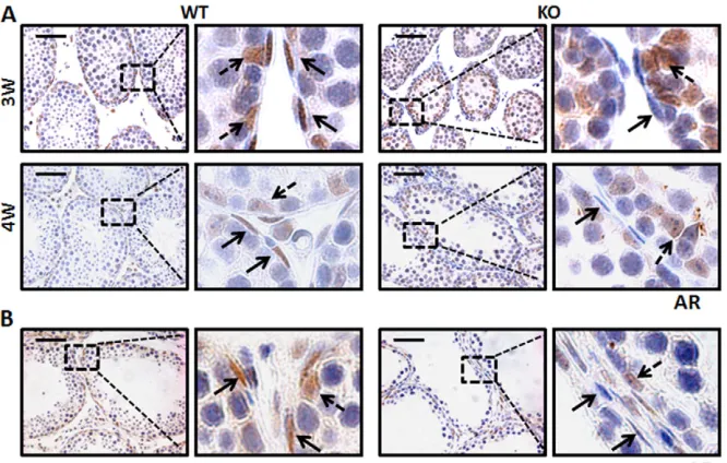
Downregulation of androgen receptor expression in Lgr4−/−PMCs
In Lgr4 mutant mice the localization of Sertoli cell nuclei was altered, as was the expression of certain Sertoli cell-specific genes, and a similar phenotype has been reported in a PMC-specific Ar knockout mouse strain (Welsh et al., 2009; Zhang et al., 2006). We speculated that in Lgr4mutant PMCs the expression of Ar was impaired. In keeping with PMCs and Sertoli cells being androgen target cell types, we observed Ar protein in their nuclei in WT testes (Fig. 5A). The level of Ar protein was comparable in Sertoli cells in both genotypes, although the localization of Sertoli cell nuclei was altered in the Lgr4-deficient mice. However, the level of Ar protein was dramatically decreased in Lgr4mutant PMCs (Fig. 5A, both at 3 weeks and 4 weeks). Similar results were obtained in testicular transplants (Fig. 5B). These results suggest that Lgr4
deficiency downregulates Ar expression in PMCs, but not in Sertoli cells, resulting in the malfunction of Sertoli cells in the seminiferous epithelium.
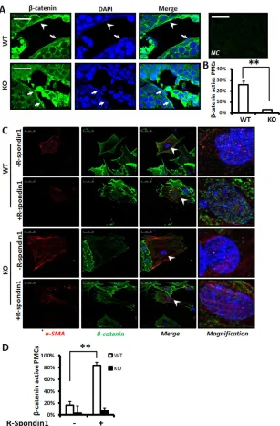
Lgr4 modulates Wnt/β-catenin signaling in PMCs In Lgr4mutant PMCs, the expression of Ar, α-SMA and numerous components of the ECM (e.g. fibronectin, laminin, collagen) was downregulated, and the majority of these genes, especially those encoding Ar, α-SMA and fibronectin, have been reported as downstream targets of Wnt/β-catenin signaling (Carthy et al., 2012; De Langhe et al., 2005; Gradl et al., 1999; Hlubek et al., 2001; Yang et al., 2002; Yang et al., 2006). We speculated that Wnt/β-catenin signaling is functioning in PMCs and that the activity of this pathway is impaired in Lgr4mutant PMCs, as Lgr4 has recently been demonstrated to be one of the receptors for R-spondins, which amplify Wnt signaling (Carmon et al., 2011; de Lau et al., 2011; Glinka et al., 2011; Hao et al., 2012). Immunostaining with an antibody against β-catenin was performed to evaluate the activity of Wnt signaling, as β-catenin localizes in nuclei upon canonical Wnt signaling pathway activation. In WT testes, β-catenin was often detected in the nuclei of PMCs, but the number of PMCs with nuclear β-catenin was dramatically decreased in Lgr4mutant testes (Fig. 6A,B). These data suggest that Lgr4 deficiency attenuates the activation of Wnt/β-catenin signaling in PMCs.
As R-spondins have recently been identified as ligands for Lgr4, we used a primary cell culture system to test whether R-spondin 1/Lgr4 functions in PMCs. WT and Lgr4 mutant seminiferous tubules were dissected and testicular cells were cultured. After being challenged with or without recombinant R-spondin 1, the cells were stained with specific antibodies. α-SMA was used as the marker for PMCs. Upon R-spondin 1 administration, the number of PMCs with nuclear β-catenin was dramatically increased in WT cultures but not in Lgr4mutant cultures (Fig. 6C,D). These data imply that Lgr4 modulates R-spondin 1-mediated activation of Wnt/β-catenin signaling in PMCs.
Reactivation of Wnt/β-catenin signaling partially rescues the defects in Lgr4−/−testis
[image:7.612.52.385.518.730.2]The above data suggested that Lgr4 mediates R-spondin 1 activation of Wnt/β-catenin signaling in PMCs, but whether the phenotype
Fig. 5. Selective decrease in androgen receptor expression in Lgr4−/−PMCs.
(A) Androgen receptor (Ar) expression was reduced in Lgr4−/−PMCs (solid arrows) but not in Sertoli cells (dashed arrows) compared with WT controls at 3 and 4 weeks of age. (B) Ar expression was decreased in PMCs of transplanted Lgr4−/−testes compared with WT transplants. Note that the expression of Ar was not affected in Sertoli cells (dashed arrows). Scale bars: 50 μm.
D
E
V
E
LO
P
M
E
N
observed in Lgr4mutant testes was due to the attenuation of Wnt signaling in PMCs was still obscure. We generated Lgr4−/−;Apcmin/+ mice and examined the effects of Apcmutation in restoring the defects caused by Lgr4deletion. Apcmin/+mice contain a germline mutation in the Apcgene that spontaneously activates β-catenin (Lefebvre et al., 1998). We found that the number of PMCs with nuclear β-catenin was significantly increased in Lgr4−/−;Apcmin/+ testes (Fig. 7A), suggesting that mutation of Apcpartially restored the activation of Wnt/β-catenin signaling in the double-mutant mice. Furthermore, we treated Lgr4−/−mice with SB216763, a Gsk3β
inhibitor, and also found that Wnt/β-catenin signaling was reactivated in PMCs (Fig. 7A).
With these two strategies to reactivate Wnt signaling in Lgr4 mutant mice, we examined the expression of specific genes in order to explore whether the phenotype caused by Lgr4deletion could be restored. As shown in Fig. 7B, the expression of fibronectin was increased and the ECM formed continuous rings surrounding the seminiferous tubules in the testes of Lgr4−/−;Apcmin/+and
SB216763-treated Lgr4−/−mice. In addition, the expression of α-SMA in PMCs
[image:8.612.53.360.57.523.2]was increased in double mutants and in Gsk3β inhibitor-treated mice (Fig. 7B, α-SMA). We also found that, in the PMCs of Wnt/β-catenin reactivated Lgr4mutant mice, the expression of Ar was restored and most of the Sertoli cell nuclei localized at the basement membrane layer of the seminiferous tubules (Fig. 7B, Ar). Not only were the PMC-specific genes restored upon Wnt/β-catenin reactivation in Lgr4-deficient mice, but the differentiation of germ cells was also promoted. In SB216763-treated Lgr4mutant testes, Scp3-negative germ cells appeared in the inner layer of the seminiferous epithelium, suggesting that upon Wnt/β-catenin reactivation many primary spermatogonia went through the differentiation process. In addition, seminiferous tubule diameter and lumen size were increased in SB216763-treated Lgr4 mutant mice compared with untreated controls (Fig. 7C). These data demonstrate that reactivation of Wnt/β -catenin signaling partially rescues the phenotype of Lgr4mutant mice, suggesting that Lgr4-mediated Wnt signaling plays an important role in PMCs.
Fig. 6. Lgr4 is crucial for Wnt/β-catenin signaling activation in PMCs. (A,B) Wnt/β-catenin signaling was activated in some WT PMCs but few Lgr4−/−PMCs. (A) IF staining was performed with a β-catenin antibody. Arrowheads indicate nuclear β-catenin indicating activation of Wnt signaling in PMCs; arrows indicate Wnt/β-catenin inactive PMCs. Purified mouse IgG1 was used as an isotype control for primary antibody (NC, top right). (B) Quantitation of Wnt/β-catenin signaling activation in PMCs. (C,D) The Lgr4 ligand R-spondin 1 activates β-catenin in WT but not
Lgr4−/−PMCs. (C) Primary cultured PMCs from 3- to 4-week-old WT and Lgr4−/−mice were challenged with or without R-spondin 1, then the cells were subjected to IF with a β-catenin antibody and an antibody against α-SMA to indicate PMCs. (D) Quantitation of the effect of R-spondin 1 on Wnt/β-catenin signaling activation in PMCs. Error bars indicate the mean ± s.e.m. **P<0.01 (Student’s t-test). Scale bars: 50 μm.
D
E
V
E
LO
P
M
E
N
DISCUSSION
Four groups, including us, have shown that Lgr4 is essential for male fertility by modulation of epididymal epithelial development and function (Hoshii et al., 2007; Li et al., 2010; Mendive et al., 2006), but the distinct roles of Lgr4 in spermatogenesis have not been studied. Here we explored the physiological function of Lgr4 in testes and found that Lgr4 is specifically expressed in PMCs and modulates spermatogenesis. Disruption of Lgr4 in the testes attenuated PMC Wnt/β-catenin signaling and led to impaired germ cell differentiation and maturation.
Previous studies have shown that the degeneration of the seminiferous epithelium in Lgr4 mutants is partially due to hypoplasia of efferent ducts and epididymis and to fluid circulation blockage, which occurred after 4 weeks of age in these tubules (Li et al., 2010; Mendive et al., 2006). To elucidate whether Lgr4 plays a direct role in testes, we focused on the first 4 weeks of postnatal development and performed testes transplant experiments. In early
postnatal stages, both the diameter and the lumen size of the seminiferous tubule in Lgr4 mutant testes were significantly reduced (Fig. 1C). In addition, the growth of testes as well as germ cell differentiation were dramatically affected in Lgr4 mutant transplants (Fig. 1D,E). These data showed that the decreased spermatogenesis is due to direct Lgr4 action in the testes, rather than being a side effect of the blockage of male reproductive ducts.
[image:9.612.51.393.59.527.2]In mouse testis, PMCs are viewed as structural cells with the ability to contract (Maekawa et al., 1996; Rossi et al., 2002). In addition, PMCs secrete key basement membrane components. Although we did not provide direct evidence that Lgr4 regulates PMC contraction, the expression of contractile ability-associated genes, such as those encoding α-SMA and desmin, was selectively downregulated in Lgr4−/−PMCs, suggesting that deletion of Lgr4 might affect PMC contractility and the transport of spermatozoa through seminiferous tubules. Whether PMCs have physiological functions beyond the two major roles remains obscure. SSCs reside Fig. 7. Forced activation of Wnt/β-catenin signaling in Lgr4−/−mice partially rescues
PMC defects. (A) β-catenin was activated in PMCs of Lgr4−/−;Apcmin/+mice and in Lgr4−/− mice after Gsk3β-specific inhibitor (SB216763) treatment. IF with β-catenin antibody and nuclei counterstained with DAPI. PMCs with nuclear β-catenin are indicated by arrows, whereas PMCs with cytoplasmic β-catenin are indicated with arrowheads. (B) Forced activation of Wnt/β-catenin signaling in
Lgr4−/−mice increased the expression of Wnt target genes that play essential roles in PMCs. IHC and IF were performed against testis sections of the indicated genotypes using antibodies against fibronectin, Ar or α-SMA. Arrows indicate positive signals typical for each antibody. (C) The differentiation of germ cells was also promoted in SB216763-treated
Lgr4mutant testis. Differentiated germ cells (Scp3 negative) appeared in the inner layer of the seminiferous epithelium in SB216763-treated Lgr4–/–mice Scale bars: 25 μm in A; 50 μm in B,C.
D
E
V
E
LO
P
M
E
N
in the niche formed by somatic cells in the basal compartments (surrounded by Sertoli cells and PMCs) of the seminiferous tubules. Sertoli cells are widely studied niche cells that stimulate either self-renewal or differentiation of the SSCs through secretion of growth factors such as Gdnf and SCF (Kitl – Mouse Genome Informatics) (Blume-Jensen et al., 2000; Meng et al., 2000; Ohta et al., 2000). It is believed that Gdnf is mainly produced in Sertoli cells, but it is also reported that Gdnf is constitutively produced by PMCs and may contribute to the SSC niche (Spinnler et al., 2010). It is difficult to demonstrate which cell type is more important for the elevation of the Gdnf level in Lgr4−/−mice, as Sertoli cells are also affected indirectly through mesenchymal-epithelial cell interactions. However, it is clearly demonstrated that Lgr4 is specifically expressed in PMCs, and disrupting the gene resulted in an increase of undifferentiated spermatogonia and in an attenuation of germ cell differentiation. This strongly suggested that Lgr4 signaling in PMCs also plays a role in regulating SSC differentiation and self-renewal through either direct or indirect mechanisms and provided genetic evidence that PMCs might also contribute to the SSC niche.
PMCs regulate Sertoli cell function through the production of a paracrine protein, termed PModS, which modulates Sertoli cell function including the secretion of key factors (Anthony et al., 1991; Norton and Skinner, 1989). However, PModS is as yet unidentified, with undefined functions and mechanisms. With PMC-specific Ar knockout mouse models, researchers have demonstrated the importance of androgen-driven mesenchymal-epithelial cell interactions for the regulation of spermatogenesis (Welsh et al., 2009; Zhang et al., 2006). In this study, we reported that Lgr4 signaling is a second pathway in PMCs to modulate Sertoli cell function during spermatogenesis. Similar phenotypes were observed in PMC-specific Ar mutant mice, and most of the genes downregulated in Armutant mice were also observed in Lgr4 -deficient animals. Although we do not have direct evidence to show how Lgr4 signaling regulates the expression of Ar in PMCs, a previous publication reported that Wnt signaling directly regulates Ar expression through three functional LEF1/TCF binding elements within the Arpromoter in prostate cancer cells (Yang et al., 2006). Since Lgr4 is the newly identified receptor for Wnt activator R-spondins, we hypothesize that Lgr4 regulates Ar expression through Wnt signaling. In Lgr4 mutant mice upon Wnt/β-catenin reactivation (Lgr4−/−;Apcmin/+ double-mutant strain and Gsk3β inhibitor-treated Lgr4−/−mice), Ar expression is restored in Lgr4
mutant PMCs, further indicating that Lgr4 is an upstream regulator of Ar expression. We do not consider Lgr4 regulation of Ar expression to be PMC specific. Our previous study and unpublished data have shown that Ar expression is also reduced in Lgr4mutant epididymal epithelium (Li et al., 2010). Epididymal-specific deletion of Arresulted in epithelial hypoplasia and hypotrophy and led to male infertility, similar to the phenotype observed in adult Lgr4mutant mice (Krutskikh et al., 2011; Mendive et al., 2006).
In conclusion, our results demonstrate that Lgr4 plays an indispensable role in PMCs to modulate spermatogenesis. Using differentiation strategies, we also demonstrated for the first time that Wnt/β-catenin is not only activated but also directly modulated by Lgr4 in PMCs. Our observations provide deeper insights into the functions and the regulatory molecular mechanisms of this particular cell type. Since Lgr4 is a seven-transmembrane receptor that is accessible to small molecules, the identification of an Lgr4 antagonist might have potential clinical value for contraception.
Acknowledgements
We thank Meizhen Liu for technical assistance and Stefan Siwko and Zhiyv Niu for comments and advice.
Funding
This work was partially supported by grants from the State Key Development Programs of China [2012CB910400, 2010CB945403]; grants from the National Natural Science Foundation of China [31171318 and 30930055]; and a grant from the Science and Technology Commission of Shanghai
Municipality [11DZ2260300].
Competing interests statement
The authors declare no competing financial interests.
Supplementary material
Supplementary material available online at
http://dev.biologists.org/lookup/suppl/doi:10.1242/dev.093641/-/DC1
References
Albrecht, M.(2009). Insights into the nature of human testicular peritubular cells. Ann. Anat.191, 532-540.
Anthony, C. T., Rosselli, M. and Skinner, M. K.(1991). Actions of the testicular paracrine factor (P-Mod-S) on Sertoli cell transferrin secretion throughout pubertal development. Endocrinology129, 353-360.
Blume-Jensen, P., Jiang, G., Hyman, R., Lee, K. F., O’Gorman, S. and Hunter, T.(2000). Kit/stem cell factor receptor-induced activation of
phosphatidylinositol 3⬘-kinase is essential for male fertility. Nat. Genet.24, 157-162.
Boyer, A., Hermo, L., Paquet, M., Robaire, B. and Boerboom, D.(2008). Seminiferous tubule degeneration and infertility in mice with sustained activation of WNT/CTNNB1 signaling in sertoli cells. Biol. Reprod.79, 475-485.
Carmon, K. S., Gong, X., Lin, Q., Thomas, A. and Liu, Q.(2011). R-spondins function as ligands of the orphan receptors LGR4 and LGR5 to regulate Wnt/beta-catenin signaling. Proc. Natl. Acad. Sci. USA108, 11452-11457.
Carthy, J. M., Luo, Z. and McManus, B. M.(2012). WNT3A induces a contractile and secretory phenotype in cultured vascular smooth muscle cells that is associated with increased gap junction communication. Lab. Invest.92, 246-255.
Clevers, H.(2006). Wnt/beta-catenin signaling in development and disease. Cell
127, 469-480.
De Langhe, S. P., Sala, F. G., Del Moral, P. M., Fairbanks, T. J., Yamada, K. M., Warburton, D., Burns, R. C. and Bellusci, S.(2005). Dickkopf-1 (DKK1) reveals that fibronectin is a major target of Wnt signaling in branching
morphogenesis of the mouse embryonic lung. Dev. Biol.277, 316-331.
de Lau, W., Barker, N., Low, T. Y., Koo, B. K., Li, V. S., Teunissen, H., Kujala, P., Haegebarth, A., Peters, P. J., van de Wetering, M. et al.(2011). Lgr5 homologues associate with Wnt receptors and mediate R-spondin signalling.
Nature476, 293-297.
Fernández, D., Bertoldi, M. V., Gómez, L., Morales, A., Callegari, E. and Lopez, L. A.(2008). Identification and characterization of Myosin from rat testicular peritubular myoid cells. Biol. Reprod.79, 1210-1218.
Glinka, A., Dolde, C., Kirsch, N., Huang, Y. L., Kazanskaya, O., Ingelfinger, D., Boutros, M., Cruciat, C. M. and Niehrs, C.(2011). LGR4 and LGR5 are R-spondin receptors mediating Wnt/β-catenin and Wnt/PCP signalling. EMBO Rep.12, 1055-1061.
Gradl, D., Kühl, M. and Wedlich, D.(1999). The Wnt/Wg signal transducer beta-catenin controls fibronectin expression. Mol. Cell. Biol.19, 5576-5587.
Hao, H. X., Xie, Y., Zhang, Y., Charlat, O., Oster, E., Avello, M., Lei, H., Mickanin, C., Liu, D., Ruffner, H. et al.(2012). ZNRF3 promotes Wnt receptor turnover in an R-spondin-sensitive manner. Nature485, 195-200.
Hlubek, F., Jung, A., Kotzor, N., Kirchner, T. and Brabletz, T.(2001). Expression of the invasion factor laminin gamma2 in colorectal carcinomas is regulated by beta-catenin. Cancer Res.61, 8089-8093.
Holstein, A. F., Maekawa, M., Nagano, T. and Davidoff, M. S.(1996). Myofibroblasts in the lamina propria of human semi-niferous tubules are dynamic structures of heterogeneous phenotype. Arch. Histol. Cytol.59, 109-125.
Honaramooz, A., Snedaker, A., Boiani, M., Schöler, H., Dobrinski, I. and Schlatt, S.(2002). Sperm from neonatal mammalian testes grafted in mice.
Nature418, 778-781.
Hoshii, T., Takeo, T., Nakagata, N., Takeya, M., Araki, K. and Yamamura, K.
(2007). LGR4 regulates the postnatal development and integrity of male reproductive tracts in mice. Biol. Reprod.76, 303-313.
Jeays-Ward, K., Dandonneau, M. and Swain, A.(2004). Wnt4 is required for proper male as well as female sexual development. Dev. Biol.276, 431-440.
Johnston, H., Baker, P. J., Abel, M., Charlton, H. M., Jackson, G., Fleming, L., Kumar, T. R. and O’Shaughnessy, P. J.(2004). Regulation of Sertoli cell number and activity by follicle-stimulating hormone and androgen during postnatal development in the mouse. Endocrinology145, 318-329.
Kato, S., Matsubara, M., Matsuo, T., Mohri, Y., Kazama, I., Hatano, R., Umezawa, A. and Nishimori, K.(2006). Leucine-rich repeat-containing G protein-coupled receptor-4 (LGR4, Gpr48) is essential for renal development in mice. Nephron Exp. Nephrol.104, e63-75.
D
E
V
E
LO
P
M
E
N
Krutskikh, A., De Gendt, K., Sharp, V., Verhoeven, G., Poutanen, M. and Huhtaniemi, I.(2011). Targeted inactivation of the androgen receptor gene in murine proximal epididymis causes epithelial hypotrophy and obstructive azoospermia. Endocrinology152, 689-696.
Lefebvre, A. M., Chen, I., Desreumaux, P., Najib, J., Fruchart, J. C., Geboes, K., Briggs, M., Heyman, R. and Auwerx, J.(1998). Activation of the peroxisome proliferator-activated receptor gamma promotes the
development of colon tumors in C57BL/6J-APCMin/+ mice. Nat. Med.4, 1053-1057.
Li, X. Y., Lu, Y., Sun, H. Y., Wang, J. Q., Yang, J., Zhang, H. J., Fan, N. G., Xu, J., Jiang, J. J., Liu, R. Y. et al.(2010). G protein-coupled receptor 48 upregulates estrogen receptor alpha expression via cAMP/PKA signaling in the male reproductive tract. Development137, 151-157.
Luo, J., Zhou, W., Zhou, X., Li, D., Weng, J., Yi, Z., Cho, S. G., Li, C., Yi, T., Wu, X. et al.(2009). Regulation of bone formation and remodeling by G-protein-coupled receptor 48. Development136, 2747-2756.
Maekawa, M., Kamimura, K. and Nagano, T.(1996). Peritubular myoid cells in the testis: their structure and function. Arch. Histol. Cytol.59, 1-13.
Mazerbourg, S., Bouley, D. M., Sudo, S., Klein, C. A., Zhang, J. V., Kawamura, K., Goodrich, L. V., Rayburn, H., Tessier-Lavigne, M. and Hsueh, A. J.
(2004). Leucine-rich repeat-containing, G protein-coupled receptor 4 null mice exhibit intrauterine growth retardation associated with embryonic and perinatal lethality. Mol. Endocrinol.18, 2241-2254.
Mendive, F., Laurent, P., Van Schoore, G., Skarnes, W., Pochet, R. and Vassart, G.(2006). Defective postnatal development of the male reproductive tract in LGR4 knockout mice. Dev. Biol.290, 421-434.
Meng, X., Lindahl, M., Hyvönen, M. E., Parvinen, M., de Rooij, D. G., Hess, M. W., Raatikainen-Ahokas, A., Sainio, K., Rauvala, H., Lakso, M. et al.(2000). Regulation of cell fate decision of undifferentiated spermatogonia by GDNF.
Science287, 1489-1493.
Mustata, R. C., Van Loy, T., Lefort, A., Libert, F., Strollo, S., Vassart, G. and Garcia, M. I.(2011). Lgr4 is required for Paneth cell differentiation and maintenance of intestinal stem cells ex vivo. EMBO Rep.12, 558-564.
Norton, J. N. and Skinner, M. K.(1989). Regulation of Sertoli cell function and differentiation through the actions of a testicular paracrine factor P-Mod-S.
Endocrinology124, 2711-2719.
Oatley, M. J., Racicot, K. E. and Oatley, J. M.(2011). Sertoli cells dictate spermatogonial stem cell niches in the mouse testis. Biol. Reprod.84, 639-645.
Ohta, H., Yomogida, K., Dohmae, K. and Nishimune, Y.(2000). Regulation of proliferation and differentiation in spermatogonial stem cells: the role of c-kit and its ligand SCF. Development127, 2125-2131.
Richardson, L. L., Kleinman, H. K. and Dym, M.(1995). Basement membrane gene expression by Sertoli and peritubular myoid cells in vitro in the rat. Biol. Reprod.52, 320-330.
Rossi, F., Ferraresi, A., Romagni, P., Silvestroni, L. and Santiemma, V.(2002). Angiotensin II stimulates contraction and growth of testicular peritubular myoid cells in vitro. Endocrinology143, 3096-3104.
Santiemma, V., Rossi, F., Guerrini, L., Markouizou, A., Pasimeni, G., Palleschi, S. and Fabbrini, A.(2001). Adrenomedullin inhibits the contraction of cultured rat testicular peritubular myoid cells induced by endothelin-1. Biol. Reprod.64, 619-624.
Schlatt, S., Honaramooz, A., Boiani, M., Schöler, H. R. and Dobrinski, I.
(2003). Progeny from sperm obtained after ectopic grafting of neonatal mouse testes. Biol. Reprod.68, 2331-2335.
Shinohara, T., Avarbock, M. R. and Brinster, R. L.(1999). beta1- and alpha6-integrin are surface markers on mouse spermatogonial stem cells. Proc. Natl. Acad. Sci. USA96, 5504-5509.
Song, H., Luo, J., Luo, W., Weng, J., Wang, Z., Li, B., Li, D. and Liu, M.(2008). Inactivation of G-protein-coupled receptor 48 (Gpr48/Lgr4) impairs definitive erythropoiesis at midgestation through down-regulation of the ATF4 signaling pathway. J. Biol. Chem.283, 36687-36697.
Spinnler, K., Köhn, F. M., Schwarzer, U. and Mayerhofer, A.(2010). Glial cell line-derived neurotrophic factor is constitutively produced by human testicular peritubular cells and may contribute to the spermatogonial stem cell niche in man. Hum. Reprod.25, 2181-2187.
Tanwar, P. S., Kaneko-Tarui, T., Zhang, L., Rani, P., Taketo, M. M. and Teixeira, J.(2010). Constitutive WNT/beta-catenin signaling in murine Sertoli cells disrupts their differentiation and ability to support spermatogenesis. Biol. Reprod.82, 422-432.
Thompson, E. W., Blackshaw, A. W. and Raychoudhury, S. S.(1995). Secreted products and extracellular matrix from testicular peritubular myoid cells influence androgen-binding protein secretion by Sertoli cells in culture. J. Androl.16, 28-35.
Tung, P. S. and Fritz, I. B.(1991). Transforming growth factor-beta and platelet-derived growth factor synergistically stimulate contraction by testicular peritubular cells in culture in serum-free medium. J. Cell. Physiol.146, 386-393.
Van Schoore, G., Mendive, F., Pochet, R. and Vassart, G.(2005). Expression pattern of the orphan receptor LGR4/GPR48 gene in the mouse. Histochem. Cell Biol.124, 35-50.
Vergouwen, R. P., Jacobs, S. G., Huiskamp, R., Davids, J. A. and de Rooij, D. G.(1991). Proliferative activity of gonocytes, Sertoli cells and interstitial cells during testicular development in mice. J. Reprod. Fertil.93, 233-243.
Verhoeven, G., Hoeben, E. and De Gendt, K.(2000). Peritubular cell-Sertoli cell interactions: factors involved in PmodS activity. Andrologia32, 42-45.
Wang, R. S., Yeh, S., Chen, L. M., Lin, H. Y., Zhang, C., Ni, J., Wu, C. C., di Sant’Agnese, P. A., deMesy-Bentley, K. L., Tzeng, C. R. et al.(2006). Androgen receptor in sertoli cell is essential for germ cell nursery and junctional complex formation in mouse testes. Endocrinology147, 5624-5633.
Wang, J., Li, X., Ke, Y., Lu, Y., Wang, F., Fan, N., Sun, H., Zhang, H., Liu, R., Yang, J. et al.(2011). GPR48 increases mineralocorticoid receptor gene expression. J. Am. Soc. Nephrol. 23, 281-293
Welsh, M., Saunders, P. T., Atanassova, N., Sharpe, R. M. and Smith, L. B.
(2009). Androgen action via testicular peritubular myoid cells is essential for male fertility. FASEB J.23, 4218-4230.
Weng, J., Luo, J., Cheng, X., Jin, C., Zhou, X., Qu, J., Tu, L., Ai, D., Li, D., Wang, J. et al.(2008). Deletion of G protein-coupled receptor 48 leads to ocular anterior segment dysgenesis (ASD) through down-regulation of Pitx2. Proc. Natl. Acad. Sci. USA105, 6081-6086.
Yang, F., Li, X., Sharma, M., Sasaki, C. Y., Longo, D. L., Lim, B. and Sun, Z.
(2002). Linking beta-catenin to androgen-signaling pathway. J. Biol. Chem.277, 11336-11344.
Yang, X., Chen, M. W., Terry, S., Vacherot, F., Bemis, D. L., Capodice, J., Kitajewski, J., de la Taille, A., Benson, M. C., Guo, Y. et al.(2006). Complex regulation of human androgen receptor expression by Wnt signaling in prostate cancer cells. Oncogene25, 3436-3444.
Yeh, J. R., Zhang, X. and Nagano, M. C.(2011). Wnt5a is a cell-extrinsic factor that supports self-renewal of mouse spermatogonial stem cells. J. Cell Sci.124, 2357-2366.
Yuan, L., Liu, J. G., Zhao, J., Brundell, E., Daneholt, B. and Höög, C.(2000). The murine SCP3 gene is required for synaptonemal complex assembly, chromosome synapsis, and male fertility. Mol. Cell5, 73-83.
Zhang, C., Yeh, S., Chen, Y. T., Wu, C. C., Chuang, K. H., Lin, H. Y., Wang, R. S., Chang, Y. J., Mendis-Handagama, C., Hu, L. et al.(2006). Oligozoospermia with normal fertility in male mice lacking the androgen receptor in testis peritubular myoid cells. Proc. Natl. Acad. Sci. USA103, 17718-17723.