1891
Introduction
SOX genes are developmental regulators characterized by the presence of an HMG (high mobility group) DNA-binding domain with >50% homology to the sex-determining gene SRY. The SOX gene family can be further subdivided into twelve subgroups defined by additional homologies outside of the DNA-binding domain (Bowles et al., 2000). The E subgroup of SOX genes consists of three members: SOX8, SOX9 and SOX10. The gene studied in most detail in this subgroup is SOX9, heterozygous mutations of which cause campomelic dysplasia (CD) in humans, a syndrome characterized by abnormal bone development, perinatal lethality and other abnormalities (Houston et al., 1983). Seventy-five percent of XY SOX9 heterozygous individuals with campomelic dysplasia also show defects in testicular differentiation or complete sex reversal (i.e. ovaries instead of testes). In addition, a chromosomal duplication encompassing the SOX9 locus has been found in one patient with XX sex reversal further supporting a role for SOX9 during the sex determination process in man (Huang et al., 1999).
Campomelic dysplasia is caused by heterozygous loss-of-function mutations typically affecting the DNA-binding or transcriptional-activation domain (HMG box) of SOX9 (Foster et al., 1994; Wagner et al., 1994). In addition, translocations or
inversions with breakpoints several hundred kilobases (kb) upstream of SOX9 have been identified (Wagner et al., 1994; Wirth et al., 1996), and transgenic analysis suggested that upstream sequences contain important regulatory elements for the SOX9 gene (Bishop et al., 2000; Wunderle et al., 1998). Studies in vitro and in vivo have demonstrated that SOX9 can bind and bend DNA, and can act as a transcriptional activator on target promoters (Bell et al., 1997; Lefebvre et al., 1997). Consistent with these findings, mutations in the transactivation domain at the C terminus have been found in individuals with campomelic dysplasia (Sudbeck et al., 1996). Moreover, recent in vitro data suggest that this gene may have an additional function during pre-mRNA splicing (Ohe et al., 2002).
Sex determination in the mouse occurs at E11.5, when an initially bipotential gonad becomes determined to develop either along the male or female axis depending on the presence or absence of the Y chromosome-encoded gene Sry. The expression of Sry is highly dynamic: very low levels of Sry transcripts can be detected as early as E10.5, which then sharply increase to a peak at E11.5, only to become repressed again by E12 (Hacker et al., 1995; Jeske et al., 1995). The regulation of Sry is largely unknown, but it has been shown that the Wilms’ tumour suppressor Wt1 is required for the presence of high levels of Sry RNA (Hammes et al., 2001;
Sex determination in mammals directs an initially bipotential gonad to differentiate into either a testis or an ovary. This decision is triggered by the expression of the sex-determining gene Sry, which leads to the activation of male-specific genes including the HMG-box containing gene Sox9. From transgenic studies in mice it is clear that Sox9 is sufficient to induce testis formation. However, there is no direct confirmation for an essential role for Sox9 in testis determination. The studies presented here are the first experimental proof for an essential role for Sox9 in mediating a switch from the ovarian pathway to the testicular pathway. Using conditional gene targeting, we show that homozygous deletion of Sox9 in XY gonads
interferes with sex cord development and the activation of the male-specific markers Mis and P450scc, and leads to the expression of the female-specific markers Bmp2 and follistatin. Moreover, using a tissue specific knock-out approach, we show that Sox9 is involved in Sertoli cell differentiation, the activation of Mis and Sox8, and the inactivation of Sry. Finally, double knock-out analyses suggest that Sox8 reinforces Sox9 function in testis differentiation of mice.
Key words: Sex determination, Gonads, Conditional gene targeting, Mouse
Summary
Functional analysis of Sox8 and Sox9 during sex determination in
the mouse
Marie-Christine Chaboissier1, Akio Kobayashi2,3, Valerie I. P. Vidal1, Susanne Lützkendorf1,
Henk J. G. van de Kant4, Michael Wegner5, Dirk G. de Rooij4, Richard R. Behringer3and Andreas Schedl1,*
1INSERM U470, Centre de Biochimie, Parc Valrose, 06108 Nice Cedex 2, France
2Program in Developmental Biology, Baylor College of Medicine, Houston, TX 77030, USA
3Department of Molecular Genetics, University of Texas, MD Anderson Cancer Center, Houston, TX 77030, USA 4Departments of Endocrinology and of Cell Biology, Utrecht University, 3584-CH Utrecht, The Netherlands 5Institute for Biochemistry, University of Erlangen, D-91054 Erlangen, Germany
*Author for correspondence (e-mail: schedl@unice.fr)
Accepted 7 January 2004
Development 131, 1891-1901
Published by The Company of Biologists 2004 doi:10.1242/dev.01087
Hossain and Saunders, 2001). Moreover, steroidogenic factor 1 (Sf1; Nr5a1 – Mouse Genome Informatics) seems to be able to activate the Sry promoter at least in pig (Pilon et al., 2003). One of the first genes activated in male gonads after the expression of Sry is Sox9. Although small amounts of cytoplasmic Sox9 can be detected in both male and female gonads at E10.5, the production of high levels of nuclear Sox9 protein is triggered through the expression of Sry in XY gonads (de Santa Barbara et al., 2000; Morais da Silva et al., 1996). Based on this early appearance of Sox9 after the onset of Sry, its association with sex reversal in man, and as it can functionally substitute for Sry in transgenic mice (Vidal et al., 2001), it has been proposed that Sry directly regulates Sox9 (Canning and Lovell-Badge, 2002).
Sox9 can function as a transcriptional activator and there is strong evidence that it regulates the Mullerian-inhibiting substance Mis (Amh – Mouse Genome Informatics). First, co-transfection experiments show that Sox9 can bind and transactivate the Mis promoter, at least in vitro (De Santa Barbara et al., 1998). Second, mutations in the potential Sox9-binding site upstream of the Mis coding sequence result in a lack of Mis expression in vivo (Arango et al., 1999). Interestingly, a recent in vitro study by Schepers et al. indicated that Sox8, a close homologue of Sox9, can also activate the Mis promoter, albeit to a somewhat lower extent (Schepers et al., 2003). Sox8 also shows a Sertoli cell-specific expression pattern in the developing testis, with an onset of expression at E12, just after the activation of Sox9. However, homozygous Sox8 knock-out mice did not show a gonadal phenotype, suggesting either functional redundancy with another gene or that Sox8 does not play a role in the development of this organ (Sock et al., 2001).
Heterozygous mutations in the human SOX9 gene are sufficient to induce sex reversal. By contrast, sex determination in heterozygous Sox9 knock-out mice occurs normally, suggesting that this pathway is less sensitive to gene dosage in mice than in humans (Bi et al., 2001). We were interested in investigating the role of Sox8 and Sox9 during sex determination and testis differentiation in more detail. The perinatal lethal phenotype of heterozygous Sox9 knock-out mice precludes the generation of homozygous null animals using standard genetic approaches. To overcome this problem we have generated a conditional knock-out allele of Sox9 (Sox9flox) (Akiyama et al., 2002). Using transgenic lines
expressing the Cre recombinase either in the germline or within the developing gonad, we show that Sox9 is essential for Sertoli cell differentiation and seminiferous tubule formation. The Sertoli cell-specific markers Mis and Sox8 directly depend on Sox9 levels, whereas high Sry expression persists in Sox9 knock-out mice, indicating that Sox9 activation leads to the downregulation of this gene. Finally, experiments with Sox8/Sox9 double knock-out mice suggest that Sox8 reinforces Sox9 function in testis formation.
Materials and methods
Construction of the Sf1-Cre plasmid
A 674 bp promoter fragment of the Sf1 gene (Wilhelm and Englert, 2002) was amplified by PCR using the primers 5′ -TTATCA-TGCCATGGTCACACCCTTAGCCCAGCAGTC-3′ and 5′ -TTATC-ATGCCATGGTCCCAGGCCTCAGGTAGGGCA-3′, and cloned in
front of the gene encoding the Cre-recombinase from the plasmid pPGKcrebpA (Gu et al., 1993). Microinjections in the pronucleus of one-cell embryos heterozygous for the Sox9floxallele (Akiyama et al.,
2002) were carried out as described (Hogan et al., 1994).
Mouse strains and genetic background
Sox8 and Sox9 knock-out mice were kept on a mixed 129/C57Bl6 background. The Sf1:Cre transgene was injected into fertilized oocytes derived from C57Bl6/CBA females mated with C57Bl6/CBA males, or with males homozygous for the Sox9flox allele (mixed
129/C57Bl6 background). Prm1:Cre and Zp3:Cre mice were maintained on a 129/C57Bl6 mixed background and C57Bl6 inbred background, respectively. Mice homozygous for the Sox8 mutation and heterozygous for the Sox9 deletion (Fig. 9) were generated using either the Prm1:Cre or the deleter:Cre strain (Schwenk et al., 1995). Both crosses gave similar results.
Genotyping of embryos
The presence of the Y chromosome was determined using primers for the Zfy and Sry genes (Hogan et al., 1994). Wild-type and Sox9flox
alleles were identified using the primers 5′ -GGGGCTTGTCTC-CTTCAGAG-3′, or 5′-ACACAGCATAGGCTACCTG-3′and 5′ -TG-GTAATGAGTCATACACAGTAC-3′, respectively. The Sox9 knock-out allele was identified using the primers: 5′ -GTCAAGCGACCC-ATG-3′ and 5′-TGGTAATGAGTCATACACAGTAC-3′. Genotyping for the Sox8 knock-out allele was performed as described (Sock et al., 2001). The presence of the Sf1-Cre transgene was determined using primers specific for the Cre gene, 5′ -CAATTTACTGACCGTACAC-3′and 5′-AGCTGGCCCAAATGTTGCTG-3′.
Histological analysis
Visceral organs were dissected free of the urogenital system that remained associated with the carcass, fixed overnight at 4°C in Bouin’s solution and embedded in paraffin wax. Sections were cut at 5-6 µm thickness and stained with haematoxylin and eosin.
Whole mount in situ hybridization
Embryos were fixed with 4% paraformaldehyde (PFA) in 1×PBS at 4°C overnight. Further processing of embryos and in situ hybridization were carried out essentially as described (Wilkinson, 1992). Sox9 riboprobes were synthesized according to Morais da Silva et al. (Morais da Silva et al., 1996).
Quantitative RT-PCR analysis
Individual urogenital ridges (mesonephros+gonad) were dissected in PBS from E13.5 embryos and immediately frozen at –70°C. RNA was prepared using the Absolutely RNA RT-PCR kit (Stratagene). Each sample was divided into two aliquots, one of which was reverse transcribed using the MMLV (Gibco), following the manufacturers instructions. The second aliquot was used as a control (RT-PCR without reverse transcription) to identify samples with DNA contamination that could not be included in the analysis of the expression from the intron-less Sry gene. Primers and probes for all other genes were designed to cover introns. All real time PCR assays were carried out using the LC-Faststart DNA Master kit (Roche). A standard curve for each gene was generated using serial dilutions of cDNA. Relative expression levels of each sample were determined in the same run and normalized by measuring the amount of Gapdh (Gapd – Mouse Genome Informatics) cDNA.
Primers for real-time PCR analysis were:
Gapdh cDNA, 5′-ATTCAACGGCACAGTCAAGG-3′and 5′
-TG-GATGCAGGGATGATGTTC-3′, hybridization probes 5′ -CCAGAA-GACTGTGGATGGCCCCT-X and LC-Red640-TGGAAAGCTGT-GGCGTGATGGC-p;
Sox9, 5′-GACAAGCGGAGGCCGAA-3′and 5′
-CTTGCAGCGCCTTGAA-GATAGCATTAG-X and LC-Red640-GAGATGTGAGTCTGTTCC-GTGGCCTC-p; and
Mis, 5′-GTCCTACATCTGGCTGAAGTGAT-3′ and 5′
-CCGAG-TAGGGCAGAGGTTCT-3′(overlapped the junction between exon 1 and 2, and exon 2 and 3, respectively), hybridization probes 5′ -GGC-CCTGTTAGTGCTATACTCTGGACC-X and LC-Red640-GCCCC-CAGGTCACAGTCACAGG-p.
We also used the following primers:
Sf1, 5′-TTCTGAGAGCCCGCTAGCC-3′and 5′
-CCTCGTCGTA-CGAGTAGTCCATG-3′, hybridization probes 5′ -CCTGGTGTCC-AGTGTCCACCCT-X and LC-Red640-TCCGGCTGAGAATTCTC-CTTCCG-p;
Sox8, 5′-CAGAGCTCAGCAAGACCCTA-3′ and 5′
-GGGTGGT-GGCCCAGTT-3′, hybridization probes 5′ -TTACAAATACCAGC-CAAGGCGAAG-X and LC-Red640-AAGAGTGTGAAGACTGGC-CGGAGC-p; and
Sry, 5′-AGCCTATGTGTAGTTCCTTGGTC-3′ and 5′
-TGCATA-AGGAGTCACATTTTGCT-3′, hybridization probes 5′ -CAATCTG-GCAGTTGAGTTAATGTGCAGAT-X and LC-Red640-CCATTCAT-TCATCCCACATATACTTGCCC-p.
Detection of the linear Sry transcript was performed as described (Toyooka et al., 1998). Each sample of RNA was checked for DNA contamination using the same amount of starting material without reverse transcription.
Immunohistochemistry
Tissues were isolated at E15.5, fixed for 2 hours in 4% PFA, washed twice for 5 minutes in 1×PBS and, after equilibration in 30% sucrose, frozen in OCT on dry ice. Sectioning and staining were carried out as described by Hammes et al. (Hammes et al., 2001). The following dilutions of primary antibodies were used: Sox9 (provided by Michael Wegner), 1:500; Mis (C-20, cat# sc-6886, Santa Cruz Biotechnology), 1:100; Wt1 (C-19, cat# sc-192 Santa Cruz Biotechnology), 1:100; and laminin (L-9393 Sigma), 1:50.
For laminin staining of cultured gonads, tissues were fixed with 4% PFA overnight and paraffin embedded. Sections were generated at 7
µm. After re-hydration, slides were placed in boiling 10 mM sodium citrate (pH 6.0) in a microwave oven for 20 minutes for antigen retrieval, and soaked in 3% hydrogen peroxide in methanol for 10 minutes at room temperature to block endogenous peroxidases. Immunodetection was performed by Vectastain Elite ABC Kit (PK-6100, Vector Laboratories) and substrate development by AEC Substrate System (K3464, DakoCytomation). Laminin α-1 antibody (M-20, cat# sc-6017 Santa Cruz Biotechnology) was used as the primary antibody at a 1:350 dilution.
In vitro organ cultures
The culture medium was Dulbecco’s modified Eagle’s medium (DMEM) with 10% fetal calf serum, 0.1 mM 2-mercaptoethanol, 2 mM glutamine, 0.5 mM pyruvate, 100 units/ml penicillin and 0.05 mg/ml streptomycin (McLaren and Southee, 1997). The urogenital organs from Sox9flox/∆ and Sox9∆/∆ embryos were dissected in the
culture media containing 20 mM HEPES. The isolated organs were cultured on polycarbonate membranes (Transwell #3403, Coster, NY) coated with 1% agarose in phosphate-buffered saline (PBS), with 500
µl of culture media per well at 37°C with 5% CO2in air for 2 or 3 days. After culture the organs were fixed in 4% PFA at 4°C overnight. For histological analysis, the fixed tissues were paraffin wax-embedded and sectioned at 7 µm.
Results
Heterozygous Sox9 mutations in mice cause perinatal lethality due to severe bone malformations and respiratory deficiency. To analyze Sox9 function during gonad formation we made use of our recently generated conditional Sox9floxallele (Akiyama
et al., 2002). Two complementing strategies were employed. First, we produced Sf1:Cre transgenic lines, which allowed the tissue-specific analysis of Sox9 in the developing gonad. Second, we generated homozygous knock-out animals by employing transgenic lines expressing the CRE recombinase within the male/female germ cell lineage, thus avoiding the lethality associated with heterozygous mutations in Sox9.
Sox9 ablation in developing gonads interferes with Sertoli cell differentiation
To address Sox9 function in gonad development in vivo we deleted the Sox9floxallele in a tissue specific manner. To direct
Cre expression to the developing gonad, we chose regulatory regions of the Sf1 gene, which in vivo is expressed in the gonadal primordium from E9.5 onwards. Using a 674 bp promoter fragment that has previously been shown to direct gonad-specific expression in transgenic mice (Wilhelm and Englert, 2002), 17 Sf1:Cre transgenic animals were generated. Expression in a number of these transgenic lines was early/ectopic, which resulted in complete deletion of the loxP-flanked Sox9 gene and concomitant perinatal lethality in the heterozygous state (Sf1:Cre; Sox9flox/+). However, bigenic
mice from several lines survived and line Sf1:Cre (5) was selected for all following analysis based on its efficient ablation of Sox9 expression (Fig. 1).
Sf1:Cre (5) transgenic animals were bred with Sox9flox/+
animals. A small proportion of double heterozygous mice (Sf1:Cre; Sox9flox/+) died soon after birth with a phenotype
similar to campomelic dysplasia, suggesting early excision of the floxed Sox9 allele in these animals. Surviving Sf1:Cre; Sox9flox/+ animals were further bred with homozygous
Sox9flox/Sox9floxmice, and embryos analyzed for deletion of the
floxed allele on the genomic level (Fig. 1A,B) and for Sox9 expression by in situ hybridization (Fig. 1C-E). Detection of Sox9 mRNA in Sf1:Cre; Sox9flox/Sox9floxembryos was variable
ranging from almost normal expression to a complete absence of signals at E13.5, reflecting the efficiency of Cre-mediated Sox9floxexcision in individual gonads (Fig. 1). Similarly,
real-time PCR analysis performed on individual urogenital ridges at E13.5 (left or right) showed variable Sox9 expression ranging from 50% of wild-type male expression to amounts comparable to that found in female mice (see later).
At E13.5, male testis development can be easily recognized by the formation of sex cords (Fig. 2B). In XY Sf1:Cre; Sox9flox/Sox9flox gonads we always detected some sex cord
Sox9flox/Sox9flox at E18.5 a significant number of meiotic
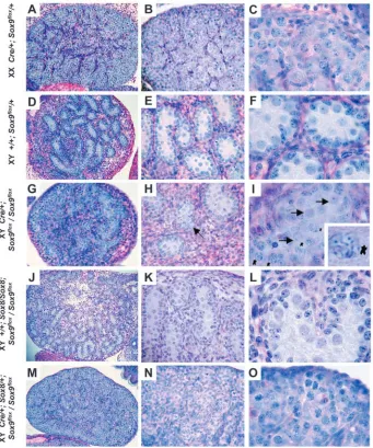
gonocytes (prophase) were detectable (Fig. 3I). Interestingly, these cells seemed to be located within seminiferous tubules next to quiescent gonocytes. Primordial germ cells (PGCs) develop according to their somatic cell environment and it has been proposed that an as yet unidentified signal produced by the Sertoli cells inhibits PGCs from entering meiosis (Adams and McLaren, 2002; Dolci and De Felici, 1990; McLaren and Southee, 1997). The presence of meiotic cells in XY testes of the tissue-specific knock-out may suggest that Sox9 is also required for proper functioning of Sertoli cells, including for the suppression of meiosis in male gonocytes.
Molecular analysis of Sox9 knock-out gonads
To investigate the effect of Sox9 ablation on the expression of
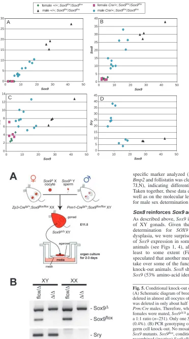
[image:4.612.46.325.66.302.2]genes involved in the sex determination process, we performed real-time PCR analyses with RNA isolated from individual E13.5 urogenital ridges. First we analyzed the expression of the Mullerian-inhibiting substance (Mis/Amh), which has been suggested to be a direct target of Sox9 (Arango et al., 1999; De Santa Barbara et al., 1998). Indeed, Mis expression in our conditional knock-out mice showed a linear relationship with Sox9 expression (Fig. 4A; r=0.9905; P<0.0001), supporting the theory that Mis represents a direct target of this transcription factor. Similarly, Sox8, which belongs to the same subgroup of Sox genes as Sox9, and which also shows Sertoli cell-specific expression from E12.5 days onwards (Schepers et al., 2003), was directly dependent on the level of Sox9 (Fig. 4B; r=0.8444; P<0.0001). Remarkably, even in cases where Sox9 expression was as low as that of wild-type female gonads, both the expression of Mis and Sox8 in XY knock-out gonads was still higher than in XX tissues. By contrast, expression of Sf1, which in vitro can be activated by the Sox9 protein (Shen and Ingraham, 2002), did not show a clear dependence on Sox9 expression levels (Fig. 4C; P=0.27), which may partly be due to the expression of Sf1 in other cell types, including progenitor cells and Leydig cells.
Fig. 1. Analysis of tissue specific deletion of Sox9 in Sf1:Cre; Sox9flox/Sox9floxanimals. Transgenic lines
carrying the Cre recombinase under a 674 bp Sf1 promoter fragment were crossed onto the homozygous Sox9flox/Sox9floxbackground. (A) The floxed and deleted
Sox9 locus. Orange arrows indicate the position of primers used for the detection of the Sox9floxand the
Sox9∆allele. (B) Detection of Sox9 deletion in urogenital ridges (E13.5) on the genomic level. Only mice carrying the Sf1:Cre transgene (lanes 3 and 4) show the deleted band Sox9∆. Incomplete deletion of the floxed allele may be due to inefficient expression of Sf1:Cre or may reflect the contamination of cells from the mesonephros. (C-E) In situ hybridization analysis for Sox9 at E13.5. Dotted lines indicate the outline of the developing gonads. Embryos homozygous for the Sox9floxallele and carrying
the Sf1:Cre transgene (Cre) show reduced (D) or absent (E) expression of Sox9 in the developing gonad. Note the persistent signal of Sox9 in other tissues (e.g. arrow in panel E indicates expression in the neural tube).
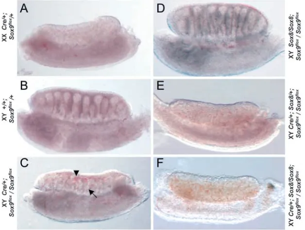
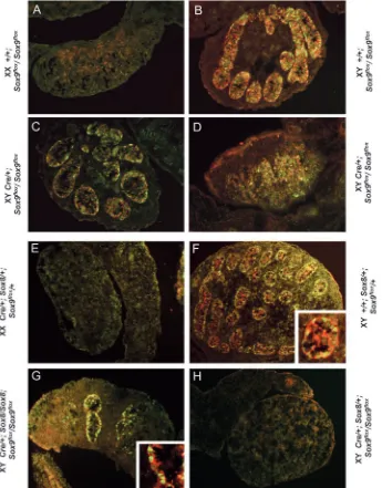
Fig. 2. Sox9 or Sox8/Sox9 knock-out mice show defects in sex cord formation. E13.5 gonads from Sox9 tissue-specific knock-out animals show a variable degree of sex cord formation ranging from normal (not shown) to severely abnormal (arrow in C). Note the presence of abnormal vascularization (arrowhead in C). Sox8/Sox8 knock-out gonads develop normal testis cords (D). By contrast, Sox9 tissue-specific knock-out animals (Cre/+
Sox9flox/Sox9flox) either heterozygous (E) or
[image:4.612.44.358.503.740.2]In wild-type mice, expression of Sry shows a very dynamic pattern with upregulation at E10.5, a peak at E11.5 and almost complete repression at E12.5 (Hacker et al., 1995). Hence, the rapid decrease of Sry expression occurs just after Sox9 activation and it has been suggested that Sry repression is mediated by expression of this gene. Indeed, Sry expression in our knock-out mice was significantly increased over wild-type levels. It should be noted that Sry repression was not linearly dependent on Sox9 expression, but occurred only in mice were Sox9 transcripts were significantly reduced (Fig. 4D).
Homozygous deletion of Sox9 interferes with sex cord formation and the expression of male specific markers
Although our real-time PCR analysis at E13.5 suggested that, at least in some gonads, Sox9 expression was very low and comparable to the level of expression in female gonads, we could not exclude the possibility that the lack of sex reversal in our tissue-specific knock-out animals was due to incomplete deletion of Sox9 at the time of sex determination (E11.5). To address this possibility we generated homozygous knock-out animals, making use of germ-line specific Cre transgenic
mouse lines. Males carrying the spermatogenesis-specific Prm1:Cre transgene (O’Gorman et al., 1997) on a floxed Sox9 background (Prm1:Cre; Sox9flox/Sox9flox) were crossed with
female Sox9flox/Sox9floxmice transgenic for the oocyte-specific
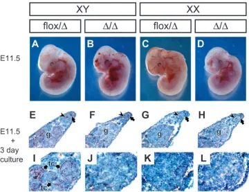
ZP3:Cre transgene (de Vries et al., 2000) (Fig. 5). Unfortunately, homozygous Sox9 knock-out animals die at E11.5 as a result of cardiac failure (Fig. 6A-D) (H. Akiyama et al., unpublished), which prohibited the in vivo analysis of the fate of XY gonads in these embryos at later stages. Hence, to investigate the function of Sox9 during sex determination, gonads were isolated at E11.5, placed into organ culture and analyzed after two to three days in culture using in situ hybridization or immunohistochemistry, respectively.
As expected, cultured XY gonads heterozygous for the Sox9 deletion (Sox9flox/∆) formed sex cords, which could be
visualized using laminin antibodies (Fig. 6E,I), and expressed the Sertoli cell-specific marker Mis and the Leydig cell-specific marker P450scc (also known as Cyp11a1) (Fig. 7A,E). XX gonads expressed the female-specific marker Bmp2 and follistatin (Fst) (Fig. 7K,O). By contrast, XY gonads carrying a homozygous deletion of Sox9 showed no signs of sex cord formation (Fig. 6F,J), and did not express any of the
[image:5.612.56.397.75.484.2]specific marker analyzed (Fig. 7B,F). Instead, expression of Bmp2 and follistatin was clearly activated in these gonads (Fig. 7J,N), indicating differentiation along the female pathway. Taken together, these data demonstrate that phenotypically, as well as on the molecular level, expression of Sox9 is required for male sex determination and testis differentiation.
Sox8 reinforces Sox9 action in testis formation
As described above, Sox9 is essential for male differentiation of XY gonads. Given the dosage sensitivity of male sex determination for SOX9 in humans with campomelic dysplasia, we were surprised that despite the very low levels of Sox9 expression in some of our tissue-specific knock-out animals (see Figs 1, 4), all of them developed sex cords at least to some extent (Fig. 2C; Fig. 3H). We therefore speculated that another member of the Sox gene family might take over some of the function of Sox9 in our tissue-specific knock-out animals. Sox8 shows a high degree of homology to Sox9 (53% amino-acid identity; 67% homology) (Schepers et 0
5 10 15 20 25 30
0 10 20 30 40 50
Sox9
Mis
0 5 10 15 20 25 30 35 40
So
x8
0 5 10 15 20 25 30 35 40 45
Sr
y
0 2 4 6 8 10 12 14
Sf1
A B
C D
0 10 20 30 40 50
Sox9
0 10 20 30 40 50
Sox9
0 10 20 30 40 50
Sox9
female Cre/+; Sox9flox/Sox9flox
male Cre/+; Sox9flox/Sox9flox
female +/+; Sox9flox
/Sox9flox
male +/+; Sox9flox/Sox9flox
Fig. 4. Real-time RT-PCR analysis of markers involved in the sex
[image:6.612.50.422.70.737.2]determination process. Sox9 expression (shown on the X axis) in individual E13.5 knock-out gonads was variable with levels in some gonads being comparable to those in female mice. (A) Mis expression was directly dependent on Sox9 levels indicating direct activation of its promoter (correlation coefficient r=0.9905; P<0.0001). Note that Mis expression in mice with low levels of Sox9 was still higher than that in control females. (B) Expression of Sox8 also showed a clear dependence on Sox9 levels (r=0.8444; P<0.0001). (C) No such direct relationship for the steroidogenic factor Sf1 was observed (r=0.3173; P=0.2689). (D) Gonads with very low levels of Sox9 showed persistent expression of the Sry gene.
Fig. 5. Conditional knock-out of Sox9 in male and female germ cells. (A) Schematic diagram of breeding strategy. Although Sox9 was deleted in almost all oocytes of Sox9flox/flox; Zp3:Cre females, Sox9
was deleted in only about half of the sperm of in Sox9flox/flox;
Prm:Cre males. Therefore, when Prm:Cre males and Zp3:Cre females were mated, Sox9∆/∆and Sox9flox/∆embryos were obtained at
a 1:1 ratio (n=251). Only one Sox9flox/floxembryo was obtained
(0.4%). (B) PCR genotyping of Sox9 mutants obtained from the germ cell knock-out. No mosaicism was observed in homozygous Sox9 mutants. Sox9flox, conditional (active) Sox9 allele; Sox9∆,
al., 2000) and is switched on in the male gonad soon after the
onset of Sox9 expression
(Schepers et al., 2003). Moreover, Sox8 knock-out mice do not show gonadal defects, which may also suggest functional redundancy with other genes (Sock et al., 2001). Our real-time PCR analysis indicated that, although
Sox8 levels depend on the
expression of Sox9, tissue-specific knock-out mice still express significantly more Sox8 mRNA than XX littermates (Fig. 4B). To determine whether this
Sox8 expression may have
compensated for Sox9 function, we crossed the Sox8 knock-out
allele onto the Sox9flox
background. As expected, Sox8 knock-out mice homozygous for the conditional Sox9floxallele but
lacking Cre recombinase developed normal testes (Fig. 2D; Fig. 3J,K). By contrast, animals carrying the Sox8 knock-out allele in addition to the tissue-specific Sox9 knock-out (Sf1:Cre; Sox8/+; Sox9flox/Sox9flox or
Sf1:Cre; Sox8/Sox8; Sox9flox/
Sox9flox) showed a much more
dramatic phenotype, and several gonads developed very few or no sex cords (Fig. 2E,F; Table 1). Immunohistochemical analysis at E15.5 confirmed this observation, and demonstrated a complete
absence of Sox9 and Mis
expression in some of these gonads (Fig. 8H). Macroscopic analysis at E18.5 showed that these knock-out gonads did not descend caudally, suggesting reduction or a complete lack of male hormone production. Finally, histological analysis showed the absence of Sertoli and Leydig cells, and the presence of ovarian differentiation (Fig. 3M-O). Thus, on all accounts these gonads appeared to be completely sex reversed. Not surprisingly, the phenotype was not completely penetrant (~40% sex reversal; Table 1), as Sox9 expression in some gonads remained high because of inefficient Cre-mediated deletion of the Sox9flox
allele. Importantly, sex reversal was only detected in mice
Fig. 6. Organ culture of urogenital tissues from Sox9 mutants. (A-D) Lateral view of embryos at E11.5. (E-H) Laminin immunostaining of sectioned cultured urogenital systems. (I-L) High magnification of the gonad of cultured urogenital systems. No cord formation was observed in gonads from XY Sox9∆/∆embryos. g, gonad; tc, testis cords; arrowhead, Wolffian duct; arrow,
Müllerian duct.
[image:7.612.206.566.71.348.2] [image:7.612.205.554.426.704.2]homozygous for the Sox9 mutation and at least heterozygous for the Sox8 knock-out.
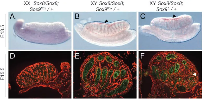
To further support the hypothesis that functional complementation rather than genetic background caused the sex reversal in our Sox8/Sox9 double knock-out model, we investigated whether the Sox8 mutation would provoke a gonadal phenotype in a Sox9 heterozygous knock-out
background. Sox9 heterozygous-mutant mice wild-type for Sox8 did not show a gonadal phenotype (Bi et al., 2001). Similarly, Sox8 knock-out mice wild-type for Sox9 showed normal sex cord development and a clearly outlined coelomic vessel (Fig. 9B). By contrast, three out of three animals (6 gonads) heterozygous for the Sox9 deleted allele, and homozygous for the Sox8 mutation (Sox8/Sox8; Sox9∆/+), showed abnormal sex cord formation and defects in coelomic vessel formation (Fig. 9C). Moreover, Sox8/Sox8; Sox9∆/+ testes at E15.5 showed a reduced number of seminiferous tubules and contained areas that resembled an ovary lacking Mis expression (Fig. 9F).
Discussion
[image:8.612.42.564.86.150.2]Since its cloning Sox9 has attracted considerable interest because of its association with campomelic syndrome in man (Foster et al., 1994; Wagner et al., 1994). Although there has been a substantial amount of evidence supporting an important role for Sox9 in the sex determination process (Kent et al., 1996; Morais da Silva et al., 1996; de Santa Barbara et al., 2000; Hanley et al., 2000; Huang et al., 1999; Bishop et al., 2000; Vidal et al., 2001), direct proof of its essential function during sexual differentiation has been missing.
Table 1. Overview of gonads analyzed
Sox9flox/ Sox8/+; Sox8/+; Sox8/Sox8; Sox8/Sox8;
Sox9flox/+ Sox9flox Sox9flox/+ Sox9flox/Sox9flox Sox9flox/+ Sox9flox/Sox9flox
XX 40 28 9 1 5 18
XY 33 31 13 7 8 6
XX (Sf1:Cre) 37 27 6 4 7 9
XY (Sf1:Cre) 30 31 11 9 (4SR) 5 3 (1SR)
Gonads were analyzed at various stages of development and judged by the presence of sex cords (E13.5), or their external appearance and descent status (E15.5 and E18.5). Sex-reversed gonads or those with abnormal appearance were sectioned (cryostat or paraffin-wax embedded) to confirm the finding histologically. Complete sex reversal (SR) was only detected in mice homozygous for the Sox9 knock-out and heterozygous/homozygous for the Sox8 mutation. Note, table only includes gonads with complete sex reversal (no sex cords); gonads with present but abnormal sex cords were scored as testes.
[image:8.612.46.391.298.739.2]Here, we have used conditional gene targeting to analyze the developmental function of Sox9 during mouse testis formation. Gonads completely lacking Sox9 expression do not show any signs of male specific differentiation, lack the expression of testis-specific genes, such as Mis and Scc, and instead express the female-specific markers Bmp2 and follistatin (Fig. 7). These data, together with our earlier studies showing that expression of Sox9 in XX gonads can substitute for Sry function in male sex determination, demonstrate that Sox9 is both required and sufficient for male testis differentiation in mice. These findings are also in agreement with a model in which Sry serves simply as a molecular switch to activate the Sox9 gene, which in turn directs gonadal cells to differentiate into Sertoli cells leading to the formation of a testis. However, the regulation of Sox9 is likely to be more complex than a simple activation through Sry, as mice lacking the orphan nuclear receptor gene Dax1 (Nr0b1 – Mouse Genome Informatics) on a Mus poschiavinus background do not express Sox9, despite apparently normal levels of Sry (Meeks et al., 2003). A detailed analysis will require the identification of gonad-specific regulatory elements at the Sox9 locus.
The fact that we did not observe a complete sex reversal in our tissue-specific knock-out mice using the Sf1:Cre transgene was probably due to incomplete inactivation of Sox9 at the time of sex determination, suggesting that the remaining small amounts of this protein may have been sufficient to activate male-specific genes. However, these variations in Sox9 levels allowed us to draw a number of important conclusions. First, using real-time PCR analysis, we could relate the expression of varying amounts of Sox9 with the expression of several genes known to be involved in the process of sexual differentiation. This analysis demonstrated a clear dependence of Mis expression on Sox9 levels, thus supporting in vivo the findings of earlier studies that described Mis as a direct transcriptional target of Sox9 (Arango et al., 1999; De Santa Barbara et al., 1998). Similarly, the transcription factor Sox8 directly depends on expression levels of Sox9. At present we
cannot clearly distinguish whether this dependence is due to a direct regulation of Sox8 by Sox9, or whether it simply reflects a reduction of the number of Sertoli cells in knock-out animals. In contrast to the direct dependence of Mis and Sox8 on Sox9 expression levels, Sry expression was higher in gonads with low levels of Sox9. At present there is no evidence for a repressive function of the Sox9 protein, and the downregulation of Sry may occur through the interaction of Sox9 with other factors, or through the activation of a transcriptional repressor. The fact that Sry levels were only increased in cases where Sox9 expression dropped below a certain threshold may suggest that persistent expression is an indirect event, possibly because of a continuous presence of Sry-positive Sertoli precursor cells originating from the coelomic epithelium (Karl and Capel, 1998; Schmahl et al., 2000) that fail to differentiate.
The incomplete inactivation of Sox9 in our tissue-specific analysis also allowed us to look at the differentiation of testes with reduced expression levels of Sox9 at later stages of development. In our histological analysis of E18.5 XY gonads, we observed meiotic gonocytes, a hallmark of female differentiation. This may suggest that Sox9 is important to maintain a signal that suppresses meiosis in XY gonocytes. Further analysis may allow us to identify this signal.
Maybe the most surprising finding in our studies was the absence of sex reversal in tissue-specific knock-out animals (Sf1:Cre; Sox9flox/Sox9flox), despite the significant reduction of
[image:9.612.110.502.72.262.2]seen following Sox9 induction. Based on their studies, Schepers et al. (Schepers et al., 2003) speculated that Sox8 expression in the gonad might only represent an evolutionary remnant of a duplicated gene, which is in the process of adopting new functions. However, another possible explanation would be that upregulation of Sox8 by Sox9 (directly or indirectly) soon after the commencement of sex determination is used to reinforce and imprint male sex determination and testis differentiation on the developing gonad. This hypothesis is further strengthened by the fact that Sox8 expression seems to depend on Sox9 levels rather than the expression of Sry, which may suggest that the male-specific regulatory elements of Sox8 were acquired independently of that of Sox9.
When interpreting results in a tissue-specific knock-out it is important to consider the inactivation of Sox9 on a cellular level. In any given cell, Sox9 is either wild-type (100%), heterozygous (50%) or homozygous (0%) for the mutation. Cells that are heterozygous for the Sox9 knock-out allele seem to differentiate normally into Sertoli cells (at least in mice), as heterozygous mutants do not show a gonadal phenotype (Bi et al., 2001). However, in the case of an additional heterozygous or homozygous deletion of Sox8, the overall Sox8/Sox9 gene dosage in a given cell would be further reduced, which may interfere with the activation of their transcriptional targets and, as a consequence, block the differentiation of this cell into a Sertoli cell.
Taken together, our data suggest the following model. Sox9 is activated through the expression of Sry, and is both essential and sufficient to induce testis formation. Sox9, either directly or indirectly, represses expression of Sry and activates Sox8. A certain threshold of Sox8 and Sox9, or Sox9 on its own, is then required for gonadal precursor cells to differentiate into Sertoli cells.
Our finding that Sox8 may reinforce Sox9 function during gonad formation may also have an impact on human genetics. Only 75% of XY individuals with campomelic dysplasia show sex reversal. The reason for this variable penetrance of the phenotype is presently unknown. Our data suggest that the gonadal phenotype in individuals with campomelic dysplasia may be influenced by the expression of SOX8, which could be variable because of either mutations at the SOX8 locus or polymorphism of SOX8, which influence its expression level or function. Moreover, given its ability to at least partially take over SOX9 function, it is conceivable that ectopic activation of SOX8 in XX gonads may cause sex reversal in human patients similar to findings for the SOX9 gene.
We thank Danilo Landrock for help with the generation of transgenic lines, Christoph Englert for communicating his Sf1 promoter studies before publication and Kay Wagner for critically reading this manuscript. We are also grateful to Olfert Landt (TIB Molbiol) for help with the LightCycler probe design, and to Blanche Capel for communicating unpublished information about Bmp2 and follistatin. These studies were supported by the Volkswagenstiftung, EC-grant QLG2-CT-1999-00741, and the National Institutes of Health (NIH) HD30184. Veterinary resources were supported by the NIH Cancer Center Support Grant CA16672.
References
Adams, I. R. and McLaren, A. (2002). Sexually dimorphic development of
mouse primordial germ cells: switching from oogenesis to spermatogenesis. Development 129, 1155-1164.
Akiyama, H., Chaboissier, M. C., Martin, J. F., Schedl, A. and De Crombrugghe, B. (2002). The transcription factor Sox9 has essential roles in successive steps of the chondrocyte differentiation pathway and is required for expression of Sox5 and Sox6. Genes Dev. 16, 2813-2828. Arango, N. A., Lovell-Badge, R. and Behringer, R. R. (1999). Targeted
mutagenesis of the endogenous mouse Mis gene promoter: in vivo definition of genetic pathways of vertebrate sexual development. Cell 99, 409-419. Bell, D. M., Leung, K. K., Wheatley, S. C., Ng, L. J., Zhou, S., Ling, K.
W., Sham, M. H., Koopman, P., Tam, P. P. and Cheah, K. S. (1997). SOX9 directly regulates the type-II collagen gene. Nat. Genet. 16, 174-178. Bi, W., Huang, W., Whitworth, D. J., Deng, J. M., Zhang, Z., Behringer, R. R. and De Crombrugghe, B. (2001). Haploinsufficiency of Sox9 results in defective cartilage primordia and premature skeletal mineralization. Proc. Natl. Acad. Sci. USA 98, 6698-6703.
Bishop, C. E., Whitworth, D. J., Qin, Y., Agoulnik, A. I., Agoulnik, I. U., Harrison, W. R., Behringer, R. R. and Overbeek, P. A. (2000). A transgenic insertion upstream of sox9 is associated with dominant XX sex reversal in the mouse. Nat. Genet. 26, 490-494.
Bowles, J., Schepers, G. and Koopman, P. (2000). Phylogeny of the SOX family of developmental transcription factors based on sequence and structural indicators. Dev. Biol. 227, 239-255.
Canning, C. A. and Lovell-Badge, R. (2002). Sry and sex determination: how lazy can it be? Trends Genet. 18, 111-113.
De Santa Barbara, P., Bonneaud, N., Boizet, B., Desclozeaux, M., Moniot, B., Sudbeck, P., Scherer, G., Poulat, F. and Berta, P. (1998). Direct interaction of SRY-related protein SOX9 and steroidogenic factor 1 regulates transcription of the human anti-Mullerian hormone gene. Mol. Cell. Biol. 18, 6653-6665.
De Santa Barbara, P., Moniot, B., Poulat, F. and Berta, P. (2000). Expression and subcellular localization of SF-1, SOX9, WT1, and AMH proteins during early human testicular development. Dev. Dyn. 217, 293-298.
de Vries, W. N., Binns, L. T., Fancher, K. S., Dean, J., Moore, R., Kemler, R. and Knowles, B. B. (2000). Expression of Cre recombinase in mouse oocytes: a means to study maternal effect genes. Genesis 26, 110-112. Dolci, S. and De Felici, M. (1990). A study of meiosis in chimeric mouse fetal
gonads. Development 109, 37-40.
Foster, J. W., Dominguez Steglich, M. A., Guioli, S., Kowk, G., Weller, P. A., Stevanovic, M., Weissenbach, J., Mansour, S., Young, I. D., Goodfellow, P. N. et al. (1994). Campomelic dysplasia and autosomal sex reversal caused by mutations in an SRY-related gene. Nature 372, 525-530.
Gu, H., Zou, Y. R. and Rajewsky, K. (1993). Independent control of immunoglobulin switch recombination at individual switch regions evidenced through Cre-loxP-mediated gene targeting. Cell 73, 1155-1164. Hacker, A., Capel, B., Goodfellow, P. and Lovell-Badge, R. (1995).
Expression of Sry, the mouse sex determining gene. Development 121, 1603-1614.
Hammes, A., Guo, J. K., Lutsch, G., Leheste, J. R., Landrock, D., Ziegler, U., Gubler, M. C. and Schedl, A. (2001). Two splice variants of the Wilms’ tumor 1 gene have distinct functions during sex determination and nephron formation. Cell 106, 319-329.
Hanley, N. A., Hagan, D. M., Clement-Jones, M., Ball, S. G., Strachan, T., Salas-Cortes, L., McElreavey, K., Lindsay, S., Robson, S., Bullen, P. et al. (2000). SRY, SOX9, and DAX1 expression patterns during human sex determination and gonadal development. Mech. Dev. 91, 403-407. Hogan, B. L., Beddington, R. S., Constantini, F. and Lacy, E. (1994).
Manipulating the Mouse Embryo. Cold Spring Harbor: Cold Spring Harbor Laboratory Press.
Hossain, A. and Saunders, G. F. (2001). The human sex-determining gene sry is a direct target of wt1. J. Biol. Chem. 276, 16817-16823.
Houston, C. S., Opitz, J. M., Spranger, J. W., Macpherson, R. I., Reed, M. H., Gilbert, E. F., Herrmann, J. and Schinzel, A. (1983). The campomelic syndrome: review, report of 17 cases, and follow-up on the currently 17-year-old boy first reported by Maroteaux et al. in 1971. Am. J. Med. Genet. 15, 3-28.
Huang, B., Wang, S., Ning, Y., Lamb, A. N. and Bartley, J. (1999). Autosomal XX sex reversal caused by duplication of SOX9. Am. J. Med. Genet. 87, 349-353.
Karl, J. and Capel, B. (1998). Sertoli cells of the mouse testis originate from the coelomic epithelium. Dev. Biol. 203, 323-333.
Kent, J., Wheatley, S. C., Andrews, J. E., Sinclair, A. H. and Koopman, P. (1996). A male-specific role for SOX9 in vertebrate sex determination. Development 122, 2813-2822.
Lefebvre, V., Huang, W., Harley, V. R., Goodfellow, P. N. and De Crombrugghe, B. (1997). SOX9 is a potent activator of the chondrocyte-specific enhancer of the pro alpha1(II) collagen gene. Mol. Cell. Biol. 17, 2336-2346.
McLaren, A. and Southee, D. (1997). Entry of mouse embryonic germ cells into meiosis. Dev. Biol. 187, 107-113.
Meeks, J. J., Weiss, J. and Jameson, J. L. (2003). Dax1 is required for testis determination. Nat. Genet. 34, 32-33.
Morais da Silva, S., Hacker, A., Harley, V., Goodfellow, P., Swain, A. and Lovell-Badge, R. (1996). Sox9 expression during gonadal development implies a conserved role for the gene in testis differentiation in mammals and birds. Nat. Genet. 14, 62-68.
O’Gorman, S., Dagenais, N. A., Qian, M. and Marchuk, Y. (1997). Protamine-Cre recombinase transgenes efficiently recombine target sequences in the male germ line of mice, but not in embryonic stem cells. Proc. Natl. Acad. Sci. USA 94, 14602-14607.
Ohe, K., Lalli, E. and Sassone-Corsi, P. (2002). A direct role of SRY and SOX proteins in pre-mRNA splicing. Proc. Natl. Acad. Sci. USA 99, 1146-1151.
Pilon, N., Daneau, I., Paradis, V., Hamel, F., Lussier, J. G., Viger, R. S. and Silversides, D. W. (2003). Porcine SRY promoter is a target for steroidogenic factor 1. Biol. Reprod. 68, 1098-1106.
Schepers, G. E., Bullejos, M., Hosking, B. M. and Koopman, P. (2000). Cloning and characterisation of the Sry-related transcription factor gene Sox8. 28, 1473-1480.
Schepers, G., Wilson, M., Wilhelm, D. and Koopman, P. (2003). SOX8 is expressed during testis differentiation in mice and synergises with SF1 to activate the Amh promoter in vitro. J. Biol. Chem. 278, 28101-28108. Schmahl, J., Eicher, E. M., Washburn, L. L. and Capel, B. (2000). Sry
induces cell proliferation in the mouse gonad. Development 127, 65-73.
Schwenk, F., Baron, U. and Rajewsky, K. (1995). A cre-transgenic mouse strain for the ubiquitous deletion of loxP-flanked gene segments including deletion in germ cells. Nucl. Acids. Res. 23, 5080-5081.
Shen, J. H. and Ingraham, H. A. (2002). Regulation of the orphan nuclear receptor steroidogenic factor 1 by Sox proteins. Mol. Endocrinol. 16, 529-540.
Sock, E., Schmidt, K., Hermanns-Borgmeyer, I., Bosl, M. R. and Wegner, M. (2001). Idiopathic weight reduction in mice deficient in the high-mobility-group transcription factor Sox8. Mol. Cell. Biol. 21, 6951-6959. Sudbeck, P., Schmitz, M. L., Baeuerle, P. A. and Scherer, G. (1996). Sex
reversal by loss of the C-terminal transactivation domain of human SOX9. Nat. Genet. 13, 230-232.
Toyooka, Y., Tanaka, S. S., Hirota, O., Tanaka, S., Takagi, N., Yamanouchi, K., Tojo, H. and Tachi, C. (1998). Wilms’ tumor suppressor gene (WT1) as a target gene of SRY function in a mouse ES cell line transfected with SRY. Int. J. Dev. Biol. 42, 1143-1151.
Vidal, V. P., Chaboissier, M. C., de Rooij, D. G. and Schedl, A. (2001). Sox9 induces testis development in XX transgenic mice. Nat. Genet. 28, 216-217. Wagner, T., Wirth, J., Meyer, J., Zabel, B., Held, M., Zimmer, J., Pasantes, J., Bricarelli, F. D., Keutel, J., Hustert, E. et al. (1994). Autosomal sex reversal and campomelic dysplasia are caused by mutations in and around the SRY-related gene SOX9. Cell 79, 1111-1120.
Wilhelm, D. and Englert, C. (2002). The Wilms tumor suppressor WT1 regulates early gonad development by activation of Sf1. Genes Dev. 16, 1839-1851.
Wilkinson, D. G. (1992). Whole mount in situ hybridization of vertebrate embryos. In In situ hybridization (ed. D. G. Wilkinson), pp. 75-83. Oxford: Oxford University Press.
Wirth, J., Wagner, T., Meyer, J., Pfeiffer, R. A., Tietze, H. U., Schempp, W. and Scherer, G. (1996). Translocation breakpoints in three patients with campomelic dysplasia and autosomal sex reversal map more than 130 kb from SOX9. Hum. Genet. 97, 186-193.