doi:10.1093/jxb/ern153 Advance Access publication 23 June, 2008
This paper is available online free of all access charges (see http://jxb.oxfordjournals.org/open_access.html for further details)
RESEARCH PAPER
Hormonal changes during salinity-induced leaf senescence
in tomato (
Solanum lycopersicum
L.)
Michel Edmond Ghanem1,*, Alfonso Albacete2,*, Cristina Martı´nez-Andu´jar2, Manuel Acosta3, Remedios Romero-Aranda4, Ian C. Dodd5, Stanley Lutts1and Francisco Pe´rez-Alfocea2,† 1
Groupe de Recherche en Physiologie Ve´ge´tale, Universite´ catholique de Louvain (UCL), Croix du Sud 5, boıˆte 13, B-1348 Louvain-la-Neuve, Belgium
2
Departamento de Nutricio´n Vegetal, Centro de Edafologı´a y Biologı´a Aplicada del Segura (CEBAS), Consejo Superior de Investigaciones Cientı´ficas (CSIC), Campus Universitario de Espinardo, PO Box 164, E-30100 Murcia, Spain
3
Departamento de Biologı´a Vegetal–Fisiologı´a Vegetal, Facultad de Biologı´a, Universidad de Murcia, Campus Universitario de Espinardo, E-30100 Murcia, Spain
4
Departamento de Mejora Vegetal, Estacio´n Experimental ‘La Mayora’ (EELM), Consejo Superior de Investigaciones Cientı´ficas (CSIC), E-29750 Algarrobo-Costa, Ma´laga, Spain
5
Department of Biological Sciences, The Lancaster Environment Centre, Lancaster University, Lancaster LA1 4YQ, UK
Received 19 February 2008; Revised 30 April 2008; Accepted 8 May 2008
Abstract
Leaf senescence is one of the most limiting factors to plant productivity under salinity. Both the accumula-tion of specific toxic ions (e.g. Na+) and changes in leaf hormone relations are involved in the regulation of this process. Tomato plants (Solanum lycopersicum L. cv Moneymaker) were cultivated for 3 weeks under high salinity (100 mM NaCl) and leaf senescence-related parameters were studied during leaf development in relation to Na+ and K+ contents and changes in abscisic acid (ABA), cytokinins, the ethylene precursor 1-aminocyclopropane-1-carboxylic acid (ACC), and the auxin indole-3-acetic acid (IAA). Na+ accumulated to a similar extent in both leaves 4 and 5 (numbering from the base of the plant) and more quickly during the third week, while concurrently K+ contents sharply de-creased. However, photosystem II efficiency, mea-sured as the Fv/Fm ratio, decreased from the second week of salinization in leaf 4 but only at the end of the third week in the younger leaf 5. In the prematurely senescent leaf 4, ABA content increased linearly while IAA strongly decreased with salinization time. Al-though zeatin (Z) levels were scarcely affected by
salinity, zeatin-riboside (ZR) and the total cytokinin content (Z+ZR) progressively decreased by 50% from the imposition of the stress. ACC was the only hormonal compound that increased in leaf tissue coincident with the onset of oxidative damage and the decline in chlorophyll fluorescence, and prior to massive Na+ accumulation. Indeed, (Z+ZR) and ACC contents and their ratio (Z+ZR/ACC) were the hormonal parameters best correlated with the onset and pro-gression of leaf senescence. The influence of different hormonal changes on salt-induced leaf senescence is discussed.
Key words: Abscisic acid, 1-aminocyclopropane-1-carboxylic acid, indole-3-acetic acid, plant hormones, salt stress, senescence, sodium chloride, tomato (Solanum lycopersicum L.), zeatin, zeatin-riboside.
Introduction
Tomato (Solanum lycopersicum L.) is the world’s most important vegetable crop in economic terms (Nuez et al., 2004), and is considered moderately salt tolerant, with
* These authors contributed equally to this work. y
To whom correspondence should be addressed. E-mail: alfocea@cebas.csic.es
yield reduced by 50% under a moderately saline regime of ;8 dS m1 (Subbarao and Johansen, 1994). Since available soil and water resources are becoming increas-ingly salinized, the negative impact on crop productivity is continuously increasing. Hence, efforts have been made during the last 50 years to improve the salt tolerance of tomato, but with little success due in part to insufficient knowledge about the mechanisms involved in limiting yield (Cuartero et al., 2005). Since cellular events tri-ggered by salinity, such as salt compartmentation, osmotic adjustment, changes in metabolic fluxes, and cell wall hardening, are connected to whole plant responses, such as growth reduction, changes in biomass allocation and phenology, leaf senescence, and finally plant death (Volkmar et al., 1998), the candidate genes to increase salt tolerance are those involved in regulating such processes (Munns, 2005). Therefore, salt tolerance could be improved by avoiding or delaying senescence, allow-ing plant resource acquisition for a longer period, to generate new growth and maintain defence mechanisms such as ionic regulation to avoid the massive entry of toxic ions to the plant
Plant senescence is an internally programmed degrada-tion leading to cell death. Since chloroplasts appear to be the initial target of senescence, and chlorophyll breakdown is so prominent, chlorophyll loss and the associated yellowing of the leaves are commonly used as indicators of plant senescence (Noode´n et al., 1997). The onset and progression of leaf senescence are regulated by both environmental and endogenous factors and their interac-tions. Environmental cues include stress factors that adversely affect plant development and productivity, such as drought, waterlogging, high or low solar radiation, extreme temperatures, ozone and other air pollutants, excessive soil salinity, and inadequate mineral nutrition in the soil (Munne´-Bosch and Alegre, 2004). Endogenous factors include age, reproductive development, and level of plant hormones. These environmental cues may accelerate leaf senescence by affecting those endogenous factors (Munne´-Bosch and Alegre, 2004).
Salinity reduces plant productivity first by reducing plant growth during the phase of osmotic stress and subsequently by inducing leaf senescence during the phase of toxicity when excessive salt is accumulated in transpiring leaves (Munns, 2002a). Indeed, in spite of the positive effect of Na+ compartmentation into vacuoles to maintain turgor (Flowers, 2004), premature senescence caused by salinity seems to occur more quickly in glycophytic plants having a higher Na+uptake (Fortmeier and Schubert, 1995; Munns, 2002a, b). However, as previously stated, growth inhibition and metabolic changes occurring during the osmotic phase of salinity are similar to those triggered by drought, which can also lead to premature senescence (Munns, 2002a, b). In turn, senescence induced by the osmotic component of salinity
probably follows the same physiological events as drought-induced senescence (Pic et al., 2002). The initial reduction in shoot growth followed by accelerated senescence is probably related to hormonal signals generated in response to the stress. Much evidence suggests that cytokinins (CKs) are the major leaf senescence-inhibiting hormones, since senescence is de-layed after the exogenous application of CKs (Van Staden et al., 1988) or the overproduction of CKs in transgenic plants transformed with the CK biosynthetic genes, isopentenyl transferase (IPT) (Gan and Amasino, 1995; Cowan et al., 2005; Rivero et al., 2007) and zeatin O-glucosyltransferase (ZOG1) (Havlova et al., 2008), and senescence is correlated with a decline in endogenous CK levels (Van Staden et al., 1988; Singh et al., 1992; Gan and Amasino, 1995). Leaf senescence can only be initiated when leaf CK levels fall below a threshold (Noode´n et al., 1997). Maintaining CK levels above this threshold inhibits transcriptional regulation of senescence-related genes and prevents the onset of senescence in the entire plant (Gan and Amasino, 1995).
Although some studies have addressed how phytohor-mones were affected by salinity (reviewed by Naqvi, 1994; Munns, 2002a), there has been little attention paid to the role of changes in endogenous hormonal balance in the onset and progression of salt stress-induced leaf senescence in an important horticultural crop such as tomato. Therefore, the aim of this study was to examine the dynamic changes of selected endogenous hormones [CKs, ABA, ACC, and indole-3-acetic acid (IAA)] in relation to the salt-induced senescence of tomato leaves (S. lycopersicum L.). Such information is a necessary preliminary to modifying hormone contents in vivo with the aim of decreasing salt-induced senescence.
Materials and methods
Plant material and culture conditions
Seeds of tomato (S. lycopersicum L.) cv. Moneymaker were sown and seedlings allowed to grow in a germination chamber in trays filled with a perlite–vermiculite mix (1:3 v/v) moistened regularly with a half-strength modified Hoagland nutrient solution. Fourteen days after sowing, seedlings were transferred into a growth chamber and fixed on polyvinyl chloride plates floating on aerated half-strength modified Hoagland nutrient solution. The nutrient solution contained the following chemicals (in mM): 5 KNO3, 1 NH4H2PO4,
0.5 MgSO4, 5.5 Ca(NO3)2 and (in lM) 25 KCl, 10 H3BO3,
1 MnSO4, 1 ZnSO4, 0.25 CuSO4, 10 Na2MoO4, and 1.87 g l 1
Fe-EDDHA. Solutions were refilled every 2 d and renewed every week.
Plants were grown in a growth chamber under a 16 h daylight period. The air temperature ranged from 25C to 28C during the day and from 17C to 18C during the night. Relative humidity was maintained at 7065% during the night and at 5065% during the day. Light intensity at the top of the canopy was;245lmol m2s1[photosynthetic photon flux density (PPFD)]. After 4 d of acclimatization in control conditions (18 d after sowing), the seedlings were exposed to 0 (control) or 100 mM NaCl added to the nutrient solution for 22 d. Three replications with eight plants per replication and salt treatment were used in this study for the measurement of different parameters. Two specific leaves per plant were chosen when salinity was imposed to monitor senescence and for subsequent biochemical determinations: a young expanding leaf (;100 cm2), identified as leaf number 4 (from the bottom), and leaf number 5. Leaf material from six plants was harvested for different analyses at 1, 9, 15, and 22 d of salt treatment.
Chlorophyll fluorescence
Modulated chlorophyll fluorescence was measured in tagged and dark-adapted (30 min) leaves in 6–10 plants per treatment, using a chlorophyll fluorometer OS-30 (OptiSciences, Herts, UK) with an excitation source intensity of 3000lmol m2 s1. The minimal fluorescence intensity (F0) in a dark-adapted state was measured in
the presence of a background far-red light to favour rapid oxidation of intersystem electron carriers. The maximal fluorescence in-tensities in the dark-adapted state (Fm) and after adaptation to white
actinic light (Fm#) were measured by 0.8 s saturating pulses (3000lmol m2s1). After theFm#measurement, the actinic light (400 mol m2 s1) was switched off, and the far-red light was applied for 3 s in order to measure the minimal fluorescence intensity in the light-adapted state (F0#). The maximum quantum
yield of open photosystem II (PSII) (Fv/Fm) and the
non-photochemical quenching (NPQ) were calculated as (Fm–F0)/Fm
and (Fm–Fm#)/Fm#, respectively (Maxwell and Johnson, 2000).
Oxidative damage
The level of lipid peroxidation was measured as 2-thiobarbituric acid-reactive substances (TBARs), mainly malondialdehyde (MDA), following the modified method of Heath and Parker (1968). Frozen leaf samples (0.25 g) were homogenized in a pre-chilled mortar with 5 ml of ice-cold 5% (w/v) trichloroacetic acid (TCA) and centrifuged at 12 000gfor 15 min at 4C. The assay mixture containing a 2 ml aliquot of supernatant and 2 ml of 0.67% (w/v) thiobarbituric acid was heated to 100C for 30 min and then rapidly cooled to 4C in an ice-bath. After centrifugation (10 000g
for 1 min at 4C), the supernatant absorbance was read (532 nm) and values corresponding to non-specific absorption (600 nm) were subtracted. The MDA concentration was calculated using its molar extinction coefficient (155 mM1cm1).
Ion concentration
For K+and Na+quantification, leaf tissues were frozen with liquid nitrogen. After thawing, the samples were centrifuged for 10 min at 10 000gand then for 5 min at 20 000gto obtain the bulk tissue sap. Necessary dilutions were performed in order to measure K+and Na+ concentrations, determined by using a Shimadzu AA-680 atomic absorption spectrophotometer (Shimadzu Ltd, Kyoto, Japan). All measurements were performed in three replicates.
Hormone extraction and analysis
Mobile phase A, consisting of water/acetonitrile/formic acid (94.9:5:0.1 by vol.), and mobile phase B, consisting of water/ acetonitrile/formic acid (10:89.9:0.1 by vol.), were used for the chromatographic separation. The elution program maintained 100% A during 5 min, then a linear gradient from 0% to 6% B in 10 min, followed by another linear gradient from 6% to 100% B in 5 min, and finally maintained at 100% B for another 5 min. The column was equilibrated with the starting composition of the mobile phase for 30 min before each analytical run. The UV chromatogram was recorded at 280 nm with the DAD module (Agilent Technologies). The mass spectrometer was operated in the positive mode with a capillary spray voltage of 3500 V, and a scan speed of 22 000 (m/z) s1from 50 to 500m/z. The nebulizer gas (He) pressure was set to 30 psi, whereas the drying gas was set to a flow of 6 l min1
at a temperature of 350C. Mass spectra were obtained using the DataAnalysis program for LC/MSD Trap Version 3.2 (Bruker Daltonik GmbH, Germany). For quantification of Z, ZR, ABA, and IAA, calibration curves were constructed for each analysed component (0.05, 0.075, 0.1, 0.2, and 0.5 mg l1) and corrected for 0.1 mg l1 internal standards: [2H5]trans-zeatin, [2H5]trans-zeatin
riboside, [2H6]cis,trans-abscisic acid (Olchemin Ltd, Olomouc,
Czech Republic), and [13C6]indole-3-acetic acid (Cambridge
Iso-tope Laboratories Inc., Andover, MA, USA). Recovery percentages ranged between 92% and 95%.
ACC was determined after conversion into ethylene by gas chromatography using an activated alumina column and an FID detector (Cromatix-KNK-2000, Konik, Barcelona, Spain). ACC was extracted with 80% (v/v) ethanol and assayed by degradation with alkaline hypochlorite in the presence of 5 mM HgCl2(Casas
et al., 1989). A preliminary purification step was performed by passing the extract through a Dowex 50W-X8, 50–100 mesh, H+-form resin and later recovered with 0.1 N NH4OH. The
conversion efficiency of ACC into ethylene was calculated separately by using a replicate sample containing 2.5 nmol of ACC as internal standard, and used for correction of data.
Enzyme extraction and assay
To measure cytokinin oxidase (CKX, EC 1.5.99.12) activity, leaf samples were cut into small pieces, powdered with liquid nitrogen using a pestle and mortar, and extracted with a 1.5-fold excess (v/w) of 0.2 M TRIS-HCl buffer, pH 8, containing 1 mM phenyl-methylsulphonyl fluoride and 3% Triton X-100. Tissue debris was removed by centrifugation at 12 000gfor 10 min. The extract was loaded onto a Sephadex G-25 (5032.5 cm) column equilibrated with 0.1 M TRIS-HCl, pH 8, to remove the low molecular mass fraction. The protein fraction was then used to assay CKX activity. The assay was performed according to the method described by Fre´bort et al. (2002), with some modifications. Samples were incubated in a reaction mixture (total volume 0.6 ml in an Eppendorf tube) of 100 mM reaction buffer (imidazole/HCl buffer, pH 6), 0.5 mM electron acceptor [2, 6-dichloroindophenol] and 0.5 mM substrate, for 0.5–12 h at 37C.
To measure cell wall invertase (CWIN, EC 3.2.1.25) activity, the enzyme extracts were prepared essentially as described in Balibrea
et al.(1999). Fresh leaf tissue samples (100 mg) were frozen with liquid nitrogen and stored at 20C until analysis. Samples containing polyvinylpyrrolidone and Fontainebleau sand were homogenized in 1 ml of extraction buffer containing 50 mM HEPES-KOH (pH 7), 10 mM MgCl2, 1 mM Na2EDTA, 2.6 mM
dithiothreitol (DTT), 10% ethylene glycol, and 0.02% Triton X-100. After centrifugation at 20 000g, the supernatant was discarded and the pellet was washed three times and re-suspended in 30 mM acetate buffer (pH 5). The amount of hexoses was determined through an enzyme-linked assay monitoring NADH formation at 340 nm, after adding 25ll of 0.6 M sucrose and incubating at
30C for 15 min. The proteins were analysed in the pellet after solubilization with 1 M NaCl, and the specific enzymatic activities were expressed as nkat mg1protein.
Statistical analysis
Data were subjected to an analysis of variance (ANOVA II) using the SAS software (SAS System for Windows, version 8.02). The statistical significance of the results was analysed by the Student– Newman–Keuls test at the 5% level.
Results
Chlorophyll fluorescence and leaf senescence-related parameters
The evolution of chlorophyll fluorescence and senescence-related parameters during salt stress is shown in Fig. 1. In leaf 4 of control plants, the maximum quantum efficiency of PSII (Fv/Fm) was almost constant during the growing period, whereas in salt-stressed plants, these values sharply decreased below those of control plants 14 d after salinization (Fig. 1A). However, in the younger leaf 5, this salinity-induced decrease inFv/Fmwas delayed by 1 week (Fig. 1B).
From the first day of salt treatment, NPQ increased significantly in leaf 4, reaching a maximum by day 15 before declining during the last week (Fig. 1C) together withFv/Fm. In control plants, NPQ increased as the leaves aged. In leaf 5, NPQ also increased during the first week of salinization but less than in the older leaf 4, and it reached control values on day 18 before strongly in-creasing on day 22, also coinciding with a significant decline in Fv/Fm(Fig. 1D).
The level of lipid peroxidation slowly increased with age in both control leaves 4 and 5, but the salt stress significantly increased it from day 15 and 22, respectively (Fig. 1E, F), coinciding with the decrease in Fv/Fm (Fig. 1A, B).
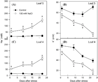
Sodium and potassium content
Sodium was accumulated to a similar extent in both leaves 4 and 5 (50–80 mM) during the first 2 weeks of salinization, and also a similar strong increase (until 150 mM) occurred at the end of the third week in both leaves (Fig. 2A, C). Compared with Na+, salinity induced an earlier and stronger reduction in K+ content in both leaves 4 and 5 (Fig. 2B, D). In the younger leaf 5 of control plants, K+ content decreased during the first 2 weeks but recovered during the third week, probably due to a remobilization from older leaves such as the senescent leaf 4.
Leaf hormonal profiling
salinity increased leaf 4 Z content by 2-fold 1 d after salinization, for the remainder of experiment the values were only slightly lower (averaging 80% of the control) than control plants (Fig. 3A). However, salinity progres-sively decreased leaf ZR contents: by 30% 1 d after salinization and by 60% at the end of the experiment (Fig. 3B). Consequently the total (Z+ZR) CK content decreased with leaf development more dramatically under salinity. At the onset of the salt-induced leaf senescence (by day 15, according to the decreasedFv/Fmand MDA accumulation),
the Z+ZR CK content was;50% lower than in the control leaves, and 70% lower than before salinization (Fig. 3).
[image:5.612.141.476.58.538.2]ACC, ABA, and IAA: The concentration of the ethylene precursor ACC remained almost constant (;5 ng g1 FW) in control leaves during leaf development, without any significant change that could be related to the onset of natural senescence (Fig. 4A). However, in leaf 4 of the salinized plants, the ACC levels increased during the second week. At the end of the experiment, the ACC
Fig. 1. Evolution of leaf senescence-related parameters. Maximum photochemical efficiency (Fv/Fm) (A, B), non-photochemical quenching (NPQ)
content in salinized leaves was 12-fold higher than in the control leaves.
Leaf ABA content increased linearly with time in both control and salinized plants, but the increase was four times greater in salinized plants during the experiment (Fig. 4B).
Leaf IAA concentration increased between days 1 and 15 in both control and salinized plants, before decreasing to the initial levels by the end of the experiment. Salinity
dramatically decreased IAA content (by 80%) on the first day of salt treatment, and this percentage reduction was maintained almost unchanged during the experiment (Fig. 4C). A replicate experiment showed a 50% decrease in leaf IAA content after 3 d of salinization (data not shown).
[image:6.612.139.473.60.338.2]Endogenous hormonal ratios: Although hormonal ratios, rather than absolute contents, could provide some insights
Fig. 2. Evolution of sodium (A, C) and potassium (B, D) concentrations in leaves 4 (C, D) and 5 (A, B) of tomato plants grown for 3 weeks on half-strength Hoagland medium in the absence (filled circles) or presence (open circles) of 100 mM NaCl. Data are means of six plants6SE. Asterisks indicate significant differences between control and salinized leaves according to Student–Newman–Keuls test atP<0.05.
[image:6.612.72.543.402.543.2]into the evolution of the salt-induced senescence, only two such ratios showed significant correlations with any indicator of senescence (Table 1). Thus, the (Z+ZR)/ABA ratio decreased linearly during leaf development under control conditions (from 5 at the start of salt treatment to 1 at the end of the experiment) (Fig. 5A). However, 1 d of salt treatment induced a similar effect as 3 weeks of development under non-stress conditions. After 3 weeks of salinization the (Z+ZR)/ABA ratio was close to zero.
The (Z+ZR)/ACC ratio decreased with age in control leaves (Fig. 5B). Salinity decreased this ratio from the first day of salinization and more drastically by day 15, coinciding with the onset of leaf senescence.
Effects of salinity on cytokinin dehydrogenase and cell wall invertase activities
CKX activity increased linearly as the leaves aged, irrespective of the growth conditions, but the activity was 2-fold higher in salinized plants by day 22 (Fig. 6A). Salt stress significantly decreased CWIN activity in leaf 4 from the first day of salt treatment (Fig. 6B).
Correlation analysis
CKs (Z+ZR) and the ethylene precursor ACC contents and their ratio (Z+ZR/ACC) were the best hormonal parameters related to senescence in leaf 4 (Table 1). However, the influence of both hormones was opposite:
whileFv/Fmwas positively correlated with the CK content and the (Z+ZR)/ACC ratio, a strong negative correlation was found with ACC contents. On the other hand, the evolution of the protective parameter NPQ was correlated positively with ABA but negatively with CK contents and their ratios with ACC and ABA. The indicator of oxidative stress MDA was inversely related to the indicator of PSII efficiencyFv/Fm(r ¼–0.84,P >0.01). In turn, oxidative damage (MDA content) was positively related to ABA and especially to ACC contents, while a strong and inverse correlation was found with CK content and the (Z+ZR)/ACC ratio. Finally, while the contents of the major nutrient K+ were positively correlated withFv/Fm and negatively with both NPQ and MDA in leaf 4, the contents of the toxic ion Na+ were only significantly correlated with the progression of the oxidative stress (Table 1).
Discussion
The onset of salt-induced leaf senescence seems independent of bulk leaf Na+accumulation
[image:7.612.73.539.408.553.2]In leaf 4 of salt-stressed plants, a considerable decline in Fv/Fmresulting from a loss of PSII efficiency was evident after 15 d of salt treatment, indicating the onset of salt-induced senescence (Guiame´t et al., 2002), while in the younger leaf 5 it was only observed after 22 d under
Fig. 4. Evolution of ACC (A), ABA (B), and IAA (C) contents during the development of leaf 4 in tomato plants grown for 3 weeks on half-strength Hoagland medium in the absence (filled circles) or presence (open circles) of 100 mM NaCl. Data are means of three plants6SE. Asterisks indicate significant differences between control and salinized leaves according to Student–Newman–Keuls test atP<0.05.
Table 1. Linear correlation coefficients between senescence-related parameters (Fv/Fm, NPQ, MDA) and hormonal, Na+, and K+ contents, and hormonal ratios in leaves developed on plants grown for 3 weeks on half-strength Hoagland medium in the absence or presence of 100 mM NaCl
Z+ZR ACC ABA IAA Z+ZR/ACC Z+ZR/ABA Z+ZR/IAA IAA/ACC ACC/ABA Na+ K+
Fv/Fm 0.75* –0.85** –0.59 0.24 0.70* 0.41 0.21 0.20 –0.55 –0.56 0.70*
NPQ –0.84** 0.63 0.77* –0.30 –0.90** –0.70* –0.25 –0.56 0.10 0.66 –0.77* MDA –0.87** 0.96*** 0.86** –0.49 –0.91** –0.61 –0.10 –0.58 0.46 0.80* –0.84**
[image:7.612.50.559.649.701.2]salinity. Since Na+accumulated to a similar extent in both leaf 4 and leaf 5, salt-induced senescence cannot be wholly explained by the putative toxic effect of this ion. Although the presumably higher Na+ accumulation in vacuoles of the younger and more actively growing leaf 5 could partially account for these differences (Flowers, 2004; Munnset al., 2006), severe toxic levels (>100 mM) were only found when massive Na+ influx occurred in both leaves during the last week of salinization. This could be related to a general failure of the control of Na+ exclusion at the root level and its translocation to the shoot, contributing to the late enhanced oxidative stress in leaf 4 and 5, as suggested by the positive correlation between Na+contents and lipid peroxidation (measured as
MDA production). Nevertheless, the decrease in K+ content from the first day of salinization was more related than Na+ to the different senescence-related parameters (Table 1).
However, before the onset of the salt-induced senes-cence in leaf 4, the NPQ was strongly induced from day 1 of salt treatment. This parameter is considered a protective process minimizing the photooxidative damage resulting from the absorption of excess radiation in chloroplasts (Gilmore, 1997). Indeed, diminished capacity of NPQ in leaf 4 could be related to the oxidative damage by day 15 (Mytinovaet al., 2006) and with the subsequent decrease in Fv/Fm values and massive Na+ accumulation in both leaves.
[image:8.612.330.544.58.433.2]Hence, other processes occurring from the first days of salinization, when the osmotic effect predominates, are
[image:8.612.64.283.61.454.2]Fig. 5. Evolution of some hormonal ratios: (Z+ZR)/ABA (A) and (Z+ZR)/ACC (B) during the development of leaf 4 in tomato plants grown for 3 weeks on half-strength Hoagland medium in the absence (filled circles) or presence (open circles) of 100 mM NaCl. Data are means of three plants 6SE. Asterisks indicate significant differences between control and salinized leaves according to Student–Newman– Keuls test atP<0.05.
probably influencing the evolution of leaf senescence and the capacity of the plant to regulate the accumulation of toxic ions. These processes resulting in photooxidative damage and premature leaf senescence are common to several abiotic stresses such as cold, drought, and salinity (Hareet al., 1997; Huneret al., 1998), while the different responses found between leaves could be related to the different sink–source transitions during leaf development (Guiame´t et al., 2002; Cowan et al., 2005). Therefore, other endogenous factors influencing those processes, such as hormones, are likely to be influencing the onset and progression of the salt-induced senescence.
Low cytokinin levels promote salt-induced senescence but are not the only signal
The changing levels of major CKs in tomato (Z+ZR) was the hormonal parameter best related toFv/Fm, NPQ, and MDA (Table 1). The involvement of cytokinins in the prevention of leaf senescence has been extensively studied in different plant species (Gan and Amasino, 1995; Balibrea et al., 2004; He et al., 2005; Guo and Gan, 2007; Rivero et al., 2007). As a consequence, it is very likely that the large drop in CK contents (especially ZR) in salinized leaves (Fig. 3C) could contribute to the progression of senescence under stress. This decrease may be the result of less synthesis, reduced transport of root-synthesized CKs, and/or increased breakdown of CKs (Werneret al., 2006; Havlovaet al., 2008). Initially, decreased transport of CKs from the roots could be responsible for the decrease in the leaves, as suggested by the decrease of both CKs in root xylem sap under salinity (data not shown). However, the strong decrease observed in leaf ZR content by day 15 of salt treatment can also be explained by an induction of CKX activity (Fig. 6A). The role of CKX could be the maintenance of an optimal level of CKs for growth adaptation to the stress conditions (e.g. shift in shoot to root assimilate partitioning) and/or resetting a CK signalling system to a basal level (Werner et al., 2001; Roitsch et al., 2003). In this regard, the concomitant decrease of the CWIN activity (Fig. 6B), a key factor in the control of senescence by CKs (Balibrea et al., 2004), also argues in favour of the role of the low content of these hormones in the progression of salt-induced senescence (Gan and Amasino, 1995), but it is not a major signal triggering its onset (Werneret al.2003; Munne´-Bosch and Alegre, 2004).
IAA and ABA are not the major signals for salt-induced senescence
Although auxin effects on abscission and senescence were first reported >50 years ago (for a review, see Noode´n, 1988), the involvement of this hormone in senescence is much less understood than that of ABA, ethylene, or CKs, and it is difficult to conclude that it antagonizes
senescence (Gan, 2004). In the present study, the highest IAA levels in salinized leaves coincided with the onset of senescence, suggesting it promoted this process. In re-buttal of this notion, IAA levels of salinized plants were 50% lower than in control leaves. Therefore, although a low IAA content could stimulate senescence on the basis that this compound has been generally seen as a senes-cence-retarding factor (Schippers et al., 2007), it seems unlikely to be a primary factor in the onset of salt-induced senescence, as suggested by the lack of correlation with any of the related parameters (Table 1), and more work is needed to answer this question.
ABA increase in leaves could be partially responsible for the decreased CK contents, thus indirectly enhancing the salt-induced senescence. Moreover, since CKs counteract ABA-induced stomatal closure (Dodd, 2005) and inhibit the glucose repression response on photosynthesis (Moore et al., 2003), a low (Z+ZR)/ABA ratio would enhance the non-stomatal photosynthetic inhibition under salinity (Balibrea et al., 2000), as suggested by the negative correlation found with NPQ (Table 1).
However, the effect of ABA on the progress of senescence remains unclear and seems to depend on the plant species, growth conditions, and interactions with other hormones. In the ABA-deficient tomato mutant sitiens, salinity promoted leaf senescence more than in the wild type (Ma¨ke¨la et al., 2003), presumably due to a higher ethylene evolution (Sharpet al., 2000), while the higher incidence of leaf death was not explained by Na+ accumulation (Ma¨ke¨laet al., 2003). Moreover, it has also been proposed that ABA could provide a protective role against salinity and high intensity light stress in Arabi-dopsis (Cramer, 2002), which can be related to the induction of antioxidant defences (Hu et al., 2006). On the other hand, various Arabidopsis mutants defective in either ABA biosynthesis or signalling have not shown measurable effects on the timing or progression of leaf senescence under different environmental stresses (Hensel et al., 1993; Guo and Gan, 2007). Further studies are required to gain insights into the role of ABA in salt-induced leaf senescence.
Increase in the ethylene precursor ACC coincides with Fv/Fmdecrease and oxidative damage in salinized leaves
Ethylene has long been considered a key hormone in regulating the onset of senescence (Zacarias and Reid, 1990), and is also suspected to be involved in salt-induced senescence (Munns, 2002a; Wi and Park, 2002). In the present study, leaf ACC concentration sharply increased after 15 d of salt treatment, coincident with the decline in bothFv/Fmand NPQ and with the appearance of oxidative damage, which indicate the onset of salt-induced leaf senescence in that leaf. Although increases in ACC, ethylene evolution, and the activity of the enzyme responsible for this conversion (ACC oxidase) are usually concomitant (Go´mez-Cadenas et al., 1998; Dodd, 2005; van der Graaf et al., 2006), the relationship between increased ACC levels and onset of senescence should be considered with caution since leaf ethylene evolution was not directly measured. However, the relationship between ACC, ethylene, and senescence has been widely supported using physiological (Zacarias and Reid, 1990; Ella et al., 2003) and genetic approaches. Leaf senescence has been delayed in either transgenic tomato plants with reduced ethylene biosynthesis (Johnet al., 1995) or mutants with reduced sensitivity to ethylene (Zacarias and Reid, 1990;
Grbic and Bleecker, 1995). However, constitutive over-production of ethylene in Arabidopsis and tomato plants did not cause precocious senescence, suggesting that ethylene alone is not sufficient to initiate leaf senescence (Grbic and Bleecker, 1995). It has been postulated that age-dependent factors are required for ethylene-regulated leaf senescence (He and Gan, 2002). In this regard, the prior decreases in CK content (Fig. 3) could sensitize the senescence process to ethylene. Indeed, the (Z+ZR)/ACC ratio was strongly correlated with the senescence-related parameters (Table 1), suggesting that both hormones are important in this process. It is also interesting to note that increased ACC concentration was prior (day 15) to massive Na+entry into the leaves (day 22) and it has been reported that ethylene receptors (e.g. NTHK1) affect sensitivity to salinity and modify Na+/K+ homeostasis (Caoet al., 2006). These data suggest that ethylene and its precursor ACC might play a role in the onset of salt-induced leaf senescence, as suggested by the strong correlation found with the decline both in Fv/Fm and in MDA production (Figs 1A, C, E and 4A, Table 1).
Conclusion
Acknowledgements
The authors are very grateful to Dr A Torrecillas (CAID, Universidad de Murcia) for his help with hormonal analyses, to the Fonds National de la Recherche Scientifique-FNRS (Belgium) and to the Fundacio´n Se´neca (Comunidad Auto´noma de Murcia, Spain) for a travel grant to M.E.G. and F.P.A., respectively, and to the CSIC (Spain) for a research grant to AA (I3P Program). The research was supported by the Fundacio´n Se´neca (Comunidad Auto´noma de Murcia, Spain) (project 03011/PI/05) and by the CICYT-FEDER project AGL2007-63610/AGR. The authors dedi-cate this paper to the memory of the late Professors Manuel Caro (CEBAS-CSIC) and Gilles Guerrier (Universite´ d’Orle´ans, France).
References
Balibrea ME, Dell’Amico J, Boları´n MC, Pe´rez-Alfocea F.
2000. Carbon partitioning and sucrose metabolism in tomato plants growing under salinity.Physiologia Plantarum110,503– 511.
Balibrea ME, Gonza´lez MC, Fatima T, Lee TK, Proels R, Tanner W, Roitsch T. 2004. Extracellular invertase is an essential component of cytokinin-mediated delay of senescence.
The Plant Cell16,1276–1287.
Balibrea ME, Parra M, Boları´n MC, Pe´rez-Alfocea F. 1999. Cytoplasmic sucrolytic activity controls tomato fruit growth under salinity.Australian Journal of Plant Physiology26,561–568.
Brugie`re N, Jiao S, Hantke S, Zinselmeier C, Roessler JA, Niu X, Jones RJ, Habben JE. 2003. Cytokinin oxidase gene expression in maize is localized to the vasculature, and is induced by cytokinins, abscisic acid and abiotic stress.Plant Physiology 132,1228–1240.
Casas JL, Del Rı´o JA, Serrano M, Acosta M.1989. Comparison of 4 methods for the extraction and quantification of 1-amino-cyclopropane-1-carboxylic acid (ACC) in tomato fruits. Revista de Agroquı´mica y Tecnologı´a de Alimentos29,191–198.
Cao WH, Liu J, Zhou QY, Zheng SF, Du BX, Zhang JS, Chen SY.2006. Expression of tobacco ethylene receptor NTHK1 alters plants responses to salt stress.Plant, Cell and Environment 29,1210–1219.
Chen W, Provart NJ, Glazebrook J, et al. 2002. Expression profile matrix of Arabidopsis transcription factor genes suggests their putative functions in response to environmental stresses.The Plant Cell14,559–574.
Cowan AK, Freeman M, Bjo¨rkman PO, Nicander B, Sitbon F, Tillberg E. 2005. Effects of senescence-induced alteration in cytokinin metabolism on source–sink relationships and ontogenic and stress-induced transitions in tobacco.Planta221,801–814.
Cuartero J, Boları´n MC, Asins MJ, Moreno V.2005. Increasing salt tolerance in the tomato.Journal of Experimental Botany57,
1045–1058.
Cramer GR. 2002. Response of abscisic acid mutants of Arabidopsis to salinity.Functional Plant Biology29,561–567.
Dobrev PI, Kaminek M. 2002. Fast and efficient separation of cytokinins from auxin and abscisic acid and their purification using mixed-mode solid-phase extraction.Journal of Chromatog-raphy950,21–29.
Dodd IC.2005. Root-to-shoot signalling: assessing the roles of ‘up’ in the up and down world of long-distance signalling in planta.
Plant and Soil74,257–275.
Ella ES, Kawano N, Yamauchi Y, Tanaka K, Ismail AM.2003. Blocking ethylene perception enhances flooding tolerance in rice seedlings.Functional Plant Biology30,813–819.
Estruch JJ, Pereto JG, Vercher Y, Beltran JP. 1989. Sucrose loading in isolated veins ofPisum sativum: regulation by abscisic acid, gibberellic acid, and cell turgor. Plant Physiology 91,
259–265.
Flowers TJ.2004. Improving salt tolerance.Journal of Experimen-tal Botany55,307–319.
Fortmeier R, Schubert S.1995. Salt tolerance of maize (Zea mays
L.): the role of sodium exclusion.Plant, Cell and Environment 18,1041–1047.
Fre´bort I, Sebela M, Galuszka P, Werner T, Schmulling T, Pec P.
2002. Cytokinin oxidase/cytokinin dehydrogenase assay: optimized procedures and applications.Analytical Biochemistry306,1–7.
Gan S. 2004. The hormonal regulation of senescence. In: Davies PJ, ed.Plant hormones: biosynthesis, signal transduction, action!
Dordrecht: Kluwer Academic Publishers, 561–581.
Gan S, Amasino RM. 1995. Inhibition of leaf senescence by autorregulated production of cytokinin.Science270,1986–1988.
Gilmore AM. 1997. Mechanistic aspects of xanthophyll cycle-dependent photoprotection in higher plant chloroplast and leaves.
Physiologia Plantarum99,197–209.
Go´mez-Cadenas A, Tadeo FR, Primo-Millo E, Talo´n M. 1998. Involvement of abscisic acid and ethylene in the responses of citrus seedlings to salt shock.Physiologia Plantarum103,475–484.
Grbic V, Bleecker AB.1995. Ethylene regulates the timing of leaf senescence inArabidopsis.The Plant Journal8,595–602.
Guiame´t JJ, Tyystja¨rvi E, Tyystja¨rvi T, John I, Kairavuo M, Pichersky E, Noode´n LD. 2002. Photoinhibition and loss of photosystem II reaction centre proteins during senescence of soybean leaves. Enhancement of photoinhibition by the ‘stay-green’ mutation cytG.Physiologia Plantarum115,468–478.
Guo YF, Gan SS.2007. Genetic manipulation of leaf senescence. In: Gan S, ed.Senescence processes in plants. Oxford: Blackwell Publishing, 304–322.
Hare PD, Cress WA, Van Staden J. 1997. The involvement of cytokinins in plant responses to environmental stress. Plant Growth Regulation23,79–103.
Havlova´ M, Dobrev PI, Motyka V, Storchova´ H, Libus J, Dobra´ J, Malbeck J, Gaudinova´ H, Vankova´ R.2008. The role of cytokinins in responses to water deficit in tobacco plants over-expressing trans-zeatin O-glucosyltransferase gene under 35S or SAG12 promoters.Plant, Cell and Environment31,341–353.
He P, Osaki M, Takebe M, Shinano T, Wasaki J. 2005. Endogenous hormones and expression of senescence-related genes in different senescent types of maize. Journal of Experi-mental Botany414,1117–1128.
He YH, Fukushige H, Hildebrand DF, Gan SS.2002. Evidence supporting a role of jasmonic acid inArabidopsisleaf senescence.
Plant Physiology128,876–884.
He YH, Gan SS. 2002. A gene encoding an acyl hydrolase is involved in leaf senescence in Arabidopsis. The Plant Cell 14,
805–815.
Heath RL, Parker L. 1968. Photoperoxidation in isolated chloroplasts. I. Kinetics and stoichiometry of fatty acid perox-idation.Archives of Biochemistry and Biophysics125,189–198.
Hensel LL, Grbic V, Baumgarten DA, Bleecker AB. 1993. Developmental and age-related processes that influence the longevity and senescence of photosynthetic tissues in Arabidop-sis.The Plant Cell5,553–564.
Hu X, Jiang M, Zhang J, Zhang A, Lin F, Tan M. 2007. Calcium–calmodulin is required for abscisic acid-induced antiox-idant defense and functions both upstream and downstream of H2O2 production in leaves of maize (Zea mays) plants. New
Phytologist173,27–38.
John I, Drake R, Farrel A, Cooper W, Lee P, Horton P, Grierson D.1995. Delayed leaf senescence in ethylene-deficient ACC-oxidase antisense tomato plants—molecular and physiolog-ical analysis.The Plant Journal7,483–490.
Kefu Z, Munns R, King RW.1991. Abscisic-acid levels in NaCl-treated barley, cotton and saltbush. Australian Journal of Plant Physiology18,17–24.
Lacombe B, Pilot G, Michard E, Gaymard F, Sentenac H, Thibaud JB. 2000. A shaker-like K+ channel with weak rectification is expressed in both source and sink phloem tissues ofArabidopsis.The Plant Cell12,837–851.
Ma¨ke¨la P, Munns R, Colmer TD, Peltonen-Sainio P. 2003. Growth of tomato and an ABA-deficient mutant (sitiens) under salinity.Physiologia Plantarum117,58–63.
Maxwell K, Johnson GN. 2000. Chlorophyll fluorescence—a practical guide.Journal of Experimental Botany51,659–668.
Moore B, Zhou L, Rolland F, Hall Q, Cheng W-H, Liu Y-H, Hwang I, Jones T, Sheen J. 2003. Role of the Arabidopsis
glucose sensor in nutrient, light and hormonal signaling.Science 300,332–336.
Munne´-Bosch S, Alegre L.2004. Die and let live: leaf senescence contributes to plant survival under drought stress. Functional Plant Biology31,203–216.
Munns R.2002a. Salinity, growth and phytohormones. In: La¨uchli A, Lu¨ttge U, eds. Salinity: environment—plants—molecules. Dordrecht: Kluwer Academic Publishers, 271–290.
Munns R.2002b. Comparative physiology of salt and water stress.
Plant, Cell and Environment25,239–250.
Munns R.2005. Genes and salt-tolerance: bringing them together.
New Phytologist3,645–663.
Munns R, James RA, La¨uchli A. 2006. Approaches to increase the salt-tolerance of wheat and other cereals. Journal of Experimental Botany57,1025–1043.
Mytinova Z, Haisel D, Wilhelmova N.2006. Photosynthesis and protective mechanisms during ageing in transgenic tobacco leaves with over-expressed cytokinin oxidase/dehydrogenase and thus lowered cytokinin content.Photosynthetica44,599–605.
Naqvi SSM. 1994. Plant hormones and stress phenomena. In: Pessarakli M, ed.Handbook of plant and crop stress. New York: Marcel Dekker, 383–400.
Noh YS, Amasino RM. 1999. Regulation of developmental senescence is conserved between Arabidopsis and Brassica napus.Plant Molecular Biology41,195–206.
Noode´n LD.1988. The phenomena of aging and senescence. In: Noode´n LD, Leopold A, eds. Senescence and aging in plants. London: Academic Press, 1–50.
Noode´n LD, Guiame´t JJ, John I.1997. Senescence mechanisms.
Physiologia Plantarum101,746–753.
Nuez F, Prohens J, Blanca JM.2004. Relationships, origin, and diversity of Galapagos tomatoes: implications for the conserva-tion of natural populaconserva-tions.American Journal of Botany91,86– 99.
Pe´rez-Alfocea F, Balibrea ME, Alarco´n JJ, Boları´n MC.2000. Composition of xylem and phloem exudates in relation to the salt-tolerance of domestic and wild tomato species. Journal of Plant Physiology156,367–374.
Pe´rez-Alfocea F, Larher F. 1995. Effects of phlorizin and P -chloromercuri-benzenesulfonic acid on sucrose and proline accumulation in detached tomato leaves submitted to NaCl and osmotic stresses.Journal of Plant Physiology145,367–373.
Pic E, Teyssendier B, Tardieu F, Turc O.2002. Leaf senescence induced by mild water deficit follows the same sequence of macroscopic, biochemical, and molecular events as monocarpic senescence in Pea1.Plant Physiology128,236–246.
Rivero RM, Kojima M, Gepstein A, Sakakibara H, Mittler R, Gepstein S, Blumwald E. 2007. Delayed leaf senescence induces extreme drought tolerance in a flowering plant. Proceed-ings of the National Academy of Sciences, USA 104, 19631– 19636.
Roitsch T, Balibrea ME, Hofmann M, Proels R, Sinha AK.
2003. Extracellular invertase: key metabolic enzyme and PR protein.Journal of Experimental Botany54,513–524.
Schippers JHM, Jing HC, Hille J, Dijkwel PP. 2007. De-velopmental and hormonal control of leaf senescence. In: Gan S, ed. Senescence processes in plants. Oxford: Blackwell Publish-ing, 145–170.
Sharp R, LeNoble ME, Else MA, Thorne ET, Gherardi F.2000. Endogenous ABA maintains shoot growth in tomato indepen-dently of effects on plant water balance: evidence for an interaction with ethylene. Journal of Experimental Botany 51,
1575–1584.
Singh S, Letham DS, Palni LMS.1992. Cytokinin biochemistry in relation to leaf senescence. VIII. Translocation, metabolism and biosynthesis of cytokinins in relation to sequential leaf senes-cence of tobacco.Physiologia Plantarum86,398–406.
Subbarao GV, Johansen C. 1994. Strategies and scope for improving salinity tolerance in crop plants. In: Pessarakli M, ed.
Handbook of plant crop stress. New York: Marcel Dekker, Inc., 559–579.
Van der Graaff E, Schwacke R, Schneider A, Deesimone M, Flu¨ gge U-I, Kunze R.2006. Transcription analysis of Arabidop-sis membrane transporters and hormone pathways during de-velopmental and induced leaf senescence.Plant Physiology141,
776–792.
Van Staden J, Cook E, Noode´n LD. 1988. Cytokinins and senescence. In: Noode´n LD, Leopold A, eds. Senescence and aging in plants. London: Academic Press, 281–328.
Volkmar KM, Hu Y, Steppuhn H.1998. Physiological responses of plants to salinity: a review.Canadian Journal of Plant Science 78,19–27.
Werner T, Ko¨llmer I, Bartrina I, Holst K, Schmu¨lling T.2006. New insights into the biology of cytokinin degradation. Plant Biology8,371–381.
Werner T, Motyka V, Laucou V, Smets R, Van Onckelen H, Schmulling T. 2003. Cytokinin-deficient transgenicArabidopsis
plants show multiple developmental alterations indicating oppo-site functions of cytokinins in the regulation of shoot and root meristem activity.The Plant Cell15,2532–2550.
Werner T, Motyka V, Srnad M, Schmu¨lling T.2001. Regulation of plant growth by cytokinin. Proceedings of the National Academy of Sciences, USA98,10487–10492.
Wi SJ, Park KY. 2002. Antisense expression of carnation cDNA encoding ACC synthase or ACC oxidase enhances polyamine content and abiotic stress tolerance in transgenic tobacco plants.
Molecules and Cells13,209–222.
Yang JC, Zhang JH, Wang ZQ, Zhu QS, Liu LJ.2002. Abscisic acid and cytokinins in the root exudates and leaves and their relationship to senescence and remobilization of carbon reserves in rice subjected to water stress during grain filling.Planta215,
645–652.
Young TE, Meeley RB, Gallie DR. 2004. ACC synthase expression regulates leaf performance and drought tolerance in maize.The Plant Journal40,813–825.
![Fig. 1. Evolution of leaf senescence-related parameters. Maximum photochemical efficiency ((C, D), and oxidative damage [malondialdehyde (MDA) accumulation] (E, F) in leaves 4 (A, C, E) and 5 (B, D, F) of tomato plants grown for3 weeks on half-strength Hoag](https://thumb-us.123doks.com/thumbv2/123dok_us/7975013.755281/5.612.141.476.58.538/evolution-senescence-parameters-photochemical-efciency-oxidative-malondialdehyde-accumulation.webp)