R E S E A R C H
Open Access
Lipoic acid plays a role in scleroderma: insights
obtained from scleroderma dermal fibroblasts
Pei-Suen Tsou
1,2*, Beatrix Balogh
2, Adam J Pinney
2, George Zakhem
2, Ann Lozier
2, M Asif Amin
3,
William A Stinson
2, Elena Schiopu
1,3, Dinesh Khanna
1,3, David A Fox
3and Alisa E Koch
4,3Abstract
Introduction:Systemic sclerosis (SSc) is a connective tissue disease characterized by fibrosis of the skin and organs. Increase in oxidative stress and platelet-derived growth factor receptor (PDGFR) activation promote type I collagen (Col I) production, leading to fibrosis in SSc. Lipoic acid (LA) and its active metabolite dihydrolipoic acid (DHLA) are naturally occurring thiols that act as cofactors and antioxidants and are produced by lipoic acid synthetase (LIAS). Our goals in this study were to examine whether LA and LIAS were deficient in SSc patients and to determine the effect of DHLA on the phenotype of SSc dermal fibroblasts.N-acetylcysteine (NAC), a commonly used thiol antioxidant, was included as a comparison.
Methods:Dermal fibroblasts were isolated from healthy subjects and patients with diffuse cutaneous SSc. Matrix metalloproteinase (MMPs), tissue inhibitors of MMPs (TIMP), plasminogen activator inhibitor 1 (PAI-1) and LIAS were measured by enzyme-linked immunosorbent assay. The expression of Col I was measured by immunofluorescence, hydroxyproline assay and quantitative PCR. PDGFR phosphorylation andα-smooth muscle actin (αSMA) were measured by Western blotting. Student’st-tests were performed for statistical analysis, andP-values less than 0.05 with two-tailed analysis were considered statistically significant.
Results:The expression of LA and LIAS in SSc dermal fibroblasts was lower than normal fibroblasts; however, LIAS was significantly higher in SSc plasma and appeared to be released from monocytes. DHLA lowered cellular oxidative stress and decreased PDGFR phosphorylation, Col I, PAI-1 andαSMA expression in SSc dermal fibroblasts. It also restored the activities of phosphatases that inactivated the PDGFR. SSc fibroblasts produced lower levels of MMP-1 and MMP-3, and DHLA increased them. In contrast, TIMP-1 levels were higher in SSc, but DHLA had a minimal effect. Both DHLA and NAC increased MMP-1 activity when SSc cells were stimulated with PDGF. In general, DHLA showed better efficacy than NAC in most cases.
Conclusions:DHLA acts not only as an antioxidant but also as an antifibrotic because it has the ability to reverse the profibrotic phenotype of SSc dermal fibroblasts. Our study suggests that thiol antioxidants, including NAC, LA, or DHLA, could be beneficial for patients with SSc.
Introduction
The pathogenesis of scleroderma (that is, systemic scle-rosis (SSc)) includes impaired immunity, vascular abnor-malities and tissue fibrosis. Despite the effort expended to understand the disease, the mechanism underlying clinical manifestations of SSc remains elusive. We and
others have reported that oxidative stress plays an im-portant role in SSc pathogenesis [1,2], and researchers in a considerable number of clinical studies have also re-ported that oxidative stress is involved in SSc [3–6]. Interestingly, SSc sera have the ability to induce reactive oxygen species (ROS) production in endothelial cells and fibroblasts [5]. Allanore et al. reported that plasma markers of oxidative stress, such as protein carbonyls, nitrosothiols and malondialdehyde, were significantly higher in patients with SSc than in healthy subjects [3]. In addition, the serum levels of 8-isoprostane, another marker for oxidative stress, were found to be elevated in * Correspondence:ptsou@umich.edu
1
Scleroderma Program, University of Michigan, 300 North Ingalls St. 7C27 NIB, Ann Arbor, MI 48109, USA
2
University of Michigan Medical School, University of Michigan Medical School, 109 Zina Pitcher Pl, 4388 BSR, Ann Arbor, MI 48109, USA Full list of author information is available at the end of the article
SSc patients and correlated with the severity of pulmo-nary fibrosis, renal vascular damage and immunological abnormalities [4].
In addition to the increased markers for oxidative stress, studies of decreased antioxidant defense capacity in SSc patients have been reported [6,7]. Several clinical trials were therefore initiated to assess the efficacy of an-tioxidants in SSc [3,8–10]. Calcium channel blockers (dihydropyridines such as nifedipine) significantly low-ered ROS and increased plasma thiol levels [3]. The anti-oxidative property of nifedipine derives from its ability to reduce superoxide (O2•−) production from peripheral
blood monocytes through the inhibition of protein kinase C (PKC)–dependent protein phosphorylation and PKC activity [10]. Use of a lipid-lowering agent/antioxi-dant, probucol, not only reduced low-density lipoprotein oxidation susceptibility but also led to significant im-provement in Raynaud’s episodes [8]. However, one trial of an antioxidant mixture together with allopurinol failed to show any clinical benefit or improvement in the ROS profile in SSc patients [9]. The purpose of incor-porating of allopurinol in that study was to reduce free radical production by inhibiting xanthine oxidase. Allo-purinol also produces O2•− [11], however, which might
be one of the reasons for the negative study result. The presence of thiols is an indication of oxidative stress in biological systems, as they are very sensitive to ROS and easily oxidized, and therefore they play a cru-cial role in maintaining the redox state in cells. We showed previously that the free thiol content in SSc der-mal fibroblasts was significantly lower than that in nor-mal (NL) cells [1]. It has also been shown that total plasma thiols in SSc patients were significantly lower than in healthy subjects [3,12]. Glutathione (GSH), the most abundant thiol compound in the body, was signifi-cantly lower in erythrocytes and dermal fibroblasts from SSc patients compared to the control population [6,13].
In this study, we focused on another crucial thiol, lipoic acid (LA), and its metabolite dihydrolipoic acid (DHLA), in SSc. LA is produced in small amounts by the body via lipoic acid synthetase (LIAS) and acts as the coenzyme for pyruvate dehydrogenase andα-ketoglutarate dehydrogen-ase in the mitochondria. Together with its reduced form DHLA, it forms a redox couple, and the two act synergis-tically as biological antioxidants when given orally. In addition, they are capable of regenerating GSH, vitamin C and vitamin E from their oxidized forms [14,15]. In con-trast to N-acetylcysteine (NAC), a commonly used thiol antioxidant in numerous studies [1,16–18], the LA-DHLA pair appears to provide more benefits as it has a better an-tioxidative profile and less toxicity. In this study, we were interested in exploring whether LA and LIAS levels are different among healthy subjects and SSc patients, and, because DHLA has more antioxidant activity than LA, we
examined whether DHLA affects the profibrotic pheno-type of dermal fibroblasts isolated from patients with dif-fuse SSc.
Methods
Patients
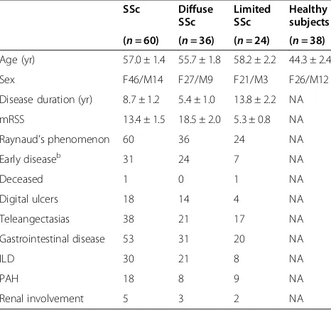
All SSc patients fulfilled the American College of Rheu-matology criteria for classification of SSc [19]. Plasma samples were collected from SSc patients and healthy subjects. Subject characteristics are listed in Table 1. Two punch biopsies (4 mm) were taken from the fore-arms of SSc patients with diffuse cutaneous variants. Normal skin tissue was obtained from healthy volunteers as well as the tissue procurement service provided by the University of Michigan Hospital. Written informed consent was obtained for all subjects, and the study was approved by the University of Michigan Institutional Review Board.
Cell culture
Both NL and SSc dermal fibroblasts were isolated from human skin. The tissue was digested using enzyme diges-tion soludiges-tion containing 2.4 U/ml dispase, 650 U/ml type II collagenase and 10,000 Dornase U/ml DNase. Dermal fibroblasts were maintained in RPMI 1640 medium with 10% fetal bovine serum (FBS), penicillin and streptomycin. Passages between 4 and 8 were used. Before experiments, NL and SSc dermal fibroblasts were switched to RPMI
1640 medium with 0.1% FBS. When needed, 500 μM
[image:2.595.305.538.490.708.2]DHLA (Santa Cruz Biotechnology, Santa Cruz, CA, USA) was added to the cell culture media. The incubation time
Table 1 SSc patient and healthy volunteer characteristicsa
SSc Diffuse SSc
Limited SSc
Healthy subjects
(n= 60) (n= 36) (n= 24) (n= 38)
Age (yr) 57.0 ± 1.4 55.7 ± 1.8 58.2 ± 2.2 44.3 ± 2.4
Sex F46/M14 F27/M9 F21/M3 F26/M12
Disease duration (yr) 8.7 ± 1.2 5.4 ± 1.0 13.8 ± 2.2 NA
mRSS 13.4 ± 1.5 18.5 ± 2.0 5.3 ± 0.8 NA
Raynaud’s phenomenon 60 36 24 NA
Early diseaseb 31 24 7 NA
Deceased 1 0 1 NA
Digital ulcers 18 14 4 NA
Teleangectasias 38 21 17 NA
Gastrointestinal disease 53 31 20 NA
ILD 30 21 8 NA
PAH 18 8 9 NA
Renal involvement 5 3 2 NA
a
ILD, Interstitial lung disease; mRSS = Modified Rodnan skin score (0 to 51); PAH, Pulmonary arterial hypertension; SSc, Scleroderma.b
for DHLA ranged from 48 to 72 hours before the cells were harvested for oxidative stress detection (48 hours)
or α-smooth muscle actin (αSMA)/collagen analysis
(72 hours). Monocytes from healthy subjects and SSc pa-tients were isolated using Percoll gradient as previously described [20]. They were plated in six-well plates, and the nonadherent cells were washed off after 2 hours of seeding. The adherent cells were monocytes and cultured in RPMI media in the presence of FBS. Before stimulation, they were switched to RPMI media without FBS. The con-ditioned media were collected for LIAS analysis.
Oxidative stress detection
Cellular O2•− was measured using dihydroethidium
(Invitrogen, Carlsbad, CA, USA). The nuclei were stained using 4′,6-diamidino-2-phenylindole (DAPI; Invitrogen). Fluorescence was detected using an Olympus FV-500 confocal microscope, and photographs were taken at 400× magnification. Cellular peroxynitrite levels were detected using 2′,7′-dichlorodihydrofluorescein diacetate [13]. Both NL and SSc dermal fibroblasts were plated in 96-well plates (5 × 10⁴ cells/well, 200 μl/well) and incu-bated in RPMI 1640 medium (0.1% FBS), with or without
20 mM NAC or 500 μM DHLA, for 48 hours at 37°C.
After the incubation, cells were then treated with 200μM 2′,7′-dichlorohihydrofluorescein diacetate in phosphate-buffered saline for 1 hour at 37°C. The fluorescence inten-sity was measured using a fluorescence plate reader, and the excitation and emission wavelengths used were 490 and 533 nm.
mRNA extraction and quantitative RT-PCR
Total RNA was isolated from dermal fibroblasts using RNeasy Mini RNA isolation kits (QIAGEN, Valencia, CA, USA). cDNA was prepared using Verso cDNA synthesis kits (Thermo Scientific, Asheville, NC, USA). Quantitative PCR was performed using SYBR Green PCR Master Mix (Applied Biosystems, Foster City, CA, USA) with specific primers for type I collagen (Col I), density-enhanced phos-phatase 1 (DEP-1), Src homology 2 domain–containing protein tyrosine phosphatase 2 (SHP-2) and β-actin. All samples were run in duplicate using Applied Biosystems Real-Time PCR System and analyzed using 7500 Applied Biosystems software.
Detection of platelet-derived growth factor receptor phosphorylation
Both NL and SSc dermal fibroblasts were incubated with or without 500μM DHLA overnight and stimulated with platelet-derived growth factor (PDGF). Cell lysates were obtained, and equal amounts of lysate proteins were incu-bated with immobilized mouse anti-human phospho-tyrosine monoclonal antibody (Cell Signaling Technology, Danvers, MA, USA) overnight at 4°C. Rabbit anti-human
platelet-derived growth factor receptor β(PDGFRβ) anti-body (Cell Signaling Technology) was used to probe for phosphorylated PDGFR after SDS-PAGE and Western blotting. The immunoprecipitated, tyrosine-phosphory-lated proteins were detected using mouse anti-human phospho-tyrosine antibody (Cell Signaling Technology).
Immunofluorescence
Cells grown in eight-well chambers were fixed and blocked with FBS before being probed with mouse anti-human Col I monoclonal antibody (Abcam, Cambridge,
MA, USA), rabbit anti-human αSMA antibody (Abcam)
or rabbit anti-LA antibody (Calbiochem, San Diego, CA, USA). Slides were subsequently incubated with Alexa Fluor 488 donkey anti-mouse antibody or Alexa Fluor 488 donkey anti-rabbit antibody (Molecular Probes/Invitrogen, Eugene, OR, USA). The nuclei were stained with DAPI.
Western blotting
Equal amounts of cell lysate were loaded onto polyacry-lamide gels and separated by SDS-PAGE. The proteins were then transferred onto nitrocellulose membranes via Western blotting. The blots were probed with antibodies to LIAS (Thermo Scientific), αSMA (Abcam) or β-actin (Sigma-Aldrich, St Louis, MO, USA).
Phosphatase activity assay
Phosphatase activity assays were carried out using the DuoSet Intracellular kits from R&D Systems (Minneapolis, MN, USA). Antibodies that capture both active and in-active protein tyrosine phosphatase 1B (PTP1B), SHP-2 or DEP-1 were immobilized. After unbound proteins were washed away, a synthetic phosphopeptide substrate that was dephosphorylated by active phosphatases was added to generate free phosphate and unphosphorylated pep-tide. The free phosphate was detected by a sensitive dye-binding assay using malachite green and molybdic acid. The activity of the phosphatase was determined by calcu-lating the rate of phosphate release.
Enzyme-linked immunosorbent assay
MMP-9 in cell culture media was detected using a Senso-Lyte Plus 520 MMP-9 assay kit (AnaSpec, Fremont, CA, USA). Endogenously active MMP-1 activity in culture media was detected using a SensoLyte Plus MMP-1 assay kit from AnaSpec.
Hydroxyproline measurement
Hydroxyproline content in cell culture media was mea-sured using a hydroxyproline assay kit (Sigma-Aldrich). Culture media were treated with concentrated hydro-chloric acid and hydrolyzed at 120°C for 3 hours. Hy-droxyproline standards, along with 50 μl of the samples, were transferred to a 96-well plate and placed in a 60°C oven until the wells were dried. Chloramine T/oxidation buffer and subsequently the diluted 4-(dimethylamino) benzaldehyde was then added to the wells and incubated for 90 minutes at 60°C. The absorbance was measured at 560 nm using a plate reader.
Statistical analysis
The results were expressed as mean ± SEM. To deter-mine the differences between the groups, Student’s t-tests were performed.P-values less than 0.05 with two-tailed analysis were considered statistically significant.
Results
LA and LIAS expression
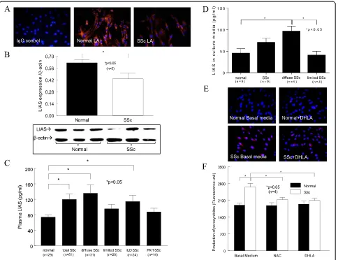
Because LA is synthesized in the body, we first examined whether LA expression differed in SSc patients com-pared to healthy subjects. We found that the cellular expression of LA in SSc dermal fibroblasts was lower compared to NL cells (Figure 1A). In addition, the enzyme that produces LA, LIAS, was significantly lower in SSc dermal fibroblasts (Figure 1B). In contrast, the plasma LIAS levels were significantly elevated in SSc patients, specifically those with diffuse SSc or intersti-tial lung disease (ILD), compared to healthy subjects (Figure 1C). Patients with limited SSc or pulmonary hypertension showed no difference in LIAS levels. To determine the source of the released LIAS levels de-tected in plasma, we measured LIAS in conditioned media collected from dermal fibroblasts, dermal endo-thelial cells and monocytes from both healthy subjects and SSc patients. Only the culture media from mono-cytes released detectable LIAS levels. Although no sta-tistical significance was reached between NL and SSc groups, there was a significant difference between NL and diffuse SSc (P< 0.05) (Figure 1D). Similar to what was seen in the plasma (Figure 1C), monocytes from limited SSc patients did not produce elevated amounts of LIAS (P< 0.05 between diffuse and limited SSc) (Figure 1D).
Oxidative stress
Because LA and its metabolite DHLA can act as anti-oxidants, we examined their antioxidative capacity in dermal fibroblasts. We found that O2•−was significantly
higher in SSc dermal fibroblasts (Figure 1E) and that DHLA had no effect. To examine whether DHLA affects other oxidative products, we measured peroxynitrite and found that it was elevated in SSc dermal fibroblasts. NAC and DHLA lowered it significantly (Figure 1F). This suggests that there is increased oxidative stress in SSc dermal fibroblasts and that different thiols affect dif-ferent forms of oxidative products.
Phosphorylation of platelet-derived growth factor receptor
Because LA was lower in SSc dermal fibroblasts and re-lieved oxidative stress, we hypothesized that adding DHLA, its active metabolite, to the cells would change their profibrotic phenotype back to normal. We first ex-amined the effect of DHLA on PDGFR activation. In NL dermal fibroblasts, PDGF-stimulated PDGFR phospho-rylation (p-PDGFR) was maximal at 30 minutes and de-creased significantly at 1 hour (Figure 2A). In contrast, p-PDGFR maximized at 10 minutes and remained phos-phorylated at 1 hour in SSc dermal fibroblasts. In the presence of DHLA, p-PDGFR peaked at 10 and 30 mi-nutes for NL fibroblasts. However, PDGF did not induce p-PDGFR in the presence of DHLA in SSc fibroblasts. The time course of p-PDGFR with or without DHLA significantly decreased the extent of p-PDGFR in both NL and SSc. This suggests that the excessive p-PDGFR seen in SSc dermal fibroblasts is due to increased oxida-tive stress, as a thiol antioxidant could reduce it.
Phosphatase activities and expression
Because DHLA affects p-PDGFR, we hypothesized that DHLA restores the activities of thiol-sensitive phospha-tases by decreasing the oxidative stress in SSc dermal fibroblasts, thereby decreasing p-PDGFR. Because PTP1B, DEP-1 and SHP-2 have been shown to dephosphorylate the PDGFR, we examined their expression and activities with or without DHLA in NL and SSc fibroblasts. SHP-2 mRNA was significantly lower in SSc (Additional file 1: Figure S1), and the presence of the antioxidants increased it, with NAC reaching statistical significance. On the other hand, DEP-1 mRNA was significantly higher in SSc dermal fibroblasts compared to NLs, whereas the thiol antioxidants did not affect the mRNA levels. Interest-ingly, DEP-1 protein expression was elevated in SSc fi-broblasts under basal conditions, and addition of NAC further increased it in both NL and SSc cells (data not shown).
significantly restored PTP1B activity. Similarly, SHP-2 activity was significantly lower in SSc dermal fibroblasts (Figure 2C). Whereas addition of NAC had no effect on SHP-2 activity in SSc fibroblasts, DHLA significantly re-stored it. On the other hand, DEP-1 activity was similar in NL and SSc fibroblasts; however, addition of antioxi-dants increased DEP-1 activity in SSc dermal fibroblasts (Figure 2D). DHLA was more effective than NAC, as it caused a significant increase in DEP-1 activity in SSc cells compared to that at the basal level. Taken together, these results imply that the increased oxidative stress in SSc inactivates phosphatases responsible for PDGFR dephosphorylation, resulting in increased p-PDGFR. The presence of antioxidants not only eliminates oxidative
substances but also restores phosphatase activities, thereby decreasing the extent of p-PDGFR.
Type I collagen expression
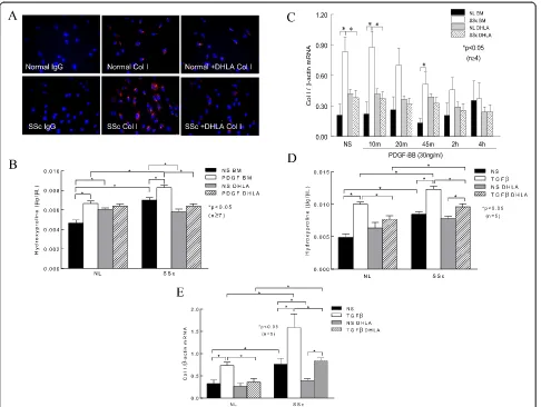
[image:5.595.58.542.88.460.2]Because PDGFR activation can lead to excess Col I pro-duction, we hypothesized that DHLA can decrease Col I in these cells. As expected, there was more Col I staining in SSc dermal fibroblasts than in NL cells (Figure 3A). In the presence of DHLA, Col I in SSc dermal fibro-blasts decreased to levels similar to those observed in NL cells. To further quantify the levels of Col I, we mea-sured hydroxyproline in the cell culture media in the presence or absence of DHLA (Figure 3B). Similar to what is shown in Figure 3A, hydroxyproline was
significantly higher in SSc than in NL cells, and addition of DHLA decreased it. Stimulating the cells with PDGF increased hydroxyproline levels significantly in both NL and SSc fibroblasts, whereas DHLA diminished this effect (Figure 3B). We also determined the effect of DHLA at the mRNA level. At basal level and at 10 and 45 minutes after PDGF stimulation, Col I mRNA was significantly higher in SSc fibroblasts (Figure 3C). After 4 hours of PDGF stimulation, Col I mRNA significantly decreased compared to NS. DHLA significantly de-creased Col I mRNA levels in SSc fibroblasts at basal levels and 10 minutes after PDGF incubation. These re-sults indicate that, in SSc, enhanced PDGFR activation leads to more Col I synthesis. By acting on scavenging ROS and deactivating the PDGFR, DHLA decreased Col I production.
To further investigate the effect of DHLA on
trans-forming growth factor β (TGF-β), NL and SSc dermal
fibroblasts were incubated with 10 ng/ml TGF-β for
48 hours and Col I production was examined by mea-suring both qPCR and hydroxyproline content. In NL fi-broblasts, the addition of TGF-β significantly increased the hydroxyproline content (Figure 3D). In contrast, the presence of DHLA significantly decreased it. Hydroxy-proline levels were significantly elevated in conditioned media from SSc dermal fibroblasts compared to NL cells, and they increased significantly after TGF-β incu-bation. Although the addition of DHLA significantly re-duced hydroxyproline levels in SSc cells, the amount of hydroxyproline was still significantly higher compared to that in NL fibroblasts.
The mRNA levels of Col I were also examined after cells were treated with TGF-β(Figure 3E). Col I mRNA levels were significantly elevated in SSc dermal
fibro-blasts compared to NL cells. TGF-β induced Col I
[image:6.595.60.539.92.440.2]mRNA in both NL and SSc dermal fibroblasts, and the
Figure 2The effect of dihydrolipoic acid on the platelet-derived growth factor receptor pathway and phosphatases. (A)Phosphorylated platelet-derived growth factor receptor (p-PDGFR) after PDGF stimulation at various time points (n= 3 NL and SSc subjects). NS, Nonstimulated. Enzymatic activities of protein tyrosine phosphatase 1B (PTP1B)(B), Src homology 2 domain–containing protein tyrosine phosphatase 2 (SHP-2)
presence of DHLA reduced the levels. These results sug-gest that DHLA not only affects PDGF-induced Col I production but also acts on TGF-β-mediated fibrotic processes.
Levels of matrix metalloproteinase, tissue inhibitor of matrix metalloproteinase 1 and plasminogen activator inhibitor 1
To further examine the effect of thiols on Col I degra-dation, levels of MMP-1 in cell culture medium were de-termined (Figure 4A). MMP-1 increased significantly after PDGF stimulation in both NL and SSc fibroblasts. In addition, PDGF-stimulated MMP-1 levels were sig-nificantly lower in SSc dermal fibroblasts, which could also contribute to the increased Col I expression seen in
these cells. Thiols did not alter MMP-1 significantly in NL cells; however, they did increase MMP-1 in SSc cells stimulated with PDGF. In particular, the presence of DHLA resulted in a significant increase in PDGF-stimulated MMP-1 levels in SSc.
[image:7.595.57.543.88.454.2]Because MMP-3 converts pro-MMP-1 and pro-MMP-9 into their active forms, we also measured MMP-3 and MMP-9 levels (Figure 4B). Similarly to MMP-1, PDGF stimulated MMP-3 production in both NL and SSc cells. In addition, MMP-3 was significantly lower in SSc fibro-blasts under basal conditions. NAC did not appear to have any effect on MMP-3, but DHLA increased MMP-3 sig-nificantly in both NL and SSc cells. The lack of MMP-3 to activate MMP-1 may exacerbate the accumulation of Col I in SSc dermal fibroblasts.
MMP-9 was significantly lower in SSc culture medium. Unlike MMP-1 and MMP-3, the antioxidants did not affect MMP-9, nor did PDGF show a consistent effect on its production (data not shown). Similarly, the an-tioxidants did not affect TIMP-1 levels (Figure 4C).
Although TIMP-1 was elevated in SSc dermal
[image:8.595.60.540.86.558.2]fibroblasts, the MMP-1/TIMP-1 ratio was significantly lower in SSc fibroblasts, suggesting a shift that favors a profibrotic phenotype in these cells (Figure 4D). DHLA, but not NAC, increased the ratio significantly, suggesting additional beneficial effects of DHLA on these cells.
To determine whether thiol antioxidants affect MMP-1 activity, we measured endogenous MMP-1 activity in cell culture media. MMP-1 activity was significantly higher in NL cells compared to SSc dermal fibroblasts with or with-out PDGF stimulation (Figure 4E). Neither NAC nor DHLA had an effect on MMP-1 activity in NL cells. In SSc dermal fibroblasts, Both DHLA and NAC increased MMP-1 activity significantly.
The levels of PAI-1, an inhibitor for urokinase/tissue type plasminogen activator which contributes to Col deg-radation, were also examined (Figure 4F). PAI-1 released from SSc fibroblasts was significantly higher, whereas the thiols significantly reduced it. Considering this informa-tion together, it is possible that DHLA decreases Col I through decreasing PAI-1 and increasing MMPs.
Expression ofα-smooth muscle actin
To examine whether DHLA affects the myofibroblast
phenotype in SSc, we examined αSMA expression. The
expression of αSMA was markedly elevated in SSc der-mal fibroblasts, and DHLA reduced it (Figures 5A and
B). In addition, we examined the effect of DHLA on TGF-β-stimulated fibroblasts. As shown in Figure 5C,
TGF-β induced αSMA expression in both NL and SSc
fibroblasts, whereas adding DHLA to the culture de-creased its expression.
Discussion
In this study, we show that the thiol antioxidant DHLA not only decreased cellular peroxynitrites but also reduced p-PDGFR, which is potentially due to its effect on the phos-phatases that are involved in this pathway. Col I, αSMA and PAI-1 decreased after DHLA treatment, whereas MMP-1 and MMP-3 increased. In addition, DHLA in-creased MMP-1 activity. It also dein-creased the ability of TGF-βto induce Col I andαSMA expression in fibroblasts. The beneficial effect of DHLA might also be due to replen-ishing the antioxidative capacity in SSc dermal fibroblasts.
[image:9.595.61.539.348.695.2]NAC is used as an antioxidant, and its effect in SSc has been examined in various studies [1,18,21–23]. Consider-ing the low oral bioavailability of NAC, which could par-tially explain its limited therapeutic efficacy in clinical
trials, the introduction of another thiol antioxidant with a much better absorption profile to treat SSc is an intriguing strategy. LA and DHLA are soluble in both lipid and aqueous environments. Because of this, LA has better bio-availability than NAC; the bioavailabilities of 200 mg of LA and NAC have been estimated to be 38% and 4%, re-spectively [24,25]. Because DHLA has more reducing ac-tivity than LA, it was chosen for our study. We were able to use lower amounts of DHLA and achieve the same or a better effect with it compared with NAC.
We attempted to measure plasma LA and DHLA levels using liquid chromatography-mass spectrometry, but they fell below the detection limit. Instead, we immunostained fibroblasts and found that LA in SSc cells was lower. We should note that the plasma levels of LA and DHLA in healthy subjects have been reported to be approximately 30 ng/ml (that is, 150 nM) and 175 ng/ml (0.84 nM), re-spectively [26,27], much lower compared to GSH (3.39 ±
1.04μM) [28], which is the predominant form of cellular
thiol. In a separate study, the plasma GSH content in SSc patients was significantly lower than in healthy controls: 170 ± 44 vs. 246 ± 46μmol/g hemoglobin, respectively [6]. Although DHLA and LA are lower in abundance than GSH, their reduction potential is more negative than that of GSH and its oxidized form (−320 mV vs. −240 mV), suggesting that DHLA can regenerate GSH and maintain the ratio between GSH and it oxidized form GSSG in cells [29,30]. Moreover, owing to their amphipathic nature and smaller molecular sizes compared to GSH, the DHLA-LA pair moves freely in both cytosol and lipid compartments and is readily accessible to more proteins and enzymes that are affected by redox signaling. Therefore, LA and DHLA, whether endogenously or given externally, are in-deed important in maintaining cellular function.
We also examined LIAS, a crucial enzyme involved in the LA synthetic pathway. The significantly lower LIAS in SSc fibroblasts might explain the lower LA levels in these cells. However, plasma LIAS was significantly elevated in patients with diffuse SSc. The reason for the discrepancy is not clear. Nonetheless, we were able to determine the source of the LIAS by measuring LIAS in culture media from dermal fibroblasts, endothelial cells and monocytes. It appears that only monocytes from healthy subjects and SSc patients released detectable amounts of LIAS (Fig-ure 1D). Similar to what we saw in the plasma, monocytes from diffuse patients produced elevated amounts of LIAS compared to healthy subjects, whereas those from limited SSc patients did not. Interestingly, we could not detect LIAS from monocytes isolated from patients with rheumatoid arthritis (data not shown). The mechanism of LIAS release in different disease settings appears to be a complicated process and remains to be explored. It is worth noting that 8-isoprostane, an oxidized lipid, was also found to be elevated in plasma obtained from patients
with diffuse SSc or ILD (data not shown). It is possible that LIAS is being produced and released from circulating immune cells to counteract the increased oxidative stress observed in SSc patients.
In recent years, the use of LA/DHLA has evolved from antioxidants to antifibrotics. LA was observed to protect against bleomycin-induced lung injury by suppressing ROS and improving the MMP-1/TIMP-1 ratio [31]. Treat-ment with LA also attenuated cardiac fibrosis in rats [32]. In a diabetic model, LA not only decreased oxidative dam-age and Col I andαSMA expression in the heart but also increased MMP-2 activity [33]. In addition, LA inhibited the development of thioacetamide-induced liver fibrosis in rats [34]. In a hepatic fibrosis mouse model, LA inhibited the expression of Col I,αSMA and PAI-1 [35]. Our study adds SSc skin fibrosis to the fibrotic diseases that have been shown to be affected by LA/DHLA.
In SSc, fibroblasts differentiate into myofibroblasts, a process characterized by excess production ofαSMA. The mechanism of DHLA’s lowering αSMA expression in our study is not known. However, LA treatment has been shown to decrease the expression of TGF-β[33] and affect the redox-sensitive transcription factors activator protein 1 (AP-1) and specificity protein 1 (Sp1) [35]. In addition, it de-creased connective tissue growth factor [32]. In hepatic stel-late cells, DHLA inhibited TGF-β/PDGF activation through the interruption of ROS-related phosphatidylinositol 3-kinase/protein kinase B (PI3K/AKT) and mitogen-activated protein kinase (MAPK) signaling [34]. Budisavljevic et al. had similar results with kidney cells, where mesangial cell transformation into myofibroblasts was completely pre-vented by LA [36]. These authors showed that LA inhibited the PDGF-activated extracellular signal-regulated kinase 1/2 (ERK1/2) pathway, suggesting that the increased expression
ofαSMA in cultured mesangial cells could be modulated by
redox-sensitive signaling pathways.
SSc fibroblasts. Although it had minimal effect on TIMP-1, the MMP-1/TIMP-1 ratio increased significantly after DHLA treatment, shifting these SSc cells from a profibro-tic state to a relatively antifibroprofibro-tic state. Moreover, DHLA not only increased the expression of MMP-1 but also in-creased MMP-1 activity after PDGF stimulation in SSc dermal fibroblasts. This suggests that DHLA can alter pathogenic processes that are important in SSc.
The expression of MMP is regulated by nuclear factor κB (NFκB) [42] and oxidative stress [43,44]; therefore, the presence of antioxidants can alter the expression of MMPs. In our hands, the incubation of thiol antioxi-dants increased the expression of MMP-1 and MMP-3 (Figure 4). However, MMP-9 expression was not altered by the thiols. It appears that the expression of MMP-9 is regulated by NFκB, AP-1 and the p38 MAPK pathway [45–47] and that LA inhibits MMP-9 expression by in-activating NFκB [48]. The involvement of these other pathways (AP-1 and p38) tightly regulates the MMP-9 expression, and this could be the reason for the ineffec-tiveness of the thiols used. On the other hand, TIMP-1 expression can be stimulated by a variety of agents, in-cluding serum, growth factors, cytokines and viruses [49,50]. Regulation of TIMP-1 expression involves the activation of transcription factors, including AP-1, Sp1, STAT (signal transducer and activator of transcription) and Pea3/Ets families, as well as signaling pathways such as the ERK1/2 pathway [51–53]. Similarly to MMP-9, the complex regulation of TIMP-1 expression makes it difficult to render its expression; hence, in our hands, the use of thiol antioxidants did not affect TIMP-1 levels in NL and SSc dermal fibroblasts.
What could be the possible mechanism by which the thiols affect the expression of the phosphatases exa-mined in this study? Transcription factors, such as NFκB and AP-1, are regulated by the intracellular redox state. They are implicated to be involved in the regulation of expression of a variety of genes [54]. It has been shown that LA and DHLA and NAC inhibit the NFκB pathway [54–56]. NAC also inhibits AP-1 activation [57], whereas the role of LA/DHLA on this pathway is unclear. It is possible that the thiol antioxidants control the expression of phosphatases through these redox-sensitive transcrip-tion factors. Moreover, the expression of phosphatases can be affected by oxidative stress [58]. The exact mechanism by which the antioxidants increase SHP-2 and DEP-1 expression requires further investigation.
The PTP family is characterized by their signature motif at their active site, HC(X)5R. This site contains an
essential cysteine residue that has a low pKa which is susceptible to oxidation. Its ability to be oxidized re-versibly acts as a redox regulatory mechanism for recep-tor tyrosine kinases to control their phosphorylation state. We and others have shown that when excessive
oxidative stress is present, the cysteine group is oxidized and the activity is inactivated [1,59,60]. We also reported that PTP1B was oxidized and subsequently inactivated in SSc dermal fibroblasts [1]. This led to prolonged p-PDGFR and increased Col I. Treatment with NAC re-stored the activity and decreased p-PDGFR and Col I. In this study, DHLA showed a similar effect. In addition to PTP1B, we examined two other thiol-sensitive phospha-tases that regulate p-PDGFR. Similar to PTP1B, these phosphatases have a cysteine residue at their active site that is required for their phosphatase activity. As shown in Figure 2, DHLA increased activities of all three phos-phatases in SSc fibroblasts. This is superior to NAC, be-cause NAC had no effect on DEP-1 and SHP-2 activities. It has been shown that thiol antioxidants are able to
re-generate enzyme−SH groups and restore the
phos-phatase activities [1,61,62]. Therefore, the mechanism by which DHLA restores the enzyme activity is through re-duction of the oxidized cysteine group at the active site. Note that the catalytic activities of these phosphatases are tightly regulated and that subcellular localization can also affect substrate specificity. In other words, the active site may have different susceptibility toward dif-ferent thiol antioxidants. In addition, the antioxidant capacity of the thiols used in this study is different (the pKa at the thiol group differs), giving them different catalytic activities toward the possible different oxidation products (for example, −SOH, −SO2H, −SSR) at the
phosphatase active sites [61,63]. Moreover, because the bioavailability of NAC is lower than that of LA/DHLA and LA/DHLA has more desirable structural properties that allow it to move freely in cellular compartments, LA/DHLA could reach a higher concentration intra-cellularly and hence be more accessible at the enzyme active site compared to NAC. We speculate that these are the reasons why NAC has no effect on SHP-2 activity (Figure 2C), but DHLA does. Nonetheless, the increase in these phosphatase activities provides a poten-tial mechanism for reducing p-PDGFR in SSc dermal fibroblasts.
Conclusion
To our knowledge, this study is the first to show that LA and LIAS are lower in SSc dermal fibroblasts. In addition, DHLA was able to reverse the profibrotic phenotype of these cells by decreasing p-PDGFR, restoring the activities of phosphatases, lowering PAI-1, and increasing both MMP-1 and MMP-3 expres-sion as well as MMP-1 activity. Moreover, DHLA
lowered the expression of αSMA, suggesting that it
Additional file
Additional file 1: Figure S1.Phosphatase expression in NL and SSc dermal fibroblasts. SHP-2 mRNA in SSc dermal fibroblasts was significantly lower than NL (P< 0.05,n= 6 subjects). On the other hand, DEP-1 mRNA was significantly elevated in SSc dermal fibroblasts compared to NLs (P< 0.05,n≥5 subjects). The presence of NAC or DHLA did not alter DEP-1 mRNA. NAC significantly increased SHP-2 mRNA in SSc cells, whereas DHLA had no significant effect. Results are expressed as mean ± SE, andP< 0.05 was considered significant.
Abbreviations
Col:Collagen; DHLA: Dihydrolipoic acid; LA: Lipoic acid; LIAS: Lipoic acid synthetase; MMP: Matrix metalloproteinase; NAC:N-acetylcysteine; PAI: Plasminogen activator inhibitor; PDGFR: Platelet-derived growth factor receptor; PTP: Protein tyrosine phosphatase;αSMA:α-Smooth muscle actin; SSc: Scleroderma; TIMP: Tissue inhibitor of matrix metalloproteinase.
Competing interests
The authors declare that they have no competing interests.
Authors’contributions
All authors agree to be accountable for all aspects of the work. PT conceived of and design the study, collected and analyzed the data and wrote the manuscript writing. BB, AP, AL, MA, WS and GZ collected and analyzed the data and critically revised the manuscript. DK, ES, DF and AEK analyzed and interpreted the data and critically revised the manuscript. All authors read and approved the final manuscript.
Acknowledgements
This work was supported by the Scleroderma Foundation (to AEK); the Office of Research and Development, Medical Research Service, Department of Veterans Affairs; the Frederick GL Huetwell and William D Robinson, MD, Professorship in Rheumatology at the University of Michigan; National Institute of Arthritis and Musculoskeletal and Skin Diseases (National Institutes of Health (NIH)) grant K24 AR063120-02 (to DK); the Arthritis Foundation (to PT); the clinical research unit at the University of Michigan; the Linda Dolce Scleroderma Research Fund; the Marvin & Betty Danto and Jonathan & Lisa Rye endowments for scleroderma research at the University of Michigan; and, in part, by the Tissue Procurement Core of the University of Michigan Comprehensive Cancer Center (grant CA46952 from the National Cancer Institute, NIH).
Author details 1
Scleroderma Program, University of Michigan, 300 North Ingalls St. 7C27 NIB, Ann Arbor, MI 48109, USA.2University of Michigan Medical School, University
of Michigan Medical School, 109 Zina Pitcher Pl, 4388 BSRB, Ann Arbor, MI 48109, USA.3Division of Rheumatology, Department of Internal Medicine,
University of Michigan Medical School, 300 North Ingalls St. 7C27 NIB, Ann Arbor, MI 48109, USA.4Department of Medical Affairs, VA Medical Service,
2215 Fuller Rd., Ann Arbor, MI 48105, USA.
Received: 20 January 2014 Accepted: 25 July 2014 Published: 15 August 2014
References
1. Tsou PS, Talia NN, Pinney AJ, Kendzicky A, Piera-Velazquez S, Jimenez SA, Seibold JR, Phillips K, Koch AE:Effect of oxidative stress on protein tyrosine phosphatase 1B in scleroderma dermal fibroblasts.Arthritis
Rheum2012,64:1978–1989.
2. Gabrielli A, Svegliati S, Moroncini G, Amico D:New insights into the role of oxidative stress in scleroderma fibrosis.Open Rheumatol J2012,6:87–95. 3. Allanore Y, Borderie D, Lemaréchal H, Ekindjian OG, Kahan A:Acute and
sustained effects of dihydropyridine-type calcium channel antagonists on oxidative stress in systemic sclerosis.Am J Med2004,116:595–600. 4. Ogawa F, Shimizu K, Muroi E, Hara T, Hasegawa M, Takehara K, Sato S:
Serum levels of 8-isoprostane, a marker of oxidative stress, are elevated in patients with systemic sclerosis.Rheumatology (Oxford)2006,
45:815–818.
5. Servettaz A, Guilpain P, Goulvestre C, Chereau C, Hercend C, Nicco C, Guillevin L, Weill B, Mouthon L, Batteux F:Radical oxygen species production induced by advanced oxidation protein products predicts clinical evolution and response to treatment in systemic sclerosis.Ann Rheum Dis2007,66:1202–1209. 6. Sfrent-Cornateanu R, Mihai C, Stoian I, Lixandru D, Bara C, Moldoveanu E:
Antioxidant defense capacity in scleroderma patients.Clin Chem Lab Med
2008,46:836–841.
7. Herrick AL, Rieley F, Schofield D, Hollis S, Braganza JM, Jayson MI:
Micronutrient antioxidant status in patients with primary Raynaud’s phenomenon and systemic sclerosis.J Rheumatol1994,21:1477–1483. 8. Denton CP, Bunce TD, Dorado MB, Roberts Z, Wilson H, Howell K,
Bruckdorfer KR, Black CM:Probucol improves symptoms and reduces lipoprotein oxidation susceptibility in patients with Raynaud’s phenomenon.Rheumatology (Oxford)1999,38:309–315. 9. Herrick AL, Hollis S, Schofield D, Rieley F, Blann A, Griffin K, Moore T,
Braganza JM, Jayson MI:A double-blind placebo-controlled trial of antioxidant therapy in limited cutaneous systemic sclerosis.Clin Exp
Rheumatol2000,18:349–356.
10. Allanore Y, Borderie D, Périanin A, Lemaréchal H, Ekindjian OG, Kahan A:
Nifedipine protects against overproduction of superoxide anion by monocytes from patients with systemic sclerosis.Arthritis Res Ther2005,
7:R93–R100.
11. Galbusera C, Orth P, Fedida D, Spector T:Superoxide radical production by allopurinol and xanthine oxidase.Biochem Pharmacol2006,
71:1747–1752.
12. Firuzi O, Fuksa L, Spadaro C, Bousova I, Riccieri V, Spadaro A, Petrucci R, Marrosu G, Saso L:Oxidative stress parameters in different systemic rheumatic diseases.J Pharm Pharmacol2006,58:951–957.
13. Marut WK, Kavian N, Servettaz A, Nicco C, Ba LA, Doering M, Chéreau C, Jacob C, Weill B, Batteux F:The organotelluride catalyst (PHTE)2NQ prevents HOCl-induced systemic sclerosis in mouse.J Invest Dermatol
2012,132:1125–1132.
14. Kagan VE, Serbinova EA, Forte T, Scita G, Packer L:Recycling of vitamin E in human low density lipoproteins.J Lipid Res1992,33:385–397.
15. Suh JH, Shenvi SV, Dixon BM, Liu H, Jaiswal AK, Liu RM, Hagen TM:Decline in transcriptional activity of Nrf2 causes age-related loss of glutathione synthesis, which is reversible with lipoic acid.Proc Natl Acad Sci U S A
2004,101:3381–3386.
16. Rosato E, Cianci R, Barbano B, Menghi G, Gigante A, Rossi C, Zardi EM, Amoroso A, Pisarri S, Salsano F:N-acetylcysteine infusion reduces the resistance index of renal artery in the early stage of systemic sclerosis.
Acta Pharmacol Sin2009,30:1283–1288.
17. Sambo P, Amico D, Giacomelli R, Matucci-Cerinic M, Salsano F, Valentini G, Gabrielli A:Intravenous N-acetylcysteine for treatment of Raynaud’s phenomenon secondary to systemic sclerosis: a pilot study.J Rheumatol
2001,28:2257–2262.
18. Yildirim Z, Kotuk M, Iraz M, Kuku I, Ulu R, Armutcu F, Ozen S:Attenuation of bleomycin-induced lung fibrosis by oral sulfhydryl containing antioxidants in rats: erdosteine andN-acetylcysteine.Pulm Pharmacol Ther2005,18:367–373. 19. LeRoy EC, Black C, Fleischmajer R, Jablonska S, Krieg T, Medsger TA Jr,
Rowell N, Wollheim F:Scleroderma (systemic sclerosis): classification, subsets and pathogenesis.J Rheumatol1988,15:202–205.
20. Ruth JH, Arendt MD, Amin MA, Ahmed S, Marotte H, Rabquer BJ, Lesch C, Lee S, Koch AE:Expression and function of CXCL16 in a novel model of gout.Arthritis Rheum2010,62:2536–2544.
21. Failli P, Palmieri L, D’Alfonso C, Giovannelli L, Generini S, Rosso AD, Pignone A, Stanflin N, Orsi S, Zilletti L, Matucci-Cerinic M:Effect ofN-acetyl-L-cysteine on peroxynitrite and superoxide anion production of lung alveolar macrophages in systemic sclerosis.Nitric Oxide2002,7:277–282.
22. Rosato E, Borghese F, Pisarri S, Salsano F:The treatment withN -acetylcysteine of Raynaud’s phenomenon and ischemic ulcers therapy in sclerodermic patients: a prospective observational study of 50 patients.
Clin Rheumatol2009,28:1379–1384.
23. Shi-wen X, Thompson K, Khan K, Liu S, Murphy-Marshman H, Baron M, Denton CP, Leask A, Abraham DJ:Focal adhesion kinase and reactive oxygen species contribute to the persistent fibrotic phenotype of lesional scleroderma fibroblasts.Rheumatology (Oxford)2012,51:2146–2154. 24. Hermann R, Niebch G, Borbe HO, Fieger-Büschges H, Ruus P, Nowak H,
25. Olsson B, Johansson M, Gabrielsson J, Bolme P:Pharmacokinetics and bioavailability of reduced and oxidizedN-acetylcysteine.Eur J Clin
Pharmacol1988,34:77–82.
26. Teichert J, Preiss R:[17] High-performance liquid chromatography methods for determination of lipoic and dihydrolipoic acid in human plasma.Methods Enzymol1997,279:159–166.
27. Khan A, Iqbal Z, Watson DG, Khan A, Khan I, Muhammad N, Muhammad S, Nasib HA, Iqbal N, Faiz-ur-rahman, Kashif M:Simultaneous determination of lipoic acid (LA) and dihydrolipoic acid (DHLA) in human plasma using high-performance liquid chromatography coupled with electrochemical detection.J Chromatogr
B Analyt Technol Biomed Life Sci2011,879:1725–1731.
28. Michelet F, Gueguen R, Leroy P, Wellman M, Nicolas A, Siest G:Blood and plasma glutathione measured in healthy subjects by HPLC: relation to sex, aging, biological variables, and life habits.Clin Chem1995,41:1509–1517. 29. Hultberg B, Andersson A, Isaksson A:Lipoic acid increases glutathione
production and enhances the effect of mercury in human cell lines.
Toxicology2002,175:103–110.
30. Krishnan CV, Garnett M:Electrochemical behavior of the super antioxidant,α-lipoic acid.Int J Electrochem Sci2011,6:3607–3630. 31. Liu R, Ahmed KM, Nantajit D, Rosenthal FS, Hai CX, Li JJ:Therapeutic
effects ofα-lipoic acid on bleomycin-induced pulmonary fibrosis in rats.
Int J Mol Med2007,19:865–873.
32. Lee JE, Yi CO, Jeon BT, Shin HJ, Kim SK, Jung TS, Choi JY, Roh GS:α-Lipoic acid attenuates cardiac fibrosis in Otsuka Long-Evans Tokushima fatty rats.Cardiovasc Diabetol2012,11:111.
33. Li CJ, Lv L, Li H, Yu DM:Cardiac fibrosis and dysfunction in experimental diabetic cardiomyopathy are ameliorated byα-lipoic acid.Cardiovasc
Diabetol2012,11:73.
34. Foo NP, Lin SH, Lee YH, Wu MJ, Wang YJ:α-Lipoic acid inhibits liver fibrosis through the attenuation of ROS-triggered signaling in hepatic stellate cells activated by PDGF and TGF-β.Toxicology2011,282:39–46. 35. Min AK, Kim MK, Seo HY, Kim HS, Jang BK, Hwang JS, Choi HS, Lee KU, Park
KG, Lee IK:α-lipoic acid inhibits hepatic PAI-1 expression and fibrosis by inhibiting the TGF-βsignaling pathway.Biochem Biophys Res Commun
2010,393:536–541.
36. Budisavljevic MN, Hodge L, Barber K, Fulmer JR, Durazo-Arvizu RA, Self SE, Kuhlmann M, Raymond JR, Greene EL:Oxidative stress in the pathogenesis of experimental mesangial proliferative glomerulonephritis.Am J Physiol
Renal Physiol2003,285:F1138–F1148.
37. Dong C, Zhu S, Wang T, Yoon W, Li Z, Alvarez RJ, ten Dijke P, White B, Wigley FM, Goldschmidt-Clermont PJ:Deficient Smad7 expression: a putative molecular defect in scleroderma.Proc Natl Acad Sci U S A2002,
99:3908–3913.
38. Wei J, Ghosh AK, Sargent JL, Komura K, Wu M, Huang QQ, Jain M, Whitfield ML, Feghali-Bostwick C, Varga J:PPARγdownregulation by TGFβin fibroblast and impaired expression and function in systemic sclerosis: a novel mechanism for progressive fibrogenesis.PLoS One2010,5:e13778. 39. Xu S, Denton CP, Holmes A, Dashwood MR, Abraham DJ, Black CM:
Endothelins: effect on matrix biosynthesis and proliferation in normal and scleroderma fibroblasts.J Cardiovasc Pharmacol1998,31:S360–S363. 40. Giannelli G, Iannone F, Marinosci F, Lapadula G, Antonaci S:The effect of
bosentan on matrix metalloproteinase-9 levels in patients with systemic sclerosis-induced pulmonary hypertension.Curr Med Res Opin2005,
21:327–332.
41. Kirk TZ, Mark ME, Chua CC, Chua BH, Mayes MD:Myofibroblasts from scleroderma skin synthesize elevated levels of collagen and tissue inhibitor of metalloproteinase (TIMP-1) with two forms of TIMP-1.J Biol Chem1995,270:3423–3428.
42. Bond M, Baker AH, Newby AC:Nuclear factorκB activity is essential for matrix metalloproteinase-1 and -3 upregulation in rabbit dermal fibroblasts.
Biochem Biophys Res Commun1999,264:561–567.
43. Shin MH, Moon YJ, Seo JE, Lee Y, Kim KH, Chung JH:Reactive oxygen species produced by NADPH oxidase, xanthine oxidase, and mitochondrial electron transport system mediate heat shock-induced MMP-1 and MMP-9 expression.Free Radic Biol Med2008,44:635–645. 44. Kar S, Subbaram S, Carrico PM, Melendez JA:Redox-control of matrix
metalloproteinase-1: a critical link between free radicals, matrix remodeling and degenerative disease.Respir Physiol Neurobiol2010,174:299–306. 45. Sato H, Seiki M:Regulatory mechanism of 92 kDa type IV collagenase
gene expression which is associated with invasiveness of tumor cells.
Oncogene1993,8:395–405.
46. Bond M, Chase AJ, Baker AH, Newby AC:Inhibition of transcription factor NF-κB reduces matrix metalloproteinase-1, -3 and -9 production by vascular smooth muscle cells.Cardiovasc Res2001,50:556–565. 47. Simon C, Simon M, Vucelic G, Hicks MJ, Plinkert PK, Koitschev A, Zenner HP:
The p38 SAPK pathway regulates the expression of the MMP-9 collagenase via AP-1-dependent promoter activation.Exp Cell Res2001,271:344–355. 48. Kim HS, Kim HJ, Park KG, Kim YN, Kwon TK, Park JY, Lee KU, Kim JG, Lee IK:
α-Lipoic acid inhibits matrix metalloproteinase-9 expression by inhibiting NF-κB transcriptional activity.Exp Mol Med2007,39:106–113.
49. Uchijima M, Sato H, Fujii M, Seiki M:Tax proteins of human T-cell leukemia virus type 1 and 2 induce expression of the gene encoding erythroid-potentiating activity (tissue inhibitor of metalloproteinases-1, TIMP-1).J Biol Chem1994,269:14946–14950.
50. Campbell CE, Flenniken AM, Skup D, Williams BR:Identification of a serum-and phorbol ester-responsive element in the murine tissue inhibitor of metalloproteinase gene.J Biol Chem1991,266:7199–7206.
51. Edwards DR, Rocheleau H, Sharma RR, Wills AJ, Cowie A, Hassell JA, Heath JK:Involvement of AP1 and PEA3 binding sites in the regulation of murine tissue inhibitor of metalloproteinases-1 (TIMP-1) transcription.
Biochim Biophys Acta1992,1171:41–55.
52. Logan SK, Garabedian MJ, Campbell CE, Werb Z:Synergistic transcriptional activation of the tissue inhibitor of metalloproteinases-1 promoter via functional interaction of AP-1 and Ets-1 transcription factors.J Biol Chem
1996,271:774–782.
53. Kwak HJ, Park MJ, Cho H, Park CM, Moon SI, Lee HC, Park IC, Kim MS, Rhee CH, Hong SI:Transforming growth factor-β1 induces tissue inhibitor of metalloproteinase-1 expression via activation of extracellular signal-regulated kinase and Sp1 in human fibrosarcoma cells.Mol Cancer Res
2006,4:209–220.
54. Sen CK, Packer L:Antioxidant and redox regulation of gene transcription.
FASEB J1996,10:709–720.
55. Packer L:α-Lipoic acid: a metabolic antioxidant which regulates NF-κB signal transduction and protects against oxidative injury.Drug Metab Rev
1998,30:245–275.
56. Zhang WJ, Frei B:α-Lipoic acid inhibits TNF-α-induced NF-κB activation and adhesion molecule expression in human aortic endothelial cells.
FASEB J2001,15:2423–2432.
57. Pinkus R, Bergelson S, Daniel V:Phenobarbital induction of AP-1 binding activity mediates activation of glutathioneS-transferase and quinone reductase gene expression.Biochem J1993,290:637–640.
58. Keyse SM, Emslie EA:Oxidative stress and heat shock induce a human gene encoding a protein-tyrosine phosphatase.Nature1992,359:644–647. 59. Denu JM, Tanner KG:Specific and reversible inactivation of protein
tyrosine phosphatases by hydrogen peroxide: evidence for a sulfenic acid intermediate and implications for redox regulation.Biochemistry
1998,37:5633–5642.
60. Meng TC, Fukada T, Tonks NK:Reversible oxidation and inactivation of protein tyrosine phosphatases in vivo.Mol Cell2002,9:387–399. 61. Parsons ZD, Gates KS:Thiol-dependent recovery of catalytic activity from
oxidized protein tyrosine phosphatases.Biochemistry2013,52:6412–6423. 62. Sommer D, Fakata KL, Swanson SA, Stemmer PM:Modulation of the
phosphatase activity of calcineurin by oxidants and antioxidantsin vitro.
Eur J Biochem2000,267:2312–2322.
63. Tanner JJ, Parsons ZD, Cummings AH, Zhou H, Gates KS:Redox regulation of protein tyrosine phosphatases: structural and chemical aspects.
Antioxid Redox Signal2011,15:77–97.
doi:10.1186/s13075-014-0411-6
Cite this article as:Tsouet al.:Lipoic acid plays a role in scleroderma:
insights obtained from scleroderma dermal fibroblasts.Arthritis Research