Introduction
Advances made in the fi eld of pediatric rheumatology over the last decades have led to a signifi cant decrease in
mortality rates. Patients are now surviving into adulthood and have to face the many challenges imposed by their chronic illness. As a result of better treatments it is likely that cardiovascular disease will emerge as a leading cause of morbidity and mortality. Th e interplay between tradi-tional cardiovascular risk factors, exposure to cortico-steroids and chronic infl ammation creates a perfect storm for early atherogenesis in this population.
Atherosclerosis is now being increasingly recognized in adults with infl ammatory rheumatic diseases but there is little information pertaining to pediatric-onset rheuma-to logic conditions [1]. Th e objectives of this review are to summarize the current state of knowledge on cardio-vascular risk and accelerated atherosclerosis in pediatric-onset systemic lupus erythematosus (pSLE), juvenile idiopathic arthritis (JIA) and juvenile dermatomyositis (JDM), and to discuss atherosclerosis preventive strate-gies that should be considered in this patient population.
Measuring atherosclerosis in children
Although cardiovascular events are the true markers of atherosclerosis in rheumatic diseases, including those in adults, examining cardiovascular risk factors and/or preventive strategies for atherosclerosis are limited by sample size, and therefore surrogate outcome markers (vascular measures of early atherosclerosis) are required. Currently there are three major vascular markers that have been validated as measures of early atherosclerosis: fl ow-mediated dilatation (FMD), carotid intima-media thickness (CIMT), and pulse wave velocity (PWV).
Endothelial injury is an important initial event in the development of atherosclerosis and therefore measure-ment of endothelial function can serve as a surrogate marker of atherosclerosis [2]. Vascular ultrasound of the brachial artery under conditions of FMD after reactive hyperemia (endothelium-dependent vasodilatation) and in response to glyceryl trinitrate (endothelium-indepen-dent vasodilatation) are noninvasive techniques to measure endothelial function. Th e severity and long-term risk of coronary artery disease has been well corre-lated with FMD [3,4].
Ultrasound study of CIMT is a reproducible, validated measurement, and increased CIMT and the presence of
Abstract
Cardiovascular morbidity and mortality are becoming major health concerns for adults with infl ammatory rheumatic diseases. The enhanced atherogenesis in this patient population is promoted by the exposure to traditional risk factors as well as nontraditional cardiovascular insults, such as corticosteroid therapy, chronic infl ammation and autoantibodies. Despite defi nite diff erences between many adult-onset and pediatric-onset rheumatologic diseases, it is extremely likely that atherosclerosis will become the leading cause of morbidity and mortality in this pediatric patient population. Because cardiovascular events are rare at this young age, surrogate
measures of atherosclerosis must be used. The three major noninvasive vascular measures of early atherosclerosis – namely, fl ow-mediated dilatation, carotid intima-media thickness and pulse wave velocity – can be performed easily on children. Few studies have explored the prevalence of cardiovascular risk factors and even fewer have used the surrogate vascular measures to document signs of early atherosclerosis in children with pediatric-onset rheumatic diseases. The objective of this review is to provide an overview on cardiovascular risk and early atherosclerosis in pediatric-onset systemic lupus erythematosus, juvenile idiopathic arthritis and juvenile dermatomyositis patients, and to review cardiovascular preventive strategies that should be considered in this population.
© 2010 BioMed Central Ltd
Cardiovascular risk in pediatric-onset
rheumatological diseases
Julie Barsalou
1, Timothy J Bradley
2,3and Earl D Silverman*
1,3,4R E V I E W
*Correspondence: earl.silverman@sickkids.ca
1Division of Rheumatology, The Hospital for Sick Children, University of Toronto, 555 University Avenue, Toronto, ON, Canada M5G 1X8
Full list of author information is available at the end of the article
plaques are predictive of future coronary artery disease and stroke [5]. Meta-analyses and reviews have repeat-edly shown that CIMT can predict the risk of future cardiac events and that change over time and a reduction in CIMT is associated with a reduction in incidence of cardiovascular disease (CVD) events [6,7].
PWV is a noninvasive, reliable and reproducible way of measuring early changes in arterial wall stiff ness and arterial distensibility [8]. Increased PWV has been
demon strated in both coronary artery disease and
cerebrovascular disease [9].
Although data are still lacking on the predictive value of these surrogate markers for future events in pediatric patients, a scientifi c statement from the American Heart Association stated that these studies detect subclinical vascular disease and therefore can identify children at risk for CVD [10]. Endothelial dysfunction may be found in multiple pediatric rheumatologic diseases and its eff ects on vascular markers need to be better character-ized. Abnormal vascular measures could result from transient, infl ammation-induced endothelial dysfunction and not from atherosclerosis per se. Interpretation of these surrogate markers should be made with caution until better methods of distinguishing these phenomena are developed.
Pediatric-onset systemic lupus erythematosus
PSLE is a life-long autoimmune disease characterized by chronic infl ammation, the production of autoantibodies and the frequent use of corticosteroid therapy. Disease onset before age 18 (pSLE) accounts for approximately 15% of all cases of systemic lupus erythe matosus (SLE) [11]. Cardio vascular disease is a leading cause of morbidity and mortality in adult SLE [12]. Although there has been a signifi cant improvement in standardized all-cause mor-tality rates in adult SLE patients over time, the mormor-tality secondary to athero sclerosis and cardiovascular diseases has not signifi cantly changed [13].
One of the few studies to report on the long-term burden of CVD in pSLE, the Lupus Outcome Study demonstrated that patients with onset of SLE during childhood not only had a similar incidence of myocardial infarction (MI) to subjects with adult-onset disease but that the fi rst MI occurred at a much earlier mean age (32 years) [14]. Using data from this study and age-matched cardiovascular data, by early adulthood pSLE patients have a 100-fold to 300-fold increased risk of death from CVD as compared with age-matched controls [15,16]. However, large prospective long-term cohort studies following patients with pSLE into adulthood are required to determine the true extent of cardiovascular morbidity and mortality. Until these collaborative studies are performed, we need to rely on markers of vascular function as surrogates for atherosclerotic events.
Evidence of early atherosclerosis in pSLE
Because many years of exposure to atherosclerosis risk factors are required before a cardiovascular event occurs, we cannot rely on the incidence of hard outcomes to assess for atherosclerosis in pSLE and other pediatric rheumatic diseases. Th e noninvasive vascular assessment techniques of CIMT, FMD and PWV described above therefore play a central role in the detection of early atherosclerosis in pSLE. Th e major problem when com-par ing and reviewing studies in pSLE is the heterogeneity of the population studied, the small sample sizes of each study, the diff erent covariates included in the analysis and the diff ering vascular study protocols used (Table 1) [17-23]. Th ese factors may lead to both type I and type II errors when determining the incidence and prevalence of early atherosclerosis in pSLE. However, despite these limitations, certain conclusions can be made. As seen in studies of adult SLE, PWV may be the best measure to detect the earliest changes of atherosclerosis in pSLE. Furthermore, it has been suggested that PWV and CIMT may measure diff erent vascular damage [24].
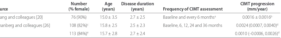
Cross-sectional studies of CIMT reported varied results as compared with age-matched controls, probably as a result of the diff ering vascular study protocols used. Despite these limitations, the average annual rate of progression of CIMT in pSLE patients is small (between
0.0016 and 0.0024 mm/year) (Table 2). Th is rate of
change in CIMT is similar to that in a study of 247 healthy Caucasian adolescents, which found a rate of change of 0.02 mm over a 10-year period (average 0.002 mm/year) [25]. In contrast, in the Atherosclerosis Prevention in Pediatric Lupus Erythematosus (APPLE) trial, the progression rates of all but one of the diverse CIMT measurements carried out on the enrolled placebo-treated pSLE patients showed higher numerical values than those reported in the healthy adolescent cohort, suggesting accelerated athero sclerosis in this large pSLE cohort [26]. More large-scale longitudinal studies are required to determine whether CIMT progression in pSLE truly follows an abnormal trajectory. Adjustment for factors that infl uence CIMT in childhood, such as age, height, body mass index and blood pressure, should be made to ensure appropriate conclusions are drawn [25].
Elevated PWV has been described in pSLE patients [17]. Interestingly, this later study was carried out on pSLE subjects relatively early in their disease course who had low disease activity. Despite this, a signifi cant diff erence from healthy controls was found. Th is might suggest that atherogenesis starts early in these patients and may aff ect even those with low disease activity.
with short disease duration and therefore they may not refl ect the long-term burden of atherosclerosis in pSLE subjects [17,23].
Risk factors for atherosclerosis
Even though factors associated with progression of subclinical atherosclerosis have not been well explored in pSLE, traditional and nontraditional risk factors are being increasingly recognized to be present in this patient population.
Traditional risk factors for cardiovascular disease in pSLE Dyslipidemia, hypertension, obesity, sedentary lifestyle, diabetes, smoking and family history of early CVD are all con sidered traditional risk factors for atherosclerosis develop ment. As compared with adult SLE patients, uncontrolled hypertension, diabetes and smoking are not commonly encountered in pediatric patients. In contrast, dyslipidemia – one of the key precursors of early atherosclerosis – has been shown to be present with increased frequency in pSLE when compared with the general pediatric population.
Dyslipidemia
As early as 1988 it was recognized that both adult and pediatric SLE patients with active disease, prior to corticosteroid therapy, had elevated triglycerides (TGs) and very-low-density lipoprotein-cholesterol and had
depressed high-density lipoprotein-cholesterol (HDL-C) and apolipoprotein A-I – often referred to as the active lupus lipid profi le [27,28]. Th e lipid profi le of elevated TGs and depressed HDL-C is not specifi c for SLE but rather is common to multiple infl ammatory states.
Later studies showed that lipid abnormalities were common in newly diagnosed pSLE patients, prior to the start of corticosteroid treatment [29]. Among a group of 54 recently diagnosed and untreated pSLE subjects, at least one lipid abnormality was found in 63% of patients – elevated TGs in 62%, abnormally low HDL-C in 24%, hypercholesterolemia in 20% and elevated low density lipoprotein cholesterol (LDL-C) in 4% – and one patient had an abnormal level of all four lipids [29].
Th e cross-sectional APPLE trial found mean TG, LDL-C and HDL-LDL-C levels that were in the normal ranges [30]. Th e most common lipid abnormality was also elevated TG levels, found in nearly 30% of subjects.
Th e diff erences between these two studies are probably the result of the lower disease activity, longer disease duration and immunosuppressive therapies of patients in the APPLE trial as compared with the untreated, active patients in the former study.
[image:3.612.64.552.101.251.2]Th e dyslipidemia seen in pSLE patients is multifactorial. Cytokines, autoantibodies, medications, dietary intake, renal disease, physical activities and genetics factors are all likely important contributors. As in other diseases, nephrotic-range proteinuria is associated with higher
Table 1. Carotid intima-media thickness in pediatric-onset systemic lupus erythematosus
Falaschi and Bowser and Huang and Schanberg and Boros and colleagues [19] colleagues [18] colleagues [20] colleagues [22] colleagues [17]
Pediatric-onset systemic lupus erythematosus
Number 26 19 76 221 31
Age (years) 17.1 ± 4.4 16.9 ± 2.3 15.0 ± 3.5 15.7 ± 2.6 15.3 ± 1.9
Disease duration (years) 5.5 ± 3.4 3.2 ± 2.5 2.6 ± 2.5 2.6 ± 2.4 2.6 ± 0.3
CIMT (mm) 0.57 ± 0.05 0.48 ± 0.049 0.63 ± 0.08 0.59 ± 0.05 Right, –0.12a; left, –0.10a
Controls
Number 26 15 38 None 60
Age (years) Matched 16.7 ± 2.1 16.4 ± 3.9 None 15.9 ± 1.9
CIMT (mm) 0.54 ± 0.03 0.454 ± 0.041 0.54 ± 0.06 None –
Values presented as mean ± standard deviation unless otherwise specifi ed. CIMT, carotid intima-media thickness. az-score.
Table 2. Serial carotid intima-media thickness measurements in pediatric-onset systemic lupus erythematosus
Number Age Disease duration CIMT progression
Source (% female) (years) (years) Frequency of CIMT assessment (mm/year)
Huang and colleagues [20] 76 (90%) 15.0 ± 3.5 2.7 ± 2.5 Baseline and every 6 monthsa 0.0016 ± 0.0016b
Schanberg and colleagues [26] 108 (82%)c 15.8 ± 2.5 2.5 ± 2.3 Baseline, 6, 12, 24 and 36 months 0.0024 (0.0007, 0.0040)d
113 (84%)e 15.7 ± 2.8 2.7 ± 2.4 0.0010 (–0.0006, 0.0026)d
Values presented as mean ± standard deviation, unless otherwise specifi ed. CIMT, carotid intima-media thickness. aMean number of CIMT assessments performed:
4.6 ± 1.6. bMean ± standard error; CIMT adjusted for age at diagnosis. cSubjects were in the placebo arm of the Atherosclerosis Prevention in Pediatric Lupus
[image:3.612.74.548.289.354.2]total cholesterol and LDL-C levels in pSLE [19]. In addition, other investigators have shown that lower levels of proteinuria (defi ned as ≥200 mg/day but <3.5 g/day) were associated with abnormal lipid levels, again demon-strating the importance of renal disease in the dys lipid-emia of pSLE [31].
Th ere are few longitudinal studies that have examined serial lipid levels in pSLE subjects [31,32]. In an inception cohort of 139 pSLE patients followed annually from diagnosis up to 3 years, it was shown that the highest levels of total cholesterol, LDL-C and TG and the lowest levels of HDL-C were found at diagnosis, prior to onset of therapy [32]. On follow-up, these same investigators found that the most important factors infl uencing lipid levels were changes in disease activity and prednisone dose [31]. When controlling for disease activity, a reduction in the prednisone dose was associated with an overall improved lipid profi le. A key concept emerges from these studies: when disease activity is brought under control, steroid therapy is being weaned and proteinuria improves, the lipid values normalize. Th ese results suggest that disease control rather than long-term lipid-lowering therapy may be the most important factor to control dyslipidemia in pSLE.
Insulin resistance and metabolic syndrome
Both insulin resistance and metabolic syndrome are present in SLE [32-35]. In pSLE, fasting insulin levels,
hemoglobin A1C and insulin C-peptide levels were
elevated, although Homeostatic Model Assessment for Insulin Resistance values were normal in the majority of patients [17,36]. Obesity, chronic infl ammation, and corticosteroid therapy all can contribute to insulin resistance [37-39]. However, elevated fasting insulin levels and hyperinsulinemia were not restricted to patients on corticosteroids or to obese patients. Insulin resistance may also be secondary to a chronic infl ammatory state including SLE. A rare cause of insulin resistance (type B insulin resistance) is the presence of antibodies against the insulin receptor. Th ese antibodies result in hyper gly cemia, insulin resistance and acanthosis nigricans, and the majority of patients with anti-insulin receptor antibodies have SLE [40]. Of note, patients with anti-insulin receptor antibodies may present with and/or develop hypoglycemia. Despite the presence of insulin resistance and/or metabolic syndrome in patients with pSLE, there is no evidence of an increased prevalence of overt type I or type II diabetes mellitus in pSLE or adult SLE [41]. Patients are, however, at risk for steroid-induced diabetes.
Nontraditional risk factors for cardiovascular disease in pSLE In adult SLE patients, traditional risk factors alone are not suffi cient to explain the enhanced risk of cardio-vascular events. Indeed, after controlling for the
Framingham risk factors, SLE patients have a 17-fold increased risk of mortality from coronary heart disease and a 10-fold increase risk of nonfatal MI [42].
Hyperhomocysteinemia
In studies of otherwise healthy individuals it is not clear whether an elevated homocysteine plasma level is a causal factor or simply a biomarker for atherosclerosis. An etiologic role of hyperhomocysteinemia in athero-sclerosis is supported by the multiple proatherogenic eff ects of hyperhomocysteinemia: induction of endo the-lial cell dysfunction; increased oxidative stress; and promotion of transcription of proinfl ammatory cytokine genes via activation of the NF-κB pathway [43-45]. However, the failure of most trials of homocysteine-lowering therapy to decrease cardiovascular risk in large randomized studies has cast doubt on the role of hyperhomocysteinemia in atherosclerosis [46,47]. Studies of both pSLE and adult SLE patients have shown elevated plasma homocysteine levels. However, there was no correlation between elevated homocysteine levels and vascular markers of early atherosclerosis [17,48,49].
Cytokines and adipokines
An imbalance between endothelial cell damage and repair has been demonstrated to occur in SLE [50,51]. Th ese abnormalities as well as recruitment of macro-phages to arteries, enhanced formation of foam cells and platelet activation have all been shown to be induced by type I interferons [52-54]. Increased serum type I inter-feron activity was shown to be a predictor of abnormal FMD and increased CIMT in adult SLE patients [55]. Similar studies have not been conducted in pSLE but, as the interferon signature is seen in pSLE [56], type I interferon may be an important disease-related factor promoting atherosclerosis.
Elevated levels of other multiple proinfl ammatory cytokines, including IL-6, IFNγ and TNFα, have been implicated in the development of atherosclerosis in other wise healthy populations [57-59]. Although elevated levels of IL-6 are present in adults with SLE and these levels have been shown to correlate with C-reactive protein and abnormal lipid levels, no association between IL-6 and the presence or progression of atherosclerosis has been found in SLE [60-64].
other infl ammatory states. However, there has been no consistent correlation between adipokine levels and early markers of atherosclerosis in adult SLE [66-69]. A pros-pective study of pSLE patients found no signifi cant diff er-ence in adiponectin levels between 105 pSLE patients and a group of healthy controls, although seven pSLE subjects had elevated levels [70]. No studies have explored factors associated with changes in adipokine levels over time or the predictive value of these molecules for the progression of atherosclerosis in pSLE.
Further studies are needed to defi ne whether cytokines, chemokines and adipokines drive the development and progression of atherosclerosis or are protective. Th ese potential biomarkers could guide physicians in stratifying CVD risk in pSLE patients.
Autoantibodies
Lupus is characterized by circulating antibodies of multiple specifi cities, including phospholipid, anti-endothelial cells, anti-apolipoprotein A-I, anti-HDL-C, anti-lipoprotein(a), anti-oxidized LDL-C and anti-lipo-pro tein lipase antibodies. Th e presence of these auto-antibodies against these key constituents in the athero-sclerosis cascade has been postulated to contribute to the pathogenesis of early atherosclerosis in SLE. In pSLE patients, both antiphos pholipid and anti-oxidized LDL-C antibodies have been found [23,71].
Chronic kidney disease
Lupus nephritis is a common disease manifestation of pSLE. In a minority of patients, renal involvement leads to altered glomerular fi ltration rate and even to end-stage renal disease. Although not a traditional cardiovascular risk factor, chronic kidney disease is a major contributor to the onset and progression of accelerated athero-sclerosis in this subgroup of individuals. Analysis of the US Renal Data System, a database comprising patients on chronic dialysis therapy or patients who had received a renal transplantation, revealed that the risk of death was twice as high in pSLE end-stage renal disease patients when compared with pediatric patients with non-SLE-related end-stage renal disease [72]. Seventy-fi ve percent of those deaths were due to cardiovascular disease and cardiac arrest.
A correlation between the duration of end-stage renal disease and CIMT as well as improvement of CIMT post renal transplantation has been described in pediatric chronic kidney disease patients [73,74]. Similar risk factors for atherosclerosis may be found in both SLE and chronic kidney disease patients: hypertension, dyslipid-emia, a chronic infl ammatory state, oxidative stress and underlying endothelial dysfunction [75-79]. Th e coexis-tence of this double hit and of other chronic kidney disease-specifi c risk factors, including activation of the
renin–angiotensin–aldosterone and sympathetic nervous systems as well as enhanced vascular calcifi cation due to metabolic derangements, lead to a strong proatherogenic milieu. Special attention should therefore be given to pSLE patients with chronic kidney disease as they are at increased risk of cardiovascular disease.
Th ere is a signifi cant gap in knowledge on factors
leading to accelerated atherosclerosis and long-term cardio vascular outcomes in pSLE. Identifying key predic-tors of CVD in this vulnerable population is crucial. Th is will enable pediatric rheumatologists to identify early on patients at highest risk and to prioritize the imple men-tation of preventive strategies.
Juvenile idiopathic arthritis
JIA is the most common rheumatologic disease of childhood. JIA encompasses diverse disease subtypes with marked variation in the level of systemic infl am-mation. Adults with multiple forms of infl ammatory arthritis have been shown to have premature athero-sclerosis and an increased morbidity and mortality secon dary to atherosclerosis [80]. In fact, it has been shown that patients with rheumatoid arthritis probably have a similar risk for atherosclerosis as those with type 2 diabetes mellitus [81]. Despite the diversity of the clinical manifestations of the diff erent JIA subsets, most patients have evidence of chronic infl ammation and therefore are probably at risk for early atherosclerosis. Diff erent disease subtypes probably have distinct risk profi les.
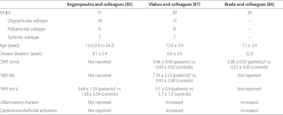
systemic JIA patients were tested [87]. Th e most recent study reported that prepubertal JIA patients (only oligoarticular and polyarticular subtypes) had signifi -cantly increased CIMT as compared with controls. At follow-up after 1 year of individualized therapy that was associated with improved disease control and decreased infl ammation, there was a signifi cant decrease in CIMT [86]. In this study, CIMT was associated with LDL-C and IL-1 levels. Overall, the small number of patients and the predominance of subjects with oligo articular JIA, the least infl ammatory subtype, make the generalizability of these fi ndings questionable. Children with systemic JIA are probably at a much higher risk of accelerated atherosclerosis than those with oligoarticular or poly-articular JIA. Future studies should address the question by specifi c disease subtypes.
Similar issues arise when studies on the prevalence of traditional risk factors for atherosclerosis are examined. Th e few studies reporting lipid levels of JIA patients included children with diff erent disease subtypes, disease activity levels and corticosteroid doses. Th is hetero-geneity probably explains the contradictory fi ndings of the active infl ammatory lipid profi le of high TGs and very-low-density lipoprotein-cholesterol and lower levels of HDL-C, LDL-C and total cholesterol found in some, but not all, studies of JIA [86-93]. Overall it is diffi cult to determine the eff ect of disease and disease activity on the lipid profi le in each distinct JIA subtype. Moreover, biologic therapies might adversely impact the lipid profi le, as seen in systemic-onset JIA patients treated with tocilizumab [94]. Further large-scale studies should be conducted to clarify the burden and trajectory of dyslipidemia in the diff erent JIA subtypes.
Similar to other rheumatic diseases, patients with JIA have evidence of elevated proinfl ammatory cytokines and chemokines. Th e degree of elevation is dependent on the subtype and level of disease activity. Most, but not all, studies have shown elevated homocysteinemia levels in JIA patients [95-97]. One study showed a correlation of homocysteine with CIMT [83]. Elevated levels of omentin, an insulin-sensitizing adipokine, were demon-strated in patients with JIA [98]. Omentin has important eff ects on endothelium as it causes vasodilatation of blood vessels and attenuates many proinfl ammatory signal ing pathways in endothelial cells, including the TNF pathway [99,100]. Th e elevated circulating levels seen in JIA may therefore act as a counter-regulatory mecha nism to attenuate the proatherosclerotic eff ects of
TNF and other mediators of chronic infl ammation. Th e
importance of leptin in atherosclerosis in JIA is not clear because the two studies reported showed opposite results [101,102].
Limitation to performing physical activities due to arthritis and its associated musculoskeletal complica-tions, as well as corticosteroid usage, renders JIA patients susceptible to weight gain. A recent study found that patients with JIA had higher body mass index, fat percentages and truncal fat than age-matched and sex-matched controls [103]. Despite the challenges to regular physical activities in this patient population, children with JIA should be strongly encouraged to engage in an exercise program.
[image:6.612.66.546.100.296.2]Data on other nontraditional risk factors in this pediatric population are lacking. As many patients will continue to have active disease into adulthood, the need to expand research on the prevalence of atherosclerosis
Table 3. Vascular measures of atherosclerosis in juvenile idiopathic arthritis
Argyropoulou and colleagues [85] Vlahos and colleagues [87] Breda and colleagues [86]
JIA (n) 31 30 38
Oligoarticular subtype 18 15 –
Polyarticular subtype 6 8 –
Systemic subtype 7 7 –
Age (years) 13.6 (3.4 to 26.2) 12.0 ± 3.0 7.1 ± 2.4
Disease duration (years) 9.1 ± 5.4 6.6 ± 3.8 22.9
CIMT (mm) Not reported 0.46 ± 0.03 (patients) vs. 0.38 ± 0.05 (patients)a vs.
0.45 ± 0.03 (controls) 0.31 ± 0.05 (controls)
FMD (%) Not reported 7.10 ± 2.23 (patients)b vs. Not reported
9.93 ± 3.90 (controls)
PWV (m/s) 3.68 ± 1.59 (patients)a vs. 5.5 ± 0.9 (patients) vs. Not reported
1.38 ± 0.54 (controls) 5.7 ± 1.0 (controls)
Infl ammatory markers Not reported Increased Increased
Cytokines/endothelial activation Not reported Increased Increased
Values are presented as mean ± standard deviation or median (range), unless otherwise specifi ed. CIMT, carotid intima-media thickness; FMD, fl ow-mediated dilatation; JIA, juvenile idiopathic arthritis; PWV, pulse wave velocity. aDiff erence from controls is statistically signifi cant (P <0.01). bDiff erence from controls is
risk factors and incidence of CVD in JIA is important. Th is is especially true for patients with systemic and polyarticular JIA subtypes, as these patients are at highest risk for chronic infl ammation and prolonged use of corticortisteroids.
Juvenile dermatomyositis
JDM is an infl ammatory disease characterized by typical cutaneous rashes, symmetrical proximal muscle weak-ness, raised serum levels of muscle enzymes and a vasculo pathy. Few studies have assessed early athero-sclerosis in JDM patients. A study of adults with dermato-myositis or polydermato-myositis demonstrated higher risks of acute MI and stroke when compared with the general population [104]. A recent study from Sweden showed that adults with polymyositis/dermatomyositis had a standardized incidence ratio of 1.92 for the development of coronary heart disease as compared with age- and sex-matched controls [105]. Th e only study to examine the risk of atherosclerosis in JDM compared CIMT and FMD of eight adults with a history of JDM with eight healthy
adults [106]. Th e CIMT was higher in JDM patients
despite the fact that they were younger and had lower body mass index than controls.
Acquired lipodystrophy has gained recognition in JDM patients with reported prevalence rates ranging from 8 to 40% [107-109]. Th is condition leads to loss of sub cu-taneous fat and is associated with diverse metabolic abnormalities such as dyslipidemia, abnormal leptin levels, insulin resistance and overt diabetes. Th e extent to which these abnormalities impact on accelerated athero-sclerosis remains unknown. Future work should evaluate whether lipodystrophy is a predictor of cardiovascular events in the JDM population.
A study of children with myositis showed that 41% had elevated fasting insulin levels with 25% meeting criteria for metabolic syndrome, 47% had elevated TG and 17% had abnormal cholesterol, LDL-C or HDL-C levels [110].
Th e previous mentioned study of CIMT was also
performed to document risk factors for atherosclerosis in
JDM [106]. Th ese same patients had higher blood
pressure values, higher prevalence of abnormal HDL-C levels and lower adiponectin levels compared with controls. Two patients fulfi lled criteria for the metabolic syndrome and 63% had lipodystrophy [107]. Atherogenic risk factors are thus clearly present in JDM children. Again, future studies should address the long-term incidence of CVD and better delineate predictors of cardio vascular morbidity and mortality in JDM.
Although JDM and pSLE are clearly two distinct entities, a parallel can be established between these conditions with respect to cardiovascular risk factors. Type I interferons have been implicated in the patho-genesis of both JDM and SLE [111]. Although no studies
have looked at the eff ect of these cytokines on the development of atherosclerosis in JDM, it is reasonable to assume that they have similar eff ects on the endo thelium to those seen in SLE. Similarly, chronic insult to the endothelium precipitated by the underlying vasculo pathy and chronic infl ammation probably leads to early athero-sclerosis in JDM, as seen in SLE. Additionally, long-term corticosteroid therapy probably results in similar meta-bolic derangements such as weight gain, insulin resis-tance and dyslipidemia. Both diseases also lead to disa-bilities resulting in a decreased ability to practice regular aerobic activities.
Prevention strategies
Atherosclerosis is known to begin in childhood. More-over, longitudinal studies have demonstrated that the presence of cardio vascular risk factors in childhood is associated with higher CIMT in young adults [112,113]. Children with infl ammatory diseases have an increased prevalence of traditional risk factors and the extra burden of non traditional risk factors for atherosclerosis. Hence, thorough ongoing cardiovascular risk assessment should be performed routinely in children with chronic rheu-matologic diseases. Long-term cardiovascular morbidity and mortality should be discussed with patients and their families soon after disclosure of the diagnosis to educate patients and facilitate the application of preventive strategies.
Traditional risk factor-related preventive measures
Overweight and obesity are common in patients with pediatric rheumatic diseases, and in particular in patients on corticosteroid therapy. Th e general unwellness secon-dary to systemic infl ammation, the arthritis, the psycho-logical impact of having a chronic disease and the development of cushingoid features all potentially contri-bute to sedentary lifestyle. Th e benefi ts of physical activity on many of the traditional cardiovascular risk factors are well established and regular exercise should
be encour aged. Children unable to perform aerobic
exercises to the desired level should be encouraged to pursue a regular exercise routine adapted to their clinical status, and intensity should be increased as tolerated. A healthy balanced diet should be promoted in all patients. Nutritional and physical activity counseling should be regarded as management priorities. Smoking status should be assessed regularly and smoking cessation support off ered.
cortico steroids, overweight or obese, or with a family history of type II diabetes mellitus.
A baseline pretreatment lipid level should be measured and serial measurements performed. Th is may not apply to certain disease subtypes, such as oligoarticular JIA, where the risk of dyslipidemia is probably similar to that in the general pediatric population. However, there are currently no clear guidelines on the frequency of
dyslipidemia screen ing and the threshold at which
specifi c treatment should be contemplated for patients with pediatric rheumatic diseases has not been defi ned [114]. Th e APPLE trial, the only study to pros pectively assess the use of a statin to reduce the progression of atherosclerosis, as measured by CIMT in pSLE, did not fi nd a signifi cant diff erence in progression of CIMT between the statin-treated and placebo-treated patient groups [26]. However, secondary analyses did show a trend in favor of atorvastatin for other CIMT endpoints. Further studies are required to determine the role of lipid-lowering therapy in pediatric rheumatology.
Th e use of antimalarial agents in pSLE and JDM
provides a dual benefi t: not only do these agents help to keep the disease inactive, but they also exert benefi cial eff ects on lipid levels and glucose tolerance [115-118].
Nontraditional risk factor-related preventive measures A key message that emanates from SLE studies and that probably applies to other infl ammatory conditions is that adequately treating the primary disease will improve many of the cardiovascular risk factors. Control of sys temic infl ammation will decrease production of pro athero genic cytokines, chemokines, adipokines and auto anti bodies, thereby decreasing the burden of pro athero genic insults. Judicious use of corticosteroid might tilt the balance in favor of benefi ts. Th is hypothesis might explain why the use of moderate doses of corticosteroid was negatively correlated with CIMT in the APPLE study [26].
Th erapies used in the treatment of rheumatic disease have been associated with a decrease in atherosclerotic burden. Anti-TNFα therapies have been associated with decreased CIMT [119]. Although B cells have been found to have atheroprotective eff ects, B-cell defi ciency in murine models of atherosclerosis results in a decrease in plaque size [120,121]. Whether this eff ect is also true in humans and whether other anti-B-cell treatments have the same eff ect are not clear. As there are increasing data on the importance of type I interferons in atherosclerosis and disease activity, it will be interestingly to see whether anti-interferon therapy will aff ect atherosclerosis. Th ese biologic thera peutic avenues off er the potential for addi-tional tools in atherosclerosis prevention and therapy. Whether the primary eff ect on atherosclerosis is secon-dary to decreas ing infl ammation or whether there are intrinsic factors related to these molecules is not clear.
Azathioprine has been linked to atherosclerosis while methotrexate and mycophenolate mofetil may off er pro-tec tion [122-125]. However, the eff ects of these three
immuno suppressive agents on atherosclerosis need
further study. Th e use of angiotensin-converting enzyme inhibitors and angiotensin receptor blocking agents in patients with proteinuria and/or hyperten sion will not only off er benefi ts by their direct action on these cardiovascular risk factors but probably also via downregulation of the renin–angiotensin system, which is also implicated in the genesis of atherosclerosis.
Conclusion
Children with chronic rheumatologic diseases are ex-posed to a vast array of proatherogenic insults, but the prevalence and natural history of accelerated athero-sclerosis in the majority of these children remains poorly defi ned. However, it is becoming more apparent that cardiovascular disease results in signifi cant morbidity and mortality in these patients in adulthood. Identifying key risk factors, developing disease-specifi c stratifi cation algorithms and implementing interventions to prevent atherosclerosis are therefore important. Th e predictive value of surrogate measures of athero sclerosis should be specifi cally studied in this pediatric population. Eff orts should be made to identify novel biomarkers that would assist us in quantifying the atherosclerotic burden and to follow its trajectory. Prospec tive, multicenter cohort studies addressing these impor tant issues are urgently needed. Pediatric rheumatology researchers have shown that assembling large national and international cohorts of patients with rheumatic diseases is feasible [15,126,127]. Hopefully, collaborative eff orts among the pediatric and adult rheumatology communities will ultimately lead to improved long-term cardiovascular outcomes in patients with pediatric-onset rheumatologic diseases.
Abbreviations
APPLE, Atherosclerosis Prevention in Pediatric Lupus Erythematosus; CIMT, carotid intima-media thickness; CVD, cardiovascular disease; FMD, fl ow-mediated dilatation; HDL-C, high-density lipoprotein-cholesterol; IFN, interferon; IL, interleukin; JDM, juvenile dermatomyositis; JIA, juvenile idiopathic arthritis; LDL-C, low-density lipoprotein-cholesterol; MI,
myocardial infarction; NF, nuclear factor; pSLE, pediatric-onset systemic lupus erythematosus; PWV, pulse wave velocity; SLE, systemic lupus erythematosus; TG, triglyceride; TNF, tumor necrosis factor.
Competing interests
JB receives funding for a Lupus Fellowship through GlaxoSmithKline Inc., the Canadian Network for Improved Outcomes in Systemic Lupus Erythematosus, the Canadian Rheumatology Association and The Arthritis Society. EDS holds the Ho Family Chair in Autoimmune Diseases. TJB declares that he has no competing interests.
Author details
Avenue, Toronto, ON, Canada M5G 1X8. 3Department of Pediatrics, University of Toronto, 555 University Avenue, Toronto, ON, Canada M5G 1X8. 4Hospital for Sick Children Research Institute, 555 University Avenue, Toronto, ON, Canada M5G 1X8.
Published: 22 May 2013
References
1. Roifman I, Beck PL, Anderson TJ, Eisenberg MJ, Genest J: Chronic infl ammatory diseases and cardiovascular risk: a systematic review. Can J Cardiol 2011, 27:174-182.
2. Cines D B, Pollak ES, Buck CA, Loscalzo J, Zimmerman GA, McEver RP, Pober JS, Wick TM, Konkle BA, Schwartz BS, Barnathan ES, McCrae KR, Hug BA, Schmidt AM, Stern DM: Endothelial cells in physiology and in the pathophysiology of vascular disorders. Blood 1998, 91:3527-3561.
3. Juonala M, Kahonen M, Laitinen T, Hutri-Kahonen N, Jokinen E, Taittonen L, Pietikainen M, Helenius H, Viikari JS, Raitakari OT: Eff ect of age and sex on carotid intima-media thickness, elasticity and brachial endothelial function in healthy adults: the cardiovascular risk in Young Finns Study.
Eur Heart J 2008, 29:1198-1206.
4. Benjami n EJ, Larson MG, Keyes MJ, Mitchell GF, Vasan RS, Keaney JF, Jr, Lehman BT, Fan S, Osypiuk E, Vita JA: Clinical correlates and heritability of fl ow-mediated dilation in the community: the Framingham Heart Study.
Circulation 2004, 109:613-619.
5. B ots ML, Grobbee DE, Hofman A, Witteman JC: Common carotid intima-media thickness and risk of acute myocardial infarction: the role of lumen diameter. Stroke 2005, 36:762-767.
6. L orenz MW, Markus HS, Bots ML, Rosvall M, Sitzer M: Prediction of clinical cardiovascular events with carotid intima-media thickness: a systematic review and meta-analysis. Circulation 2007, 115:459-467.
7. N guyen QM, Toprak A, Xu JH, Srinivasan SR, Chen W, Berenson GS:
Progression of segment-specifi c carotid artery intima-media thickness in young adults (from the Bogalusa Heart Study). Am J Cardiol 2011,
107:114-119.
8. A atola H, Hutri-Kahonen N, Juonala M, Viikari JS, Hulkkonen J, Laitinen T, Taittonen L, Lehtimaki T, Raitakari OT, Kahonen M: Lifetime risk factors and arterial pulse wave velocity in adulthood: the cardiovascular risk in young Finns study. Hypertension 2010, 55:806-811.
9. T suchikura S, Shoji T, Kimoto E, Shinohara K, Hatsuda S, Koyama H, Emoto M, Nishizawa Y: Central versus peripheral arterial stiff ness in association with coronary, cerebral and peripheral arterial disease. Atherosclerosis 2010,
211:480-485.
10. Urbina EM, Williams RV, Alpert BS, Collins RT, Daniels SR, Hayman L, Jacobson M, Mahoney L, Mietus-Snyder M, Rocchini A, Steinberger J, McCrindle B; American Heart Association Atherosclerosis, Hypertension, and Obesity in Youth Committee of the Council on Cardiovascular Disease in the Young:
Noninvasive assessment of subclinical atherosclerosis in children and adolescents: recommendations for standard assessment for clinical research: a scientifi c statement from the American Heart Association.
Hypertension 2009, 54:919-950.
11. Tucker L: Review: Making the diagnosis of systemic lupus erythematosus in children and adolescents. Lupus 2007, 16:546-549.
12. Tyrrell PN, Beyene J, Feldman BM, McCrindle BW, Silverman ED, Bradley TJ:
Rheumatic disease and carotid intima-media thickness: a systematic review and meta-analysis. Arterioscler Thromb Vasc Biol 2010, 30:1014-1026. 13. Urowitz MB, Gladman D, Ibanez D, Fortin P, Sanchez-Guerrero J, Bae S, Clarke
A, Bernatsky S, Gordon C, Hanly J, Wallace D, Isenberg D, Ginzler E, Merrill J, Alarcon G, Steinsson K, Petri M, Dooley MA, Bruce I, Manzi S, Khamashta M, Ramsey-Goldman R, Zoma A, Sturfelt G, Nived O, Maddison P, Font J, van Vollenhoven R, Aranow C, Kalunian K, Stoll T, Buyon J: Clinical manifestations and coronary artery disease risk factors at diagnosis of systemic lupus erythematosus: data from an international inception cohort. Lupus 2007,
16:731-735.
14. Hersh AO, von Scheven E, Yazdany J, Panopalis P, Trupin L, Julian L, Katz P, Criswell LA, Yelin E: Diff erences in long-term disease activity and treatment of adult patients with childhood- and adult-onset systemic lupus erythematosus. Arthritis Rheum 2009, 61:13-20.
15. Hersh AO, Trupin L, Yazdany J, Panopalis P, Julian L, Katz P, Criswell LA, Yelin E:
Childhood-onset disease as a predictor of mortality in an adult cohort of patients with systemic lupus erythematosus. Arthritis Care Res 2010,
62:1152-1159.
16. California Department of Health Statistics: Death Statistics Data Table – Leading Causes of Female Deaths by Age and Race/Ethnic Group and Rates for All Races Combined. California, USA: California Department of Health Statistics; 2010. 17. Boros CA, Bradley TJ, Cheung MM, Bargman JM, Russell JL, McCrindle BW,
Adeli K, Hamilton J, Silverman ED: Early determinants of atherosclerosis in paediatric systemic lupus erythematosus. Clin Exp Rheumatol 2011,
29:575-581.
18. Bowser CS, Kumar S, Salciccioli L, Kutlin A, Lazar J, Rahim I, Suss A, Kohlhoff S, Hammerschlag MR, Moallem HJ: Absence of Chlamydia pneumoniae and signs of atherosclerotic cardiovascular disease in adolescents with systemic lupus erythematosus. Pediatr Cardiol 2008, 29:545-551. 19. Falaschi F, Ravelli A, Martignoni A, Migliavacca D, Sartori M, Pistorio A, Perani
G, Martini A: Nephrotic-range proteinuria, the major risk factor for early atherosclerosis in juvenile-onset systemic lupus erythematosus. Arthritis Rheum 2000, 43:1405-1409.
20. Huang YL, Chung HT, Chang CJ, Yeh KW, Chen LC, Huang JL: Lymphopenia is a risk factor in the progression of carotid intima-media thickness in juvenile-onset systemic lupus erythematosus. Arthritis Rheum 2009,
60:3766-3775.
21. Nascif AK, Hilario MO, Terreri MT, Ajzen SA, D’Almeida V, Plavnik FL, de Jesus Christofalo DM: Endothelial function analysis and atherosclerotic risk factors in adolescents with systemic lupus erythematosus. Int J Adolesc Med Health 2007, 19:497-505.
22. Schanberg LE, Sandborg C, Barnhart HX, Ardoin SP, Yow E, Evans GW, Mieszkalski KL, Ilowite NT, Eberhard A, Levy DM, Kimura Y, von Scheven E, Silverman E, Bowyer SL, Punaro L, Singer NG, Sherry DD, McCurdy D, Klein-Gitelman M, Wallace C, Silver R, Wagner-Weiner L, Higgins GC, Brunner HI, Jung L, Soep JB, Reed A; Atherosclerosis Prevention in Pediatric Lupus Erythematosus Investigators: Premature atherosclerosis in pediatric systemic lupus erythematosus: risk factors for increased carotid intima-media thickness in the atherosclerosis prevention in pediatric lupus erythematosus cohort. Arthritis Rheum 2009, 60:1496-1507. 23. Soep JB, Mietus-Snyder M, Malloy MJ, Witztum JL, von Scheven E:
Assessment of atherosclerotic risk factors and endothelial function in children and young adults with pediatric-onset systemic lupus erythematosus. Arthritis Rheum 2004, 51:451-457.
24. Oren A, Vos LE, Uiterwaal CS, Grobbee DE, Bots ML: Cardiovascular risk factors and increased carotid intima-media thickness in healthy young adults: the Atherosclerosis Risk in Young Adults (ARYA) Study. Arch Intern Med 2003, 163:1787-1792.
25. Jourdan C, Wuhl E, Litwin M, Fahr K, Trelewicz J, Jobs K, Schenk JP, Grenda R, Mehls O, Tröger J, Schaefer F: Normative values for intima-media thickness and distensibility of large arteries in healthy adolescents. J Hypertens 2005,
23:1707-1715.
26. S chanberg LE, Sandborg C, Barnhart HX, Ardoin SP, Yow E, Evans GW, Mieszkalski KL, Ilowite NT, Eberhard A, Imundo LF, Kimura Y, von Scheven E, Silverman E, Bowyer SL, Punaro M, Singer NG, Sherry DD, McCurdy D, Klein-Gitelman M, Wallace C, Silver R, Wagner-Weiner L, Higgins GC, Brunner HI, Jung L, Soep JB, Reed AM, Provenzale J, Thompson SD; Atherosclerosis Prevention in Pediatric Lupus Erythematosus Investigators: Use of atorvastatin in systemic lupus erythematosus in children and adolescents.
Arthritis Rheum 2012, 64:285-296.
27. I lowite NT, Samuel P, Ginzler E, Jacobson MS: Dyslipoproteinemia in pediatric systemic lupus erythematosus. Arthritis Rheum 1988, 31:859-863. 28. B orba EF, Bonfa E: Dyslipoproteinemias in systemic lupus erythematosus: infl uence of disease, activity, and anticardiolipin antibodies. Lupus 1997,
6:533-539.
29. T yrrell PN, Beyene J, Benseler SM, Sarkissian T, Silverman ED: Predictors of lipid abnormalities in children with new-onset systemic lupus erythematosus. J Rheumatol 2007, 34:2112-2119.
30. A rdoin SP, Schanberg LE, Sandborg C, Yow E, Barnhart HX, Mieszkalski K, Ilowite NT, von Scheven E, Eberhard A, Levy DM, Kimura Y, Silverman E, Bowyer SL, Punaro L, Singer NG, Sherry DD, McCurdy D, Klein-Gitelman M, Wallace C, Silver R, Wagner-Weiner L, Higgins GC, Brunner HI, Jung LK, Imundo L,Soep JB, Reed AM; APPLE Investigators: Laboratory markers of cardiovascular risk in pediatric SLE: the APPLE baseline cohort. Lupus 2010,
19:1315-1325.
31. S arkissian T, Beyene J, Feldman B, McCrindle B, Silverman ED: Longitudinal examination of lipid profi les in pediatric systemic lupus erythematosus.
Arthritis Rheum 2007, 56:631-638.
of the interaction between disease activity and therapy on the lipid profi le in patients with pediatric systemic lupus erythematosus. Arthritis Rheum 2006, 54:1283-1290.
33. S abio JM, Zamora-Pasadas M, Jimenez-Jaimez J, Albadalejo F, Vargas-Hitos J, Rodriguez del Aguila MD, Hidalgo-Tenorio C, Gonzalez-Gay MA, Jimenez-Alonso J: Metabolic syndrome in patients with systemic lupus erythematosus from Southern Spain. Lupus 2008, 17:849-859. 34. E l Magadmi M, Ahmad Y, Turkie W, Yates AP, Sheikh N, Bernstein RM,
Durrington PN, Laing I, Bruce IN: Hyperinsulinemia, insulin resistance, and circulating oxidized low density lipoprotein in women with systemic lupus erythematosus. J Rheumatol 2006, 33:50-56.
35. Z eng YJ, Zeng FQ, Dai L, Yang C, Lin BZ, Zheng DH, Liu D, Yan L, Ren M, Cheng H: Characteristics and risk factors for hyperglycemia in Chinese female patients with systemic lupus erythematosus. Lupus 2010, 19:1344-1350. 36. P osadas-Romero C, Torres-Tamayo M, Zamora-Gonzalez J, Aguilar-Herrera BE,
Posadas-Sanchez R, Cardoso-Saldana G, Ladron de Guevara G, Solis-Vallejo E, El Hafi di M: High insulin levels and increased low-density lipoprotein oxidizability in pediatric patients with systemic lupus erythematosus.
Arthritis Rheum 2004, 50:160-165.
37. S teinberg GR, Michell BJ, van Denderen BJ, Watt MJ, Carey AL, Fam BC, Andrikopoulos S, Proietto J, Gorgun CZ, Carling, Hotamisligil GS, Febbraio MA, Kay TW, Kemp BE: Tumor necrosis factor alpha-induced skeletal muscle insulin resistance involves suppression of AMP-kinase signaling. Cell Metab 2006, 4:465-474.
38. E manuela F, Grazia M, Marco de R, Maria Paola L, Giorgio F, Marco B:
Infl ammation as a link between obesity and metabolic syndrome. J Nutr Metab 2012, 2012:476380.
39. v an Raalte DH, Ouwens DM, Diamant M: Novel insights into glucocorticoid-mediated diabetogenic eff ects: towards expansion of therapeutic options? Eur J Clin Invest 2009, 39:81-93.
40. A rioglu E, Andewelt A, Diabo C, Bell M, Taylor SI, Gorden P: Clinical course of the syndrome of autoantibodies to the insulin receptor (type B insulin resistance): a 28-year perspective. Medicine (Baltimore) 2002, 81:87-100. 41. C ortes S, Chambers S, Jeronimo A, Isenberg D: Diabetes mellitus
complicating systemic lupus erythematosus – analysis of the UCL lupus cohort and review of the literature. Lupus 2008, 17:977-980.
42. Es daile JM, Abrahamowicz M, Grodzicky T, Li Y, Panaritis C, du Berger R, Cote R, Grover SA, Fortin PR, Clarke AE, Senécal JL: Traditional Framingham risk factors fail to fully account for accelerated atherosclerosis in systemic lupus erythematosus. Arthritis Rheum 2001, 44:2331-2337.
43. Wei ss N, Keller C, Hoff mann U, Loscalzo J: Endothelial dysfunction and atherothrombosis in mild hyperhomocysteinemia. Vasc Med 2002,
7:227-239.
44. Ung vari Z, Csiszar A, Edwards JG, Kaminski PM, Wolin MS, Kaley G, Koller A:
Increased superoxide production in coronary arteries in
hyperhomocysteinemia: role of tumor necrosis factor-alpha, NAD(P)H oxidase, and inducible nitric oxide synthase. Arterioscler Thromb Vasc Biol 2003, 23:418-424.
45. Au- Yeung KK, Woo CW, Sung FL, Yip JC, Siow YL, O K: Hyperhomo-cysteinemia activates nuclear factor-kappaB in endothelial cells via oxidative stress. Circ Res 2004, 94:28-36.
46. Kal ra DK: Homocysteine and cardiovascular disease. Curr Atheroscler Rep 2004, 6:101-106.
47. Sap osnik G, Ray JG, Sheridan P, McQueen M, Lonn E: Homocysteine-lowering therapy and stroke risk, severity, and disability: additional fi ndings from the HOPE 2 trial. Stroke 2009, 40:1365-1372.
48. Nar shi CB, Giles IP, Rahman A: The endothelium: an interface between autoimmunity and atherosclerosis in systemic lupus erythematosus?
Lupus 2011, 20:5-13.
49. do Prado R, D’Almeida VM, Guerra-Shinohara E, Galdieri LC, Terreri MT, Hilario MO: Increased concentration of plasma homocysteine in children with systemic lupus erythematosus. Clin Exp Rheumatol 2006, 24:594-598. 50. Lee PY, Li Y, Richards HB, Chan FS, Zhuang H, Narain S, Butfi loski EJ, Sobel ES,
Reeves WH, Segal MS: Type I interferon as a novel risk factor for endothelial progenitor cell depletion and endothelial dysfunction in systemic lupus erythematosus. Arthritis Rheum 2007, 56:3759-3769.
51. Tha cker SG, Berthier CC, Mattinzoli D, Rastaldi MP, Kretzler M, Kaplan MJ:
The detrimental eff ects of IFN-α on vasculogenesis in lupus are mediated by repression of IL-1 pathways: potential role in atherogenesis and renal vascular rarefaction. J Immunol 2010, 185:4457-4469.
52. Li J, Fu Q, Cui H, Qu B, Pan W, Shen N, Bao C: Interferon-alpha priming
promotes lipid uptake and macrophage-derived foam cell formation: a novel link between interferon-alpha and atherosclerosis in lupus.
Arthritis Rheum 2011, 63:492-502.
53. Gooss ens P, Gijbels MJ, Zernecke A, Eijgelaar W, Vergouwe MN, van der Made I, Vanderlocht J, Beckers L, Buurman WA, Daemen MJ, Daemen MJ, Kalinke U, Weber C, Lutgens E, de Winther MP: Myeloid type I interferon signaling promotes atherosclerosis by stimulating macrophage recruitment to lesions. Cell Metab 2010, 12:142-153.
54. Lood C, Amisten S, Gullstrand B, Jonsen A, Allhorn M, Truedsson L, Sturfelt G, Erlinge D, Bengtsson AA: Platelet transcriptional profi le and protein expression in patients with systemic lupus erythematosus: up-regulation of the type I interferon system is strongly associated with vascular disease.
Blood 2010, 116:1951-1957.
55. Somer s EC, Zhao W, Lewis EE, Wang L, Wing JJ, Sundaram B, Kazerooni EA, McCune WJ, Kaplan MJ: Type I interferons are associated with subclinical markers of cardiovascular disease in a cohort of systemic lupus erythematosus patients. PLoS One 2012, 7:e37000.
56. Benne tt L, Palucka AK, Arce E, Cantrell V, Borvak J, Banchereau J, Pascual V:
Interferon and granulopoiesis signatures in systemic lupus erythematosus blood. J Exp Med 2003, 197:711-723.
57. Danes h J, Kaptoge S, Mann AG, Sarwar N, Wood A, Angleman SB, Wensley F, Higgins JP, Lennon L, Eiriksdottir G, Rumley A, Whincup PH, Lowe GD, Gudnason V: Long-term interleukin-6 levels and subsequent risk of coronary heart disease: two new prospective studies and a systematic review. PLoS Med 2008, 5(4):e78.
58. Ridke r PM, Rifai N, Stampfer MJ, Hennekens CH: Plasma concentration of interleukin-6 and the risk of future myocardial infarction among apparently healthy men. Circulation 2000, 101:1767-1772.
59. Svenu ngsson E, Fei GZ, Jensen-Urstad K, de Faire U, Hamsten A, Frostegard J:
TNF-alpha: a link between hypertriglyceridaemia and infl ammation in SLE patients with cardiovascular disease. Lupus 2003, 12:454-461.
60. Asanu ma Y, Chung CP, Oeser A, Shintani A, Stanley E, Raggi P, Stein CM:
Increased concentration of proatherogenic infl ammatory cytokines in systemic lupus erythematosus: relationship to cardiovascular risk factors.
J Rheumatol 2006, 33:539-545.
61. Chung CP, Oeser A, Solus J, Avalos I, Gebretsadik T, Shintani A, Linton MF, Fazio S, Stein CM: Infl ammatory mechanisms aff ecting the lipid profi le in patients with systemic lupus erythematosus. J Rheumatol 2007,
34:1849-1854.
62. Rua-F igueroa I, Arencibia-Mireles O, Elvira M, Erausquin C, Ojeda S, Francisco F, Naranjo A, Rodriguez-Gallego C, Garcia-Laorden I, Rodriguez-Perez J, Rodríguez-Lozano C: Factors involved in the progress of preclinical atherosclerosis associated with systemic lupus erythematosus: a 2-year longitudinal study. Ann Rheum Dis 2010, 69:1136-1139.
63. Sabio JM, Vargas-Hitos J, Zamora-Pasadas M, Mediavilla JD, Navarrete N, Ramirez A, Hidalgo-Tenorio C, Jaimez L, Martin J, Jimenez-Alonso J: Metabolic syndrome is associated with increased arterial stiff ness and biomarkers of subclinical atherosclerosis in patients with systemic lupus erythematosus.
J Rheumatol 2009, 36:2204-2211.
64. Roman MJ, Shanker BA, Davis A, Lockshin MD, Sammaritano L, Simantov R, Crow MK, Schwartz JE, Paget SA, Devereux RB, Salmon JE: Prevalence and correlates of accelerated atherosclerosis in systemic lupus erythematosus.
N Engl J Med 2003, 349:2399-2406.
65. Pischo n T, Girman CJ, Hotamisligil GS, Rifai N, Hu FB, Rimm EB: Plasma adiponectin levels and risk of myocardial infarction in men. JAMA 2004,
291:1730-1737.
66. Chung CP, Long AG, Solus JF, Rho YH, Oeser A, Raggi P, Stein CM:
Adipocytokines in systemic lupus erythematosus: relationship to infl ammation, insulin resistance and coronary atherosclerosis. Lupus 2009,
18:799-806.
67. Reynol ds HR, Buyon J, Kim M, Rivera TL, Izmirly P, Tunick P, Clancy RM:
Association of plasma soluble E-selectin and adiponectin with carotid plaque in patients with systemic lupus erythematosus. Atherosclerosis 2010,
210:569-574.
68. McMaho n M, Skaggs BJ, Sahakian L, Grossman J, Fitzgerald J, Ragavendra N, Charles-Schoeman C, Chernishof M, Gorn A, Witztum JL, Wong WK, Weisman M, Wallace DJ, La Cava A, Hahn BH: High plasma leptin levels confer increased risk of atherosclerosis in women with systemic lupus erythematosus, and are associated with infl ammatory oxidised lipids.
Ann Rheum Dis 2011, 70:1619-1624.
Feldt JM: Resistin levels in lupus and associations with disease-specifi c measures, insulin resistance, and coronary calcifi cation. J Rheumatol 2011,
38:2369-2375.
70. Al M, Ng L, Tyrrell P, Bargman J, Bradley T, Silverman E: Adipokines as novel biomarkers in paediatric systemic lupus erythematosus. Rheumatology (Oxford) 2009, 48:497-501.
71. Svenun gsson E, Jensen-Urstad K, Heimburger M, Silveira A, Hamsten A, de Faire U, Witztum JL, Frostegard J: Risk factors for cardiovascular disease in systemic lupus erythematosus. Circulation 2001, 104:1887-1893. 72. Sule S , Fivush B, Neu A, Furth S: Increased risk of death in pediatric and
adult patients with ESRD secondary to lupus. Pediatr Nephrol 2011,
26:93-98.
73. Rinat C, Becker-Cohen R, Nir A, Feinstein S, Shemesh D, Algur N, Ben Shalom E, Farber B, Frishberg Y: A comprehensive study of cardiovascular risk factors, cardiac function and vascular disease in children with chronic renal failure. Nephrol Dial Transplant 2010, 25:785-793.
74. Litwin M, Wuhl E, Jourdan C, Niemirska A, Schenk JP, Jobs K, Grenda R, Wawer ZT, Rajszys P, Mehls O, Schaefer F: Evolution of large-vessel arteriopathy in paediatric patients with chronic kidney disease. Nephrol Dial Transplant 2008, 23:2552-2557.
75. Tucker PS, Dalbo VJ, Han T, Kingsley MI: Clinical and research markers of oxidative stress in chronic kidney disease. Biomarkers 2013, 18:103-115. 76. Luksha N, Luksha L, Carrero JJ, Hammarqvist F, Stenvinkel P, Kublickiene K:
Impaired resistance artery function in patients with end-stage renal disease. Clin Sci 2011, 120:525-536.
77. Yilmaz MI, Stenvinkel P, Sonmez A, Saglam M, Yaman H, Kilic S, Eyileten T, Caglar K, Oguz Y, Vural A, Çakar M, Altun B, Yenicesu M, Carrero JJ: Vascular health, systemic infl ammation and progressive reduction in kidney function; clinical determinants and impact on cardiovascular outcomes. Nephrol Dial Transplant 2011, 26:3537-3543.
78. Moody W E, Edwards NC, Madhani M, Chue CD, Steeds RP, Ferro CJ, Townend JN: Endothelial dysfunction and cardiovascular disease in early-stage chronic kidney disease: cause or association? Atherosclerosis 2012,
223:86-94.
79. Muntner P, Hamm LL, Kusek JW, Chen J, Whelton PK, He J: The prevalence of nontraditional risk factors for coronary heart disease in patients with chronic kidney disease. Ann Internal Med 2004, 140:9-17.
80. Han C, Robinson DW Jr, Hackett MV, Paramore LC, Fraeman KH, Bala MV:
Cardiovascular disease and risk factors in patients with rheumatoid arthritis, psoriatic arthritis, and ankylosing spondylitis. J Rheumatol 2006,
33:2167-2172.
81. Nurmoha med MT: Cardiovascular risk in rheumatoid arthritis. Autoimmun Rev 2009, 8:663-667.
82. Gorska A, Rutkowska-Sak L, Musiej-Nowakowska E, Chlabicz S, Gorski S:
[Nailfold videocapillaroscopy – a useful tool for screening patients with juvenile idiopathic arthritis at the risk of development of premature atherosclerosis]. Postepy Hig Med Dosw (Online) 2010, 64:296-302. 83. Pietrewi cz E, Urban M: [Early atherosclerosis changes in children with
juvenile idiopathic arthritis]. Pol Merkur Lekarski 2007, 22:211-214. 84. Urban M, Pietrewicz E, Gorska A, Szczepanski W, Baran M: [Correlation
between intima-media thickness in carotid artery and markers of epithelial cell dysfunction in patients with juvenile idiopathic arthritis].
Med Wieku Rozwoj 2009, 13:277-282.
85. Argyropo ulou MI, Kiortsis DN, Daskas N, Xydis V, Mavridis A, Efremidis SC, Siamopoulou A: Distensibility and pulse wave velocity of the thoracic aorta in patients with juvenile idiopathic arthritis: an MRI study. Clin Exp Rheumatol 2003, 21:794-797.
86. Breda L, Di Marzio D, Giannini C, Gaspari S, Nozzi M, Scarinci A, Chiarelli F, M ohn A: Relationship between infl ammatory markers, oxidant-antioxidant status and intima-media thickness in prepubertal children with juvenile idiopathic arthritis. Clin Res Cardiol 2013, 102:63-71.
87. Vlahos AP, Theocharis P, Bechlioulis A, Naka KK, Vakalis K, Papamichael ND, Alfa ntaki S, Gartzonika K, Mavridis A, Michalis LK, Siamopoulou A: Changes in vascular function and structure in juvenile idiopathic arthritis. Arthritis Care Res (Hoboken) 2011, 63:1736-1744.
88. Honkanen VE, Pelkonen P, Konttinen YT, Mussalo-Rauhamaa H, Lehto J, Westermarck T: Serum cholesterol and vitamins A and E in juvenile chronic arthritis. Clin Exp Rheumatol 1990, 8:187-191.
89. Marangoni RG, Hayata AL, Borba EF, Azevedo PM, Bonfa E, Schainberg CG:
Decreased high-density lipoprotein cholesterol levels in polyarticular juvenile idiopathic arthritis. Clinics (Sao Paulo) 2011, 66:1549-1552.
90. Musiej-Nowakowska E, Zawadzka F, Wesolowska H, Mikolajew M: Serum lipid concentr ations in juvenile rheumatoid arthritis. Acta Univ Carol Med (Praha) 1991, 37:46-49.
91. Tselepis AD, Elisaf M, Besis S, Karabina SA, Chapman MJ, Siamopoulou A:
Associat ion of the infl ammatory state in active juvenile rheumatoid arthritis with hypo-high-density lipoproteinemia and reduced lipoprotein-associated platelet-activating factor acetylhydrolase activity.
Arthritis Rheum 1999, 42:373-383.
92. Ilowite NT, Samuel P, Beseler L, Jacobson MS: Dyslipoproteinemia in juvenile rhe umatoid arthritis. J Pediatr 1989, 114:823-826.
93. Bakkaloglu A, Kirel B, Ozen S, Saatci U, Topaloglu, Besbas N: Plasma lipids and lipoproteins in juvenile chronic arthritis. Clin Rheumatol 1996, 15:341-345. 94. De Benedetti F, Brunner HI, Ruperto N, Kenwright A, Wright S, Calvo I, Cuttica
R , Ravelli A, Schneider R, Woo P, Wouters C, Xavier R, Zemel L, Baildam E, Burgos-Vargas R, Dolezalova P, Garay SM, Merino R, Joos R, Grom A, Wulff raat N, Zuber Z, Zulian F, Lovell D, Martini A; PRINTO; PRCSG: Randomized trial of tocilizumab in systemic juvenile idiopathic arthritis. N Engl J Med 2012,
367:2385-2395.
95. Goncalves M, D’Almeida V, Guerra-Shinohara EM, Galdieri LC, Len CA, Hilario MO: Homocysteine and lipid profi le in children with Juvenile Idiopathic Arthritis. Pediatr Rheumatol Online J 2007, 5:2.
96. Huemer M, Fodinger M, Huemer C, Sailer-Hock M, Falger J, Rettenbacher A, Berneck er M, Artacker G, Kenzian H, Lang T, Stöckler-Ipsiroglu S:
Hyperhomocysteinemia in children with juvenile idiopathic arthritis is not infl uenced by methotrexate treatment and folic acid supplementation: a pilot study. Clin Exp Rheumatol 2003, 21:249-255.
97. Huemer M, Huemer C, Ulmer H, Crone J, Fodinger M, Falger J, Sailer-Hock M:
No evi dence for hyperhomocysteinemia or increased prevalence of genetic polymorphisms in the homocysteine pathway in patients with moderate juvenile idiopathic arthritis. J Rheumatol 2005, 32:170-174. 98. Cantarini L, Simonini G, Fioravanti A, Generoso M, Bacarelli MR, Dini E, Galeazzi M, Cimaz R: Circulating levels of the adipokines vaspin and omentin in patients with juvenile idiopathic arthritis, and relation to disease activity. Clin Exp Rheumatol 2011, 29:1044-1048.
99. Tan BK, Adya R, Randeva HS: Omentin: a novel link between infl ammation, diabesity , and cardiovascular disease. Trends Cardiovasc Med 2010,
20:143-148.
100. Yamawaki H, Kuramoto J, Kameshima S, Usui T, Okada M, Hara Y: Omentin, a novel ad ipocytokine inhibits TNF-induced vascular infl ammation in human endothelial cells. Biochem Biophys Res Commun 2011, 408:339-343. 101. Elwakkad AS, Said RN, Muhammad SI, Saleh MT, Elhamshary A: Role for leptin
and p rolactin in human juvenile rheumatic diseases. Pak J Biol Sci 2007,
10:1984-1989.
102. Perfetto F, Tarquini R, Simonini G, Bindi G, Mancuso F, Guiducci S, Matucci-Ceri nic M, Falcini F: Circulating leptin levels in juvenile idiopathic arthritis: a marker of nutritional status? Ann Rheum Dis 2005, 64:149-152.
103. Caetano MC, Sarni RO, Terreri MT, Ortiz TT, Pinheiro M, de Souza FI, Hilario MO:
Excess of adiposity in female children and adolescents with juvenile idiopathic arthritis. Clin Rheumatol 2012, 31:967-971.
104. Tisseverasinghe A, Bernatsky S, Pineau CA: Arterial events in persons with derma tomyositis and polymyositis. J Rheumatol 2009, 36:1943-1946. 105. Zoller B, Li X, Sundquist J, Sundquist K: Risk of subsequent coronary heart
dise ase in patients hospitalized for immune-mediated diseases: a nationwide follow-up study from Sweden. PLoS One 2012, 7:e33442. 106. Eimer MJ, Brickman WJ, Seshadri R, Ramsey-Goldman R, McPherson DD,
Smulevitz B, Stone NJ, Pachman LM: Clinical status and cardiovascular risk profi le of adults with a history of juvenile dermatomyositis. J Pediatr 2011,
159:795-801.
107. Bingham A, Mamyrova G, Rother KI, Oral E, Cochran E, Premkumar A, Kleiner D, Jam es-Newton L, Targoff IN, Pandey JP, Carrick DM, Sebring N, O’Hanlon TP, Ruiz-Hidalgo M, Turner M, Gordon LB, Laborda J, Bauer SR, Blackshear PJ, Imundo L, Miller FW, Rider LG; Childhood Myositis Heterogeneity Study Group: Predictors of acquired lipodystrophy in juvenile-onset dermatomyositis and a gradient of severity. Medicine (Baltimore) 2008,
87:70-86.
108. McCann LJ, Juggins AD, Maillard SM, Wedderburn LR, Davidson JE, Murray KJ, Pilki ngton CA: The Juvenile Dermatomyositis National Registry and Repository (UK and Ireland) – clinical characteristics of children recruited within the fi rst 5 yr. Rheumatology (Oxford) 2006, 45:1255-1260.
110. Coyle K, Rother KI, Weise M, Ahmed A, Miller FW, Rider LG: Metabolic abnormalitie s and cardiovascular risk factors in children with myositis.
J Pediatr 2009, 155:882-887.
111. Baechler EC, Bilgic H, Reed AM: Type I interferon pathway in adult and juvenile d ermatomyositis. Arthritis Res Ther 2011, 13:249.
112. Li S, Chen W, Srinivasan SR, Bond MG, Tang R, Urbina EM, Berenson GS:
Childhood c ardiovascular risk factors and carotid vascular changes in adulthood: the Bogalusa Heart Study. JAMA 2003, 290:2271-2276. 113. Raitakari OT, Juonala M, Kahonen M, Taittonen L, Laitinen T, Maki-Torkko N,
Jarvi salo MJ, Uhari M, Jokinen E, Ronnemaa T, Akerblom HK, Viikari JS:
Cardiovascular risk factors in childhood and carotid artery intima-media thickness in adulthood: the Cardiovascular Risk in Young Finns Study.
JAMA 2003, 290:2277-2283.
114. Kavey RE, Allada V, Daniels SR, Hayman LL, McCrindle BW, Newburger JW, Parekh RS, Steinberger J: Cardiovascular risk reduction in high-risk pediatric patients: a scientifi c statement from the American Heart Association Expert Panel on Population and Prevention Science; the Councils on Cardiovascular Disease in the Young, Epidemiology and Prevention, Nutrition, Physical Activity and Metabolism, High Blood Pressure Research, Cardiovascular Nursing, and the Kidney in Heart Disease; and the Interdisciplinary Working Group on Quality of Care and Outcomes Research: endorsed by the American Academy of Pediatrics. Circulation 2006, 114:2710-2738.
115. Hodis HN, Quismorio FP, Jr, Wickham E, Blankenhorn DH: The lipid, lipoprotein, an d apolipoprotein eff ects of hydroxychloroquine in patients with systemic lupus erythematosus. J Rheumatol 1993, 20:661-665. 116. Petri M, Lakatta C, Magder L, Goldman D: Eff ect of prednisone and
hydroxychloroqu ine on coronary artery disease risk factors in systemic lupus erythematosus: a longitudinal data analysis. Am J Med 1994,
96:254-259.
117. Tam LS, Gladman DD, Hallett DC, Rahman P, Urowitz MB: Eff ect of antimalarial agen ts on the fasting lipid profi le in systemic lupus erythematosus. J Rheumatol 2000, 27:2142-2145.
118. Penn SK, Kao AH, Schott LL, Elliott JR, Toledo FG, Kuller L, Manzi S, Wasko MC:
H ydroxychloroquine and glycemia in women with rheumatoid arthritis and systemic lupus erythematosus. J Rheumatol 2010, 37:1136-1142. 119. Westlake SL, Colebatch AN, Baird J, Curzen N, Kiely P, Quinn M, Choy E, Ostor
AJK , Edwards CJ: Tumour necrosis factor antagonists and the risk of cardiovascular disease in patients with rheumatoid arthritis: a systematic literature review. Rheumatology 2011, 50:518-531.
120. Major AS, Fazio S, Linton MF: B-lymphocyte defi ciency increases atherosclerosis i n LDL receptor-null mice. Arterioscler Thromb Vasc Biol 2002,
22:1892-1898.
121. Ait-Oufella H, Herbin O, Bouaziz JD, Binder CJ, Uyttenhove C, Laurans L, Taleb S, Van Vre E, Esposito B, Vilar J, Sirvent J, Van Snick J, Tedgui A, Tedder TF, Mallat Z: B cell depletion reduces the development of atherosclerosis in mice.
J Exp Med 2010, 207:1579-1587.
122. Bulgarelli A, Martins Dias AA, Caramelli B, Maranhao RC: Treatment with methotrex ate inhibits atherogenesis in cholesterol-fed rabbits.
J Cardiovasc Pharmacol 2012, 59:308-314.
123. Toloza SM, Uribe AG, McGwin G, Jr, Alarcon GS, Fessler BJ, Bastian HM, Vila LM, W u R, Shoenfeld Y, Roseman JM, Reveille JD; LUMINA Study Group: Systemic lupus erythematosus in a multiethnic US cohort (LUMINA). XXIII. Baseline predictors of vascular events. Arthritis Rheum 2004, 50:3947-3957. 124. van Leuven SI, Mendez-Fernandez YV, Wilhelm AJ, Wade NS, Gabriel CL,
Kastelein JJ , Stroes ES, Tak PP, Major AS: Mycophenolate mofetil but not atorvastatin attenuates atherosclerosis in lupus-prone LDLr−/− mice. Ann Rheum Dis 2012, 71:408-414.
125. van Leuven SI, van Wijk DF, Volger OL, de Vries JP, van der Loos CM, de Kleijn DV, Horrevoets AJ, Tak PP, van der Wal AC, de Boer OJ, Pasterkamp G, Hayden MR, Kastelein JJ, Stroes ES: Mycophenolate mofetil attenuates plaque infl ammation in patients with symptomatic carotid artery stenosis.
Atherosclerosis 2010, 211:231-236.
126. Watson L, Leone V, Pilkington C, Tullus K, Rangaraj S, McDonagh JE, Gardner-Medwin J, Wilkinson N, Riley P, Tizard J, Armon K, Sinha MD, Ioannou Y, Archer N, Bailey K, Davidson J, Baildam EM, Cleary G, McCann LJ, Beresford MW; UK Juvenile-Onset Systemic Lupus Erythematosus Study Group: Disease activity, severity, and damage in the UK Juvenile-Onset Systemic Lupus
Erythematosus Cohort. Arthritis Rheum 2012, 64:2356-2365.
127. Hasija R, Pistorio A, Ravelli A, Demirkaya E, Khubchandani R, Guseinova D, Malattia C, Canhao H, Harel L, Foell D, Wouters C, De Cunto C, Huemer C, Kimura Y, Mangge H, Minetti C, Nordal EB, Philippet P, Garozzo R, Martini A, Ruperto N; Pediatric Rheumatology International Trials Organization:
Therapeutic approaches in the treatment of juvenile dermatomyositis in patients with recent-onset disease and in those experiencing disease fl are: an international multicenter PRINTO study. Arthritis Rheum 2011,
63:3142-3152.
doi:10.1186/ar4212