IJPSR (2014), Vol. 5, Issue 3 (Research Article)
Received on 24 September, 2013; received in revised form, 10 December, 2013; accepted, 17 February, 2014; published 01 March, 2014 DEVELOPMENT AND CHARACTERIZATION OF SOLID LIPID NANOPARTICLES BY SOLVENT DIFFUSION- EVAPORATION METHOD FOR TOPICAL DELIVERY
Poonam Yadav*, Girish Soni, Raghuveer Irchhaiya, Alok Mahor and Shashi Alok
Institute of Pharmacy, Bundelkhand University, Jhansi- 284 128, Uttar Pradesh, India
ABSTRACT: The aim of the present study was to prepare solid lipid nanoparticles (SLNs) for the topical delivery. Tristearin was used as solid lipid with soya lecithin by using surfactant, Poloxamer 188 (1%) and Tween 80 (0.5%). Solid lipid nanoparticles (SLN) loaded with Flucanazole were prepared by solvent diffusion- emulsification method. The properties of the SLNs such as particle size, zeta potential (ZP), Polydispersity index (PI) and drug % entrapment efficiency (% EE) were investigated. The morphology of SLNs was observed by transmission electron microscopy (TEM) and Scanning electron microscopy (SEM). The drug release behavior was studied by in vitro method using franz diffusion cell with dialysis membrane. The results show the formulation F2 had smallest particle size of 122±3.42 nm with Zeta potential -24.03±1.84 and Polydispersity index 0.668±3.21. The % Entrapment efficiency of formulation F2 was found to be 76.53±0.24. The average particles sizes of the nanoparticles were found to increase on storage, which may be due to aggregation of particles. This effect was encountered lower in the case of formulation stored at 4oC, which signify that aggregation can be regulated by regulating temperature and hence ideal storage condition of SLNs are at 4oC than those stored at 27oC. Fluconazole-SLNs in vitro drug release was conducted in phosphate-buffered saline (pH 7.4) at 37oC. In vitro cumulative % drug release from F2 SLN formulation was found 56 % in PBS (pH-7.4) over 48 h.
INTRODUCTION: Over the last two decades, there has been a dramatic increase in the rate of
superficial and invasive fungal infections.
Treatment of severe life threatening skin fungal infections with fluconazole (FLZ) has shown to be emerge as an efficient therapy and occupies a prominent position among the alternatives of treatment 1.
QUICK RESPONSE CODE
DOI:
10.13040/IJPSR.0975-8232.5(3).1028-34
Article can be accessed online on:
www.ijpsr.com
DOI link: http://dx.doi.org/10.13040/IJPSR.0975-8232.5(3).1028-34
However, topical delivery of FLZ resulted in systemic absorption, skin irritation and therefore
failed to achieve mycological eradication 2.
Therefore, these problems create the poor patient compliance and compromising the efficacy of the therapy. Moreover, the topical administration of bioactives is however a challenging field in drug delivery with the intricacy in controlling and not determining the exact amount of drug that reach the different skin layers. Fluconazole (FLZ) has emerged as the primary treatment option for
virtually all forms of susceptible Candida
infections in both immunocompetent and
immunocompromised hosts. The hydrophobic nature of FLZ poses problems in a suitable topical
dosage form for topical delivery 3.
Keywords:
solid lipid nanoparticles, Tristearin, In vitro cumulative % drug release,
Antifungal
Correspondence to Author:
Poonam Yadav
From the recent past, biocompatible lipids have been attracting the attention of the formulation scientist as carrier for the delivery of poorly soluble
drugs 4. Among them, lipid nanoparticles
formulations with solid matrix have gained huge popularity.
A distinct advantage of solid lipid nanoparticles (SLN) over polymeric nanoparticles is the fact that the lipid matrix is made from physiologically tolerated lipid components, which decreases the
potential acute and chronic toxicity 5. SLNs are
prepared using solid lipid (i.e., lipids that are solid at room temperature as well as body temperature). These lipids are biocompatible and biodegradable with GRAS (Generally Recognized as safe) status.
SLNs are beneficial in many aspects 6, 7 such as;
1. Use of organic solvents can be avoided to
produce SLNs,
2. Possess negligible toxicity,
3. Lipophilic compounds can be easily
encapsulated,
4. Bioavailability of highly lipophilic molecules
can be increased via lymphatic uptake,
5. Degradation of chemical/moisture/light/
oxidation sensitive molecules can be prevented by their incorporation in the nanoparticles matrix,
6. Sustained drug release from the nanoparticle
matrix is possible due to solid nature of the matrix leading to prolonged drug release and minimization of the adverse side effects of the encapsulated drug molecule,
7. Penetration through skin or mucus barrier is
possible due to nano size.
8. Coupling of ligands with lipids (e.g. lectins) 14
Nevertheless, polymorphic transition of the lipid may occur with time due to the crystalline structure of solid lipid 8, 9.
The objective of this study was to prepare solid lipid nanoparticles of Fluconazole by using Solvent Diffusion – Emulsification method. Tristearin and Soya lecithin are used as lipids with poloxamer and
Tween 80 as surfactants. Fluconazole is a poorly water-soluble drug. The various formulatios were prepared with different ratios of lipids and surfactants. The Particle size, polydispersity index, zeta potential, entrapment efficiency, Transmission Electron Microscopy (TEM), Scanning electron
microscopy (SEM) and In- vitro release of
Fluconazole loaded solid lipid nanoparticles were investigated.
MATERIALS AND METHODS:
Materials: Tristearin (purchased from Hi‐media, India) was used as solid lipid for the preparation of
SLNs. Soya lecithin (LECIVA-S70) was
generously provided as a gift from VAV life sciences Pvt. Ltd. Mumbai, India. Fluconazole was obtained as gift sample from Maxtar Biogenics pvt. Ltd. Himachal Pradesh, India. Poloxamer 188 was received as gift samples BASF, India. Tween 80
(Polyoxyethylene sorbital monoleate) was
purchased from Himedia, India. Other chemicals used were all analytical grade.
Preparation of SNLs by Solvent Diffusion- Evaporation method: The Fluconazole loaded SLN’s were prepared according to a modified
Solvent Diffusion- Evaporation method 10. In brief,
Fluconazole, Tristearin and Soya lecithin were
dissolved in ethanol at 60o C. An aqueous phase
was prepared by dissolving Poloxamer 188 (1%) and Tween 80 (0.5%) in distilled water and heated to the same temperature of the oil phase. The oil phase was dropped into hot aqueous phase under rapid stirring at 2000 rpm (Remi equipment, India). Then the heating is off and the homogeneous suspension allowed for continuously stirring until
the complete ethanol is evaporated. The
fluconazole loaded SLN’s were probe sonicated (Qsonica, sonicator Q125, MISONIX sonicators) before centrifuged (Tomy MX-305, high speed refrigerated Micro Centrifuge) at 15000 rpm for 30
min at 4oC. Then the resulting pellet was
lyophilized.
Characterization of SLN’s:
Instruments, UK) at 25oC 11. Zetasizer potential (ZP) of the nanoparticles Dispersions was also measured by the same instrument. Before measurement, the nanoparticles dispersion was appropriately diluted to yield a suitable scattering intensity with ultra-pure water. For
particle size and polydispersity index
measurement, the diluted nanoparticles
dispersion was poured into the cuvette which was placed in the cuvette holder of the instrument and analyzed using the zetasizer software (DTS v 6.12, Malvern Instrument, UK). For zeta potential measurement folded capillary cuvette was used. All measurements were performed in triplicate.
2. Determination of entrapment efficiency: The entrapment efficiency of SLN dispersion was determined by centrifugation method [12]. SLN dispersion (containing an equivalent to 20 mg of drug) was centrifuged at 15000 rpm for 30 min in a refrigerated centrifuge to collect the supernatant liquid. The collected liquid was filtered to measure the free drug concentration after suitable dilution with a fresh phosphate buffer saline pH 7.4. The absorbance was measured at 261 nm in a UV spectrophotometer to calculate the entrapment efficiency using the following formula:
% Entrapment efficiency =
Wt. of drug incorporated/Wt. of drug initially taken × 100
3. Transmission Electron Microscopy (TEM):
The morphology of Fluconazole SLNs was examined using an electronic transmission microscope (model JEM-1230, Jeol, Tokyo, Japan) at 70 kV. After 50-fold dilution with the original dispersion medium of the preparation, the samples were stained with 1% (w/v) EDTA for observation.
4. Scanning electron microscopy (SEM):
Prepared nanoparticles were characterized for shape by scanning electron microscopy (SEM, Leo 435 VP 501B, Philips).
5. Stability studies: The purpose of stability testing is to provide evidence on how the quality of a formulation varies with time under
the influence of a variety of environmental factors such as temperature, humidity, and light. Degradation is likely to occur under
tropical conditions of higher ambient
temperature and humidity. Fluconazole loaded solid lipid nanoparticles prepared by solvent diffusion- Evaporation method were stored at
4oC and room temprature for a period of 10, 20,
30, 45 and 60 days. Average size, zeta potential and % Entrapment efficiency were determined.
6. In- vitro release of Fluconazole loaded solid lipid nanoparticles: Fluconazole release rates from the solid lipid nanoparticles under investigation were measured through Dialysis membrane having pore size of 2.4 nm and with molecular weight cut off 12,000-14,000 was used. The membrane was soaked in distilled water for 12 h before mounting in a franz diffusion cell. Phosphate buffer saline (pH 7.4) was used as receptor fluid. The solid lipid nanoparticles were diluted by addition of appropriate volume of phosphate buffer saline. 1ml of diluted solid lipid nanoparticles was
applied to upper donor chamber and
temperature was maintained at 37±5oC. An
aliquot of 100μl of samples was withdrawn from receiver compartment through side tube over 48 h. the fresh medium was replaced each time to maintain constant volume. The
percentage of release fluconazole was
calculated by determination of the amount of
fluconazole in receiver medium. The
concentration of fluconazole in receiver
medium was determined by UV-VIS
Spectrophotometer (UV-1800, 240V,
Shimadzu, Japan).
RESULTS AND DISCUSSION:
The all formulations are probe sonicated and there is huge decrease in the particle size after sonication the average sonication time is 3.6 mins (data not shown) with amplification 35%. The different formulations were prepared (table 1) using varying
[image:4.612.47.569.134.197.2]ratios of lipids (tristearin and soya lecithin) and surfactants (poloxamer and tween 80). The best formulation was selected after the characterization of formulations.
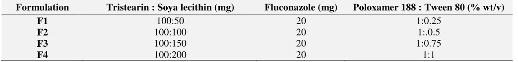
TABLE 1: COMPOSITION OF SOLID LIPID NANOPARTICLES.
Formulation Tristearin : Soya lecithin (mg) Fluconazole (mg) Poloxamer 188 : Tween 80 (% wt/v)
F1 100:50 20 1:0.25
F2 100:100 20 1:.0.5
F3 100:150 20 1:0.75
F4 100:200 20 1:1
Particle size: Particle size of the nanoparticles is presented as z-average diameter, which is basically mean hydrodynamic diameter of the particles. Particle size measurement was required to confirm the production of the particles in nano-rang. The result indicates that particle size was significantly influenced by most of the formulation and process variables. Starting with lipids the tristearin amount was kept constant in all formulations with varying amount of Egg lecithin among all these formulations the F2 with same amount of tristearin and soya lecithin was consider the ideal formulation.
Poloxamer 188 and tween 80 both of them were used as surfactant with varying concentrations. F2 containing surfactant Poloxamer 188 (1%) and Tween 80 (0.5%) with small particle size of
122±3.42 nm in comparison with other
formulations. Particles size decreased as follows: F2 > F3 > F1 > F4. All the formulation within the
250 nm range is given in table 2.
Polydispersity index: Polydispersity index (PI) indicates the width size of the particle size
distribution, which range from 0 to 1.
Theoretically, monodisperse population indicates PI = 0. However PI < 0.6 is considered as narrow size distribution. Therefore, PI measurement was
essential to confirm the narrow size distribution of the particles. Among all the formulations F2 produced SLNs with lowest PI and F4 produced SLNs with high PI. Values are given in table 2.
Zeta potential: Zeta potential (ZP) refers to the surface charge of the particles. ZP (±) indicates the degree of repulsion between close and similarly charged particles in the dispersion. This repulsion force prevents aggregation of the particles. Therefore, ZP is a useful parameter to predict the stability of the solid lipid nanoparticles dispersions. The Zeta potential of F1 was found to be -24.03±1.84 given in table 2.
Entrapment efficiency: The EE% of the
developed SLNs was shown in Table 2. A high amount of drug could be incorporated in nanoparticle dispersion. It can be seen that the encapsulated moiety in the SLNs in formulation F2 (76.53±0.24) is the highest entrapment efficiency among all the other formulations. The drug
entrapment efficiency was measured using
centrifugation method and all the Fluconazole
-SLN formulations had average entrapment
efficiency. The high EE might be beneficial to reduce the skin irritation of drug due to avoid the direct contact between drug and skin surface.
TABLE 2: PHYSIOCHEMICAL PROPERTIES OF THE INVESTIGATED SOLID LIPID NANOPERTICLES
Formulations Particle size (nm) Polydispersity index Zeta potential (mv) % Entrapment efficiency
F1 189±4.62 0.763±3.69 -27±3.26 72.24±2.36
F2 122±3.42 0.668±3.21 -24.03±1.84 76.43±0.24
F3 173±1.23 0.788±4.02 -27±1.83 73.50±0.28
F4 226±2.61 0.897±3.61 -30.11±1.35 70.36±2.63
Transmission Electron Microscopy (TEM): The
TEM imaging of Fluconazole-SLN is shown in Fig.
1. The particle size of Fluconazole-SLN from TEM
images accords with that from that from PCS. The imaging showed that Fluconazole-SLN exhibited a spherical shape and had a narrow size distribution.
FIG. 1: TEM IMAGE OF FORMULATION F2 FLUCONAZOLE LOADED SLN
Scanning electron microscopy (SEM): The SEM image revealed that the particle size was in nanometric range and that the particles had nearly
spherical morphology shown in Fig. 2.
FIG 2: SEM IMAGE OF FORMULATION F2 FLUCONAZOLE LOADED SLN
Stability study: The present study is desired to test
the stability of fluconazole loaded SLN
formulation. Stability test of SLNs was performed
in terms of particle size, zeta potential and % entrapment efficiency during storage.
All formulations were stored in screw capped, amber colored small glass bottles at 4±1°C and room temperature. Analysis of the samples was made Average particle size, zeta potential and % Entrapment efficiency after a period of 10, 20, 30,
45 and 60 days. The change in particle size of all
formulations was observed at temp 4oC and at
27oC.
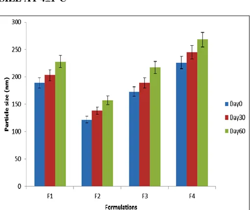
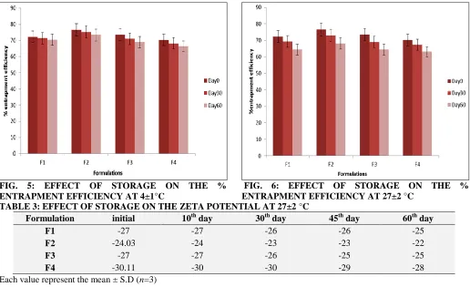
Result shows that the particle sizes increases with increase in the temperature given in fig 3 & 4 and the zeta potential and % entrapment efficiency decreases with increases in temperature shown in fig 5and 6 under storage condition at 4°C and at room temperature. The effect of storage on the zeta
potential at 27±2 °C and 4±1°C is given in tables 3
& 4.
FIG. 3: EFFECT OF STORAGE ON THE PARTICLE SIZE AT 4±1°C
[image:5.612.47.300.130.366.2] [image:5.612.314.573.279.718.2] [image:5.612.313.573.284.473.2] [image:5.612.50.299.444.673.2] [image:5.612.315.570.497.711.2]FIG. 5: EFFECT OF STORAGE ON THE % ENTRAPMENT EFFICIENCY AT 4±1°C
FIG. 6: EFFECT OF STORAGE ON THE % ENTRAPMENT EFFICIENCY AT 27±2 °C
TABLE 3: EFFECT OF STORAGE ON THE ZETA POTENTIAL AT 27±2 °C
Formulation initial 10th day 30th day 45th day 60th day
F1 -27 -27 -26 -26 -25
F2 -24.03 -24 -23 -23 -22
F3 -27 -27 -26 -25 -25
F4 -30.11 -30 -30 -29 -28
Each value represent the mean ± S.D (n=3)
TABLE 4: EFFECT OF STORAGE ON THE ZETA POTENTIAL AT 4±1°C
Formulation initial 10th day 30th day 45th day 60th day
F1 -27 -27 -27 -27 -24
F2 -24.03 -24 -24 -23 -23
F3 -27 -27 -27 -27 -26
F4 -30.11 -30 -30 -30 -28
Each value represent the mean ± S.D (n=3)
In- vitro release of Fluconazole loaded solid lipid nanoparticles: The in vitro release of Fluconazole from different SLN formulations was determined using a dialysis membrane with franz diffusion cell. In order to evaluate the SLNs formulation release for topical use was investigated over 48 h. Release rate of Fluconazole was supposed to occur only from the lipid phase of Solid lipid nanoparticle dispersion because of the poor water solubility of
this drug that prevented its solution in water, as reported for other lipophilic drugs loaded into SLN
13
. The cumulative release of Fluconazole from lipid nanoparticles was in the following order: F1 (42%) < F2 (56%) < F3 (63%) < F4 (69%). The release of Fluconazole from formulation F4 was fastest among other formulations. F1 showed the slowest release of Fluconazole.
TABLE 5: CUMULATIVE % DRUG RELEASE OF FLUCONAZOLE LOADED SLN GEL.
Time after Cumulative % Drug release of F1
Cumulative % Drug release of F2
Cumulative % Drug release of F3
Cumulative % Drug release of F4
0 min 0 0 0 0
30 min 08 15 19 24
1h 12 25 28 38
2h 20 33 36 43
3h 24 38 42 48
4h 27 41 45 50
6h 32 47 52 58
12h 36 50 56 61
24h 40 54 62 67
48h 42 56 63 69
[image:6.612.49.565.38.353.2]CONCLUSIONS: The present research work could be concluded as successful development of solid lipid particles of an antifungal drug fluconazole using solid lipid (tristearin) and co-lipid (soya lecithin) by solvent diffusion-emulsification method. The fluconazole loaded SLNs presented a suitable particle size, zeta
potential polydispersity index, entrapment
efficiency and in vitro drug release. The SEM and
TEM images also revealed the formation of SLNs in nano-sized spherical particles with smooth surface. The SLNs were more stable at 4°C then 27°C, the particle size increased more with decrease in entrapment efficiency at 27°C then 4°C.
ACKNOWLEDGEMENT: The author is thankful
to Ms. Mangal Chaudari Vav Life Sciences Pvt.Ltd. Mumbai for providing free sample of LECIVA-S70. Dr. Farhan Jalees Ahmad, Associate Professor, Faculty of Pharmacy, Jamia Hamdard, Hamdard University, New Delhi for providing
Probsonicator, Zetasizer and Centrifugation
facilities. All India Institute of Medical Sciences, New Delhi, India for TEM facilities. The authors are thankful to the authorities of Institute of Pharmacy, Bundelkhand University, Jhansi for providing Laboratory support and a special thanks to Mr. Alok Mahor for his kind guidance.
REFRENCES:
1. Gupta M, Goyal AK, Paliwal SR, Paliwal R, Mishra N, Vaidya B, et al., Development and characterization of effective topical liposomal system for localized treatment of cutaneous candidiasis. J. Liposome Res 2010; 20: 341– 350.
2. Bidkar S, Jain D, Padsalg A, Patel K, Mokale V. Formulation development and evaluation of fluconazole gel in various polymer bases. As. J. Pharm 2007; 1:63–68. 3. Gupta M, Vyas SP. Development, characterization and in
vivo assessment of effective lipidic nanoparticles for dermal delivery of fluconazole against cutaneous candidiasis. Chemistry and Physics of Lipids 2012; 165: 454– 461.
4. Pouton CW. Formulation of poorly water-soluble drugs for oral administration: physicochemical and physiological issues and the lipid formulation classification system. Eur. J. pharm. Sci 2006; 29: 278-287.
5. Muller RH, Mader k, Gohla S. Solid lipid nanoparticles (SLN) for controlled drug delivery– a review of the state of the art. Eur. J. Pharm. Biopharm 2000; 50: 161-177. 6. Das S, Chaudhury A. Recent advances in lipid
nanoparticles formulations with solid matrix for drug delivery. AAPS PharmSciTech 2011; 12: 62-76.
7. Gershkovich P, Hoffman A. Effect of a high-fat meal on absorption and disposition of lipophilic compounds: importance of degree of association with triglyceride-rich lipoproteins. Eur. J.Pharm.Sci 2007; 32: 24-32.
8. Muller RH, Radtke M, Wissing SA. Nanostructured lipid matrices for improved microencapsulation of drugs. Int. J. Pharma 2002a; 242: 121-128.
9. Muller RH, Radtke M, Wissing SA. Solid lipid nanoparticles and Nanostructured lipid carriers (NLC) in cosmetics and dermatologocal preparations. Adv. Drug Deliv. Rev 2002b; 54: S131-S155.
10. Ye J, Wang Q, Zhou X, Zhang N. Injectable actarit-loaded solid lipid nanoparticles as passive targeting therapeutic agent for rheumatoid arthritis. International Journal of Pharmaceutics 2008; 352: 273-279.
11. Das S, Ng WK, Tan RBH. Development and in vitro evaluation of a lipid nanoparticles formulations containing tretinoin. In: Tiddy, G., Tan, R.B.H. (Eda.), Nanoformulations. RSC Publishing 2012: 38-52.
12. Luo Y, Chen D, Ren L, Zhao X, Qin J. Solid lipid nanoparticles for enhancing vinpocetine's oral bioavailability. J Control Release 2006; 114: 53–9. 13. Jenning V, Schafer-Korting M, Colha S. Vitamin A-loaded
solid lipid nanoparticles for topical use: drug release properties. J. Control. Release 2000; 66: 115-126. 14. Mahor A, Alok S, Gupta Y, Jain SK: Body distribution
and stability studies on Mitoxantrone loaded solid lipid nanoparticles conjugated with Concanavalin A. IJPPS 2010; 2(2): 39-42.
All © 2013 are reserved by International Journal of Pharmaceutical Sciences and Research. This Journal licensed under a Creative Commons Attribution-NonCommercial-ShareAlike 3.0 Unported License.
This article can be downloaded to ANDROID OS based mobile. Scan QR Code using Code/Bar Scanner from your mobile. (Scanners are available on Google Playstore)
How to cite this article: