IJPSR (2017), Volume 8, Issue 11 (Research Article)
Received on 05 February, 2017; received in revised form, 21 June, 2017; accepted, 27 October, 2017; published 01 November, 20 17
ANTIPROLIFERATIVE ACTIVITY OF RUTIN ON HELA CELL LINE INDUCED CERVICAL CANCER IN RATS
U. Vadapalli*1, S. Muvvala 1, R. Alluri 2 and B. V. S. Lakshmi 1
Department of Pharmacology 1, Malla Reddy College of Pharmacy, Dhulapally, Maisammaguda, Secunderabad - 500100, Telangana, India.
Department of Pharmacology 2, Vishnu Institute of Pharmaceutical Education and Research, Narsapur, Hyderabad - 502313, India.
ABSTRACT: In present study antiproliferative activity of Rutin was evaluated on HeLa cell line induced cervical cancer in rats. For this study, 30 rats were divided into 5 groups and each group containing 6 rats each. Group I- normal saline treatment for 45 days, Group II- cancer cells (1×10⁶ cells in 0.1ml/rat), Group III– 5-Fluorouracil (20mg/kg + 1×10⁶ cells in 0.1ml/rat), Group IV- Rutin (50mg/kg + 1×10⁶ cells in 0.1ml/rat), Group V- Rutin (70mg/kg + 1×10⁶ cells in 0.1ml/rat). After 24 h of tumour inoculation intraperitoneally, Rutin was administered daily for 45 days. After administration of last dose followed by 18 hrs fasting, rats were sacrificed for observation of antiproliferative activity. The change in body weight, body circumference of tumour bearing hosts and simultaneous alterations in haematological profile, serum (Triglycerides, Total protein, Total cholesterol, GGT, ALP and glucose) and liver biochemical parameters (lipid peroxidation, GSH and antioxidant CAT, GPx) were estimated. The changes in tissue enzymes-Glucose-6 phosphate dehydrogenase, Hexokinase, Succinate dehydrogenase and CytochromeP450 levels were also estimated. Rutin maintained the body circumference and body weight of proliferation bearing rat. Haematological profile reverted towards normal levels in Rutin treated rat. Treatment with Rutin restored serum biochemical parameters towards normal levels and decreased levels of lipid peroxidation and increased levels of reduced glutathione and other antioxidant enzymes. The Rutin treatment restored Glucose-6 phosphate dehydrogenase, Hexokinase, Succinate dehydrogenase and CytochromeP450 levels in proliferation induced rat. Rutin exhibited antiproliferative effect by modulating haematological parameters, lipid peroxidation and augmenting antioxidant defense system in proliferation bearing rat.
INTRODUCTION: Cervical cancer is the third most common type of cancer in women worldwide1. This cancer develops slowly; starting from a precancerous dysplasia designated cervical intraepithelial neoplasia that may further develop to invasive cervical carcinoma.
QUICK RESPONSE CODE
DOI:
10.13040/IJPSR.0975-8232.8(11).4803-11
Article can be accessed online on:
www.ijpsr.com
DOI link: http://dx.doi.org/10.13040/IJPSR.0975-8232.8 (11).4803-11
Several molecules present in the diet, including flavonoids, can inhibit the growth of cancer cells with an ability to act as “chemopreventers” 2
. Their cancer-preventive effects have been attributed to various mechanisms, including the induction of cell-cycle arrest and/or apoptosis as well as the antioxidant functions. The antioxidant activity of chemo preventers has recently received a great interest, essentially because oxidative stress participates in the initiation and progression of different pathological conditions, including cancer. Since antioxidants are capable of preventing oxidative damage, the wide use of natural
food-Keywords:
Rutin, HeLa cell line, Antiproliferative effect,
Antioxidant activity
Correspondence to Author: V. Uma Rani
Assistant Professor,
Department of Pharmacology, Malla Reddy College of Pharmacy, Dhulapally, Maisammaguda, Secunderabad - 500100, Telangana, India.
derived antioxidants is receiving greater attention as potential anti-carcinogens 3. Among flavonoids, Rutin is considered an excellent free-radical scavenging agent.
Rutin is a plant pigment (flavonoid) that is found in certain fruits and vegetables. Rutin is used to make medicine. The major sources of rutin for medical use include buckwheat, Japanese pagoda tree, and Eucalyptus macrorhyncha. Other sources of rutin include the leaves of several species of eucalyptus, lime tree flowers, elder flowers, hawthorn leaves and flowers, Ginkgo biloba, apples, and other fruits and vegetables. Epidemiological studies have pointed out their possible role in preventing cardiovascular disease and cancer. This health-promoting activity seems to be related to the free-radical scavenging activity of flavonoids. Rutin has the maximum antioxidant potential 4. Rutin is known to affect antioxidant enzymes superoxide dismutase (SOD) and cytochrome oxidase that have the potential to convert reactive oxygen species (ROS) to a hydrogen peroxide and an oxygen molecule. There are reports on the antimutagenic effect of oxidative DNA damage towards benzopyrene induced mutagenicity 5. It has been shown that rutin contain significant amounts of phenolic compounds including flavonoids 6. Phenolic compounds of plant products are mainly responsible for the antioxidant activity to reverse the effect of ROS mechanism by various pathways, and they have a potent effect to reduce incidence of cancer 7. Reactive oxygen species (ROS) are produced as a by-product of various metabolic processes, mainly during respiration, in living organisms. Normal physiological concentrations of ROS usually have a role of regulation of cell activities, whereas higher concentrations cause oxidative damage 8. Rutin has been shown to possess anti-cancer activity 9, antioxidant activity 10
, anti diabetic activity 11,and anti- inflammation activity 12.
Although there are some reports on inhibition of in vivo metabolic activation of carcinogens by rutin and the antimutagenic effect of oxidative DNA
damage towards benzopyrene induced mutagenicity, there are no reports on whether rutin has an effect on human cervical cancer cell lines. Considering the rich antioxidant status of rutin, this study investigated possible antiproliferative effects and
antioxidant status of Rutin on the HeLa cell line induced cervical cancer in rat.
MATERIALS AND METHODS:
Rutin: Rutin is in the form of rutin hydrate. It was suspended in distilled water and freshly prepared just before the administration. It was orally administered by gastric tube at a dose level of 50 mg/kg and 70mg/kg b. w. /day for 45 days.
Cell Lines: HeLa cervical cancer cell line. The cell line was obtained from National Institute of Nutrition, Hyderabad. These cells were maintained in bovine serum albumin medium at 37 °C in a humidified atmosphere of 5% CO2 in air.
Animals: Female Wistar Albino rats weighing 180-250g were obtained from Albino Research Institute, Bachupally (V), Quthbullapur (M), Hyderabad. The rats were housed in polypropylene cages and maintained under standard conditions (12 h light and dark cycles, at 25 ± 3 °C and 35-60% humidity). Standard pelletized feed and tap water were provided ad libitum. All the pharmacological experimental protocols were approved by the Institutional Animal Ethics Committee (Reg no: MRCP/CPCSEA/IAEC/2013-14/MPCOL/09).
Anti-proliferative Activity of Rutin Against HeLa Cell Line Induced Cervical Cancer in Rats: Thirty Female Wistar Albino rats weighing 180-250g were divided into five groups of six animals each.
Group 1: normal saline treatment for 45 days.
Group 2: cancer cells (1×10⁶/rat).
Group 3: 5-fluorouracil (20mg/kg b.w.) + cells (1×10⁶/rat).
Group 4: 50mg/kg dose of rutin+cells (1×10⁶/rat).
Group 5: 70mg/kg dose of rutin+cells (1×10⁶/rat).
Rutin 50 mg/kg p.o. for 45 days. Group 5 animals were given HeLa cell line 1×10⁶ cells/rat i.p. and Rutin 70 mg/kg until 45th day respectively.
Blood Sample Preparation: The animals were sacrificed on 45th day using ether anesthesia, blood was collected by carotid bleeding and transferred to anticoagulant EDTA tubes for the estimation of haematological parameters like Hb, RBC and WBC.
Serum Sample Preparation: The animals were sacrificed using ether anaesthesia; blood was collected by carotid bleeding and was centrifuged using Remi cool centrifuge at 4000 rpm for 15 minutes. Serum was separated for the estimation of various biochemical parameters like serum alkaline phosphatase, Triglycerides, Total cholesterol, Total protein, GGT and glucose.
Tissue Sample Preparation: At the end of the experiment, animals were sacrificed with light ether anaesthesia. Cervical tissue was separated and washed with phosphate buffer saline (0.05M, pH 7.4). The cervix was taken later and minced into small pieces and homogenized in ice cold phosphate buffer saline (0.05M, pH 7.4) using tissue homogenizer to obtain 1:9 (w/v) (10%) whole homogenate. A part of the cervix homogenate was taken and mixed with equal volume of 10% Trichloro acetic acid (TCA) for the estimation of malondialdehyde. Homogenate was centrifuged using Remi cool centrifuge at 8000 rpm for 30 min. The supernatant was separated and used for estimation of anti-oxidant levels of different enzymes i.e. Catalase and reduced glutathione, malondialdehyde and glutathione peroxidase. The supernatant was also analyzed for tissue enzymes-Glucose-6 phosphate dehydrogenase, Hexokinase, Succinate dehydrogenase and Cytochrome P450 levels.
Histopathological Studies: At the end of the experimental period, the rats were sacrificed and cervix was removed. The tissue sample from each group was selected and stored in 10% buffered formalin solution and further embedded in paraffin with wax. The blocks were processed for sectioning; the sections were then stained with haematoxylin and eosin as nuclear and cytoplasmic stains, respectively to assess the activity.
Pathological changes, if any, were viewed under light microscope and recorded.
Statistical Analysis: The experimental results were expressed as the Mean ± SEM with six rats in each group. Statistical significance of difference between groups was determined by one way ANOVA followed by unpaired t-test.
RESULTS:
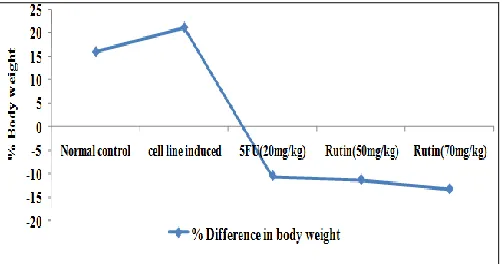
Effect of Rutin on Body Weight and Body Circumference of Cervical Cancer Induced Rat:
[image:3.612.315.566.318.450.2]There was an increase in the body weight and body circumference of cervical cancer induced rat from second week onwards during a growth period of 45 days when compared to normal group. Treatment with 5-Fluorouracil and Rutin maintained the body weight and body circumference of cervical cancer induced rat (Fig. 1 and 2).
FIG. 1: EFFECT OF RUTIN ON BODY WEIGHT
Values are expressed as mean ± SEM; 5-FU= 5 Fluro Uracil.
FIG. 2: EFFECT OF RUTIN ON % INCREASE IN CIRCUMFERENCE
Values are expressed as mean ± SEM. (n = 6); FU 5 Fluro Uracil.
[image:3.612.315.565.486.622.2]and 100mg/kg doses maintained the cervix weight as compared to the cell line induced group (Fig. 3).
FIG. 3: EFFECT OF RUTIN ON ORGAN WEIGHT
Values are expressed as mean ± SEM, (n = 6), Data was analyzed by one way ANOVA followed by unpaired t test. ap < 0.001 when compared to the normal control.***p <0.001 as compared with cell line induced group, 5-FU= 5 Fluro Uracil
Effect of Rutin on Serum Biochemical Enzymes of Cervical Cancer Induced Rat: There was a significant (P < 0.001) decrease in serum glucose,
cholesterol, triglycerides and total protein, significant (P < 0.001) increase in serum GGT and ALP activity of cervical cancer group when compared to normal group and treatment with 5-Fluorouracil and Rutin 50mg/kg and 100mg/kg significantly increased the glucose level, cholesterol, triglycerides, total protein levels, significantly decreased the enzyme activity as compared to cervical cancer group and restored to normal levels (Table 1).
Effect of Rutin on Catalase, MDA, GSH and GPx in Cervical Cancer Induced Rat: Catalase, GSH, and GPx were significantly (P < 0.001) decreased and MDA levels were significantly (P < 0.001) increased in the cervical cancer group when compared to the normal group. Treatment with 5 fluorouracil and Rutin significantly (P < 0.001) decreased the MDA levels and increased the Catalase, GSH, and GPx levels towards the normal (Table 2).
TABLE 1: EFFECT OF RUTIN ON SERUM BIOCHEMICAL PARAMETERS ON HELA CELL LINE INDUCED
CERVICAL CANCER IN RATS
Groups TP
(IU/L)
GGT (IU/L)
ALP (IU/L)
Glucose (mg/dl)
TG (mg/dl)
Cholesterol (mg/dl)
Group I Normal control
17.89±1.9 26.20±0.73 17.33±0.67 63.5±4.01 326.8±7.28 95.2±7.28
Group II Cell line induced
10.74±1.6c 29.6±0.55 c 24.50±0.99b 34.14±5.66 c 143.1±7.14b 67.1±6.57 b
Group III 5- FU (20mg/kg)
15.70±1.6** 30.8±0.60 19.00±0.68** 92.83±6.68*** 344.8±8.85*** 76.6±6.65***
Group IV Rutin (50mg/kg)
11.19±1.0 25.5±0.76*** 17.12±0.28** 102.17±8.27** 224.4±5.73*** 85.7±2.24**
Group V Rutin (70mg/kg)
21.5±1.6** 26.6±0.67*** .67±0.67** 116.67±8.64** 253.4±6.68** 123.7±11.6*
Values are expressed as mean ± SEM, (n=6). Data was analyzed by one way ANOVA fallowed by unpaired t test. cp < 0.001,
bp<0.01as compared with normal control; ***p<0.001 **p<0.01and *p<0.05 as compared with cell line induced group, 5-FU= 5
Fluro Uracil.
TABLE 2:EFFECT OF RUTINON ANTIOXIDANT PARAMETERS ON HELA CELL LINE INDUCED CERVICAL
CANCER IN RATS
Groups Catalase
(µ mole / min / mg)
GPx (µ mole / min / mg)
GSH (µ mole / min / mg)
MDA (nmol/g)
Group I Normal control
0.41±0.03 4.99±0.67 14.67±0.70 53.41±3.74
Group II Cell line induced
0.24±0.01c 2.87±0.34c 7.57±0.13c 91.70±1.59c
Group III 5- FU (20mg/kg)
0.34±0.02*** 4.61±0.64* 12.68±0.93*** 64.92±1.49***
Group IV Rutin (50mg/kg)
0.29±0.02* 4.30±0.60* 10.21±1.20 77.09±2.55***
Group V Rutin (70mg/kg)
0.30±0.02** 5.07±0.43** 10.25±1.10 69.66±0.66***
[image:4.612.50.566.383.521.2] [image:4.612.48.565.592.727.2]Effect of Rutin on Hematological Parameters of Cervical Cancer Induced Rat: Haemoglobin content and RBC count were significantly (P < 0.001) decreased and total WBC count was significantly (P < 0.001) increased in the cervical
[image:5.612.46.569.137.284.2]cancer group as compared to the Normal group. Treatment with 5-Fluorouracil and Rutin restored the RBC and haemoglobin levels towards the cell line induced group (Table 3).
TABLE 3: EFFECT OF RUTIN ON HEMATOLOGICAL ESTIMATIONS OF CERVICAL CANCER
Values are expressed as mean ± SEM., Data was analyzed by one way ANOVA followed by unpaired t test. cp < 0.001, bp < 0.01 and ap < 0.05as compared with the normal control; ***p < 0.001,**p < 0.01 and*p < 0.05 as compared with cell line induced group, 5-FU= 5 Fluro Uracil.
FIG. 4: EFFECT OF RUTIN ON HEXOKINASE, GLUCOSE- 6- PHOSPHATE DEHYDROGENATE, SUCCINATE DEHYDROGENATE ON HELA CELL LINE INDUCED CERVICAL CANCER IN RATS
Values are expressed as mean ± SEM, (n=6). Data was analyzed by one way ANOVA fallowed by unpaired t test. cp < 0.001, bp < 0.01as compared with normal control; ***p < 0.001, **p < 0.01 and *p < 0.05 as compared with cell line induced group, 5-FU= 5 Fluro Uracil.
FIG. 5: EFFECT OF RUTIN ON CYTOCHROME P450 ON HELA CELL LINE INDUCED CERVICAL CANCER IN RATS
Values are expressed as mean ± SEM, (n = 6). Data was analyzed by one way ANOVA fallowed by unpaired t test. bp < 0.01as compared with normal control;*p < 0.05 as compared with cell line induced group, 5-FU = 5 Fluro Uracil.
Parameters Group I
Normal control
Group II Cell line Induced
Group III Cell line + 5-FU (20mg/kg)
Group IV Cell line + Rutin
(50mg/kg)
Group V Cell line + Rutin
(70mg/kg)
Haemoglobin (g%) 15.48±0.48 10.26±0.4c 12.8±0.97* 11.0±0.72 13.0±0.59**
Haemotocrit (vol) 49.0±0.49 33.0±0.36c 40.0±0.93 37.50±0.43*** 40.00±0.73***
RBC (M/Cmm) 8.24±0.36 3.6±0.57c 4.7±0.70 4.16±0.27 4.25±0.27
Platelet count (Lakhs/Cmm) 3.82±0.29 2.18±0.36b 3.33±0.35** 3.11±0.69** 3.75±0.10** WBC(thousands of cells/Cmm) 4.5±18.5 9.4±39.6c 5.3±31.5*** 6.9±42.49*** 6.1±38.36***
Neutrophils 46.17±1.9 30.3±1.61a 19.0±0.93 20±1.15 21.1±1.76*
Lymphocytes (%) 82.17±2.6 64.1±3.5c 73.1±3.7 72.0±2.59 73.3±2.87
Eisonophils (%) 1.66.±0.21 1.16±0.27 1.66±1.67 2.80±0.37 2.41±0.37
[image:5.612.149.467.327.486.2] [image:5.612.152.461.554.703.2]Effect of Rutin on Glucose-6 Phosphate dehydrogenase, Hexokinase, Succinate dehydro-genase and Cytochrome P450 Levels in Cervical Cancer Induced in Rats: Glucose-6 phosphate dehydrogenase (P < 0.01), hexokinase levels (P < 0.001) were significantly increased in the cervical cancer induced group compared to the normal control group. Treatment with 5 fluorouracil and Rutin significantly (P < 0.01) restored the levels of Glucose-6 phosphate dehydrogenase towards the normal. Succinate dehydrogenase (P < 0.001), cytochrome P450 (P < 0.01) levels were significantly (P < 0.001) decreased in cervical cancer induced group compared to the normal control group. Treatment with 5 flurouracil and Rutin (P < 0.01) significantly restored the levels of cytochrome P450 (Fig. 4 and 5).
DISCUSSION: In recent years, it has increasingly been recognized that malignancy may not exclusively result from enhanced cell proliferation but also from decreased physiological cell death, i.e. apoptosis. Apoptotic induction has been a new target for innovative mechanism-based drug discovery. Chemoprevention, a relatively new strategy to prevent cancer, depends on the use of nontoxic chemical substances, to block, reverse, or retard the process of carcinogenesis. Plant-based diet is regarded one of the potential chemo preventive agents.
If a plant-derived extract induces apoptosis and has anti-proliferative and antioxidant effects, it might protect normal cells from the damage caused by ROS while inducing apoptosis and inhibiting proliferation in tumour cells. In this study, we investigated in vivo effects of rutin on the human HeLa cell lines and further investigated the effect of rutin in rat 13. Our results showed that Rutin inhibited the growth of cervical cancer cells in a concentration dependent manner, compared to the controls. Rutin at a concentration of 70mg/kg exhibited a maximum of 95% inhibition of growth of cervical cancer (HeLa) cells. With its antioxidant potential, Rutin can be used as a dietary supplement in some diseases as well as in cancer. In other previous studies, CAT and SOD activities were increased in the rutin added groups; it seemed that this compensatory change could not prevent cell death.
Thus, the mechanism of the apoptosis might be based on some reasons other than oxidative stress. There is increasing evidence suggesting that certain antioxidants compounds act as preventive or protective factors. The present study is preliminary to measure the antioxidant levels and Haematological parameters cervical cancer induced rat and in 5FU and Rutin treated groups.
The decrease in Hb (haemoglobin) concentration and RBC (red blood cells) value indicates the presence of anaemia in all cancer induced rat. This anaemia may be caused by GSH depletion in cancer induced rat which is important as cellular antioxidant so its depletion lead to red blood cell destruction which lead to decreased Hb value and the other cause may be the bone marrow failure which is caused by replacement of its normal elements by cancer cells in varying degrees. These levels were restored to normal in Rutin treated groups; A significant difference in WBC count between cancer induced rat and control was noticed in this study. The WBC count in cervical cancer induced rat was low as compared to their controls. The reduced number of WBC count may be due to the loss of blood in stools and may be due to the replacement of normal bone marrow cells by the cancer cells. The levels of WBC were restored to normal in Rutin treated and 5-fluorouracil treated groups.
Enhanced lipid peroxidation and impairment in antioxidant defense mechanisms were demonstrated in patients with lung and cervical cancers 14. Serum MDA levels were significantly elevated in cervical cancer induced groups when compared to controls. Increased lipid peroxidation in serum and tissues has been reported in cervical cancer induced rat. The lipid peroxidation products such as MDA can structurally alter DNA, proteins and other bio molecules. Our findings are in agreement with most of the earlier studies suggesting that cervical cancer induced rat might be at risk from oxidative cell damage. Oxidative stress arises when there is an imbalance between oxygen-free radical (OFR) formation and scavenging by antioxidants.
Excess generation of free radical can cause oxidative damage to bio-molecules resulting in lipid peroxidation. OFR-induced lipid peroxidation has been implicated in neoplastic transformation. The increase in the rate of lipid peroxidation causes the increased production of MDA that leaks into the blood stream, consequently causing increased levels of MDA in rat induced with cervical cancer. Rutin (50mg/kg and 70mg/kg) significantly (p < 0.001) reduced the levels of MDA in cervical cancer treated groups; this may be due to the Super oxide dismutase present in the Rutin that converts oxygen free radicals in to hydrogen peroxide and water.
Gamma GT is an oncofoetal protein, a glycoprotein whose levels have been shown to be altered during development and carcinogenesis. In most of the liver diseases, both malignant and non-malignant, GGT estimation has been reported to be a sensitive but nonspecific indicator of the disease 15. Some studies have shown that GGT levels are also elevated in malignant tumours of the other tissues. This increase in serum GGT activity in cancer induced rat is due to rapid turnover of malignant cells, which release the enzymes in to blood streams. The levels of GGT were restored to normal in Rutin treated groups.
The levels of Total protein and Glucose were significantly (P < 0.001) changed in cervical cancer induced groups compared to their respective controls and these levels were restored in the Rutin treated groups.
The levels of Triglycerides, Total cholesterol and ALP were significantly (p < 0.01) changed in cervical cancer induced groups compared to their respective controls and these levels were restored in the Rutin treated groups. Early attempts which were moderately successful were to examine vaginal fluid samples for enzyme activity, especially the activity of glucose-6-phosphate. It is known that proliferating cells have a high pentose phosphate. Numerous early studies have shown that this metabolic pathway is increased in many types of tumour. Because major functions of the non-oxidative and non-oxidative sequences of the pentose phosphate pathways are the supply of ribose-5-phosphate for incorporation into ribonucleic acid and coenzymes, and the reduction of NADP+ to NADPH for metabolic synthetic reactions respectively, it can be expected that the pentose phosphate pathways play important roles in the metabolism of tumours and rapidly dividing cells. The first enzyme of the pentose pathway is glucose-6-phosphate dehydrogenase (G6PD) and the second 6-phosphogluconate dehydrogenase (6PGD).
G6PD is known to be increased in tumour cells generally and in other dividing cells. The enzyme is important not only for participating in the supply of pentose sugars for nucleic acid synthesis but also for producing NADPH and thus changing the redox couple NADP+/NADPH. Glucose-6-phosphate dehydrogenase (G6PD) functions to catalyze the oxidation of glucose 6-phosphate to 6-phosphogluconolactone and the reduction of NADP+ to NADPH. In this way, G6PD provides cells with NADPH as a reducing power that maintains the sulfhydryl groups of cellular proteins and aids in detoxification of free radicals and peroxides. Although G6PD is expressed in all tissues, its deficiency causes severe effects in erythrocytes and renders these cells more susceptible to oxidative stress.
with high glucose requirements such as erythrocytes, endothelial cells and the brain 17. Hexokinase (HK)-There are four important mammalian HK isoforms. Besides HK-1, an isoenzyme found in all mammalian cells, tumour cells predominantly express HK-2. Expression studies revealed an approximately 100-fold increase in the mRNA levels for HK-2 18. The prominent role of HK-2 for the accomplishment of the Warburg effect has been demonstrated by Wolf
et al., who found that inhibition of HK-2, but not
HK-1, in a human glioblastoma multiforme resulted in the restoration of normal oxidative glucose metabolism with decreased extracellular lactate and increased O2 consumption 19.
Mutations in TCA cycle enzymes can lead to tumorigenesis 20. Mutations of the succinate dehydrogenase (SDH) and the fumarate hydratase (FH) have been shown to result in paragangliomas and pheochromocytomas. The succinate dehydrogenase complex assembly factor 2 (SDHAF2⁄SDH5), responsible for the incorporation of the co-factor FAD into the functional active SDH, was recently shown to be a paraganglioma-related tumour suppressor gene 21. FH mutations have been found in cutaneous and uterine leiomyomas, leiomyosarcomas and renal cell cancer 22- 24. Two mechanisms have been suggested to account for the connection between loss of function of SDH or FH and tumorigenesis. (a) Redox stress due to generation of ROS by mutant SDH proteins causes an inhibition of HIF-dependent prolyl hydroxylase (PHD), an enzyme targeting under normoxic conditions the a-subunit of HIF for degradation.
According to this explanation ROS can lead to pseudo-hypoxia in tumours with SDH mutations
via stabilization of HIF. (b) Metabolic signalling in SDH-deficient tumors via increased succinate levels inhibits the PHD and therefore leads to stabilization of the HIF-1a subunit at normal oxygen levels. A similar mechanism was proposed for the consequences of FH deficiency: accumulating fumarate can act as a competitive inhibitor of PHD leading to a stabilization of HIF-1 25
. Cytochrome P450 (CYP) is a multi-gene superfamily of heme-containing enzymes catalyzing the oxidative metabolism of many compounds 26. CYP families 1, 2 and 3, which are
the main CYP families participating in the metabolism of xenobiotics, are highly expressed within the liver.
High expression levels of various CYPs have been found previously in many tumours. In addition to biotransformation of carcinogenic compounds, CYPs have also been suggested to convert endogenous substrates to metabolites that facilitate cancer development and to participate in the metabolism of anticancer drugs. Of the different CYP isoforms, over expression of CYP1B1 is the one most often detected in various tumours. Strong expression was also observed in the squamous cell carcinoma of the uterine cervix as well as in the two tumors detected in the ovaries (i.e., a metastatic adenocarcinoma from the appendix and a mucinous cystadenocarcinoma) 27.
From the histopathology study, proliferation of mucosal gland in sub mucosal layer, diffused fibrosis, active proliferation of fibrous tissue was observed in cervical cancer induced group. Normal mucosal epithelium and muscular region was observed in the 5-FU, Rutin (50mg/kg) and Rutin (70mg/kg) treated groups.
CONCLUSION: In conclusion our present study demonstrated that, Rutin was found to be potent antioxidant, anti-tumor agent against in vivo cervical cancer. The Rutin significantly acted as a free radical scavenging agent.
ACKNOWLEDGEMENT: The authors are thankful to the authorities of Malla Reddy College of Pharmacy, Secunderabad, for providing support to this study.
CONFLICT OF INTEREST: The authors declare that there are no conflicts of interest.
REFERENCES:
1. Jemal A, Center MM, DeSantis C et al.: Cancer Epidemiol Biomarkers Prev 2010; 19(8): 1893.
2. Harborne JB: The Flavonoids: Advances in Research Since 1986. Chapman and Hall, London 1993; 87. 3. Duthie G and Crozier A: Curr. Opin. Clin. Nut. 2000; 3:
447.
4. Pathak S, Multani AS and Banerji P: Int J Oncol 2003; 23: 975.
5. Ahmed MI, Fayed ST, Hossein H and Tash FM: Dis Markers 1999, 15, 283.
7. Bahar G, Feinmesser R, Shpitzer T, Popovtzer A Nagler RM: Cancer 2007; 109(1): 54.
8. Mathew BB, Tiwari A and Jatawa SK: J Pharm. Res 2011; 4(12): 4340.
9. Hertog MG, Hollman PC and Katan MB: J Agric Food Chemist 1992; 40: 2379.
10. Hertog MG, Feskens EJ, Hollmann PC, Katan MB and Kronthout D: Lancet 1993; 342: 1007.
11. Ahmed OM, Moneim AA, Yazid IA and Mahmoud AM: 2010; 15: 35.
12. Raghav SK, Gupta B, Agrawal C, Goswami K and Das HR: J Ethnopharmacol 2006; 104: 234.
13. Cook JA, Gius D, Wink DA, Krishna MC, Russo A and
Mitchell JV: Semin Radiat Oncol, 2004; 14: 259.
14. Manju V, Kalaivani Sailaja J, and N. Nalini: Clin Biochem 2002; 35: 621.
15. Corti A, Franzini M, Paolicchi A and Pompella A: Anti- cancer Res 2010; 30(4): 1169.
16. Rodriguez-Enriquez S, Marin-Hernandez A, Gallardo-Perez JC and Moreno-Sanchez R: J Cell Physiol 2009; 221: 352.
17. Scheepers A, Joost HG and Schurmann A: Parenter Enteral Nutr 2004; 28: 364.
18. Mathupala SP, Ko YH and Pedersen PL: Semin Cancer Biol 2009; 19: 17.
19. Johansson T, Berrez JM and Nelson BD: Biochem Biophys Res Commun 1985; 133: 608.
20. Baines CP, Kaiser RA, Sheiko T, Craigen WJ and Molkentin JD: Nat Cell Biol 2007; 9: 550.
21. Bayley JP, Kunst HP, Cascon A, Sampietro ML, Gaal J, Korpershoek E, Hinojar-Gutierrez A, Timmers HJ, Hoefsloot LH, Hermsen MA et al.: Lancet Oncol 2010; 11: 366.
22. Hao HX, Khalimonchuk O, Schraders M, Dephoure N, Bayley JP, Kunst H, Devilee P, Cremers CW, Schiffman JD, Bentz BG etal.: Science 2009; 325: 1139.
23. Selak MA, Armour SM, MacKenzie ED, Boulahbel H, Watson DG, Mansfield KD, Pan Y, Simon MC, Thompson CB and Gottlieb E: Cancer Cell 2005; 7: 77.
24. Astuti D, Latif F, Dallol A, Dahia PL, Douglas F, George E, Skoldberg F, Husebye ES, Eng C and Maher ER: Am J Hum Genet 2001; 69:49.
25. Niemann S and Muller U: Nat Genet 2000; 26: 268. 26. Sheweita SA: Curr Drug Metab 2000; 1: 107. 27. Murray GI: J Pathol 2000; 192: 419.
All © 2013 are reserved by International Journal of Pharmaceutical Sciences and Research. This Journal licensed under a Creative Commons Attribution-NonCommercial-ShareAlike 3.0 Unported License.
This article can be downloaded to ANDROID OS based mobile. Scan QR Code using Code/Bar Scanner from your mobile. (Scanners are available on Google Playstore)
How to cite this article: