R E S E A R C H A R T I C L E
Open Access
Nongenotoxic effects and a reduction of the
DXR-induced genotoxic effects of
Helianthus
annuus
Linné (sunflower) seeds revealed by
micronucleus assays in mouse bone marrow
Marcelo Fabiano Gomes Boriollo
1,2,3*, Luiz Silva Souza
1,2, Marielly Reis Resende
4, Thaísla Andrielle da Silva
1,2,
Nelma de Mello Silva Oliveira
1,4, Maria Cristina Costa Resck
1,3, Carlos Tadeu dos Santos Dias
5and João Evangelista Fiorini
1,2Abstract
Background:This research evaluated the genotoxicity of oil and tincture ofH. annuusL. seeds using the micronucleus assay in bone marrow of mice. The interaction between these preparations and the genotoxic effects of doxorubicin (DXR) was also analysed (antigenotoxicity test).
Methods:Experimental groups were evaluated at 24-48 h post treatment with N-Nitroso-N-ethylurea (positive control–NEU), DXR (chemotherapeutic), NaCl (negative control), a sunflower tincture (THALS) and two sources of sunflower oils (POHALS and FOHALS). Antigenotoxic assays were carried out using the sunflower tincture and oils separately and in combination with NUE or DXR.
Results: For THALS, analysis of the MNPCEs showed no significant differences between treatment doses (250– 2,000 mg.Kg−1) and NaCl. A significant reduction in MNPCE was observed when THALS (2,000 mg.Kg−1) was administered in combination with DXR (5 mg.Kg−1). For POHALS or FOHALS, analysis of the MNPCEs also showed no significant differences between treatment doses (250–2,000 mg.Kg−1) and NaCl. However, the combination DXR + POHALS (2,000 mg.Kg−1) or DXR + FOHALS (2,000 mg.Kg−1) not contributed to the MNPCEs reduction.
Conclusions:This research suggests absence of genotoxicity of THALS, dose-, time- and sex-independent, and its combination with DXR can reduce the genotoxic effects of DXR. POHALS and FOHALS also showed absence of genotoxicity, but their association with DXR showed no antigenotoxic effects.
Keywords:Bone marrow,Helianthus annuus L. (sunflower), Micronucleus assay, Rodents, Tincture, Oil
Background
The cultivated sunflower (Helianthus annuus L.) is one of 67 species in the genus Helianthus. It is a di-cotyledonous plant and a member of the Compositae
(Asteraceae) family, having a typical composite flower [1]. The composition of the seed is markedly affected by
the sunflower variety [2,3]. Nevertheless, the composition ranges of sunflower dehulled seeds (on a percentage dry weight basis) is as follows [4]: protein 20.4–40.0%; peptides, amino acids and other non–protein nitrogen1–13%; carbo-hydrates4–10%; lipids47–65%; fatty acids (palmitic acid5–7%, atearic2–6%; arachidic acid0.0–0.3%, oleic acid15–37%; linoleic acid 51–73%, and linolenic acid <0.3%); tocopherol 0.07%; carotenoids 0.01–0.02%; vitamin B1 0.002%; chlorogenic acid (CGA)0.5–2.4%; quinic acid (QA)0.12–0.25%; caffeic acid (CA) 0.05–0.29%; total minerals 3–4%; potassium 0.67–0.75%; phosphorus0.60–0.94%; sulphur0.26–0.32%; magnesium0.35–0.41%; calcium0.08–0.10%; and sodium0.02%.
* Correspondence:marcelo.boriollo@unifenas.br
1Laboratório de Farmacogenômica e Biologia Molecular, Faculdade de
Ciências Médicas & Centro de Pesquisa e Pós–graduação, Universidade José do Rosário Vellano (UNIFENAS), Campus Universitário, Rod. MG 179, Km 0, Alfenas, MG CEP: 37130-000, Brasil
2Centro de Pesquisa e Pós–graduação em Ciência Animal, Área de Patologia
e Farmacologia Animal, Universidade José do Rosário Vellano (UNIFENAS), Alfenas, Minas Gerais, Brasil
Full list of author information is available at the end of the article
Tocopherols are excellent natural antioxidants that protect oils against oxidative rancidity. The α form has the highest biological vitamin E activity, and theγ form has been reported to have the highest antioxidant ac-tivity [5]. The sterols found in sunflower oils include
β-sitosterol, stigmasterol, campesterol,δ-5-avenasterol, and δ-7-stigmasterol [6,7]. Plant sterols are only min-imally absorbed by humans, and their ingestion appears to inhibit intestinal cholesterol and bile acid absorption [8]. Most trace metals in refined, bleached and deodor-ized sunflower seed oil are removed during processing. It is particularly important that copper and iron be re-moved because these metals greatly reduce the oxida-tive stability of the oil [9]. Other metals, such as lead and cadmium, are of particular concern due to their toxicity and their supposed link to coronary heart dis-ease and hypertension [10].
In drug development, the genotoxicity assays represent a considerable effort, as most pharmaceutical organiza-tions evaluate a new therapeutic agent based onin vitro
and in vivodata genotoxic [11]. In this context, tests to evaluate the genotoxic activity of the plants used by the population as well as their isolated compounds are ne-cessary and important for establishing control measures in widespread use. Furthermore, it is necessary to clarify the mechanisms and conditions that mediate the pro-posed biological effect before plants are considered as therapeutic agents [12]. As far as genotoxicity studies are concerned, the in vivo micronucleus (MN) assay in rodent bone marrow plays a crucial role in the test bat-tery aimed at identifying hazardous mutagens [13]; this assay is especially suited to assessing genotoxic hazards because it allows consideration of multiple factors, such as in vivo metabolism, pharmacokinetics and DNA re-pair processes, even though these processes vary among species, among tissues and among genetic endpoints [14-17]. In addition, understanding the genotoxic effects induced by phytotherapeutics and foods employing the mammalian in vivo MN assay has been the goal of sev-eral researchers groups [18-20].
In order to contribute to the information on the geno-toxic potential of herbal extracts and food, the present study evaluated the genotoxic effects of two sources of oil and tincture ofH. annuusL. (sunflower) seeds using
in vivomicronucleus assays in mouse bone marrow. The effect of the maximum permissible concentration of
H. annuus L. (oils and tincture) on the doxorubicin (DXR)–induced genotoxic effects in mice bone marrow was also studied (i.e., antigenotoxicity assay).
Methods Phytotherapeutics
Tincture and oil of sunflower seeds were purchased com-mercially and stored according to the manufacturer's
recommendations [tincture of H. annuus L. seeds (THALS) – Yod Comércio de Produtos Naturais Ltda., cat. # 544606, Campinas, SP, Brazil; pharmaceutical oil of H. annuus L. seeds (POHALS) – Farmácia de Manipulação Alfenense Ltda., Alfenas, MG, Brazil; food oil ofH. annuusL. seeds (FOHALS)–Agricultural Cargill S.A., Mairinque, SP, Brazil]. Aliquots (1.5 L) of this tinc-ture were submitted to solvent removal proceedings by rotary evaporation (40 rpm) (Rotavapor Model R-215) coupled in bath heating systems 50–60°C (Bath Heating model B-491), vacuum pump500 mmHg (Vacuum Pump V-700 with Automatic Vacuum Controller V-855), recir-culator (Recirrecir-culator Chiller F-100) and evaporation bottle (Büchi Labortechnik AG, Switzerland). The final product was transferred to a reaction bottle 1 L (SCHOTT® DURAN®) and kept at −20°C for 24 hours in order to evaluate the freezing of the final product and the ef-ficacy of the solvent evaporation process [21]. Then, al-iquots (40 mL) of this final product was transferred into glass vials penicillin type (50 mL) and lyophilized (Lyophilizer model Alpha 1–2 LDPlus, Martin Christ Gefriertrocknungsanlagen GmbH©, Germany) and their dry mass were measured (Electronic Analytical Balance AUW-220D, Shimadzu Corp., Kyoto, Japan). The lyophi-lized final product was prepared in aqueous solvent (150 mM NaCl in water type 1) at concentrations of 2×, sterilized by filtration (Millipore Corporation, hydro-philic Durapore®PVDF, 0.22 μm,∅47 mm, cat. # GVWP 047 00), and stored in sterile polypropylene tubes (50 mL) at−70°C until moment of use.
System–testin vivo
Healthy, heterogeneous, young adult male and female
the experimental treatment, the animals were euthanized by CO2asphyxiation in adapted acrylic chambers [14]. This
research was approved by Committee of Ethics in Research Involving Animals of UNIFENAS (CEPEAU Protocol No. 04A/2008).
Experimental groups
Groups of animals (consisting of 3 males and 3 females each) were treated using a single dosing regimen ad-ministered by gavage (phytotherapeutic and negative control) or intraperitoneally (chemotherapeutic and posi-tive control) and two euthanasia times (24 and 48 h), based on a regulatory recommendation regarding thein vivo mi-cronucleus assay [14,17]:
▪Control groups: 150 mM NaCl (negative control), 50 mg.Kg−1 of N-Nitroso-N-ethylurea (positive control: NEU, Sigma N8509, CAS no. 759-73-9) and 5 mg.Kg−1 of doxorubicin hydrochloride [20] (chemotherapeutic: DXR, Eurofarma Laboratórios Ltda., CAS no. 23214-92-8).
▪Genotoxicity test (phytotherapeutics): THALS (250–2,000 mg.Kg−1), POHALS (250–2,000 mg.Kg−1) and FOHALS (250–2,000 mg.Kg−1). The maximum tolerated dose (MTD) was defined as (i) the highest dose that can be administered without inducing lethality or excessive toxicity during the study causing moribund euthanasia, or (ii) a dose that produces some indication of toxicity of the bone marrow (e.g. a reduction in the proportion of immature erythrocytes among total erythrocytes in the bone marrow), or (iii) 2,000 mg.Kg−1 [14,17]. ▪Antigenotoxicity test 1 (phytotherapeutics + chemotherapeutic) [20]: THALS (2,000 mg.Kg−1) + DXR (5 mg.Kg−1), FOHALS (2,000 mg.Kg−1) + DXR (5 mg.Kg−1) and FOHALS (2,000 mg.Kg−1) + DXR (5 mg.Kg−1).
▪Antigenotoxicity test 2 (phytotherapeutics + positive control): THALS (2,000 mg.Kg−1) + NEU (50 mg.Kg−1), POHALS (2,000 mg.Kg−1) + NEU (50 mg.Kg−1) and POHALS (2,000 mg.Kg−1) + NEU (50 mg.Kg−1).
Processing the bone marrow and cell analysis
Shortly after euthanasia, the femora were surgically and aseptically removed, and the animals appropriately dis-carded. Each femur was sectioned at the proximal end and the contents of the spinal canal were washed with 1.5 mL of 150 mM NaCl solution and transferred to a 15 mL centrifuge tube [14,17,23]. This material was re-suspended with a Pasteur pipette to ensure a random distribution of bone marrow cells. The suspension was then centrifuged at 1,000 rpm (Centrífuga de Bancada
Microprocessada, Mod. NT 810, Nova Técnica Ind. e
Com. de Equip. para Laboratório Ltda., Piracicaba, SP,
Brazil) for 5 minutes. The supernatant was discarded and the resulting sediment was resuspended in 500 μL of 150 mM NaCI solution added 4% formaldehyde. The slides were prepared by smearing (2 slides per animal), dried at room temperature for 24 h and stained with Leishman's eosin methylene blue dye [pure dye for 3 min, followed by diluted dye in water type 1 (1:6) for 15 min] to differentiate polychromatic erythrocyte (PCE) from normochromatic erythrocyte (NCE).
Polychromatic erythrocytes (PCEs) were observed at a magnification of 1000× using optical microscopy (Nikon Eclipse E–200), counted (at least 2000 polychromatic erythrocytes anucleated per animal were scored for the incidence of micronucleated polychromatic erythrocytes) with the aid of a digital cell counter (Contador Diferencial CCS02, Kacil Indústria e Comércio Ltda., PE, Brasil Contador Diferencial CCS02, Kacil Indústria e Comércio Ltda., PE, Brazil) and photographed using an 8.1 Mega-pixel Digital Camera (DC FWL 150). The number of PCEs and NCEs, the number and frequency of micro-nucleated polychromatic erythrocytes (MNPCEs) were reported. In order to evaluate bone-marrow toxicity, the ratio of PCE to NCE was also observed [14,17]. This PCE/NCE ratio is an indicator of the acceleration or in-hibition of erythropoiesis and it has been reported to vary with scoring time. A continuous decline in the PCE/NCE ratio may be due to the inhibition of cell div-ision, the killing of erythroblasts, the removal of dam-aged cells, or dilution of the existing cell pool with newly formed cells [20].
Statistical analysis
The data obtained in the micronucleus assay were submit-ted toone–wayanalysis of variance (ANOVA), using a fac-torial scheme of 10 × 2 × 2 (treatment × sex × euthanasia time), and medium comparison with Tukey's test (α= 0.05) using SAS®version 9.2 computer software.
Results and discussion
H. annuus L. has been considered an important source of natural oil for centuries and has been used as a pre-ventive medicine against diuresis, diarrhoea, and various inflammatory diseases [24], and has also been used for the relief of asthmatic symptoms [25], gastric protection [26,27], its healing properties [28], anti-inflammatory ac-tion [29] and antimicrobial properties [26,28]. However, studies aimed at understanding the genotoxic and muta-genic effects of H. annuus L. were subject of compara-tively little research [19,30], which drove us to evaluate the harmful genotoxic and antigenotoxic properties (i.e., clastogenicity and/or aneugenicity) of oil and tincture of
H. annuusL. seeds using the MN assayin vivo.
statistically for each one of the animal groups treated with only tincture (THALS) or oils (POHALS or FOHALS) of sunflower seeds–genotoxic assays–and for each one of the groups treated with phytotherapeutics and chemother-apeutic agent DXR (THALS + DXR, POHALS + DXR or FOHALS + DXR)–antigenotoxic assays–, as well as con-trol groups.
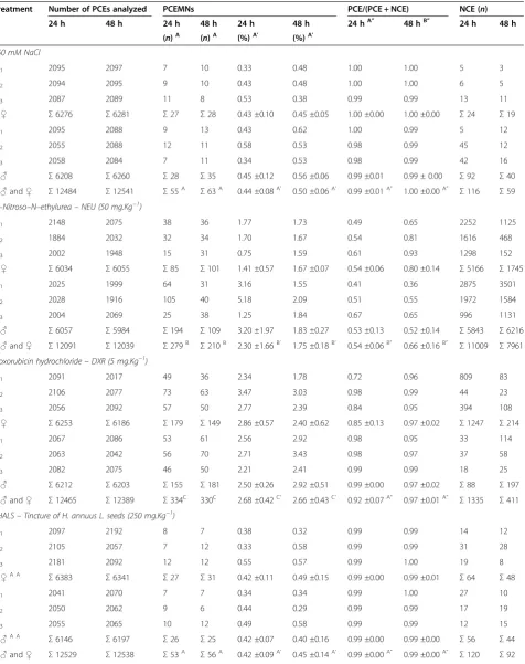
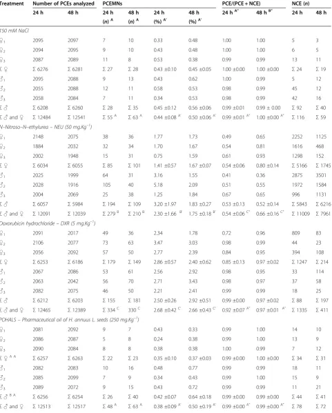
For animal groups treated with THALS, analysis of the MNPCEs showed no significant differences (p < 0.05) between all the treatment doses (250–2,000 mg.Kg−1) and negative control (NaCl). These results suggest ab-sence of genotoxicity of THALS, regardless of the dose of phytotherapic administration (250–2,000 mg.Kg−1), the treatment time (24 and 48 h) or the sex of the animal (male and female). Treatment of mice with 5 mg.Kg−1 DXR significantly induced MNPCE at 24 and 48 h post treatment and for both sexes, whose MNPCE frequencies were significantly above (p< 0.05) those observed in the positive NEU control (50 mg.Kg−1). However, the reduc-tion in MNPCE (p < 0.05) was observed when THALS (2,000 mg.Kg−1) is administered in combination with the chemotherapy agent DXR (5 mg.Kg−1), suggesting antigenotoxic effects (anticlastogeny and/or antianeugeny). Therefore, THALS provides a partial protection against the genotoxic effects induced by DXR in the bone marrow of mice, regardless of the treatment time (24 and 48 h) or the sex of the animal, although the genotoxic effect observed in this treatment combination has is similar (i.e., numbers and frequencies of MNPCEs) to that observed in NEU-treated animals. The analysis obtained from the PCE/NCE ratio showed no significant differences (p< 0.05) between all doses of THALS (250–2,000 mg.Kg−1), THALS (2,000 mg.Kg−1) + DXR (5 mg.Kg−1) and negative controls. These results suggest that there is not systemic toxicity of THALS and/or DXR under the MN assay conditions, regardless of the phytotherapeutic doses and times, but sex-dependent (Table 1).
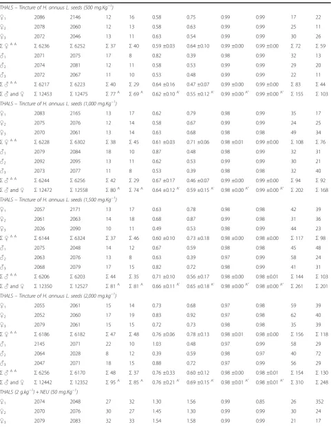
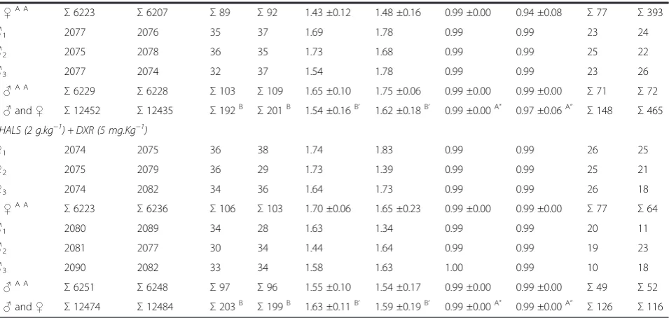
For animal groups treated with POHALS (Table 2) or FOHALS (Table 3), analysis of the MNPCEs showed no significant differences (p < 0.05) between all the treatment doses (250–2,000 mg.Kg−1) and nega-tive control (NaCl). These results suggest absence of genotoxicity for both sources of sunflower oil (pharma-ceutical and food), regardless of the dose of oil admin-istration (250–2,000 mg.Kg−1) or treatment time (24 and 48 h), but it was sex-dependent. Treatment of mice with DXR (5 mg.Kg−1) + POHALS (2,000 mg.Kg−1) or DXR (5 mg.Kg−1) + FOHALS (2,000 mg.Kg−1) not con-tribute to the MNPCEs reduction at 24 and 48 h post treatment and for both sexes, suggesting that both sources of sunflower oil not decrease the DXR-induced genotoxic effects and therefore they do not have antige-notoxic effects (anticlastogeny and/or antianeugeny). The analysis obtained from the PCE/NCE ratio showed
no significant differences (p< 0.05) between all doses of POHALS (250–2,000 mg.Kg−1) and negative controls, time-dependent and sex-independent. For FOHALS, the PCE/NCE ratio showed significant differences (p< 0.05) only in the highest dose (2,000 mg.Kg−1) tested, time-independent and sex-dependent. These results suggest that the systemic toxicity of sunflower oil can be dependent on its source and its highest dose used. In addition, treat-ments with DXR (5 mg.Kg−1) + POHALS (2,000 mg.Kg−1) or DXR (5 mg.Kg−1) + FOHALS (2,000 mg.Kg−1) signifi-cantly decrease the PCE/NCE ratio in mouse bone mar-row. These results suggests that the association sunflower oil and chemotherapeutic agent DXR can potentize the systemic toxicity, regardless of the sex (only POHALS) and time (only FOHALS).
For the first time, this research has provided informa-tion on the genotoxic and antigenotoxic effects of THALS. However, genotoxic studies of sunflower oil and oil sunflower ozonized (at a dose limit of 2 g.kg−1.d−1, based on evidence of toxicity from subchronic studies via intragastric administration of the product) were pre-viously carried out using the MN assay in the bone mar-row of mice using male and female Cenp: NMRI mice [31]. In this study, the treatment with sunflower oil did not cause cytotoxic damage to erithrocytes, as reported in the analyses of the PCE/NCE ratio, which corroborate with our findings from the pharmaceutical oil and par-tially with food oil. Likewise, that research proposes the hypothesis that no clastogenic effect occurs in the bone marrow of animals treated with the sunflower oil under experimental conditions [31].
Table 1 The incidence of MNPCEs and PCE/NCE ratio in bone marrow of male and femaleSwiss albinusmice after testing for 24 h and 48 h
Treatment Number of PCEs analyzed PCEMNs PCE/(PCE + NCE) NCE (n)
24 h 48 h 24 h 48 h 24 h 48 h 24 hA” 48 hB” 24 h 48 h
(n)A (n)A (%)A’ (%)A’
150 mM NaCl
♀1 2095 2097 7 10 0.33 0.48 1.00 1.00 5 3
♀2 2094 2095 9 10 0.43 0.48 1.00 1.00 6 5
♀3 2087 2089 11 8 0.53 0.38 0.99 0.99 13 11
Σ♀ Σ6276 Σ6281 Σ27 Σ28 0.43 ±0.10 0.45 ±0.05 1.00 ±0.00 1.00 ±0.00 Σ24 Σ19
♂1 2095 2088 9 13 0.43 0.62 1.00 0.99 5 12
♂2 2055 2088 12 11 0.58 0.53 0.98 0.99 45 12
♂3 2058 2084 7 11 0.34 0.53 0.98 0.99 42 16
Σ♂ Σ6208 Σ6260 Σ28 Σ35 0.45 ±0.12 0.56 ±0.06 0.99 ±0.01 0.99 ± 0.00 Σ92 Σ40
Σ♂and♀ Σ12484 Σ12541 Σ55A Σ63A 0.44 ±0.08A’ 0.50 ±0.06A’ 0.99 ±0.01A” 1.00 ±0.00A” Σ116 Σ59 N–Nitroso–N–ethylurea–NEU (50 mg.Kg−1)
♀1 2148 2075 38 36 1.77 1.73 0.49 0.65 2252 1125
♀2 1884 2032 32 34 1.70 1.67 0.54 0.81 1616 468
♀3 2002 1948 15 31 0.75 1.59 0.61 0.93 1298 152
Σ♀ Σ6034 Σ6055 Σ85 Σ101 1.41 ±0.57 1.67 ±0.07 0.54 ±0.06 0.80 ±0.14 Σ5166 Σ1745
♂1 2025 1999 64 31 3.16 1.55 0.41 0.36 2875 3501
♂2 2028 1916 105 40 5.18 2.09 0.51 0.55 1972 1584
♂3 2004 2069 25 38 1.25 1.84 0.67 0.65 996 1131
Σ♂ Σ6057 Σ5984 Σ194 Σ109 3.20 ±1.97 1.83 ±0.27 0.53 ±0.13 0.52 ±0.14 Σ5843 Σ6216
Σ♂and♀ Σ12091 Σ12039 Σ279B Σ210B 2.30 ±1.66B’ 1.75 ±0.18B’ 0.54 ±0.06B” 0.66 ±0.16B” Σ11009 Σ7961 Doxorubicin hydrochloride–DXR (5 mg.Kg−1)
♀1 2091 2017 49 36 2.34 1.78 0.72 0.96 809 83
♀2 2106 2077 73 63 3.47 3.03 0.98 0.99 44 23
♀3 2056 2092 57 50 2.77 2.39 0.84 0.95 394 108
Σ♀ Σ6253 Σ6186 Σ179 Σ149 2.86 ±0.57 2.40 ±0.62 0.85 ±0.13 0.97 ±0.02 Σ1247 Σ214
♂1 2067 2086 53 61 2.56 2.92 0.98 0.95 33 114
♂2 2063 2042 56 70 2.71 3.43 0.98 0.97 37 58
♂3 2082 2075 46 50 2.21 2.41 0.99 0.99 18 25
Σ♂ Σ6212 Σ6203 Σ155 Σ181 2.50 ±0.26 2.92 ±0.51 0.99 ±0.00 0.97 ±0.02 Σ88 Σ197
Σ♂and♀ Σ12465 Σ12389 Σ334C 330C 2.68 ±0.42C’ 2.66 ±0.43C’ 0.92 ±0.07A” 0.97 ±0.01A” Σ1335 Σ411 THALS–Tincture of H. annuus L. seeds (250 mg.Kg−1)
♀1 2097 2192 8 7 0.38 0.32 0.99 0.99 14 12
♀2 2105 2057 7 12 0.33 0.58 0.99 0.99 31 28
♀3 2181 2092 12 12 0.55 0.57 0.99 1.00 19 8
Σ♀A A
Σ6383 Σ6341 Σ27 Σ31 0.42 ±0.11 0.49 ±0.15 0.99 ±0.00 0.99 ±0.01 Σ64 Σ48
♂1 2041 2070 7 7 0.34 0.34 0.99 1.00 27 10
♂2 2050 2062 9 6 0.44 0.29 0.99 0.99 17 19
♂3 2055 2065 10 12 0.49 0.58 0.99 0.99 12 15
Σ♂A A
Σ6146 Σ6197 Σ26 Σ25 0.42 ±0.07 0.40 ±0.16 0.99 ±0.00 0.99 ±0.00 Σ56 Σ44
Table 1 The incidence of MNPCEs and PCE/NCE ratio in bone marrow of male and femaleSwiss albinusmice after testing for 24 h and 48 h(Continued)
THALS–Tincture of H. annuus L. seeds (500 mg.Kg−1)
♀1 2086 2146 12 16 0.58 0.75 0.99 0.99 17 22
♀2 2078 2060 12 13 0.58 0.63 0.99 0.99 25 11
♀3 2072 2046 13 11 0.63 0.54 0.99 0.99 30 26
Σ♀A A
Σ6236 Σ6252 Σ37 Σ40 0.59 ±0.03 0.64 ±0.10 0.99 ±0.00 0.99 ±0.00 Σ72 Σ59
♂1 2071 2075 17 8 0.82 0.39 0.98 0.99 32 13
♂2 2074 2081 12 11 0.58 0.53 0.99 0.99 29 20
♂3 2072 2067 11 10 0.53 0.48 0.99 0.99 22 11
Σ♂A A
Σ6217 Σ6223 Σ40 Σ29 0.64 ±0.16 0.47 ±0.07 0.99 ±0.00 0.99 ±0.00 Σ83 Σ44
Σ♂and♀ Σ12453 Σ12475 Σ77A Σ69A 0.62 ±0.10A’ 0.55 ±0.12A’ 0.99 ±0.00A” 0.99 ±0.00A” Σ155 Σ103 THALS–Tincture of H. annuus L. seeds (1,000 mg.Kg−1)
♀1 2083 2165 13 17 0.62 0.79 0.98 0.99 35 17
♀2 2075 2076 12 14 0.58 0.67 0.99 0.99 24 25
♀3 2070 2061 13 14 0.63 0.68 0.98 0.98 49 34
Σ♀A A
Σ6228 Σ6302 Σ38 Σ45 0.61 ±0.03 0.71 ±0.06 0.98 ±0.01 0.99 ±0.00 Σ108 Σ76
♂1 2079 2084 18 10 0.87 0.48 0.98 0.99 32 31
♂2 2092 2095 13 11 0.62 0.53 0.99 0.99 30 21
♂3 2073 2077 11 8 0.53 0.39 0.98 0.98 32 40
Σ♂A A
Σ6244 Σ6256 Σ42 Σ29 0.67 ±0.17 0.46 ±0.07 0.99 ±0.00 0.99 ±0.00 Σ94 Σ92
Σ♂and♀ Σ12472 Σ12558 Σ80A Σ74A 0.64 ±0.12A’ 0.59 ±0.15A’ 0.98 ±0.00A” 0.99 ±0.00A” Σ202 Σ168 THALS–Tincture of H. annuus L. seeds (1,500 mg.Kg−1)
♀1 2057 2171 13 17 0.63 0.78 0.98 0.98 42 39
♀2 2061 2063 14 18 0.68 0.87 0.99 0.98 31 36
♀3 2026 2090 10 11 0.49 0.53 0.98 0.99 44 23
Σ♀A A
Σ6144 Σ6324 Σ37 Σ46 0.60 ±0.10 0.73 ±0.18 0.98 ±0.00 0.98 ±0.00 Σ117 Σ98
♂1 2075 2048 14 12 0.67 0.59 0.98 0.98 45 48
♂2 2063 2076 13 8 0.63 0.39 0.97 0.99 58 24
♂3 2068 2079 17 15 0.82 0.72 0.98 0.99 41 31
Σ♂A A
Σ6206 Σ6203 Σ44 Σ35 0.71 ±0.10 0.56 ±0.17 0.98 ±0.00 0.98 ±0.01 Σ144 Σ103
Σ♂and♀ Σ12350 Σ12527 Σ81A Σ81A 0.66 ±0.11A’ 0.65 ±0.18A’ 0.98 ±0.00A” 0.98 ±0.00A” Σ261 Σ201 THALS–Tincture of H. annuus L. seeds (2,000 mg.kg−1)
♀1 2055 2061 15 14 0.73 0.68 0.97 0.98 59 39
♀2 2052 2060 17 19 0.83 0.92 0.97 0.98 62 40
♀3 2079 2061 15 15 0.72 0.73 0.98 0.98 35 39
Σ♀A A
Σ6186 Σ6182 Σ47 Σ48 0.76 ±0.06 0.78 ±0.13 0.98 ±0.01 0.98 ±0.00 Σ156 Σ118
♂1 2145 2071 22 10 1.03 0.48 0.97 0.99 58 29
♂2 2064 2028 8 12 0.39 0.59 0.98 0.97 40 72
♂3 2047 2071 18 15 0.88 0.72 0.97 0.99 56 29
Σ♂A A
Σ6256 Σ6170 Σ48 Σ37 0.76 ±0.33 0.60 ±0.12 0.98 ±0.00 0.98 ±0.01 Σ154 Σ130
Σ♂and♀ Σ12442 Σ12352 Σ95A Σ85A 0.76 ±0.21A’ 0.69 ±0.15A’ 0.98 ±0.01A” 0.98 ±0.01A” Σ310 Σ248 THALS (2 g.kg−1) + NEU (50 mg.Kg−1)
♀1 2074 2048 27 32 1.30 1.56 0.99 0.85 26 352
♀2 2070 2076 30 27 1.45 1.30 0.99 0.99 30 24
specific DNA–adducts. Such results were in general agreement with evidence from experimental and epi-demiological studies summarized by Bartsch and col-laborators (1999) [38]: n–PUFAs are related to the generation of oxidative DNA damage, a high intake of n–6 PUFAs is implicated in some types of cancers, and n–9 MUFAs and n–3 PUFAs may have a role in cancer prevention. Additionally, it was suggested that the relative concentrations of short–chain C18:3 n–3 linolenic acid, C18:2 n–6 linoleic acid, and polyphenols are the major factors responsible for the genotoxicity of cooking oils in the SMART assay [30]. Despite the ex-istence of this information, contradictory or inconclu-sive data were found in the literature. For instance, one study reported that linoleic acid (C18:2 n–6 PUFA) suppressed cancer cell proliferation [39], while other studies indicated an enhancing effect on carcinogenesis [40,41]. Oleic acid (C18:1, n–9 MUFA), a promoter of cancer cell proliferation [39], has also been reported to be an effective anticancer and antigenotoxic agent [42,43]. Linolenic acid (C18:3 short–chain n–3 PUFA) had anticancer activity in some studies [39,44], but pro-moted cancer in other studies [41,45]. Phenolic com-pounds, another important constituent of vegetable oils, are present in the unsaponifiable lipid phase. Phenolics are involved in both extra- and intracellular processes, inducing cytosolic detoxifying mechanisms, microsomal enzyme activation, and the scavenging of free radicals [46,47]. Evidence indicates that polyphenols can inhibit
the genotoxicity of genotoxic agents [48,49] and function as anticancer agents [50].
[image:7.595.61.539.112.339.2]The clastogenic and cytotoxic effects from heated sunflower oil were studied in lymphocytes, hepatocytes (HepG2) and in human umbilical vein endothelial cells (HUVEC) [19]. In lymphocytes incubated with water extract of heated sunflower oil containing 0.075 or 0.15 μM of thiobarbituric acid–reactive substances (this extract has a high content in polar aldehydes), the rate of chromosomal breakage was 18.4% and 23.1%, compared to 8.7% and 6.6%, or 8.1% and 9.2%, respectively in lymphocytes incubated with the same volume of a water extract from non–heated oil or dis-tilled water. In HepG2 or HUVEC cells, the cytotoxic properties of heated sunflower oil were dose dependent, and the cytotoxicity occurred at concentrations as low as 0.25 μM. In contrast, the same volume of non– heated oil or distilled water was non–toxic for these cells. The results show that a water extract obtained from heated oil is clastogenic and, in higher doses, cytotoxic. These data also suggested that a water ex-tract, obtained from culinary oils submitted to heat stress, with a high content of aldehydes is clastogenic. It was speculated that the ingestion of large amounts of these products may also impact human health, es-pecially in those diseases secondary to chromosomal breakage such as certain congenital malformations and certain types of cancer. This last fact can be corroborated by previous reports indicating that the administration Table 1 The incidence of MNPCEs and PCE/NCE ratio in bone marrow of male and femaleSwiss albinusmice after testing for 24 h and 48 h(Continued)
Σ♀A A
Σ6223 Σ6207 Σ89 Σ92 1.43 ±0.12 1.48 ±0.16 0.99 ±0.00 0.94 ±0.08 Σ77 Σ393
♂1 2077 2076 35 37 1.69 1.78 0.99 0.99 23 24
♂2 2075 2078 36 35 1.73 1.68 0.99 0.99 25 22
♂3 2077 2074 32 37 1.54 1.78 0.99 0.99 23 26
Σ♂A A
Σ6229 Σ6228 Σ103 Σ109 1.65 ±0.10 1.75 ±0.06 0.99 ±0.00 0.99 ±0.00 Σ71 Σ72
Σ♂and♀ Σ12452 Σ12435 Σ192B Σ201B 1.54 ±0.16B’ 1.62 ±0.18B’ 0.99 ±0.00A” 0.97 ±0.06A” Σ148 Σ465 THALS (2 g.kg−1) + DXR (5 mg.Kg−1)
♀1 2074 2075 36 38 1.74 1.83 0.99 0.99 26 25
♀2 2075 2079 36 29 1.73 1.39 0.99 0.99 25 21
♀3 2074 2082 34 36 1.64 1.73 0.99 0.99 26 18
Σ♀A A
Σ6223 Σ6236 Σ106 Σ103 1.70 ±0.06 1.65 ±0.23 0.99 ±0.00 0.99 ±0.00 Σ77 Σ64
♂1 2080 2089 34 28 1.63 1.34 0.99 0.99 20 11
♂2 2081 2077 30 34 1.44 1.64 0.99 0.99 19 23
♂3 2090 2082 33 34 1.58 1.63 1.00 0.99 10 18
Σ♂A A
Σ6251 Σ6248 Σ97 Σ96 1.55 ±0.10 1.54 ±0.17 0.99 ±0.00 0.99 ±0.00 Σ49 Σ52
Σ♂and♀ Σ12474 Σ12484 Σ203B Σ199B 1.63 ±0.11B’ 1.59 ±0.19B’ 0.99 ±0.00A” 0.99 ±0.00A” Σ126 Σ116 Means with the same letter are not significantly different (p< 0.05).
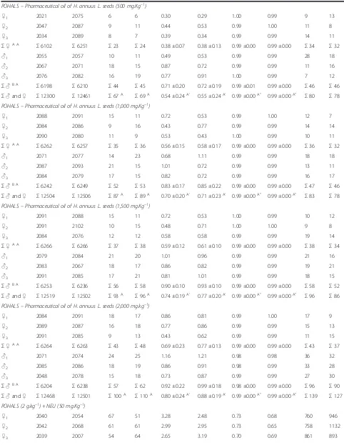
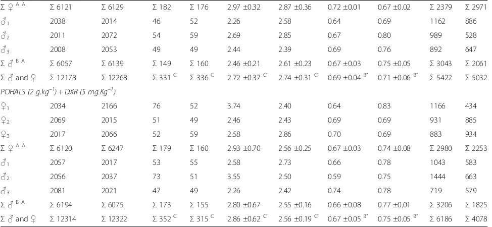
Table 2 The incidence of MNPCEs and PCE/NCE ratio in bone marrow of male and femaleSwiss albinusmice after testing for 24 h and 48 h
Treatment Number of PCEs analyzed PCEMNs PCE/(PCE + NCE) NCE (n)
24 h 48 h 24 h 48 h 24 h 48 h 24 hA” 48 hB” 24 h 48 h
(n)A (n)A (%)A’ (%)A’
150 mM NaCl
♀1 2095 2097 7 10 0.33 0.48 1.00 1.00 5 3
♀2 2094 2095 9 10 0.43 0.48 1.00 1.00 6 5
♀3 2087 2089 11 8 0.53 0.38 0.99 0.99 13 11
Σ♀ Σ6276 Σ6281 Σ27 Σ28 0.43 ±0.10 0.45 ±0.05 1.00 ±0.00 1.00 ±0.00 Σ24 Σ19
♂1 2095 2088 9 13 0.43 0.62 1.00 0.99 5 12
♂2 2055 2088 12 11 0.58 0.53 0.98 0.99 45 12
♂3 2058 2084 7 11 0.34 0.53 0.98 0.99 42 16
Σ♂ Σ6208 Σ6260 Σ28 Σ35 0.45 ±0.12 0.56 ±0.06 0.99 ±0.01 0.99 ± 0.00 Σ92 Σ40
Σ♂and♀ Σ12484 Σ12541 Σ55A Σ63A 0.44 ±0.08A’ 0.50 ±0.06A’ 0.99 ±0.01A” 1.00 ±0.00A” Σ116 Σ59 N–Nitroso–N–ethylurea–NEU (50 mg.Kg–1)
♀1 2148 2075 38 36 1.77 1.73 0.49 0.65 2252 1125
♀2 1884 2032 32 34 1.70 1.67 0.54 0.81 1616 468
♀3 2002 1948 15 31 0.75 1.59 0.61 0.93 1298 152
Σ♀ Σ6034 Σ6055 Σ85 Σ101 1.41 ±0.57 1.67 ±0.07 0.54 ±0.06 0.80 ±0.14 Σ5166 Σ1745
♂1 2025 1999 64 31 3.16 1.55 0.41 0.36 2875 3501
♂2 2028 1916 105 40 5.18 2.09 0.51 0.55 1972 1584
♂3 2004 2069 25 38 1.25 1.84 0.67 0.65 996 1131
Σ♂ Σ6057 Σ5984 Σ194 Σ109 3.20 ±1.97 1.83 ±0.27 0.53 ±0.13 0.52 ±0.14 Σ5843 Σ6216
Σ♂and♀ Σ12091 Σ12039 Σ279B Σ210B 2.30 ±1.66 B’ 1.75 ±0.18B’ 0.54 ±0.06C” 0.66 ±0.16C” Σ11009 Σ7961 Doxorubicin hydrochloride–DXR (5 mg.Kg–1)
♀1 2091 2017 49 36 2.34 1.78 0.72 0.96 809 83
♀2 2106 2077 73 63 3.47 3.03 0.98 0.99 44 23
♀3 2056 2092 57 50 2.77 2.39 0.84 0.95 394 108
Σ♀ Σ6253 Σ6186 Σ179 Σ149 2.86 ±0.57 2.40 ±0.62 0.85 ±0.13 0.97 ±0.02 Σ1247 Σ214
♂1 2067 2086 53 61 2.56 2.92 0.98 0.95 33 114
♂2 2063 2042 56 70 2.71 3.43 0.98 0.97 37 58
♂3 2082 2075 46 50 2.21 2.41 0.99 0.99 18 25
Σ♂ Σ6212 Σ6203 Σ155 Σ181 2.50 ±0.26 2.92 ±0.51 0.99 ±0.00 0.97 ±0.02 Σ88 Σ197
Σ♂and♀ Σ12465 Σ12389 Σ334C 330C 2.68 ±0.42C’ 2.66 ±0.43C’ 0.92 ±0.07A” 0.97 ±0.01 A” Σ1335 Σ411 POHALS–Pharmaceutical oil of H. annuus L. seeds (250 mg.Kg–1)
♀1 2081 2092 9 7 0.43 0.33 0.99 1.00 14 10
♀2 2086 2087 5 8 0.24 0.38 0.99 1.00 13 9
♀3 2090 2084 8 8 0.38 0.38 1.00 0.99 7 12
Σ♀A A
Σ6257 Σ6263 Σ22 Σ23 0.35 ±0.10 0.37 ±0.03 0.99 ±0.00 1.00 ±0.00 Σ34 Σ31
♂1 2082 2083 10 16 0.48 0.77 0.99 0.99 18 11
♂2 2085 2099 7 9 0.34 0.43 0.99 1.00 15 9
♂3 2089 2072 9 15 0.43 0.72 0.99 0.99 11 21
Σ♂B A
Σ6256 Σ6254 Σ26 Σ40 0.42 ±0.07 0.64 ±0.18 0.99 ±0.00 0.99 ±0.00 Σ44 Σ41
Table 2 The incidence of MNPCEs and PCE/NCE ratio in bone marrow of male and femaleSwiss albinusmice after testing for 24 h and 48 h(Continued)
POHALS–Pharmaceutical oil of H. annuus L. seeds (500 mg.Kg–1)
♀1 2021 2075 6 6 0.30 0.29 1.00 0.99 9 13
♀2 2047 2087 9 11 0.44 0.53 0.99 1.00 11 8
♀3 2034 2089 8 7 0.39 0.34 0.99 0.99 14 11
Σ♀A A
Σ6102 Σ6251 Σ23 Σ24 0.38 ±0.07 0.38 ±0.13 0.99 ±0.00 0.99 ±0.00 Σ34 Σ32
♂1 2055 2057 10 11 0.49 0.53 0.99 0.99 28 18
♂2 2067 2071 18 15 0.87 0.72 0.99 0.99 11 16
♂3 2076 2082 16 19 0.77 0.91 1.00 0.99 7 12
Σ♂B A
Σ6198 Σ6210 Σ44 Σ45 0.71 ±0.20 0.72 ±0.19 0.99 ±0.01 0.99 ±0.00 Σ46 Σ46
Σ♂and♀ Σ12300 Σ12461 Σ67A Σ69A 0.54 ±0.24A’ 0.55 ±0.24A’ 0.99 ±0.00A” 0.99 ±0.00A” Σ80 Σ78 POHALS–Pharmaceutical oil of H. annuus L. seeds (1,000 mg.Kg–1)
♀1 2088 2091 15 11 0.72 0.53 0.99 1.00 12 7
♀2 2084 2086 9 16 0.43 0.77 0.99 0.99 14 14
♀3 2090 2080 11 9 0.53 0.43 1.00 0.99 10 11
Σ♀A A
Σ6262 Σ6257 Σ35 Σ36 0.56 ±0.15 0.58 ±0.17 0.99 ±0.00 0.99 ±0.00 Σ36 Σ32
♂1 2071 2077 14 23 0.68 1.11 0.99 0.99 18 18
♂2 2087 2093 21 15 1.01 0.72 0.99 0.99 13 11
♂3 2084 2079 17 15 0.82 0.72 0.99 0.99 16 17
Σ♂B A
Σ6242 Σ6249 Σ52 Σ53 0.83 ±0.17 0.85 ±0.22 0.99 ±0.00 0.99 ±0.00 Σ47 Σ46
Σ♂and♀ Σ12504 Σ12506 Σ87A Σ89A 0.70 ±0.20A’ 0.71 ±0.23A’ 0.99 ±0.00A” 0.99 ±0.00A” Σ83 Σ78 POHALS–Pharmaceutical oil of H. annuus L. seeds (1,500 mg.Kg–1)
♀1 2091 2088 15 11 0.72 0.53 1.00 0.99 10 12
♀2 2091 2102 10 15 0.48 0.71 1.00 1.00 9 8
♀3 2084 2076 12 12 0.58 0.58 0.99 0.99 19 14
Σ♀A A
Σ6266 Σ6266 Σ37 Σ38 0.59 ±0.12 0.61 ±0.10 0.99 ±0.00 0.99 ±0.00 Σ38 Σ34
♂1 2079 2084 21 20 1.01 0.96 0.99 0.99 21 16
♂2 2083 2067 18 17 0.86 0.82 0.99 0.99 19 21
♂3 2091 2085 17 21 0.81 1.01 0.99 0.99 18 15
Σ♂B A
Σ6253 Σ6236 Σ56 Σ58 0.90 ±0.10 0.93 ±0.10 0.99 ±0.00 0.99 ±0.00 Σ58 Σ52
Σ♂and♀ Σ12519 Σ12502 Σ93A Σ96A 0.74 ±0.19A’ 0.77 ±0.20A’ 0.99 ±0.00A” 0.99 ±0.00A” Σ96 Σ86 POHALS–Pharmaceutical oil of H. annuus L. seeds (2,000 mg.kg–1)
♀1 2084 2091 18 17 0.86 0.81 0.99 1.00 17 9
♀2 2089 2087 16 18 0.77 0.86 0.99 0.99 15 13
♀3 2091 2085 9 13 0.43 0.62 0.99 0.99 11 15
Σ♀A A
Σ6264 Σ6263 Σ43 Σ48 0.69 ±0.23 0.77 ±0.13 0.99 ±0.00 0.99 ±0.00 Σ43 Σ37
♂1 2071 2074 24 25 1.16 1.21 0.98 0.98 36 32
♂2 2085 2086 18 19 0.86 0.91 0.98 0.99 33 28
♂3 2048 2078 15 18 0.73 0.87 0.99 0.99 27 30
Σ♂B A
Σ6204 Σ6238 Σ57 Σ62 0.92 ±0.22 0.99 ±0.18 0.98 ±0.00 0.99 ±0.00 Σ96 Σ90
Σ♂and♀ Σ12468 Σ12501 Σ100A Σ110A 0.80 ±0.24A’ 0.88 ±0.19A’ 0.99 ±0.00A” 0.99 ±0.00A” Σ139 Σ127 POHALS (2 g.kg–1) + NEU (50 mg.Kg–1)
♀1 2040 2054 67 51 3.28 2.48 0.73 0.68 760 946
♀2 2042 2068 61 61 2.99 2.95 0.73 0.65 758 1132
of thermally stressed sunflower oil to rats is terato-genic [51].
Doxorubicin (DXR) is an important anthracyclines an-ticancer agent. It is a valuable component of various chemotherapeutic regimens for breast carcinoma and small-cell lung carcinoma. In metastatic thyroid carcin-oma, DXR is most likely the best available agent [20]. However, DXR has been reported to induce micronuclei, chromatid and chromosome aberrations, and DNA single- and double-strand breaks in vitro and in vivo
[52-56]. The genotoxicity of anticancer drugs is of special interest because of the risk of inducing secondary malig-nancies. Therefore, it is essential to screen for newer pharmacological agents that can protect the normal cells against DXR–induced cumulative (geno) toxicity. Many plants that have been widely used in traditional medi-cine are less toxic than pharmaceutical agents and have recently attracted the attention of researchers around the world. Plants contain many compounds, and it is likely that these can provide better protective effects than a single molecule [57]. The presence of many mol-ecules in plants may be advantageous, as some of them may counteract the toxicity of others, and as a result, the net effect may be beneficial for therapeutic pur-poses. For example, the effect of various concentrations (200, 250, 300, 350, and 400 mg/kg body weight) ofAegle marmelos on the doxorubicin (DXR)–induced genotoxic effects in mice bone marrow was studied [20]. Treatment of mice with different concentrations of DXR (5, 10, or
15 mg.kg−1body weight) resulted in a dose–dependent elevation in the frequency of micronucleated polychro-matic and normochropolychro-matic erythrocytes in mouse bone marrow, and it was accompanied by a DXR dose– dependent decline in the PCE/NCE ratio. The treat-ment of mice with Aegle marmelos, orally once daily for 5 consecutive days before DXR treatment, signifi-cantly reduced the frequency of DXR–induced micro-nuclei and significant increased the PCE/NCE ratio at all scoring times. This observed chemoprotective effect may be due to the sum total of interaction between dif-ferent ingredients of this complex mixture. The degree of protection may depend on the interaction of compo-nents individually or collectively with the genotoxic agent. The plausible mechanisms of action of Aegle marmelos in protecting against DXR–induced genomic insult were scavenging of O2•– and •OH and other free
[image:10.595.56.543.114.339.2]radicals, increase in antioxidant status, restoration of topoisomerase II activity, and inhibition of the formation of DXR–iron complex [20]. Another study was under-taken to evaluate the genotoxic potential of Copaifera langsdorffiiDesf. leaf hydroalcoholic extract and its influ-ence on the genotoxicity induced by chemotherapeutic agent DXR using the Swissmouse peripheral blood mi-cronucleus test. The results of this study demonstrated thatC. langsdorffiiDesf. was not itself genotoxic and that in animals treated with C. langsdorffii Desf. and DXR, the number of micronuclei was significantly decreased compared to animals receiving DXR alone. The putative Table 2 The incidence of MNPCEs and PCE/NCE ratio in bone marrow of male and femaleSwiss albinusmice after testing for 24 h and 48 h(Continued)
Σ♀A A
Σ6121 Σ6129 Σ182 Σ176 2.97 ±0.32 2.87 ±0.36 0.72 ±0.01 0.67 ±0.02 Σ2379 Σ2971
♂1 2038 2014 46 52 2.26 2.58 0.64 0.69 1162 886
♂2 2011 2072 54 59 2.69 2.85 0.67 0.80 989 528
♂3 2008 2053 49 49 2.44 2.39 0.69 0.76 892 647
Σ♂B A
Σ6057 Σ6139 Σ149 Σ160 2.46 ±0.21 2.61 ±0.23 0.67 ±0.03 0.75 ±0.05 Σ3043 Σ2061
Σ♂and♀ Σ12178 Σ12268 Σ331C Σ336C 2.72 ±0.37C’ 2.74 ±0.31C’ 0.69 ±0.04B” 0.71 ±0.06B” Σ5422 Σ5032 POHALS (2 g.kg–1) + DXR (5 mg.Kg–1)
♀1 2034 2166 76 52 3.74 2.40 0.64 0.83 1166 434
♀2 2069 2015 51 49 2.46 2.43 0.69 0.69 931 885
♀3 2017 2066 52 59 2.58 2.86 0.70 0.69 883 934
Σ♀A A
Σ6120 Σ6247 Σ179 Σ160 2.93 ±0.70 2.56 ±0.25 0.67 ±0.03 0.74 ±0.08 Σ2980 Σ2253
♂1 2057 2017 53 55 2.58 2.73 0.66 0.78 1043 583
♂2 2056 2037 73 51 3.55 2.50 0.59 0.75 1444 663
♂3 2081 2021 47 49 2.26 2.42 0.74 0.78 719 579
Σ♂B A
Σ6194 Σ6075 Σ173 Σ155 2.80 ±0.67 2.55 ±0.16 0.66 ±0.08 0.77 ±0.01 Σ3206 Σ1825
Σ♂and♀ Σ12314 Σ12322 Σ352C Σ315C 2.86 ±0.62C’ 2.56 ±0.19C’ 0.67 ±0.05B” 0.75 ±0.05B” Σ6186 Σ4078 Means with the same letter are not significantly different (p< 0.05).
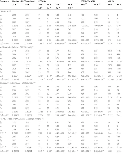
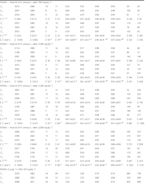
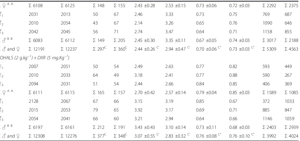
Table 3 The incidence of MNPCEs and PCE/NCE ratio in bone marrow of male and femaleSwiss albinusmice after testing for 24 h and 48 h
Treatment Number of PCEs analyzed PCEMNs PCE/(PCE + NCE) NCE (n)
24 h 48 h 24 h 48 h 24 h 48 h 24 hA” 48 hA” 24 h 48 h
(n)A (n)A (%)A’ (%)A’
150 mM NaCl
♀1 2095 2097 7 10 0.33 0.48 1.00 1.00 5 3
♀2 2094 2095 9 10 0.43 0.48 1.00 1.00 6 5
♀3 2087 2089 11 8 0.53 0.38 0.99 0.99 13 11
Σ♀ Σ6276 Σ6281 Σ27 Σ28 0.43 ±0.10 0.45 ±0.05 1.00 ±0.00 1.00 ±0.00 Σ24 Σ19
♂1 2095 2088 9 13 0.43 0.62 1.00 0.99 5 12
♂2 2055 2088 12 11 0.58 0.53 0.98 0.99 45 12
♂3 2058 2084 7 11 0.34 0.53 0.98 0.99 42 16
Σ♂ Σ6208 Σ6260 Σ28 Σ35 0.45 ±0.12 0.56 ±0.06 0.99 ±0.01 0.99 ± 0.00 Σ92 Σ40
Σ♂and♀ Σ12484 Σ12541 Σ55A Σ63A 0.44 ±0.08A’ 0.50 ±0.06A’ 0.99 ±0.01A” 1.00 ±0.00A” Σ116 Σ59 N–Nitroso–N–ethylurea–NEU (50 mg.Kg−1)
♀1 2148 2075 38 36 1.77 1.73 0.49 0.65 2252 1125
♀2 1884 2032 32 34 1.70 1.67 0.54 0.81 1616 468
♀3 2002 1948 15 31 0.75 1.59 0.61 0.93 1298 152
Σ♀ Σ6034 Σ6055 Σ85 Σ101 1.41 ±0.57 1.67 ±0.07 0.54 ±0.06 0.80 ±0.14 Σ5166 Σ1745
♂1 2025 1999 64 31 3.16 1.55 0.41 0.36 2875 3501
♂2 2028 1916 105 40 5.18 2.09 0.51 0.55 1972 1584
♂3 2004 2069 25 38 1.25 1.84 0.67 0.65 996 1131
Σ♂ Σ6057 Σ5984 Σ194 Σ109 3.20 ±1.97 1.83 ±0.27 0.53 ±0.13 0.52 ±0.14 Σ5843 Σ6216
Σ♂and♀ Σ12091 Σ12039 Σ279B Σ210B 2.30 ±1.66B’ 1.75 ±0.18B’ 0.54 ±0.06D” 0.66 ±0.16D” Σ11009 Σ7961 Doxorubicin hydrochloride–DXR (5 mg.Kg−1)
♀1 2091 2017 49 36 2.34 1.78 0.72 0.96 809 83
♀2 2106 2077 73 63 3.47 3.03 0.98 0.99 44 23
♀3 2056 2092 57 50 2.77 2.39 0.84 0.95 394 108
Σ♀ Σ6253 Σ6186 Σ179 Σ149 2.86 ±0.57 2.40 ±0.62 0.85 ±0.13 0.97 ±0.02 Σ1247 Σ214
♂1 2067 2086 53 61 2.56 2.92 0.98 0.95 33 114
♂2 2063 2042 56 70 2.71 3.43 0.98 0.97 37 58
♂3 2082 2075 46 50 2.21 2.41 0.99 0.99 18 25
Σ♂ Σ6212 Σ6203 Σ155 Σ181 2.50 ±0.26 2.92 ±0.51 0.99 ±0.00 0.97 ±0.02 Σ88 Σ197
Σ♂and♀ Σ12465 Σ12389 Σ334C 330C 2.68 ±0.42C’ 2.66 ±0.43C’ 0.92 ±0.07AB” 0.97 ±0.01AB” Σ1335 Σ411 FOHALS–Food oil of H. annuus L. seeds (250 mg.Kg−1)
♀1 2137 2019 11 9 0.51 0.45 0.99 0.99 24 14
♀2 2142 2073 7 14 0.33 0.68 1.00 1.00 8 5
♀3 2146 2016 9 7 0.42 0.35 0.99 1.00 12 6
Σ♀A A
Σ6425 Σ6108 Σ27 Σ30 0.42 ±0.09 0.49 ±0.17 0.99 ±0.00 1.00 ±0.00 Σ44 Σ25
♂1 2061 2079 6 10 0.29 0.48 0.95 0.94 98 140
♂2 2093 2093 8 7 0.38 0.33 0.99 0.99 31 20
♂3 2050 2041 8 6 0.39 0.29 0.99 0.97 30 59
Σ♂B B
Σ6204 Σ6213 Σ22 Σ23 0.35 ±0.03 0.37 ±0.10 0.98 ±0.02 0.97 ±0.03 Σ159 Σ219
Table 3 The incidence of MNPCEs and PCE/NCE ratio in bone marrow of male and femaleSwiss albinusmice after testing for 24 h and 48 h(Continued)
FOHALS–Food oil of H. annuus L. seeds (500 mg.Kg−1)
♀1 2014 2046 10 11 0.50 0.54 0.98 0.99 39 24
♀2 2007 2094 8 8 0.40 0.38 0.95 0.98 106 34
♀3 2010 2035 9 16 0.45 0.79 0.96 0.99 81 11
Σ♀A A
Σ6031 Σ6175 Σ27 Σ35 0.45 ±0.05 0.57 ±0.20 0.96 ±0.02 0.99 ±0.01 Σ226 Σ69
♂1 2037 2082 10 10 0.49 0.48 0.95 0.94 116 135
♂2 2063 2067 8 16 0.39 0.77 0.96 0.96 78 89
♂3 2053 2078 5 9 0.24 0.43 0.95 0.97 102 63
Σ♂B B
Σ6153 Σ6227 Σ23 Σ35 0.37 ±0.12 0.56 ±0.18 0.95 ±0.01 0.96 ±0.02 Σ296 Σ287
Σ♂and♀ Σ12184 Σ12402 Σ50A Σ70A 0.41 ±0.09A’ 0.57 ±0.17A’ 0.96 ±0.01AB” 0.97 ±0.02AB” Σ522 Σ356 FOHALS–Food oil of H. annuus L. seeds (1,000 mg.Kg−1)
♀1 2114 2083 11 16 0.52 0.77 0.98 0.98 36 48
♀2 2148 2058 11 13 0.51 0.63 0.96 0.97 84 72
♀3 2097 2090 8 9 0.38 0.43 0.93 0.95 149 106
Σ♀A A
Σ6359 Σ6231 Σ30 Σ38 0.47 ±0.08 0.61 ±0.17 0.96 ±0.02 0.97 ±0.01 Σ269 Σ226
♂1 2073 2026 9 17 0.43 0.84 0.90 0.96 227 74
♂2 2065 2071 12 13 0.58 0.63 0.99 0.94 20 129
♂3 2003 2084 5 8 0.25 0.38 0.95 0.91 97 207
Σ♂B B
Σ6141 Σ6181 Σ26 Σ38 0.42 ±0.17 0.62 ±0.23 0.95 ±0.02 0.94 ±0.03 Σ344 Σ410
Σ♂and♀ Σ12500 Σ12412 Σ56A Σ76A 0.45 ±0.12A’ 0.61 ±0.18A’ 0.95 ±0.03AB” 0.95 ±0.02AB” Σ613 Σ636 FOHALS–Food oil of H. annuus L. seeds (1,500 mg.Kg−1)
♀1 2065 2021 6 15 0.29 0.74 0.98 0.90 35 236
♀2 2041 2081 13 10 0.64 0.48 0.93 0.89 159 262
♀3 2068 2072 11 14 0.53 0.68 0.89 0.88 247 294
Σ♀A A
Σ6174 Σ6174 Σ30 Σ39 0.49 ±0.18 0.63 ±0.14 0.93 ±0.05 0.89 ±0.01 Σ441 Σ792
♂1 2106 2087 10 18 0.47 0.86 0.92 0.96 194 94
♂2 2011 2088 5 11 0.25 0.53 0.91 0.94 189 126
♂3 2039 2128 14 16 0.69 0.75 0.98 0.88 38 277
Σ♂B B
Σ6156 Σ6303 Σ29 Σ45 0.47 ±0.22 0.71 ±0.17 0.94 ±0.04 0.93 ±0.04 Σ421 Σ497
Σ♂and♀ Σ12330 Σ12477 Σ59A Σ84A 0.48 ±0.18A’ 0.67 ±0.15A’ 0.94 ±0.04AB” 0.91 ±0.03AB” Σ862 Σ1289 FOHALS–Food oil of H. annuus L. seeds (2,000 mg.kg−1)
♀1 2084 2015 11 13 0.53 0.65 0.89 0.86 245 325
♀2 2096 2025 13 11 0.62 0.54 0.91 0.83 216 407
♀3 2076 2002 9 17 0.43 0.85 0.95 0.94 114 126
Σ♀A A
Σ6256 Σ6042 Σ33 Σ41 0.53 ±0.09 0.68 ±0.16 0.92 ±0.03 0.88 ±0.06 Σ575 Σ858
♂1 2057 2199 16 20 0.78 0.91 0.94 0.97 143 65
♂2 2016 2158 17 12 0.84 0.56 0.95 0.86 110 347
♂3 2106 2133 11 15 0.52 0.70 0.92 0.89 194 261
Σ♂B B
Σ6179 Σ6490 Σ44 Σ47 0.71 ±0.17 0.72 ±0.18 0.93 ±0.02 0.91 ±0.06 Σ447 Σ673
Σ♂and♀ Σ12435 Σ12532 Σ77A Σ88A 0.62 ±0.16A’ 0.70 ±0.15A’ 0.92 ±0.02B” 0.89 ±0.05B” Σ1022 Σ1531 FOHALS (2 g.kg−1) + NEU (50 mg.Kg−1)
♀1 2020 2062 54 49 2.67 2.38 0.70 0.74 880 738
♀2 2080 2052 44 52 2.12 2.53 0.80 0.68 520 948
antioxidant activity of one or more of the active com-pounds ofC. langsdorffiiDesf., including two major flavon-oid heterosides (quercitrin and afzelin), may explain the effect of this plant on DXR genotoxicity [18].
Conclusions
In conclusion, this research observed an absence of gen-otoxicity of a tincture and two oils of sunflower seeds, regardless of the dose tested and the treatment time (24–48 h), but sex-independent (sunflower tincture) or sex-dependent (sunflower oils). Antigenotoxic effects (anticlastogeny and/or antianeugeny) were observed using only a dose of the sunflower tincture in association with the chemotherapy agent DXR. Therefore, the sunflower tincture can promote a partial protection against the geno-toxic effects induced by DXR. The sunflower tincture no showed systemic toxicity and it was dose- and time-independent and sex-dependent, whereas the systemic toxicity of sunflower oil can be dependent on its source and its highest dose used.
Other studies involving the genotoxicity and antigeno-toxicity of H. annuusL. extracts and oils (seeds, flowers and leaves) should be conducted [including genotoxicity assays with Salmonella typhimuriumtest (Ames test) as an indicator of potential carcinogenicity to mammals, gene mutation test in mammalian cells (mouse lymph-oma assay), cytogenetic and aneuploidy tests in vitro, micronucleus test in cultured cells in vitro, fluorescent
in situhybridization (FISH) test for mutagenesis, comet
test to detect of DNA damage and repair in individual cells, and functional genomic and proteomic tests for mutagenesis (cDNA microarrays and other array ana-lyses)], to characterize the potential effects and geno-toxic and antigenogeno-toxic mechanisms and, importantly, for the establishment of limits for human consumption, the delineation of potential risks to human health, and for rational strategies for implementing chemo-preventive measures.
Competing interest
The authors have declared no competing interest.
Authors’contributions
MFGB, JEF and NMSO wrote and revised the draft, MCCR and TAS provided animals care and revised the draft, LSS and MRR aided micronucleus assays and revised the draft, MFGB and CTSD performed statistical analysis. MFGB and JEF have given final approval of the version to be published. All authors have read and approved the final manuscript.
Acknowledgements
This research was supported byRede Mineira de Ensaios Toxicológicos e Farmacológicos de Produtos Terapêuticos(REDE MINEIRA TOXIFAR–2012), Fundação de Amparo à Pesquisa do Estado de Minas Gerais(FAPEMIG). The authors thank the Language Services of Elsevier for help in English language editing.
Author details
1Laboratório de Farmacogenômica e Biologia Molecular, Faculdade de
[image:13.595.60.541.112.339.2]Ciências Médicas & Centro de Pesquisa e Pós–graduação, Universidade José do Rosário Vellano (UNIFENAS), Campus Universitário, Rod. MG 179, Km 0, Alfenas, MG CEP: 37130-000, Brasil.2Centro de Pesquisa e Pós–graduação em Ciência Animal, Área de Patologia e Farmacologia Animal, Universidade José do Rosário Vellano (UNIFENAS), Alfenas, Minas Gerais, Brasil.3Centro de Cirurgia Experimental e Farmacologia, Universidade Estadual de Campinas,
Table 3 The incidence of MNPCEs and PCE/NCE ratio in bone marrow of male and femaleSwiss albinusmice after testing for 24 h and 48 h(Continued)
Σ♀A A
Σ6108 Σ6125 Σ148 Σ155 2.43 ±0.28 2.53 ±0.15 0.73 ±0.06 0.72 ±0.03 Σ2292 Σ2375
♂1 2031 2013 50 67 2.46 3.33 0.73 0.75 769 687
♂2 2010 2054 43 67 2.14 3.26 0.65 0.76 1090 646
♂3 2042 2045 56 71 2.74 3.47 0.64 0.71 1158 855
Σ♂B B
Σ6083 Σ6112 Σ149 Σ205 2.45 ±0.30 3.35 ±0.11 0.67 ±0.05 0.74 ±0.03 Σ3017 Σ2188
Σ♂and♀ Σ12191 Σ12237 Σ297C Σ360C 2.44 ±0.26C’ 2.94 ±0.47C’ 0.70 ±0.06C” 0.73 ±0.03C” Σ5309 Σ4563 FOHALS (2 g.kg−1) + DXR (5 mg.Kg−1)
♀1 2007 2051 50 54 2.49 2.63 0.77 0.82 593 449
♀2 2010 2033 64 49 3.18 2.41 0.77 0.88 590 267
♀3 2094 2031 51 54 2.44 2.66 0.84 0.85 406 369
Σ♀A A
Σ6111 Σ6115 Σ165 Σ157 2.70 ±0.42 2.57 ±0.14 0.79 ±0.04 0.85 ±0.03 Σ1589 Σ1085
♂1 2128 2067 67 66 3.15 3.19 0.85 0.67 372 1033
♂2 2015 2053 79 65 3.92 3.17 0.69 0.71 885 847
♂3 2054 2041 66 60 3.21 2.94 0.64 0.66 1146 1059
Σ♂B B
Σ6197 Σ6161 Σ212 Σ191 3.43 ±0.43 3.10 ±0.14 0.73 ±0.11 0.68 ±0.03 Σ2403 Σ2939
Σ♂and♀ Σ12308 Σ12276 Σ377C Σ348C 3.07 ±0.55C’ 2.83 ±0.32C’ 0.76 ±0.08C” 0.76 ±0.10C” Σ3992 Σ4024 Means with the same letter are not significantly different (p< 0.05).
Campinas (UNICAMP), São Paulo, Brasil.4Laboratório de Ecotoxicologia e Microbiologia Ambiental, Faculdade de Tecnologia, Universidade Estadual de Campinas (UNICAMP), Limeira, São Paulo, Brasil.5Departamento de Ciências Exatas, Escola de Agricultura“Luiz de Queiroz”, Universidade de São Paulo (ESALQ/USP), Piracicaba, SP, Brasil.
Received: 22 August 2013 Accepted: 27 March 2014 Published: 2 April 2014
References
1. Heiser CB:Sunflowers: Helianthus (Compositae-Heliantheae).InEvolution of Crop Plants.Edited by Simmonds NW. London: Longmans Green; 1976:36–38.
2. Earle FR, Vanetten CH, Clark TF, Wolff IA:Compositional data on sunflower seed.J Am Oil Chem Soc1968,45:876–879.
3. Salunkhe DK, Chavan JK, Adsule RN, Kadam SS:World Oilseeds: Chemistry, Technology and Utilization.InSunflower.New York: Van Nostrand Reinhold; 1992.
4. Gonzáles-Pérez S, Vereijken JM:Sunflower proteins: Overview of their physicochemical, structural and functional properties.J Sci Food Agric 2007,87:2173–2191.
5. Warner KA, Mounts TL, List GVR:Effects of added tocopherols on the flavor stability of purified vegetable oils.Inform1990,1:326. 6. Ito T, Tamura T, Matsumoto T:Sterol Composition of 19 vegetable oils.
J Am Oil Chem Soc1973,50:122–125.
7. Trost VW:Characterization of corn oil, soybean oil and sunflower oil nonpolar material.J Am Oil Chem Soc1989,66:325–333.
8. Dupont HL, Sullivan P, Evans DG, Vollet JJ, Ericsson CD, Ackerman PB, Tjoa WS:Prevention of traveler's diarrhea (emporiatric enteritis). Prophylactic administration of subsalicylate bismuth.JAMA1980,
243:237–241.
9. Cowan JC:Key factors and recent advances in the flavor stability of soybean oil.J Am Oil Chem Soc1966,43:300A, 302A, 318A, 320.
10. Bierenbaum ML, Fleischman AI, Dun J, Arnold J:Possible toxic waste factor in coronary heart disease.Lancet1975,1:1008–1010.
11. Purves D, Harvey C, Tweats D, Lumley CE:Genotoxity testing: current practices and strategies used by the pharmaceutical industry.
Mutagenesis1995,10:297–312.
12. Varanda EA:Atividade mutagênica de plantas medicinais.Rev Ciênc Farm Básica Apl2006,27:1–7.
13. Mateuca R, Lombaert N, Aka PV, Decordier I, Kirsch–Volders M:
Chromosomal changes: induction, detection methods and applicability in human biomonitoring.Biochimie2006,88:1515–1531.
14. Krishna G, Hayashi M:In vivo rodent micronucleus assay: protocol, conduct and data interpretation.Mutat Res2000,455:155–166. 15. Organisation for Economic Cooperation and Development:OECD Guideline
for the Testing of Chemicals: Bacterial reverse mutation test.Paris; 1997:Guideline 471.
16. Organisation for Economic Cooperation and Development:OECD Guideline for the Testing of Chemicals: In vitro mammalian chromosome aberration test. Paris; 1997:Guideline 473.
17. Organisation for Economic Cooperation and Development:OECD Guideline for the Testing of Chemicals: Mammalian erythrocyte micronucleus test.Paris; 1997:Guideline 474.
18. Alves JM, Munari CC, Neto MABM, Furtado RA, Senedese JM, Bastos JK, Tavares DC:In vivoprotective effect ofCopaifera langsdorffii
hydroalcoholic extract on micronuclei induction by doxorubicin.
J Appl Toxicol2013,33:854–860.
19. Indart A, Viana M, Clapés S, Izquierdo L, Bonet B:Clastogenic and cytotoxic effects of lipid peroxidation products generated in culinary oils submitted to thermal stress.Food Chem Toxicol2007,45:1963–1967. 20. Venkatesh P, Shantala B, Jagetia GC, Rao KK, Baliga MS:Modulation of
Doxorubicin–Induced Genotoxicity byAegle marmelosin Mouse Bone Marrow: A Micronucleus Study.Integr Cancer Ther2007,6:42–53. 21. Brasil:Farmacopéia Brasileira, Agência Nacional de Vigilância Sanitária
-ANVISA/Fundação Oswaldo.Brasília: Cruz - FIOCRUZ; 2010. 22. Collaborative Study Group for the Micronucleus Test (CSGMT):Sex
differences in the micronucleus test.Mutat Res1986,172:151–163. 23. Zambrano MA, Targa HJ, Rabello–Gay MN:Physiological saline solutions
as a useful tool in micronucleus and metaphase slide preparations.
Stain Technol1982,57:48–49.
24. Lewi DM, Hopp HE, Escandon AS:Sunflower (Helianthus annuusL.).
Methods Mol Biol2006,343:291–297.
25. Heo JC, Woo SUK, Kweon MA, Park JY, Lee HK, Son M, Rho JR, Lee SH:
Aqueous extract of theHelianthus annuusseed alleviates asthmatic symptomsin vivo.Int J Mol Med Chem2008,21:57–61.
26. Cardoso CC, Rodrigues KL, Pichara NL, Dall’Aglio R, Fiorini JE, Fraschini F, Diana GM, Drago L, De Vecchi E, Carvalho JCT:Olio di girasole ozonizzato associate ad acidoα-lipoico e a lattobacilli: studio pre-clinico dell’azione antiulcerosa, antinfiammatoria e antibatterica.Farmaci2004,28:97–110. 27. Ricardo GA, Zullyt ZR, Yilian L, Hernández F, Menéndez S:Efecto del
OLEOZON®frente a lesiones gástricas inducidas por indometacina en
ratas (Effect of OLEOZON®on gastric lesions induced by indomethacin in
rats).Revista electrónica de Veterinaria2007,8:1–6.
28. Rodrigues KL, Cardoso CC, Caputo LR, Carvalho JC, Fiorini JE, Schneedorf JM:
Cicatrizing and antimicrobial properties of an ozonised oil from sunflower seeds.Inflammopharmacology2004,3:261–270.
29. Akihisa T, Yasukawa K, Oinuma H, Kasahara Y, Kimura Y, Takase S, Yamanouchi S, Takido M, Kumaki K, Tamura T:Triterpene alcohols from the flowers of Compositae and their anti-inflammatory effects.
Phytochemistry1996,43:1255–1260.
30. Rojas-Molina M, Campos–Sanches J, Analla M, Serrano M, Moraga AA:
Genotoxicity of vegetable cooking oils in theDrosophilawing spot test.
Environ Mol Mutagen2005,45:90–95.
31. Montero ACR, Carvajal YG, Rodríguez ZZ, López GF, Mirabal JM:Evaluación genotóxica del OLEOZON mediante los ensayos de micronúcleos en medula ósea y sangre periférica de ratón.Revista CENIC: Ciências Biológicas1998,29:200–202.
32. Salerno JW, Smith DE:The use of sesame oil and other vegetable oils in the inhibition of human colon cancer growthin vitro.Anticancer Res1991,
11:209–215.
33. Cognault S, Jourdan ML, Germain E, Pitavy R, Morel E, Durand G, Bougnoux P, Lhuillery C:Effect of an m-linolenic acid–rich diet on rat mammary tumour growth depends on the dietary oxidative status.Nutr Cancer 2000,36:33–41.
34. Nakayama M, Ju HR, Sugano M, Hirose N, Ueki T, Doi F, Eynard AR:Effect of dietary fat and cholesterol on dimethylbenz [a]-antracene-induced mammary tumorigenesis in Sprague–Dawley rats.Anticancer Res1993,
13:691–698.
35. La Vecchia C, Negri E, Franceschi S, Decarli A, Giacosa A, Lipworth L:Olive oil, other dietary fats, and the risk of breast cancer (Italy).Cancer Cause Control1995,6:545–550.
36. Rao GN, Ney E, Herbert RA:Effect of melatonin and linolenic acid on mammary cancer in transgenic mice with c-neu breast cancer oncogen.
Breast Cancer Res2000,64:287–296.
37. Burns CP, Luttenegger DG, Specctor AA:Effect of dietary fat saturation on survival of mice with L1210 Leukemia.J Natl Cancer Inst1978,
61:513–515.
38. Bartsch H, Nai J, Owen RW:Dietary polyunsaturated fatty acids and cancer of the breast and colorectum: emerging evidence for their role as risk modifiers.Carcinogenesis1999,20:2209–2218.
39. Boovens J, Engelbrecht P, Le Roux S, Louwrens CC, Van der Merwe CF, Katzeff IE:Some effects of the essential fatty acids linoleic acid and alpha-Linolenic acid and of their metabolites gamma-Linolenic acid, arachidonic acid, eicosapentaenoic acid, docosahesaenoic acid, and of prostaglandins A1 and E1 in the proliferation of human osteogenic sarcoma cells in culture.Prostaglandins Leukot Med1984,15:15–33. 40. Johanning GL, Lin TZ:Unsaturated fatty acid effects on human breast
cancer cell adhesion.Nutr Cancer1995,24:57–66.
41. Newcomer LM, King IB, Wicklund KG, Stanford JL:The association of fatty acids with prostate cancer risk.Prostate2001,47:262–268.
42. Iwado H, Naito M, Hayatsu H:Genotoxicity and antigenotoxicity of air-borne particulates.Mutat Res1991,246:93–103.
43. Siegel I, Liu TL, Yaghoubzadeh E, Keskey TS, Gleicher N:Cytotoxic effects of free fatty acids on ascites tumor cells.J Natl Cancer Inst Monographs1987,
78:271–277.
44. Bégin LR, Clement PB, Kirk ME, Jothy S, McCaughey WT, Ferenczy A:
Aggressive angiomyxoma of pelvic soft parts: a clinicopathologic study of nine cases.Hum Pathol1985,16:621–628.
46. DeFlora S, Ramel C:Mechanisms of inhibitors of mutagenesis and carcinogenesis: classification and review.Mutat Res1988,202:285–306. 47. Visioli F, Bellosta S, Galli C:Oleuropein, the bitter principle of olives,
enhances nitric-oxide production by mouse macrophages.Life Sci1998,
62:541–546.
48. Santos JH, Graf U, Reguly ML, De Andrade HHR:The synergistic effects of vanillin on recombination predominate over its antigenotoxic action in relation to MMC-induced lesions in somatic cells ofDrosophila
melanogaster.Mutat Res1999,444:355–365.
49. Weisburger JH:Can cancer risks be altered by changing nutritional traditions?Cancer1998,83:1278–1281.
50. Katiyar SK, Mohan RR, Agarwal R, Mukhtar H:Protection against induction of mouse skin papillomas with low and high risk of conversion to malignancy by green tea polyphenols.Carcinogenesis1997,18:497–502. 51. Indart A, Viana M, Grootveld MC, Silwood CJ, Sanchez-Vera I, Bonet B:
Teratogenic actions of thermally-stressed culinary oils in rats.Free Radic Res Commun Commun2002,36:1051–1058.
52. Al-Shabanah OA:Inhibition of adriamycin-induced micronuclei by desferrioxamine inSwiss albino mice.Mutat Res1993,301:107–111. 53. Bean CL, Armstrong MJ, Galloway SM:Effect of sampling time on
chromosome aberration yield for 7 chemicals in Chinese hamster ovary cells.Mutat Res1992,265:31–44.
54. Delvaeye M, Verovski V, De Neve W, Storme G:DNA breakage, cytotoxicity, drug accumulation and retention in two human ovarian tumor cell lines AZ224 and AZ364 treated with adriamycin, modulated by verapamil.
Anticancer Res1993,13:1533–1538.
55. Dhawan A, Kayani MA, Parry JM, Parry E, Anderson D:Aneugenic and clastogenic effects of doxorubicin in human lymphocytes.Mutagenesis 2003,18:487–490.
56. Jagetia GC, Nayak V:Effect of doxorubicin on cell survival and micronuclei formation in HeLa cells exposed to different doses of gamma–radiation.Strahlenther Onkol2000,176:422–428.
57. Vidhya N, Devraj SN:Antioxidant effect of eugenol in rat intestine.Ind J Exp Biol1999,37:1192–1195.
doi:10.1186/1472-6882-14-121
Cite this article as:Boriolloet al.:Nongenotoxic effects and a reduction of the DXR-induced genotoxic effects ofHelianthus annuusLinné (sunflower) seeds revealed by micronucleus assays in mouse bone marrow.BMC Complementary and Alternative Medicine201414:121.
Submit your next manuscript to BioMed Central and take full advantage of:
• Convenient online submission
• Thorough peer review
• No space constraints or color figure charges
• Immediate publication on acceptance
• Inclusion in PubMed, CAS, Scopus and Google Scholar
• Research which is freely available for redistribution