http://dx.doi.org/10.4236/detection.2016.43009
A Mass Spectrometric Study of Kratom
Compounds by Direct Infusion Electrospray
Ionization Triple Quadrupole Mass
Spectrometry
Hanzhuo Fu
Ultimate Analysis Laboratory, Boca Raton, USA
Received 17 April 2016; accepted 20 June 2016; published 23 June 2016
Copyright © 2016 by author and Scientific Research Publishing Inc.
This work is licensed under the Creative Commons Attribution International License (CC BY).
http://creativecommons.org/licenses/by/4.0/
Abstract
Mitragynine (MG) and its major metabolites 7-hydroxymitragynine (7-OH-MG) are two of the ma-jor components of the plant extract Kratom, which is a tree planted in Southeast Asia. Kratom has long been used by opioid-dependent individuals as an alternative to their unavailable opioid of choice and chronic pain medication, as a stealth-to-urine drug screening opiate substitute while in opioid recovery treatment and recreationally, alone or as a booster. In this study, a direct infusion method was utilized and electrospray ionization triple quadrupole mass spectrometer was used as the detector for data acquisition. Pharmacokinetic study was conducted to investigate the effect of mitragynine and 7-hydroxymitragynine and major fragments of both compounds were pro-posed.
Keywords
Mitragynine, 7-Hydroxymitragynine, Kratom, HPLC-MS/MS, Pharmacokinetic, Mass Fragment
1. Introduction
Administration (USDEA) Office of Diversion Control lists kratom as a drug of concern; however, kratom re-mains legal in the U.S. and is also one of the most popular legal highs in the U.K. [9][10]. M. speciose Korth contains more than 25 alkaloids that vary quantitatively depending on geographic location.
Several unique alkaloids are present in kratom leaves and the predominant alkaloid is mitragynine. Mitragy-nine (Figure 1) is structurally similar to the aphrodisiac yohimbine, which is the most prevalent of these alkalo-ids, and mitragynine is believed to be responsible for kratom’s opioid effects [11][12]. Another less prevalent alkaloid of kratom, 7-hydroxymitragynine, has also been studied for different functionality such as analgesic ac-tivity. 7-hydroxymitragynine is orally active in animals as an analgesic, and produces normal opioid side effects including constipation, with similar chemical structure to mitragynine compound.
In recent years, the hyphenated technique of chromatography and electrophoresis coupling with mass spec-trometry has been widely used for different applications, for instance, kinetic study of cylindrospermopsin under TiO2 photocatalytic reaction [13], treatment of 6-hydroxymethyl uracil [14], Microcystis aeruginosa and micro-cystincyanotoxin [15], chiral separation of cathinone analogs [16][17], microcystins separation and detection [18], and fluorescent substrate for monitoring acid phosphatase activity [19][20].
In the previous study, it was focused on the method development and validation using LC-MS/MS [21]. A rapid and effective method for quantification of mitragynine and 7-hydroxymitragynine compounds in human urine matrix was reported. Correlation coefficients greater than 0.99 were obtained for both mitragynine and 7- hydroxymitragynine compounds in the previous study [21]. In the current study, mass spectrometry part was emphasized with mass spectrum and mass fragments information. Pharmacokinetic and metabolites were ex-amined using direct infusion electrospray ionization technique.
Mitragynine C23H30N2O4(m.w. 398.50)
7-Hydroxymitragynine C23H30N2O5(m.w. 414.49)
Mitragynine-D3
[image:2.595.179.443.339.693.2]C23H27D3N2O4 (m.w. 401.51) C23H27D3N2O5 (m.w. 417.51)7-Hydroxymitragynine-D3
Figure 1. Chemical structures of mitragynine, mitragynine-D3, 7-hydro-
2. Experimental
2.1. Reagents
Acetonitrile and HPLC-grade water are purchased from EMD Millipore (Billerica, MA, USA). Formic acid was purchased from Amresco (Solon, OH, USA). Mitragynine with a concentration of 100 µg/mL and 7-hydroxy- mitragynine with a concentration of 100 µg/mL standards were purchased from Cerilliant (Round Rock, TX, USA). Internal standards mitragynine-D3 with a concentration of 100 µg/mL and 7-hydroxymitragynine-D3 with a concentration of 100 µg/mL were purchased from Cerilliant (Round Rock, TX, USA). Internal standards were used for quantification purposes as a mean for correct the loss of analytes of interest during sample preparation process or sample injection. The phenyl-hexyl HPLC column was purchased from Phenomenex (Torrance, CA, USA).
2.2. LC-MS/MS Instrumentation
The assay was developed on a Shimadzu 20AD liquid chromatography (Columbia, MD, USA) coupled to an AB SciexQTrap 5500 quadrupole linear ion trap mass spectrometer (Framingham, MA, USA). A 2.6-µm 100 mm × 2.1 mm phenyl-hexyl analytical column was employed, and gradient elution with a 0.4-mL/min flow rate of wa-ter and acetonitrile as mobile phases was utilized. The LC-MS/MS conditions were optimized to achieve rapid and effective goals for the detection of kratom compounds.
2.3. Preparation of Standard Solutions
Stock solutions were prepared weekly to keep the active component fresh. Methanol standards and urine stan-dards were prepared for injection, respectively. The methanol stanstan-dards were stored at −8˚C, and the urine stan-dards were stored at 4˚C. For HPLC injection, 50 µL of working standards, 50 µL of 10 ng/mL internal stan-dards and 150 µL of Mobile Phase A solution were mixed as the injection stanstan-dards. For testing on urine sam-ples, working standards were substituted with human urine.
2.4. Sample Extraction
Both blank and patient urine samples were stored at −20˚C until analysis. Urine samples were thawed and 1.0 mL aliquot was transferred to a 4 mL clear glass screw-top culture tube and spiked with 50 µL of 10 ng/mL in-ternal standards. The urine samples were centrifuged at 15,000 rpm speed for 15 min and the supernatant was transferred to a clue tube. With addition of internal standards and diluent, the mixture was transferred to an HPLC vial for HPLC-MS/MS analysis.
2.5. Calibration
Quantitation of mitragynine and 7-hydroxymitragynine were calibrated by internal standard technique. Deute-rated internal standards purchased from Cerilliant (Round Rock, TX, USA) were added to the sample mixture as a calibration technique. The calibration curve was constructed by plotting the ratios of the peak area of mitragy-nine and mitragymitragy-nine internal standard against the ratios of concentration of mitragymitragy-nine and mitragymitragy-nine inter-nal standard. The 1/x regression model was employed to acquire the regression equation and coefficient (r).
3. Results and Discussion
3.1. HPLC Method Development
Analytes were eluted with gradient mobile phases of water with 0.1% formic acid (Mobile Phase A) and aceto-nitrile (Mobile Phase B). Formic acid is a commonly-used additive for reversed-phase liquid chromatography, as it provides protons and promotes ionization for analytes. Acetonitrile is an organic solvent that provides advan-tages over methanol in terms of low back pressure, high sensitivity and less ghost peak for the gradient elution program.
3.2. MS/MS Optimization
The operating conditions and parameters for the electrospray ionization source were optimized to obtain the best mass spectrometric performance for both mitragynine and 7-hydroxymitragynine. The mass spectrometry para-meters are listed as Table 1.
3.3. Mass Spectra
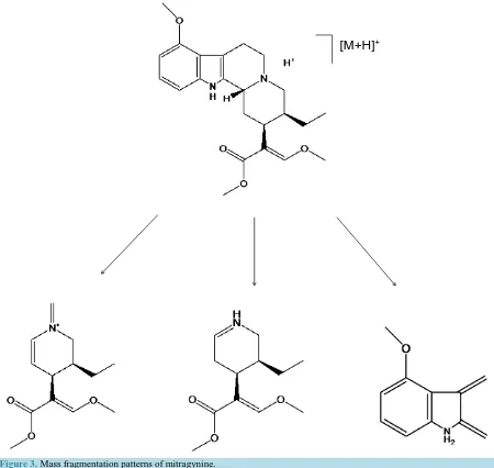
With the assistance of direct infusion electrospray ionization mass spectrometry technique, mass spectra and in-formation such as molecular weight and mass fragment were obtained. Possible cleavage and metabolites me-chanisms are proposed as Figure 3 for mitragynine and Figure 4 for 7-hydroxymitragynine. As indicated by the mass spectrum parameters in method development, the transition 174.3 is the major fragment of protonated mi-tragynine compound under instrument conditions stated in Table 1 and Table 2.
As illustrated in Figure 3, protonated mitragynine with m/z 399.5 has fragment of m/z 174.3, 226.1 and 238.2. Figure 4 demonstrated the major fragment of protonated 7-hydroxymitragynine m/z 415.5 is m/z 190.2.
3.4. Method Validation
The analytical figures of merit were assessed and presented in Table 3. Linearity in terms of slope and R- squared value was determined. Sensitivity including LOD and LOQ, intraday precision and interday precision were calculated as well. Limit of detection down to 0.0123 ng/mL for mitragynine and 0.0691 ng/mL for 7-hydroxymitragynine were achieved. The LOD values show that the established method is very sensitive for trace amount analysis.
0.2 0.4 0.6 0.8 1.0 1.2 1.4 1.6 1.8 2.0 2.2 2.4 2.6 2.8 3.0 3.2 3.4
Retention Time (min)
Ab
un
da
nc
e
6.0e5 1.0e4 Mitragynine [M+H] m/z399.5 7-hydroxymitragynine [M+H] m/z415.5Figure 2. Extracted chromatograms of mitragynine and 7-hydroxymotragyninewith 0.1% formic acid and acetonitrile as mobile phases.
Table 1. Optimized MS/MS operating conditions for mitragynine and 7-hydroxymigragynine obtained from tandem mass spectrometry.
MS/MS conditions Mitragynine 7-hydroxymitragynine
Polarity Positive Positive
Ionspray voltage 4000 V 4000 V
Temperature 550˚C 550˚C
Collision gas Medium Medium
Ion source gas 1 50.0 55.0
[M+H]
+Figure 3. Mass fragmentation patterns of mitragynine.
Table 2. Multiple reaction monitoring (MRM) parameters of mitragynine and 7-hydroxymitragynine (analytes) and mitra-
gynine-D3 and 7-hydroxymitragynine-D3 (internal standards).
MS/MS conditions MG 7-OH-MG MG-D3 7-OH-MG-D3
Precursor ion (m/z) 399.5 415.5 402.5 418.6
Product ion (m/z) 174.3 190.2 238.3 193.2
Collision energy (eV) 45 45 35 45
Table 3. Analytical figures of merit of LC-MS/MS results.
Analytes
Linearity Sensitivity Precision (intraday, n = 5)
Precision (inter-day, 3d/n = 6)
Slope R2 LOD (ng/mL) LOQ (ng/mL) RSD% RSD%
Mitragynine 92.5 0.9987 0.0123 0.0356 1.55 1.70
[image:5.595.87.537.83.509.2][M+H]
+Figure 4. fragmentation patterns of 7-hydroxymitragy-
nine.
4. Concluding Remarks
In this study, a specific detection and identification method by direct infusion electrospray ionization mass spec-trometry has been developed for analysis of mitragynine and 7-hydroxymitragynine. The method demonstrates a rapid and precise route for the detection and identification in pharmaceutical and biomedical applications. This approach provides capability of mass spectrum and mass fragments information, which facilitates the pharma-cokinetic study of mitragynine and 7-hydroxymitragynine compounds.
References
[1] Ward, J., Rosenbaum, C., Hernon, C., McCurdy, C. and Boyer, E. (2011) Herbal Medicines for the Management of Opioid Addiction. CNS Drugs, 25, 999-1007. http://dx.doi.org/10.2165/11596830-000000000-00000
[2] Suwanlert, S. (1975) A Study of Kratom Eaters in Thailand. Bulletin on Narcotics.
[3] Jansen, K.L.R. and Prast, C.J. (1988) Psychoactive Properties of Mitragynine (Kratom). Journal of Psychoactive Drugs,
20, 455-457. http://dx.doi.org/10.1080/02791072.1988.10472519
[image:6.595.200.401.84.504.2]Antinoci-ceptive Action of Mitragynine in Mice: Evidence for the Involvement of Supraspinal Opioid Receptors. Life Sciences,
59, 1149-1155. http://dx.doi.org/10.1016/0024-3205(96)00432-8
[5] Vicknasingam, B., Narayanan, S., Beng, G.T. and Mansor, S.M. (2010) The Informal Use of Ketum (Mitragyna speci-osa) for Opioid Withdrawal in the Northern States of Peninsular Malaysia and Implications for Drug Substitution Therapy. International Journal of Drug Policy, 21, 283-288. http://dx.doi.org/10.1016/j.drugpo.2009.12.003
[6] Boyer, E.W., Babu, K.M., Macalino, G.E. and Compton, W. (2007) Self-Treatment of Opioid Withdrawal with a Die-tary Supplement, Kratom. The American Journal on Addictions, 16, 352-356.
http://dx.doi.org/10.1080/10550490701525368
[7] Phongprueksapattana, S., Putalun, W., Keawpradub, N. and Wungsintaweekul, J. (2008) Mitragyna speciosa: Hairy Root Culture for Triterpenoid Production and High Yield of Mitragynine by Regenerated Plants. Zeitschrift für
Natur-forschung C: A Journal of Biosciences, 63, 691. http://dx.doi.org/10.1515/znc-2008-9-1014
[8] Zhao, C., Arroyo-Mora, L.E., DeCaprio, A.P., Sharma, V.K., Dionysiou, D.D. and O’Shea, K.E. (2014) Reductive and Oxidative Degradation of Iopamidol, Iodinated X-Ray Contrast Media, by Fe(III)-Oxalate under UV and Visible Light Treatment. Water Research, 67, 144-153. http://dx.doi.org/10.1016/j.watres.2014.09.009
[9] Babu, K.M., McCurdy, C.R. and Boyer, E.W. (2008) Opioid Receptors and Legal Highs: Salvia divinorum and Kratom.
Clinical Toxicology, 46, 146-152. http://dx.doi.org/10.1080/15563650701241795
[10] Philipp, A.A., Wissenbach, D.K., Zoerntlein, S.W., Klein, O.N., Kanogsunthornrat, J. and Maurer, H.H. (2009) Studies on the Metabolism of Mitragynine, the Main Alkaloid of the Herbal Drug Kratom, in Rat and Human Urine Using Liq-uid Chromatography-Linear Ion Trap Mass Spectrometry. Journal of Mass Spectrometry, 44, 1249-1261.
http://dx.doi.org/10.1002/jms.1607
[11] Kong, W.M., Chik, Z., Ramachandra, M., Subramaniam, U., Aziddin, R.E.R. and Mohamed, Z. (2011) Evaluation of the Effects of Mitragyna speciosa Alkaloid Extract on Cytochrome P450 Enzymes Using a High Throughput Assay.
Molecules, 16, 7344-7356. http://dx.doi.org/10.3390/molecules16097344
[12] Chittrakarn, S., Keawpradub, N., Sawangjaroen, K., Kansenalak, S. and Janchawee, B. (2010) The Neuromuscular Blockade Produced by Pure Alkaloid, Mitragynine and Methanol Extract of Kratom Leaves (Mitragyna speciosa
Korth.). Journal of Ethnopharmacology, 129, 344-349. http://dx.doi.org/10.1016/j.jep.2010.03.035
[13] Chen, L., Zhao, C., Dionysiou, D.D. and O’Shea, K.E. (2015) TiO2 Photocatalytic Degradation and Detoxification of Cylindrospermopsin. Journal of Photochemistry and Photobiology A: Chemistry, 307-308, 115-122.
http://dx.doi.org/10.1016/j.jphotochem.2015.03.013
[14] Zhao, C., Pelaez, M., Dionysiou, D.D., Pillai, S.C., Byrne, J.A. and O’Shea, K.E. (2014) UV and Visible Light Acti-vated TiO2 Photocatalysis of 6-Hydroxymethyl Uracil, a Model Compound for the Potent Cyanotoxin Cylindrosper-mopsin. Catalysis Today, 224, 70-76. http://dx.doi.org/10.1016/j.cattod.2013.09.042
[15] Liu, S., Zhao, Y., Ma, F., Ma, L., O’Shea, K., Zhao, C., Hu, X. and Wu, M. (2015) Control of Microcystis aeruginosa
Growth and Associated Microcystin Cyanotoxin Remediation by Electron Beam Irradiation (EBI). RSC Advances, 5, 31292-31297. http://dx.doi.org/10.1039/C5RA00430F
[16] Merola, G., Fu, H., Tagliaro, F., Macchia, T. and McCord, B.R. (2014) Chiral Separation of 12 Cathinone Analogs by Cyclodextrin-Assisted Capillary Electrophoresis with UV and Mass Spectrometry Detection. Electrophoresis, 35, 3231-3241. http://dx.doi.org/10.1002/elps.201400077
[17] Merola, G., Fu, H., Tagliaro, F., Macchia, T. and McCord, B. (2014) Chiral Separation of 12 Cathinone Analogs by Cyclodextrin-Assisted Capillary Electrophoresis Time-of-Flight Mass Spectrometry. 41st International Symposium and
Exhibit on High Performance Liquid Phase Separations and Related Techniques (HPLC 2014), New Orleans.
[18] Zheng, B., Fu, H., Berry, J.P. and McCord, B. (2016) A Rapid Method for Separation and Identification of Microcys-tins Using Capillary Electrophoresis and Time-of-Flight Mass Spectrometry. Journal of Chromatography A, 1431, 205-214. http://dx.doi.org/10.1016/j.chroma.2015.11.034
[19] Yang, D., Li, Z., Diwu, Y.A., Fu, H., Liao, J., Wei, C. and Diwu, Z. (2008) A Novel Fluorogenic Coumarin Substrate for Monitoring Acid Phosphatase Activity at Low pH Environment. Current Chemical Genomics, 2, 48.
http://dx.doi.org/10.2174/1875397300802010048
[20] Fan, M., Yang, D., Wang, X., Liu, W. and Fu, H. (2014) DOSS-Based QAILs: As Both Neat Lubricants and Lubricant Additives with Excellent Tribological Properties and Good Detergency. Industrial & Engineering Chemistry Research,
53, 17952-17960. http://dx.doi.org/10.1021/ie502849w
[21] Fu, H., Cid, F.X., Dworkin, N., Cocores, J. and Shore, G. (2015) Screening and Identification of Mitragynine and 7- Hydroxymitragynine in Human Urine by LC-MS/MS. Chromatography, 2, 253-264.


