CSF-1–dependant donor-derived macrophages mediate chronic
graft-versus-host disease
Kylie A. Alexander, … , Geoffrey R. Hill, Kelli P.A. MacDonald
J Clin Invest. 2014;124(10):4266-4280. https://doi.org/10.1172/JCI75935.
Chronic GVHD (cGVHD) is the major cause of late, nonrelapse death following stem cell transplantation and characteristically develops in organs such as skin and lung. Here, we used multiple murine models of cGVHD to investigate the contribution of macrophage populations in the development of cGVHD. Using an established IL-17– dependent sclerodermatous cGVHD model, we confirmed that macrophages infiltrating the skin are derived from donor
bone marrow (F4/80+CSF-1R+CD206+iNOS–). Cutaneous cGVHD developed in a CSF-1/CSF-1R–dependent manner,
as treatment of recipients after transplantation with CSF-1 exacerbated macrophage infiltration and cutaneous pathology. Additionally, recipients of grafts from Csf1r–/– mice had substantially less macrophage infiltration and cutaneous pathology as compared with those receiving wild-type grafts. Neither CCL2/CCR2 nor GM-CSF/GM-CSFR signaling pathways were required for macrophage infiltration or development of cGVHD. In a different cGVHD model, in which bronchiolitis
obliterans is a prominent manifestation, F4/80+ macrophage infiltration was similarly noted in the lungs of recipients after transplantation, and lung cGVHD was also IL-17 and CSF-1/CSF-1R dependent. Importantly, depletion of macrophages using an anti–CSF-1R mAb markedly reduced cutaneous and pulmonary cGVHD. Taken together, these data indicate that donor macrophages mediate the development of cGVHD and suggest that targeting CSF-1 signaling after transplantation may prevent and treat cGVHD.
Research Article Immunology
Find the latest version:
Introduction
Graft-versus-host disease (GVHD) remains a major complication of allogeneic stem cell transplantation (SCT). GVHD can present in acute and chronic forms, which differ in their time of onset and symptoms. Acute GVHD (aGVHD) occurs early after trans-plantation, with target organ damage characterized by apopto-sis. In contrast, chronic GVHD (cGVHD) is a late complication of SCT and is characterized by fibrosis. Indeed, cGVHD presents with many features that overlap with certain autoimmune dis-eases. While skin is the primary organ involved in cGVHD, both lung and liver fibrosis can also occur, and these manifestations are all associated with significant morbidity and mortality. Our understanding of the pathophysiology of aGVHD is far more advanced than that of cGVHD, and it is clear that these forms are mediated by different immunological subsets and cytokine networks. Currently, the majority of clinical allogeneic
trans-plants use granulocyte colony–stimulating factor–mobilized (G-CSF–mobilized) peripheral blood (PB) stem cells, which has led to rapid hematopoietic reconstitution, improved leukemic eradication, and similar levels of aGVHD, but increased levels of cGVHD (1). Therapeutically, there are limited options available for the treatment of cGVHD, with corticosteroids representing the mainstay of treatment. Unfortunately, this is often ineffec-tive and is associated with significant morbidity, thus cGVHD represents an increasing burden in the clinic.
Macrophages play an essential role in both homeostasis and pathology and are characterized by high functional heterogeneity (2). The differentiation, proliferation, and survival of the majority of macrophage populations are dependent on colony-stimulating factor 1 (CSF-1) (3, 4). Macrophages acquire diverse phenotypic and functional forms in response to local cytokines and microbial sig-nals, which has resulted in the “M1” and “M2” macrophage clas-sifications (5, 6). Classically activated macrophages (M1) mediate defense of the host from a variety of bacteria, protozoa, and viruses and have roles in antitumor immunity. Activation of proinflamma-tory M1 macrophages is typically induced by IFN-γ, lipopolysaccha-ride, and cytokines such as granulocyte-macrophage colony-stimu-lating factor (GM-CSF) (5–7). Alternatively activated macrophages (M2) have antiinflammatory functions and regulate tissue repair and remodeling. M2 macrophage activation is induced by IL-4 and IL-13 (8) as well as immune complexes, glucocorticoids, and the
Chronic GVHD (cGVHD) is the major cause of late, nonrelapse death following stem cell transplantation and characteristically develops in organs such as skin and lung. Here, we used multiple murine models of cGVHD to investigate the contribution of macrophage populations in the development of cGVHD. Using an established IL-17–dependent sclerodermatous cGVHD model, we confirmed that macrophages infiltrating the skin are derived from donor bone marrow (F4/80+CSF-1R+CD206+iNOS–).
Cutaneous cGVHD developed in a CSF-1/CSF-1R–dependent manner, as treatment of recipients after transplantation with CSF-1 exacerbated macrophage infiltration and cutaneous pathology. Additionally, recipients of grafts from Csf1r–/– mice
had substantially less macrophage infiltration and cutaneous pathology as compared with those receiving wild-type grafts. Neither CCL2/CCR2 nor GM-CSF/GM-CSFR signaling pathways were required for macrophage infiltration or development of cGVHD. In a different cGVHD model, in which bronchiolitis obliterans is a prominent manifestation, F4/80+ macrophage
infiltration was similarly noted in the lungs of recipients after transplantation, and lung cGVHD was also IL-17 and CSF-1/ CSF-1R dependent. Importantly, depletion of macrophages using an anti–CSF-1R mAb markedly reduced cutaneous and pulmonary cGVHD. Taken together, these data indicate that donor macrophages mediate the development of cGVHD and suggest that targeting CSF-1 signaling after transplantation may prevent and treat cGVHD.
CSF-1–dependant donor-derived macrophages mediate
chronic graft-versus-host disease
Kylie A. Alexander,1 Ryan Flynn,2 Katie E. Lineburg,1 Rachel D. Kuns,1 Bianca E. Teal,1 Stuart D. Olver,1 Mary Lor,1 Neil C. Raffelt,1
Motoko Koyama,1 Lucie Leveque,1 Laetitia Le Texier,1 Michelle Melino,1 Kate A. Markey,1 Antiopi Varelias,1 Christian Engwerda,1
Jonathan S. Serody,3 Baptiste Janela,4 Florent Ginhoux,4 Andrew D. Clouston,5 Bruce R. Blazar,2 Geoffrey R. Hill,1,6
and Kelli P.A. MacDonald1
1QIMR Berghofer Medical Research Institute, Brisbane, Australia. 2Pediatric Blood and Marrow Transplantation Program, University of Minnesota, Minneapolis, Minnesota, USA. 3Lineberger Comprehensive
Cancer Center, University of North Carolina School of Medicine, Chapel Hill, North Carolina, USA. 4Singapore Immunology Network (SIgN), A*STAR (Agency for Science, Technology and Research), Biopolis,
Singapore. 5Envoi Pathology, Brisbane, Australia. 6Department of Bone Marrow Transplantation, Royal Brisbane Hospital, Brisbane, Australia.
Note regarding evaluation of this manuscript: Manuscripts authored by scientists
associated with Duke University, The University of North Carolina at Chapel Hill, Duke-NUS, and the Sanford-Burnham Medical Research Institute are handled not by members of the editorial board but rather by the science editors, who consult with selected external editors and reviewers.
Conflict of interest: The authors have declared that no conflict of interest exists. Submitted: March 4, 2014; Accepted: July 10, 2014.
The Journal of Clinical Investigation
R e s e a R c h a R t i c l eopment of cGVHD, and this was therefore the focus of this study.
Results
F4/80+ macrophages infiltrate
the skin of mice receiving either G-CSF–mobilized or –nonmobilized grafts. We investigated the kinet-ics of macrophage infiltration into the skin after transplantation in lethally irradiated mice receiving either allogeneic, G-CSF–mobilized
(G-CSF BALB/c graft → C57Bl/6
[B6] recipients; Figure 1, A–D), or semiallogeneic, nonmobilized grafts (B6 bone marrow [BM] plus
[image:3.585.42.397.58.247.2]T cell graft → B6D2F1 recipients;
Figure 1, E–H) that develop sclero-dermatous cGVHD. IHC analysis for F4/80 expression demonstrated
that F4/80+ macrophages
infil-trated the dermis of mice receiving G-CSF–mobilized grafts starting 7 days after transplantation (Figure 1,
A and B), with robust F4/80+
mac-rophage infiltration present by day 21 after transplantation throughout the dermis and s.c. fat layers (Fig-ure 1C). We noted minimal macrophage infiltration in mice that received G-CSF–mobilized T cell–depleted (TCD) grafts (Figure
1D). In mice receiving nonmobilized grafts, we noted F4/80+
mac-rophage infiltration in the dermis by day 21 after transplantation
(Figure 1E), and by day 28 after transplantation, F4/80+
macro-phages were predominantly located within the s.c. fat layer (Figure 1, F and G), with minimal macrophage infiltration noted in mice that received TCD grafts (Figure 1H). Although not examined here, our initial studies also identified neutrophils and T cells within the mononuclear infiltrate in skin during GVHD (9), however, they were far less dominant than the F4/80+ macrophage infiltrate
illus-trated in Figure 1, and only the macrophages were IL-17 dependent. Skin-infiltrating macrophages are donor M2–like CSF-1R– expressing macrophages. To examine the phenotype of these
F4/80+ macrophages, IHC was performed using markers for both
classically activated and alternatively activated macrophages (inducible nitric oxide synthase [iNOS] and mannose receptor CD206, respectively). Additionally, IHC for the congenic marker CD45.1 was used to mark donor cells. IHC in serial sections was performed 21 days after transplantation in lethally irradiated mice receiving either G-CSF–mobilized non-TCD grafts (cGVHD mice) or G-CSF–mobilized TCD grafts (no cGVHD controls). Results confirmed that the vast majority of infiltrating F4/80+ cells in mice
receiving G-CSF–mobilized grafts were donor (CD45.1+)
alter-natively activated (CD206+) macrophages, with minimal iNOS
expression (F4/80+CD45.1+CD206+iNOS–) (Figure 2A and
Sup-plemental Figure 1A; supSup-plemental material available online with this article; doi:10.1172/JCI75935DS1). We noted minimal donor macrophage infiltration in mice that received G-CSF–mobilized cytokine CSF-1 (6, 7). Although the M1 and M2 classification
sys-tem is widely used, it is increasingly clear that these macrophage populations represent the extreme ends of a wide spectrum of phe-notypes associated with macrophage activation.
Recent preclinical and clinical data from our group estab-lished a highly reproducible and informative model of cGVHD that supports a role for IL-17 as a central mediator of pathology, particularly within the skin (9). Cutaneous cGVHD was shown to be exacerbated by G-CSF mobilization in an IL-17–dependent manner, and late after transplantation, scleroderma was absent in recipients of Il17a–/– grafts compared with wild-type (WT) grafts.
Interestingly, IL-17A controlled the infiltration of F4/80+
macro-phages into skin, and this preceded the development of sclero-derma, suggesting that macrophages play important profibrotic roles in the development of cutaneous cGVHD.
There is an expanding body of evidence suggesting that mac-rophages can play both anti- (10, 11) and profibrotic roles (12, 13). To date, models of macrophage depletion in vivo have been unsat-isfactory, inducing rapid cell death and inflammation that compli-cate experimental conclusions. Nevertheless, studies in renal (13), hepatic (10), lung (12), and skin fibrosis (14, 15) suggest that macro-phage depletion can modify fibrosis. Cutaneous macromacro-phage infil-tration has been suggested to be associated with refractory aGVHD and poor prognosis (16). Alternatively, activated macrophages
(CD163+) have been previously shown to be present in both humans
(17) and rats with systemic sclerosis (18). Additionally, macrophages displaying an M2 phenotype are present in the skin (19) and lungs of patients with systemic sclerosis (20). However, there are no studies to date that demonstrate a causal role of macrophages in the
ated PB monocyte populations in
naive B6.WT, B6.Ccr2–/–, and B6
common β chain–/– mice to
exam-ine the requirement of chemokexam-ine (C-C motif) ligand 2 (CCL2) and GM-CSF, respectively. To exam-ine the requirement of CSF-1, we used fetal liver chimeras (FLCs) on a B6 background, since CSF-1R deficiency is neonatally lethal. We examined monocyte populations using a gating strategy (Figure
3A) that removed Ly6G+CD3+ and
high side-scatter cells (SSCs). As expected, compared with B6.WT mice, we observed a significant reduction in the frequency and absolute numbers of circulating
CD11b+F4/80+Ly6Chi classical
monocytes in the Ccr2–/– mice
(Figure 3, B and C; **P = 0.004, **P = 0.0081). In contrast, com-pared with B6.WT FLCs, we found that the B6 Csf1r–/– FLCs exhibited
a significant reduction in Ly6Clo
subset frequencies and abso-lute numbers (Figure 3, B and C; ***P = 0.002, *P = 0.0286). The contribution of GM-CSF to cir-culating monocyte populations was minimal, since the frequencies and absolute numbers of both Ly6Chi and Ly6Clo cell populations were not significantly altered in
the common β chain–deficient mice.
CSF-1 treatment after SCT exacerbates cutaneous cGVHD. We had previously confirmed in Figure 3 that CSF-1 was required for
the development of the Ly6Clo PB monocyte/macrophage
popu-lation, which is suggested to be a tissue macrophage precursor. Therefore, we further examined the contribution of CSF-1 to mac-rophage infiltration and cutaneous cGVHD after transplantation. Lethally irradiated B6 mice received G-CSF–mobilized BALB/c whole spleen or TCD spleen and were treated with either saline or CSF-1 for 5 days from day 14 after transplantation. Compared with saline-treated recipients, we found that CSF-1 treatment
significantly increased F4/80+ macrophages in the skin 19 days
after transplantation (Figure 4A), quantified as positive pixels/
mm2 (Figure 4B; **P = 0.0017). CSF-1 treatment preferentially
expanded donor alternatively activated macrophages, as
con-firmed via IHC (F4/80+CD45.1+CD206+iNOS–) (Figure 4C).
Semi-quantitative histopathology confirmed that recipients treated with CSF-1 had significantly higher cutaneous pathology scores than did those treated with saline (Figure 4D; **P = 0.0087).
Further analysis of PB monocyte populations 19 days after transplantation illustrated a trend toward an increase in the
fre-quency of Ly6Clo cells after 5 days of CSF-1 treatment (Figure
5A). Therefore, we carried out further monocyte/macrophage analysis of cells isolated 19 days after transplantation from the skin (ears) of mice treated with either saline or CSF-1 (gating TCD grafts. We also noted alternatively activated macrophage
infiltrate in lethally irradiated mice that received nonmobilized grafts (data not shown). B6D2F1 mice receiving BM plus T cell grafts from MacGreen mice (MacGreen mice have EGFP driven
off the Csf1r promoter) (21) confirmed that these F4/80+
macro-phages are also CSF-1R+ (Figure 2B). Using the same
transplanta-tion system, we additransplanta-tionally confirmed that donor (CSF-1R+) cells
infiltrate the skin as early as 7 days after transplantation (Supple-mental Figure 2A), and by day 21, the vast majority of F4/80+ cells
are of donor origin (Supplemental Figure 2B).
Cytokine/chemokine dependency of blood monocyte populations in naive mice. Current dogma holds that circulating monocytes can give rise to a variety of tissue-resident macrophages throughout the body, particularly during inflammation. In the steady state, circulat-ing CD11b+F4/80+ monocytes can be divided into 2 distinct cell
pop-ulations based on the expression of Ly6C (22). CD11b+F4/80+Ly6Chi
monocytes express the C-C chemokine receptor type 2 (CCR2) and are commonly referred to as classical or inflammatory
mono-cytes. In contrast, CD11b+F4/80+Ly6Clo monocytes are CCR2–,
but express the fractalkine receptor CX3CR1 and are commonly referred to as patrolling monocytes, which are widely considered to be tissue-resident macrophage precursors. To elucidate the mono-cyte precursors and cytokine dependency of the donor infiltrating macrophages following BM transplantation (BMT), we next exam-ined the cytokine and chemokine requirements for the development and migration of the distinct PB monocyte populations. For these studies, we examined both frequency (percentage) and
[image:4.585.36.403.57.289.2]The Journal of Clinical Investigation
R e s e a R c h a R t i c l eCsf1r–/– grafts had a significantly lower clinical GVHD score
through-out the time course examined compared with that of WT graft recipients (Figure 6A). IHC performed 21 days after transplanta-tion illustrated that mice receiving WT grafts (Figure 6B) had
sig-nificantly higher F4/80+ macrophage infiltration compared with
that seen in mice receiving Csf1r–/– grafts (Figure 6B), quantified as
positive pixels/mm2 (Figure 6C; **P = 0.003). Masson’s trichrome
staining (collagen fibers are stained blue) and semiquantitative his-topathology scores assessed 48 days after transplantation confirmed that mice receiving Csf1r–/– grafts had significantly lower cutaneous
pathology and fibrosis than did those receiving WT grafts (Fig-ure 6D; *P = 0.045, **P = 0.008), confirming a pathogenic role for CSF-1/CSF-1R signaling in the development of cutaneous pathol-ogy after transplantation. Analysis of PB monocyte populations 21 days after transplantation illustrated that recipients of Csf1r–/– FLC
grafts exhibited a significant increase in the frequency of Ly6Chi cells
and a significant reduction in the Ly6Clo cell population (Figure 6E;
*P = 0.0286). Other CSF-1R–dependent cells reside within the skin apart from macrophages, most notably langerin-expressing Langer-hans cells, which reside in the epidermis. In contrast to macrophages, strategy for monocyte/macrophage analysis; Figure 5B). We
per-formed analysis using a gating strategy similar to that shown in Figure 3A, with monocytes (Figure 5B, Mono) being classified as Ly6CloSSC loF4/80+CD11b+ and macrophages (Figure 5B, Mɸ) as
Ly6CloSSChiF4/80+. Data are expressed as the absolute number
of CD45+ cells for both monocytes and macrophages (Figure 5C).
We observed a significant increase in absolute numbers in both the
CD45+ monocyte population (Ly6CloCD11b+F4/80+) (Figure 5C;
**P = 0.0087) and CD45+ macrophage population (Ly6CloF4/80+)
(Figure 5C; *P = 0.026) in the CSF-1–treated mice compared with the absolute numbers detected in saline-treated mice. Overall, this confirmed that CSF-1 treatment after transplantation
signifi-cantly increased cutaneous Ly6Clo monocytes and macrophages
compared with those detected in saline-treated recipients. F4/80+ macrophage infiltration and cutaneous fibrosis after
trans-plantation is CSF-1R dependent. We subsequently investigated the requirement for donor CSF-1R signaling in the development of
cutaneous cGVHD using WT and Csf1r–/– FLCs. Lethally irradiated
B6D2F1 recipients were given BM plus T cell grafts from either B6
[image:5.585.62.535.56.400.2]WT FLC, Csf1r–/– FLC, or WT FLC TCD grafts. Mice that received
tion of GM-CSF signaling to macrophage infiltration and cutaneous GVHD. Serum GM-CSF was only detected 7 days after trans-plantation (Figure 7A; *P = 0.01), with no differ-ence in serum GM-CSF levels between mice receiving either allogeneic or TCD grafts from day 14 after transplantation.
F4/80+ macrophages were
still noted 21 days after transplantation in the skin of lethally irradiated mice that received WT or
com-mon β chain–/– BM plus
T cell grafts (Figure 7B).
The common β chain–/–
mouse has defective sig-naling for IL-3 and IL-5 as well as for GM-CSFR signaling (24), therefore, we specifically blocked GM-CSFR signaling with an anti–GM-CSF Ab (M250 mAb; Amgen). Treatment with M250 starting 7 days after trans-plantation had no effect
on F4/80+ macrophage
infiltration (Figure 7C), quantified as positive
pixels/mm2 (Figure 7D).
PB monocyte analysis 21 days after transplantation showed no change in the frequencies of Ly6Chi and
Ly6Clo cell populations in mice receiving either B6 WT or B6
com-mon B chain–/– grafts (Figure 7E). Thus, GM-CSF is not required
for the infiltration of F4/80+ macrophages and the subsequent
development of cutaneous pathology after transplantation. Development of cutaneous pathology is CCR2 independent. Monocyte chemoattractant protein 1 (MCP-1/CCL2), a multi-functional chemokine belonging to the C–C chemokine super-family, has been shown to be upregulated in a variety of fibrotic conditions (25–27). Furthermore, patients with scleroderma exhibit elevated levels of CCL2 in both serum and skin (28, 29). Similarly, we found elevated levels of CCL2 in the serum from 7 to 28 days after transplantation in lethally irradiated mice receiving BM plus T cell grafts (Allo) compared with levels detected in TCD non-GVHD controls (Figure 8A). To investigate the contribution of donor CCL2/CCR2 signaling to the development of cutaneous GVHD, lethally irradiated mice received BM from B6 WT or B6
Ccr2–/– mice with B6 WT T cells. IHC demonstrated that F4/80+
macrophages were still present in the skin 28 days after transplan-however, Langerhans cells are instructed by the alternative CSF-1R
ligand IL-34 (23). To investigate the contribution of Langerhans cells to the development of CSF-1R–dependent sclerodermatous cGVHD, mice received BM and T cell grafts from langerin-diphtheria toxin receptor–transgenic (langerin-DTR–transgenic) mice in which DTR and EGFP are expressed under the control of the Cd207 (encoding langerin) promoter. Administration of DT between 7 and 40 days after transplantation effectively depleted donor Langerhans cells
(Supplemental Figure 3A), but neither F4/80+ macrophage
infiltra-tion nor the development of cutaneous pathology was altered (Sup-plemental Figure 3, B and C), thus establishing that Langerhans cells do not contribute to cutaneous cGVHD.
F4/80+ macrophage infiltration and cutaneous fibrosis after
transplantation is GM-CSFR independent. Both CSF-1/CSF-1R and GM-CSF/GM-CSFR signaling pathways are involved in regulating the number and function of macrophage lineage populations, and both signaling pathways have been shown to contribute to mac-rophage heterogeneity (7). Therefore, we examined the
[image:6.585.38.429.57.391.2]The Journal of Clinical Investigation
R e s e a R c h a R t i c l etation in mice receiving either B6 WT or B6 Ccr2–/– grafts
(Fig-ure 8B), quantified as positive pixels/mm2 (Figure 8C). We also
observed no difference in cutaneous pathology in the recipients of
WT or Ccr2–/– grafts (Figure 8, D and E). Similarly, Ab-mediated
blockade using an anti-CCL2 Ab had no significant effect on
F4/80+ macrophage infiltration (Figure 8, F and G) or subsequent
cutaneous pathology (Figure 8H). PB monocyte analysis 21 days after transplantation illustrated no change in the frequencies of Ly6Chi or Ly6Clo cell populations in mice receiving either B6 WT
or B6 Ccr2–/– grafts (Figure 8I). Thus, CCL2/CCR2 signaling is not
required for the infiltration of F4/80+ macrophages and the
subse-quent development of cutaneous pathology after transplantation. This suggests that the chemokine CCL2 is redundant for mono-cyte recruitment in the context of the GVHD inflammatory milieu. Anti–CSF-1R Ab treatment after transplantation attenuates cutaneous GVHD. To investigate whether depletion of the CSF-1/
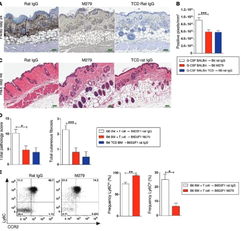
CSF-1R–dependent macrophage population after transplantation was an effective therapeutic strategy to prevent and treat cGVHD, we used an anti–CSF-1R Ab (M279; Amgen). We have previously demonstrated that M279 specifically depletes tissue-resident macrophages (30). Lethally irradiated B6 recipients of G-CSF– mobilized BALB/c grafts were treated with either rat IgG control or M279 mAb beginning 7 days after transplantation. IHC staining demonstrated that M279 mAb treatment significantly depleted
F4/80+ macrophages in the skin compared with levels detected
rat IgG–treated control mice (Figure 9A), quantified as positive
pixels/mm2 (Figure 9B; ***P = 0.002). Long-term depletion (out
to day 48) of F4/80+ macrophages in B6D2F1 mice that received
B6 BM and T cells or TCD grafts also confirmed that M279 mAb treatment significantly reduced cutaneous GVHD (Figure 9, C and D; *P = 0.012), and similar to mice receiving Csf1r–/– grafts (Figure
[image:7.585.50.535.59.429.2]6D), M279-treated mice showed no evidence of cutaneous fibrosis
(Figure 9D; ***P = 0.0006). PB analysis on day 48 after transplan-tation also confirmed that M279 mAb treatment resulted in a
sig-nificant increase in the frequency of Ly6Chi monocytes and a
sig-nificant decrease in Ly6Clo monocytes (Figure 9E; **P = 0.0012,
*P = 0.0262). Thus, CSF-1R Ab–mediated depletion of donor mac-rophages after transplantation may be an effective therapeutic strategy to prevent and/or treat cutaneous cGVHD.
Development of cutaneous GVHD is TGF-β dependent. We have
previously reported that in the B10.BR → BALB/c model of
sclero-derma, skin-infiltrating CD11b+ mononuclear cells produce high
[image:8.585.44.533.58.507.2]lev-els of TGF-β, and neutralization of this cytokine attenuated cGVHD (31). We therefore speculated that TGF-β is a mediator of fibrotic skin pathology invoked by macrophages in the current study. Thus, we examined the expression of TGF-β in the CD11b+F4/80+Ly6Clo Figure 6. F4/80+ macrophage infiltration and cutaneous fibrosis following BMT are CSF-1/CSF-1R dependent. Lethally irradiated B6D2F1 mice received BM plus T cells from WT FLCs or Csf1r–/– FLCs (KO FLCs). (A) Mice that received KO FLC grafts had significantly lower GVHD clinical scores (day 7, ***P≤ 0.0001;
The Journal of Clinical Investigation
R e s e a R c h a R t i c l eDevelopment of lung GVHD is IL-17 and CSF-1R dependent. Both
transplantation models used above (G-CSF–mobilized BALB/c →
B6 and B6 BM plus T cell → B6D2F1) resulted in the development of
scleroderma after transplantation, but neither model elicited fibrotic manifestations in other cGVHD target organs. Therefore, we next investigated the contribution of CSF-1/CSF-1R–dependent macro-phages to cGVHD using the recently described multiorgan system
cGVHD model (B6 → B10.BR) (32), in which lung injury is
associ-ated with obstructive lung disease and fibrosis, with increased col-lagen deposition surrounding bronchioles (bronchiolitis obliterans PB monocyte precursor population after transplantation. Our results
confirmed that the PB monocyte precursors to the pathogenic tissue
macrophages rapidly produced TGF-β in response to TLR4 ligation
[image:9.585.45.543.56.486.2]with LPS (Supplemental Figure 4A). Furthermore, Ab blockade of TGF-β from days 14 to 46 after transplantation significantly reduced GVHD clinical scores and, importantly, significantly decreased cutaneous pathology compared with the control mAb–treated group (Supplemental Figure 4, B and C). Taken together, the data suggest that donor CSF-1R–dependent macrophages contribute to cGVHD via the expression of TGF-β.
plus splenocyte (Sp) grafts that developed cGVHD also had elevated airway resistance, which correlated with lung constriction (Resis-tance), elevated elastance, which signifies increased stiffness or rigidity of the lungs (Elastance), and decreased total lung capacity [BO]) but without scleroderma manifestations. BO is characterized
[image:10.585.62.526.42.547.2]by airway blockage, peribronchiolar fibroproliferation, and oblitera-tion of bronchioles and is a late-stage complicaoblitera-tion of GVHD. Pul-monary function tests (PFTs) demonstrated that recipients of BM
Figure 8. F4/80+ macrophage infiltration and cutaneous fibrosis post BMT is CCL2/CCR2 independent. (A) Serum CCL2 (MCP-1; pg/ml) in lethally irradiated B6D2F1 mice that received B6 BM plus T cell (Allo) or TCD grafts 7, 14, 21, and 28 days after transplantation (days 7 and 14, *P = 0.015; day 21, *P = 0.035; day 28, **P = 0.009). n = 4–6/group. (B) Lethally irradiated B6D2F1 mice received BM plus T cell grafts from B6 or Ccr2–/– donors. IHC to detect F4/80 expression
The Journal of Clinical Investigation
R e s e a R c h a R t i c l eand that effects may be mediated by T cells, innate lymphoid cells, or another as-yet uncharacterized cytokine-producing cell population. Development of lung pathology was also confirmed to be CSF-1R dependent, as recipients of Csf1r–/– grafts had significantly better
pul-monary function compared with that of mice receiving WT grafts or those receiving WT BM with Csf1r–/– splenocytes (Figure 10B).
Recip-ients of Csf1r–/– BM plus splenocyte grafts also displayed a significant
and compliance (Compliance) (32). As with cutaneous GVHD (9), lung GVHD developed in an IL-17–dependent manner, since mice receiving grafts deficient in the retinoid-related orphan receptor γ
(Rorc–/–), which is required for lineage commitment in
[image:11.585.58.538.54.514.2]IL-17–produc-ing cells, exhibited significantly better pulmonary function relative to that of mice receiving WT grafts (Figure 10A). We acknowledge that we cannot discriminate the cellular sources of IL-17 by this approach
Figure 10. Lung GVHD develops in an IL-17– and CSF-1R–dependent manner. (A) B10.BR recipients treated with 120 mg/kg/day cyclophosphamide (days –3 and –2) and lethally irradiated (day –1; 850 cGy) were transplanted with B6 BM with either B6 or Rorc–/– Sp (n = 4/all groups). On day 60 after
trans-plantation, pulmonary function measures were performed. R, resistance; E, elastance; C, compliance. Recipients of Rorc–/– grafts exhibited significantly
improved pulmonary function compared with that of WT graft recipients (R: *P = 0.02, *P = 0.03; E: *P = 0.01, *P = 0.03; C: **P = 0.005,*P = 0.01). (B) Similarly, recipients of Csf1r–/– BM plus Csf1r–/– Sp grafts had significantly improved pulmonary function compared with that of mice receiving WT BM plus
Csf1r–/– Sp. (R: *P = 0.024,*P = 0.042,*P = 0.0431; E: *P = 0.013,*P = 0.031,*P = 0.01; C: **P = 0.001, **P = 0.0031, *P = 0.033). n = 4. (C) Trichrome staining
illustrates reduced collagen deposition in mice that received Csf1r–/– grafts compared with those that received WT grafts, quantified in D as trichrome
The Journal of Clinical Investigation
R e s e a R c h a R t i c l eenvironment including an early systemic release of MCP-1 and GM-CSF, our data surprisingly illustrate that neither CCR2 nor GM-CSF signaling is required for the development of cutaneous GVHD and that, instead, fibrosis in both skin and lung models of GVHD is CSF-1 dependent. Blocking the action of CSF-1/CSF-1R
using either Csf1r–/– mice or an anti–CSF-1R Ab resulted in a
sig-nificant decrease in Ly6Clo cell populations in both the blood and
skin. As previously observed (30), the reduction in the Ly6Clo cell
population was balanced by an increase in the Ly6Chi cell
popu-lation, supporting the proposed model that suggests a precursor relationship between the immature inflammatory monocytes and mature resident monocytes in vivo (36, 41). Therefore, we pro-pose that the precursor to the pathogenic tissue macrophages that sequester in the GVHD target organs is a nonclassical/patrolling
F4/80+CD11b+Ly6Clo donor BM–derived monocyte subset.
Macrophages and CSF-1/CSF-1R signaling have already been shown to contribute to engraftment and the development of aGVHD following transplantation (42). CSF-1 levels are substan-tially elevated in the circulation in mice with aGVHD, which could function to extend the survival of host macrophages after trans-plantation (43). We previously demonstrated that pretreatment of recipients with the anti–CSF-1R mAb M279 prior to allogeneic BMT significantly ablated macrophages in GVHD target organs, resulting in exaggerated donor T cell activation and accelerated GVHD pathology after transplantation (30). Furthermore, host intestinal macrophages were also shown to engulf and clear donor T cells, thus contributing to the attenuation of alloreactive T cell responses (44). Therefore, donor and host macrophages appear to exert opposing effects on GVHD outcomes, with host macro-phages being protective for aGVHD, while donor macromacro-phages are pathogenic for cGVHD. Thus, in contrast to the attenuation of cGVHD that CSF-1 blockade provided, interrupting this pathway in the early peritransplantation period could exacerbate aGVHD. These observations are reminiscent of our earlier findings, in which TGF-β neutralization early after transplantation resulted in a sig-nificant exacerbation of aGVHD, whereas delayed neutralization attenuated sclerodermatous cGVHD (31). In that study, a
skin-in-filtrating donor CD11b+ mononuclear cell population expressed
the highest levels of TGF-β. Here, we confirm and extend these
findings, demonstrating that blockade of TGF-β after
transplanta-tion in the B6 → B6D2F1 model significantly reduced cutaneous
pathology and further demonstrating TGF-β expression by the
F4/80+CD11b+Ly6Clo blood monocyte precursor to the pathogenic
skin macrophages. Taken together, the data support the notion that donor CSF-1–dependent skin-infiltrating macrophages contribute
to cutaneous GVHD in a TGF-β–dependent manner.
These studies highlight Ly6Clo monocyte precursors,
mac-rophages, and their effector molecules (e.g., TGF-β) as
poten-tial targets for the prevention and treatment of cGVHD. In the current study, we have used a mAb against mouse CSF-1R that blocks the actions of CSF-1 without inducing apoptosis or overt inflammation (30). While it is clear that blockage of CSF-1R signaling can have functional consequences on tissue-resident macrophages (45), treatment with the anti–CSF-1R mAb M279 following transplantation phenocopied the effects produced using a Csf1r–/– allograft (i.e., a dramatic reduction in
skin-infil-trating macrophages and attendant pathology). While we can-reduction in collagen deposition around the bronchioles compared
with recipients of WT grafts (Figure 10C), quantified as trichrome area/total area (Figure 10D). These data suggest that donor BM is the source of CSF-1R+ tissue–resident macrophages that are essential
for cGVHD-induced BO.
Last, we investigated whether M279 mAb treatment resulted in a reduction in lung cGVHD. Recipients were administered con-trol IgG or M279 mAb after transplantation from days 0 to 28. IHC to detect F4/80 expression confirmed that macrophages infiltrated the lungs of mice that received BM plus Sp grafts and rat IgG (Fig-ure 10E), with reduced F4/80+ cell infiltration in recipients treated
with M279 mAb. We noted minimal macrophage infiltration in mice that received BM-only grafts (Figure 10E). Furthermore, we found that M279 mAb treatment significantly improved all lung PFT parameters (Figure 10F) and resulted in a significant reduction in collagen deposition (Figure 10G), quantified as trichrome area/ total area (Figure 10H). Overall, these data confirm that cutane-ous and pulmonary GVHD develop in a CSF-1/CSF-1R–dependent manner, and depletion of this population after transplantation rep-resents a novel therapeutic strategy to prevent and treat cGVHD.
Discussion
Macrophages have been shown to play crucial roles in tissue repair as well in the resolution and/or progression of tissue fibro-sis (10–13). This study provides new insight into the pathogenic role of donor macrophages in the progression of fibrosis, the cardinal feature of cGVHD. We specifically demonstrate that CSF-1/CSF-1R–dependent donor BM–derived macrophages infil-trate GVHD target organs as early as 7 days after transplantation, which precedes the development of cutaneous and pulmonary GVHD. The infiltration of these macrophages was demonstrated to be both CCR2 and GM-CSF independent. Most importantly,
blocking the action of CSF-1/CSF-1R using either Csf1r–/– mice or
an anti–CSF-1R Ab depleted these donor macrophages from the skin and lung, resulting in a dramatic reduction in cGVHD.
The origin of tissue-resident macrophages remains contro-versial. During development, yolk sac and liver embryonic mac-rophage precursors seed the resident macmac-rophage populations of most tissues including the skin, liver, and lung. During the neo-natal period, these tissue-resident macrophage populations are expanded by local proliferation and maintained during adulthood by self-renewal (33). In the gut, however, the resident macrophage population is derived from BM (34, 35). During injury or infection, PB monocytes can be recruited to tissues and sites of inflamma-tion and give rise to a variety of tissue-resident macrophages. In mice, circulating monocyte populations can be divided into 2 dis-tinct groups (22). The classical/inflammatory monocytes, which
are classified as Ly6Chi, CCR2+, and Cx3CR1lo, constitute the
monocyte subset most likely to be recruited to sites of inflamma-tion. The nonclassical/patrolling monocytes are Ly6Clo/–, CCR2lo/–,
and Cx3CR1+ and are thought to contribute to tissue-resident
stitution of the immune system, and mice were confirmed to have donor engraftment greater than 98% before being used as transplant donors or recipients.
SCT and BMT. On day –1, mice received 1,000 cGy (B6) or 1,100 cGy (B6D2F1) total-body irradiation (137 Cs source at approximately 85 cGy/minute), split into 2 doses separated by 3 hours to minimize gastrointestinal toxicity. On day 0, B6 mice each received 25 × 106
donor Sp from G-GSF–mobilized BALB/c donors. Sp were depleted of T cells as previously described (52) for non-GVHD control groups. In BMT experiments, B6D2F1 recipients underwent transplantation on day 0 with 5 × 10⁶ BM with or without 1 × 10⁶ purified T cells. Mice
received BM from either B6, common β chain–/–, Ccr2–/–, WT FLC, Csfr1–/– FLC, or langerin DTR-EGFP mice. Non-GVHD control groups
were injected with TCD BM grafts. Transplanted mice were moni-tored daily, and those with GVHD clinical scores greater than or equal to 6 (53, 54) were killed and the date of death registered as the next day in accordance with institutional animal ethics committee guidelines. Recipients of langerin DTR-EGFP grafts were treated with saline or DT (0.1 μg/mouse; Sigma-Aldrich) twice weekly from days 7 to 40. For Ab-mediated depletion, from 7 days after SCT, mice received either 500 μg every 2 weeks of mouse IgG or anti–GM-CSFR (M250; Amgen) or 400 μg 3 times per week of anti–CSF-1R Ab (M279; Amgen) or rat IgG control Ab — all via i.p. injection. For TGF-β blockade, from 7 days after transplantation, mice were treated with either control or anti– TGF-β Ab (in-house, ATCC) (20 μg/3 times per week) via i.p. injec-tion. B10.BR recipients were conditioned with cyclophosphamide on days −3 and −2 (120 mg/kg/day i.p.). On day −1, mice received lethal irradiation (850 cGy). On day 0, mice received 1 × 10⁷ BM cells from
either WT B6, WT FLC, or Csf1r–/– FLC mice with 1 × 10⁶ Sp from either
WT B6 or Csf1r–/– B6 mice or from Rorc–/– B6 mice.
Assessment of GVHD. The degree of systemic GVHD was assessed using a cumulative scoring system that measures changes based on 5 clinical parameters: weight loss, posture (hunching), activity, fur texture, and skin integrity (maximum index 10). Mice were monitored daily, and those with GVHD clinical scores (53) greater than or equal to 6 were culled and the date of death recorded as the next day in accor-dance with institutional animal ethics guidelines.
Fluorescence-activated cell-sorting analysis. The following anti-bodies were purchased from BioLegend: phycoerythrin-conjugated (PE-conjugated) anti-F4/80, allophycocyanin-conjugated anti-CCR2, PE-Cy7–conjugated anti-IA/IE, APC-Cy7–conjugated anti-Ly6G, Pacific Blue–conjugated anti-Ly6, and PerCP-Cy5.5–conjugated CD11b. PE.CF594-conjugated CD3 was purchased from BD Biosci-ences. Biotinylated anti–TGF-β was purchased from R&D Systems.
Tissue digests. For ear skin digests, the ears were removed and each ear split into dorsal and ventral surfaces with forceps. Skin was floated on 2 ml of dispase solution (Gibco) and incubated for 2 hours at 37°C. Dermal and epidermal sheets were detached with forceps and trans-ferred to 2 ml of a 0.1-mg/ml collagenase solution (Sigma-Aldrich) in HBSS (Mediatech) with Ca++ supplemented with 10% FBS, and 250 μl
of 20,000 U/ml DNAse I (Roche) per 50-ml working solution. Skin was cut into small pieces and incubated for 1.5 hours at 37°C. Epider-mal and derEpider-mal cells were disaggregated by pipetting 10 times with a syringe and needle (19-gauge), filtered through nylon mesh to remove clumps, and the mesh was washed with flow buffer to reduce cell loss (1 × PBS/ethylenediaminetetraacetic acid, 2 mM BSA 0.5%), then cen-trifuged for 5 minutes at 400 g.
not completely exclude additional effects on function, the M279 mAb appears to be acting on monocytes and macrophages in more of a quantitative rather than a qualitative fashion when used after BMT. There are multiple agents that target the CSF-1R/CSF-1 pathway in clinical trials, predominantly to ablate tumor-associated macrophages as a means to limit tumor pro-gression. These include a humanized version of the rodent CSF-1R mAb used here. In addition, many small-molecule tyrosine kinase inhibitors such as imatinib, sunitinib, and sorafenib are used in clinical practice to treat various malignancies. Interest-ingly, imatinib, at least, has significant off-target effects that interrupt the CSF-1R pathway (46), and this agent has demon-strated efficacy, albeit limited, in the treatment of cGVHD (47– 49). Together, it appears that targeting the CSF-1 pathway may provide an effective means for treating chronic GVHD, and clin-ical trials to test this hypothesis could now be rapidly initiated.
Methods
Mice. Female C57Bl/6 (B6) (H-2b, CD45.2), B6.Ptprca (H-2b, CD45.1+),
BALB/c (H-2d, CD45.2), and B6D2F1 (H-2b/d, CD45.2) mice were purchased from the Animal Resources Center. Mice were housed in sterilized microisolator cages and received acidified autoclaved water (pH 2.5) after transplantation. The mice ranged in age from 8 to 14 weeks. BALB/c CD45.1+ (H-2d) mice were supplied by Mark Smyth
(QIMR Berghofer Medical Research Institute, Queensland, Australia). Csf1r–/+ mice were provided by Richard E. Stanley (Albert Einstein
Col-lege of Medicine, New York, New York, USA). C-C chemokine receptor 2 (Ccr2–/–) (50) mice were provided by Mark Smyth (QIMR Berghofer
Medical Research Institute). Common β chain–/– (Csf2rbtm1Cgh)(24)
mice were provided by Angel Lopez (Center for Cancer Biology, Ade-laide, Australia). Langerin DTR-EGFP mice (Cd207tm3(DTR/GFP)Mal) were
provided by Bernard Malissen (Centre d’Immunologie de Marseille, Luminy, France) (51). B10.BR (H-2k) mice were purchased from the Jackson Laboratory, and B6 (H2b) mice were purchased from the
National Cancer Institute.
Cytokine mobilization. Recombinant human G-CSF was adminis-tered s.c. at 10 μg/animal daily for 6 days.
Generation of Csf1r–/– FLCs. To generate FLCs, mice
heterozy-gous for Csf1r deficiency were time-mated, and at E18/19, pregnant mice were culled, pups removed, and the fetal liver harvested into a single-cell suspension. A portion of each pup’s tail was harvested into QuickExtract DNA extraction solution (Epicentre Biotech-nologies) for genomic DNA extraction, and PCR was performed on each to determine both the Csf1r genotype (WT, heterozy-gote, or homozygote) and the sex of each pup. Primer sequences for Csf1r genotyping were as follows: CSF-1R in exon 2, forward: 5′-AGACTCATTCCAGAACCAGAGC-3′; CSF-1R in exon 3, reverse: 5′-GAATTTGGAGTCCTCACCTTTG-3′; and CSF-1R neo, for-ward: 5′-CCGGTAGAATTCCTCGAGTCTA-3′. Primer sequences for sex differentiation genotyping were as follows: X chromosome, forward: 5′-GTTCTTTCGTTTTCCCCTCTC-3′; X chromosome, reverse: 5′-GGCATTATCTAAGGAGAAGCATC-3′; Y chromosome (a): 5′-GACTAGACATGTCTTAACATCTGTCC-3′; and Y chromo-some (b): 5′-CCTATTGCATGGACAGCAGCTTATG-3′. Female CSF-1R–KO and WT pups were identified, and 2 × 10⁶ FLCs from
recon-The Journal of Clinical Investigation
R e s e a R c h a R t i c l eadditional F(ab′) fragment–blocking step for CD45.1 Ab IHC](Jackson ImmunoResearch) and incubated in one of the following primary anti-bodies: rat anti-mouse F4/80 (BioLegend), rat anti-mouse CD206 (AbD Serotec); rabbit anti-mouse iNOS (Abcam), or mouse anti-mouse CD45.1 (BioLegend) or matching isotype controls, and normal Rabbit IgG (Santa Cruz Biotechnology Inc.) or mouse IgG (BioLegend). Endogenous per-oxidase activity was blocked using 3% H2O2. Cells were subsequently incubated with either goat anti-rat or goat anti-rabbit biotinylated F(ab′)2 fragment followed by HRP-conjugated streptavidin. Slides were devel-oped as described above. Immunofluorescence (IF) of frozen tissues was performed as described above, but using streptavidin-conjugated Alexa Fluor 555 tertiary Ab. Slides were then stained with DAPI. All IHC slides were viewed using an Aperio Scanscope XT microscope with Scanscope software (version 10.2.2.2352). IF slides were viewed on an LSM 710 confocal microscope (Zeiss) with ImageJ 1.44p software (NIH).
PFTs. PFTs were performed as described previously (55). Anes-thetized mice were weighed, and lung function was assessed by whole-body plethysmography using the Flexivent system (SCIREQ) and analyzed with Flexivent version 5.1 software.
Cytokine analysis. Serum cytokine concentrations were deter-mined using the BD Cytometric Bead Array system (BD Biosciences — Pharmingen) according to the manufacturer’s protocol.
Statistics. Survival curves were plotted using Kaplan-Meier esti-mates and compared by log-rank analysis using PRISM 5 (GraphPad Software). A P value of less than 0.05 was considered statistically significant. A 2-tailed Mann-Whitney U test or an unpaired t test was used to evaluate significant differences between groups, and all data represent the mean ± SEM.
Study approval. All mouse experiments were performed in accor-dance with and under the approval of the QIMR Berghofer Medical Research Institute’s Animal Ethics Committee and the IACUC of the University of Minnesota.
Acknowledgments
K.A. Markey is a National Health and Medical Research Council Clinical Training Fellow. M. Koyama is a Leukemia Foundation of Australia Postdoctoral Fellow. G.R. Hill is a National Health and Medical Research Council Australia Fellow and Queensland Health Senior Clinical Research Fellow. K.P.A. MacDonald is a Cancer Council Queensland Senior Research Fellow. This work was supported by the National Health and Medical Research Coun-cil of Australia (ID496689) and by NIH grants P01 CA142106 and P01 AI 056299 (to B.R. Blazer) and T32 AI007313 (to R. Flynn).
Address correspondence to: Kelli MacDonald, Antigen Presenta-tion and ImmunoregulaPresenta-tion Laboratory, QIMR Berghofer Medical Research Institute, 300 Herston Road, Brisbane, QLD 4006, Austra-lia. Phone: 61.7.3362.0404; E-mail: Kelli.MacDonald@qimr.edu.au.
PB in vitro stimulation. Unfractionated red cell–lysed PB was cul-tured with 100 ng LPS for 2 hours in Iscove’s modified Dulbecco’s medium (IMDM) supplemented with 10% FBS (Gibco), 1 mM sodium pyruvate (Thermo Scientific), 1 mM Pen Strep (Gibco), 1 mM gluta-mine (HyClone), 1 mM nonessential amino acids (Gibco), and 23 mM
β-mercaptoethanol (Sigma-Aldrich).
Histopathology and immunostaining of GVHD target organs. At var-ious times after transplantation, GVHD target tissues were harvested, fixed in 10% formalin for 24 hours, embedded in paraffin, and pro-cessed to generate 5-μm-thick sections. All lung tissue harvested was embedded in optimal cutting–temperature compound, snap-frozen in liquid nitrogen, and stored at −80°C. Lungs were inflated by infusion of 1 ml of optimal cutting–temperature compound/PBS (3:1) intratra-cheally before harvesting. H&E sections of skin were examined in a blinded fashion (by A.D. Clouston) using a semiquantitative scoring system for GVHD as previously published (54). Samples were scored from 0 to 4 for epidermal and dermal inflammation, dermal fibrosis and subcutaneous fibrosis (summed to give a total cutaneous fibrosis score of 8), epidermal apoptosis (total score of 4), and loss of s.c. fat (total score of 4). IHC was performed on deparaffinized and rehydrated sections. Briefly, slides were incubated with antigen retrieval solution (0.37% Carezyme Trypsin; Biocare Medical) for 10 minutes. Slides were then incubated in serum block for 60 minutes (10% FBS, 10% normal goat serum), followed by rat anti-mouse F4/80 Ab (BioLegend) or rat IgG2b isotype–matched control for 60 minutes. Endogenous per-oxidase activity was blocked using 3% H2O2. Cells were subsequently incubated with a goat anti-rat biotinylated F(ab′)2 fragment (Santa Cruz Biotechnology Inc.), followed by HRP-conjugated streptavidin (Jack-son ImmunoResearch). DAB was developed according to the manu-facturer’s instructions (Dako), and all slides were counterstained with Mayer’s hematoxylin and mounted using permanent mounting media.
For trichrome staining, 6-μm sections were cut and incubated in Bouin’s solution for 15 minutes at 55°C, washed, and stained for 5 min-utes in Weigert’s iron hematoxylin solution. Slides were washed in tap water, then distilled water for 5 minutes and stained for 5 minutes in Biebrich Scarlet-Acid Fuchsin solution (Thermo Scientific). Slides were subsequently washed in distilled water, stained in phosphotungstic/ phosphomolybdic acid solution for 5 minutes, aniline blue solution for 5 minutes, and 1% acetic acid solution for 2 minutes, and washed in dis-tilled water to remove all residue. Slides were then dehydrated through alcohol baths and xylene washes and coverslipped. Images were col-lected using a BX51 light microscope (Olympus).
Frozen tissues issues were embedded in Tissue-Tek OCT compound (Sakura Finetek) without prior fixation, and IHC was carried out on 7-μm sections fixed with 75% acetone/35% ethanol. IHC was performed as described above but with the addition of an endogenous avidin/biotin blocking step (10 minutes with avidin, 10 minutes with biotin; Vector Labs). Slides were then blocked in serum as described above [with an
1. Bensinger WI, et al. Transplantation of bone marrow as compared with peripheral-blood cells from HLA-identical relatives in patients with hematologic cancers. N Engl J Med. 2001;344(3):175–181.
2. Gordon S, Taylor PR. Monocyte and mac-rophage heterogeneity. Nat Rev Immunol. 2005;5(12):953–964.
3. Bonifer C, Hume DA. The transcriptional regula-tion of the Colony-Stimulating Factor 1 Receptor (csf1r) gene during hematopoiesis. Front Biosci. 2008;13:549–560.
4. Stanley ER, et al. Biology and action of col-ony — stimulating factor-1. Mol Reprod Dev. 1997;46(1):4–10.
5. Gordon S. Alternative activation of macrophages.
Nat Rev Immunol. 2003;3(1):23–35.
6. Mantovani A, Sica A, Sozzani S, Allavena P, Vecchi A, Locati M. The chemokine sys-tem in diverse forms of macrophage acti-vation and polarization. Trends Immunol. 2004;25(12):677–686.
2008;8(7):533–544.
8. Stein M, Keshav S, Harris N, Gordon S. Interleu-kin 4 potently enhances murine macrophage mannose receptor activity: a marker of alterna-tive immunologic macrophage activation. J Exp Med. 1992;176(1):287–292.
9. Hill GR, et al. Stem cell mobilization with G-CSF induces type 17 differentiation and promotes scleroderma. Blood. 2010;116(5):819–828. 10. Duffield JS, et al. Selective depletion of
mac-rophages reveals distinct, opposing roles during liver injury and repair. J Clin Invest. 2005;115(1):56–65.
11. Pesce JT, et al. Arginase-1-expressing mac-rophages suppress Th2 cytokine-driven inflammation and fibrosis. PLoS Pathog. 2009;5(4):e1000371.
12. Murray LA, et al. TGF-beta driven lung fibro-sis is macrophage dependent and blocked by Serum amyloid P. Int J Biochem Cell Biol. 2011;43(1):154–162.
13. Ko GJ, Boo CS, Jo SK, Cho WY, Kim HK. Macro-phages contribute to the development of renal fibrosis following ischaemia/reperfusion-induced acute kidney injury. Nephrol Dial Transplant. 2008;23(3):842–852.
14. Wang H, et al. Activated macrophages are essential in a murine model for T cell-mediated chronic psoriasiform skin inflammation.
J Clin Invest. 2006;116(8):2105–2114.
15. Stratis A, et al. Pathogenic role for skin macro-phages in a mouse model of keratinocyte- induced psoriasis-like skin inflammation.
J Clin Invest. 2006;116(8):2094–2104.
16. Nishiwaki S, et al. Impact of macrophage infiltration of skin lesions on survival after allogeneic stem cell transplantation: a clue to refractory graft-versus-host disease. Blood. 2009;114(14):3113–3116.
17. Shimizu K, et al. Increased serum levels of solu-ble CD163 in patients with scleroderma.
Clin Rheumatol. 2012;31(7):1059–1064.
18. Juniantito V, et al. Immunophenotypical charac-terization of macrophages in rat bleomycin-in-duced scleroderma. Vet Pathol. 2013;50(1):76–85. 19. Higashi-Kuwata N, et al. Characterization of
monocyte/macrophage subsets in the skin and peripheral blood derived from patients with systemic sclerosis. Arthritis Res Ther. 2010;12(4):R128.
20. Sun L, et al. New concepts of IL-10-induced lung fibrosis: fibrocyte recruitment and M2 activation in a CCL2/CCR2 axis. Am J Physiol Lung Cell Mol
Physiol. 2011;300(3):L341–L353.
21. Sasmono RT, et al. A macrophage colony-stimu-lating factor receptor-green fluorescent protein transgene is expressed throughout the mono-nuclear phagocyte system of the mouse. Blood. 2003;101(3):1155–1163.
22. Geissmann F, Jung S, Littman DR. Blood monocytes consist of two principal subsets with distinct migratory properties. Immunity. 2003;19(1):71–82.
23. Greter M, et al. Stroma-derived interleukin-34 controls the development and maintenance of langerhans cells and the maintenance of micro-glia. Immunity. 2012;37(6):1050–1060.
24. Robb L, et al. Hematopoietic and lung abnor-malities in mice with a null mutation of the common β subunit of the receptors for granulo-cyte-macrophage colony-stimulating factor and interleukins 3 and 5. Proc Natl Acad Sci U S A. 1995;92(21):9565–9569.
25. Lloyd CM, Dorf ME, Proudfoot A, Salant DJ, Gutierrez-Ramos JC. Role of MCP-1 and RANTES in inflammation and progression to fibrosis during murine crescentic nephritis.
J Leukoc Biol. 1997;62(5):676–680.
26. Hasegawa M, Sato S, Takehara K. Augmented production of chemokines (monocyte chemotac-tic protein-1 (MCP-1), macrophage inflammatory protein-1alpha (MIP-1α) and MIP-1β) in patients with systemic sclerosis: MCP-1 and MIP-1α may be involved in the development of pulmonary fibrosis. Clin Exp Immunol. 1999;117(1):159–165. 27. Distler JH, et al. Monocyte chemoattractant
protein 1 released from glycosaminoglycans mediates its profibrotic effects in systemic scle-rosis via the release of interleukin-4 from T cells.
Arthritis Rheum. 2006;54(1):214–225.
28. Yamamoto T, Eckes B, Hartmann K, Krieg T. Expression of monocyte chemoattractant pro-tein-1 in the lesional skin of systemic sclerosis.
J Dermatol Sci. 2001;26(2):133–139.
29. Scala E, et al. Cytokine and chemokine levels in systemic sclerosis: relationship with cutaneous and internal organ involvement. Clin Exp Immunol. 2004;138(3):540–546.
30. MacDonald KP, et al. An antibody against the colony-stimulating factor 1 receptor depletes the resident subset of monocytes and tis-sue- and tumor-associated macrophages but does not inhibit inflammation. Blood. 2010;116(19):3955–3963.
31. Banovic T, et al. TGF-β in allogeneic stem cell transplantation: friend or foe? Blood. 2005;106(6):2206–2214.
32. Srinivasan M, et al. Donor B-cell alloantibody deposition and germinal center formation are required for the development of murine chronic GVHD and bronchiolitis obliterans. Blood. 2012;119(6):1570–1580.
33. Ginhoux F, et al. Fate mapping analysis reveals that adult microglia derive from primitive macro-phages. Science. 2010;330(6005):841–845. 34. Bogunovic M, et al. Origin of the lamina
propria dendritic cell network. Immunity. 2009;31(3):513–525.
35. Varol C, et al. Intestinal lamina propria dendritic cell subsets have different origin and functions.
Immunity. 2009;31(3):502–512.
36. Sunderkotter C, et al. Subpopulations of mouse blood monocytes differ in maturation stage and inflammatory response. J Immunol. 2004;172(7):4410–4417.
37. Hashimoto D, et al. Tissue-resident macrophages self-maintain locally throughout adult life with minimal contribution from circulating mono-cytes. Immunity. 2013;38(4):792–804. 38. Yona S, et al. Fate mapping reveals origins and
dynamics of monocytes and tissue macrophages under homeostasis. Immunity. 2013;38(1):79–91. 39. Ramadori G, Armbrust T. Cytokines in the liver.
Eur J Gastroenterol Hepatol. 2001;13(7):777–784.
40. Luckey SW, Petersen DR. Activation of Kupffer cells during the course of carbon tetrachlo-ride-induced liver injury and fibrosis in rats.
Exp Mol Pathol. 2001;71(3):226–240.
41. Tacke F, Ginhoux F, Jakubzick C, van Rooijen N, Merad M, Randolph GJ. Immature mono-cytes acquire antigens from other cells in the bone marrow and present them to T cells after maturing in the periphery. J Exp Med. 2006;203(3):583–597.
42. Blazar BR, Aukerman SL, Vallera DA. Effect of recombinant human macrophage colony-stim-ulating factor in irradiated murine recipients of T-cell-depleted allogeneic or non-depleted syngeneic bone marrow transplants. Blood. 1992;79(6):1636–1642.
43. Praloran V, et al. Alterations in the expression of colony-stimulating factor-1 and its receptor dur-ing an acute graft-vs-host reaction in mice.
J Immunol. 1990;145(10):3256–3261.
44. Hashimoto D, et al. Pretransplant CSF-1 therapy expands recipient macrophages and ameliorates GVHD after allogeneic hematopoietic cell trans-plantation. J Exp Med. 2011;208(5):1069–1082. 45. Pyonteck SM, et al. CSF-1R inhibition alters
mac-rophage polarization and blocks glioma progres-sion. Nat Med. 2013;19(10):1264–1272. 46. Dewar AL, et al. Macrophage colony-stimulating
factor receptor c-fms is a novel target of imatinib.
Blood. 2005;105(8):3127–3132.
47. Magro L, et al. Imatinib mesylate as salvage ther-apy for refractory sclerotic chronic graft-versus-host disease. Blood. 2009;114(3):719–722. 48. Olivieri A, et al. Imatinib for refractory chronic
graft-versus-host disease with fibrotic features.
Blood. 2009;114(3):709–718.
49. de Masson A, et al. Limited efficacy and tol-erance of imatinib mesylate in steroid-refrac-tory sclerodermatous chronic GVHD. Blood. 2012;120(25):5089–5090.
50. Boring L, et al. Impaired monocyte migration and reduced type 1 (Th1) cytokine responses in C-C chemokine receptor 2 knockout mice.
J Clin Invest. 1997;100(10):2552–2561.
51. Kissenpfennig A, et al. Dynamics and function of Langerhans cells in vivo: dermal dendritic cells colonize lymph node areas distinct from slower migrating Langerhans cells. Immunity. 2005;22(5):643–654.
52. Burman AC, et al. IFNγ differentially controls the development of idiopathic pneumonia syndrome and GVHD of the gastrointestinal tract. Blood. 2007;110(3):1064–1072.
53. Cooke KR, et al. An experimental model of idio-pathic pneumonia syndrome after bone marrow transplantation: I. The roles of minor H antigens and endotoxin. Blood. 1996;88(8):3230–3239. 54. Hill GR, Crawford JM, Cooke KR, Brinson YS, Pan L, Ferrara JL. Total body irradiation and acute graft-versus-host disease: the role of gas-trointestinal damage and inflammatory cytok-ines. Blood. 1997;90(8):3204–3213. 55. Panoskaltsis-Mortari A, Tram KV, Price AP,