R E S E A R C H A R T I C L E
Open Access
Development of hypobranchial muscles
with special reference to the evolution of
the vertebrate neck
Noritaka Adachi
1*, Juan Pascual-Anaya
1, Tamami Hirai
1, Shinnosuke Higuchi
1,2and Shigeru Kuratani
1Abstract
Background:The extant vertebrates include cyclostomes (lamprey and hagfish) and crown gnathostomes (jawed vertebrates), but there are various anatomical disparities between these two groups. Conspicuous in the
gnathostomes is the neck, which occupies the interfacial domain between the head and trunk, including the occipital part of the cranium, the shoulder girdle, and the cucullaris and hypobranchial muscles (HBMs). Of these, HBMs originate from occipital somites to form the ventral pharyngeal and neck musculature in gnathostomes. Cyclostomes also have HBMs on the ventral pharynx, but lack the other neck elements, including the occipital region, the pectoral girdle, and cucullaris muscles. These anatomical differences raise questions about the evolution of the neck in vertebrates.
Results:In this study, we observed developing HBMs as a basis for comparison between the two groups and show that the arrangement of the head–trunk interface in gnathostomes is distinct from that of lampreys. Our
comparative analyses reveal that, although HBM precursors initially pass through the lateral side of the pericardium in both groups, the relative positions of the pericardium withrespect to the pharyngeal arches differ between the two, resulting in diverse trajectories of HBMs in gnathostomes and lampreys.
Conclusions:We suggest that a heterotopic rearrangement of early embryonic components, including the pericardium and pharyngeal arches, may have played a fundamental role in establishing the gnathostome HBMs, which would also have served as the basis for neck formation in the jawed vertebrate lineage.
Keywords:Head–trunk interface, Hypobranchial muscles, Pericardium, Pharyngeal arch
Background
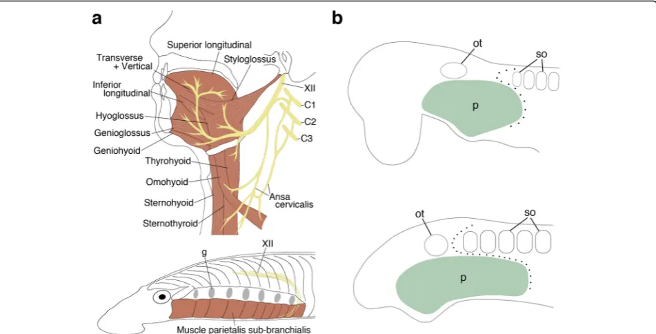
Based on diverse anatomical features, such as the jaw, nostril, and paired appendages, all living vertebrates are categorized into two major groups, cyclostomes and gnathostomes. The neck is one of the gnathostome features, found caudal to the interface of the head and trunk [1], defined as a domain between the occipital part of the cranium and the shoulder girdle of the gnathos-tomes, and occupied by cucullaris and hypobranchial muscles (HBMs) [2–6]. The HBMs are positioned in the ventral aspect of the neck and pharynx, and are collect-ively innervated by the hypoglossal and/or the occipi-tospinal nerves. These muscles function in jaw opening,
swallowing, and respiration in the gnathostomes. In tet-rapods, the rostral part of the muscles constitutes the tongue and the posterior part forms the infrahyoid mus-culatures, performing diverse functions from feeding to speech (Fig.1a) [7–15].
Morphologically, cyclostomes do not have an evident neck domain, as the occipital region, shoulder girdle, and cucullaris muscles are not present in these animals [3, 16]. In contrast, both lampreys and hagfishes have HBMs on the ventral part of the pharynx; these are in-nervated by the homologue of the hypoglossal nerve (Fig.1a) [3,17]. However, these muscles lie superficial to the pharynx, not inside the mouth and pharynx as they do in the gnathostomes [18, 19]. These anatomical dis-parities raise questions about the evolutionary origins of the neck in gnathostomes; however, the mechanisms leading to these disparities remain largely unknown. * Correspondence:[email protected]
1Evolutionary Morphology Laboratory, RIKEN center for Developmental Biology, 2-2-3 Minatojima-minami, Chuo-ku, Kobe 650-0047, Japan Full list of author information is available at the end of the article
The anatomical elements constituting the neck originate in the vicinity of the head–trunk interface of gnathostome embryos. The interface consists of the anterior extremity of somites and the posterior extremity of pharyngeal arches, and the circumpharyngeal crest cells pass through this interfacial domain (Fig.1b) [1,20]. Notably, the anterior oc-cipital somites and circumpharyngeal crest cells give rise to HBMs and their connective tissues, respectively [4,21,22] also see below); thus, the interfacial domain is a key embry-onic region for understanding neck morphogenesis [1].
As noted, although anatomical disparities exist be-tween cyclostomes and gnathostomes, a number of em-bryological studies have observed resemblances in the head–trunk interface and HBMs. For example, it has been shown that the lamprey embryo also possesses the interfacial domain [16, 23]. Studies have revealed that the occipital somites give rise to HBM precursors, which migrate once posterior to the pharyngeal arches and then turn rostrally to reach the ventral pharynx in verte-brates, including the lamprey [4,9,22,24–33]. Molecu-lar analyses have revealed the involvement of Pax3, a paired box transcription factor-encoding gene, in the de-velopment of HBMs [26, 28, 29, 34]. Similarly, lamprey
HBMs are marked by Pax3/7, a homolog of Pax3[35].
Thus, cyclostomes and gnathostomes have similar embryonic components and diverse anatomical out-comes in the head–trunk interface. This suggests that
developmental differences of components in the
interfacial domain account for the anatomical disparities between the two groups, and comparative embryological analysis of this domain is crucial to understanding verte-brate neck evolution [3]. The analysis of HBMs is espe-cially crucial, as muscles found in both groups would be a useful developmental landmark for comparing the interfacial domains in these animals. However, no detailed comparison of the HBM pathway between lam-prey and gnathostome embryos has been reported to date. Furthermore, the migration trajectory of HBMs relative to the pharynx remains obscure, even in em-bryos of gnathostomes. In the present study, we investi-gated the development of HBMs and the embryonic morphology of the head–trunk interface in chicken, mouse, shark, and lamprey embryos to gain insight into the evolution of the vertebrate neck.
Methods
Sample collection
Mature adult lampreys (Lethenteron camtschaticum, synonym for Lethenteron japonicum) were collected in Hokkaido, Japan, and the fertilized eggs were obtained as described previously [36]. Adult cloudy catsharks (Scylior-hinus torazame) were captured in Ibaraki, Japan. They laid fertilized eggs in saltwater tanks and the eggs were incu-bated at 16 °C. Fertilized chicken eggs (Gallus gallus) were purchased from local suppliers and incubated at 38 °C. C57BL/6 mouse embryos (Mus musculus) were obtained
from CDB animal facilities. Wnt1-Cre heterozygous mice [37] and R26R-H2B-EGFP homozygous mice (Acc No. CDB0203K: http://www2.clst.riken.jp/arg/reporter_mice. html) [38] were crossed and the offspring were geno-typed by PCR to obtain Wnt1-Cre/R26R-H2B-EGFP embryos. Noon on the day that the vaginal plug was observed was defined as E0.5. Embryos of lamprey, shark, chicken, and mouse were staged, following the systems described in [39–42], respectively.
Histological analysis and 3D reconstruction
Lamprey and shark embryos were fixed in Bouin’s fixa-tive, sectioned, and stained as described previously [43]. At least three embryos were observed for each develop-mental stage. Carazzi’s hematoxylin was used for lam-prey sections, Mayer’s hematoxylin for shark sections, and Alcian blue for cartilaginous staining. 3D recon-struction of embryos was performed by the method described previously [43].
Molecular cloning and phylogenetic analysis
Embryonic total RNA was extracted by TRIzol Reagent (Life Technologies), and cDNA was synthesized by SuperScript IV Reverse Transcriptase (Thermo Fisher Scientific). RNA-sequence analysis of embryonic lamprey and shark was performed and the sequences of lamprey Nkx2–3/2–5/2–6, Tbx1/10A, and Tbx4/5, and shark Nkx2–5 and Tbx5were found in the assembled data set (to be published elsewhere). DNA fragments were ampli-fied, cloned, and sequenced as described previously [44]. The accession numbers and primers used in this study are listed in the Additional file 1. The sequences of lamprey DlxB, Hox2α, Hox3α, andPax3/7, and sharkDlx5,Pax3, and Tbx1 have been described previously [35, 44–48]. Shark Hoxa2 sequence has been described recently in a separate study (Pascual-Anaya et al., under preparation) and is publicly available in GenBank under the accession number MF398238. For phylogenetic analyses, amino acid sequences of orthologous genes’from different vertebrate species were compiled from GenBank (http://www.ncbi. nlm.nih.gov/) and Ensembl (http://www.ensembl.org/in dex.html). Multiple alignment of protein sequences were performed with MAFFT [49] as implemented at the European Bioinformatics Institute’s web server (http:// www.ebi.ac.uk/Tools/msa/mafft/), or MUSCLE [50] as im-plemented in MEGA v7 [51], release 7,161,111-i38651, with default parameters, and saved in FASTA format. Resulting alignments were then trimmed by trimAl version 1.2rev59 [52] using the‘–automated1’ parameter and then formatted into NEXUS format by readAl v.1.2 (bundled with the trimAl package) (Suppl. Molecular phylogenetic trees of lamprey were inferred using Bayesian inference with MrBayes 3.2.6 [53] under the assumption of an LG + I + G evolutionary model and were performed
with two independent MrBayes runs, 4 chains each. Convergence was considered when the standard deviation < 0.01 for more than 1 million generations. To build the consensus tree, a burn-in of the 25% of the trees was per-formed (see Additional file1: Figure S1 for the number of generations of each gene’s phylogenetic analysis).
In situ hybridization
Lamprey and shark embryos were fixed in MEMFA fixa-tive. Chicken and mouse embryos were fixed in 4% PFA/ PBS. Whole-mount in situ hybridization was performed as described in [54]. The method of two-color whole-mount in situ hybridization has been described by [55]. Embryos were imaged using a Leica MZ16FA. After whole-mount in situ hybridization, lamprey embryos were immersed in MEMFA fixative with 2.5% glutaralde-hyde solution, dehydrated, and embedded in paraffin. Sections were prepared at 6 μm, and counterstained in eosin solution. The protocol of in situ hybridization on shark sections was described in [36]. Adjacent paraffin sections were used to compare gene expression patterns. Sections were imaged with an Olympus BX53. The expression pattern of each gene was investigated in > 3 embryos.
Immunohistochemistry
Three Wnt1-Cre/R26R-H2B-EGFP embryos were
obtained and utilized in the analysis. The embryos were fixed in 4%PFA/PBS, dehydrated, and embedded in paraf-fin. Sections were prepared at 6 μm. Deparaffinized sec-tions were autoclaved in 1 mM citrate buffer (pH 6.0) to unmask epitopes [56]. Anti-PAX3 mouse monoclonal antibody (1/100 dilution, ab69856, Abcam) and anti-GFP rabbit polyclonal antibody (1/500 dilution, ab290, Abcam) were used for the primary antibody. Goat anti-mouse IgG Alexa Fluor 647 (1/200 dilution, A21237, Thermo Fisher Scientific) and goat anti-rabbit IgG Alexa Fluor 488 (1/500 dilution, A11034, Thermo Fisher Scientific) were used for the secondary antibody. The signal detec-tion was performed with a confocal laser microscope Leica TCS SP8X and Spectral Dye Separation in LAS AF software.
Results
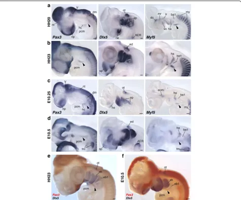
so-called hypoglossal cord [26–29].Myf5expression was only detected in the anteroventral compartment of the cord. The hypoglossal cord circumvented the posterior boundary of the pharynx withDlx5-positive mesenchyme, and the rostral part of the cord reached to the ventrocau-dal aspect of the pharyngeal arches (Fig.2a, c).
Subsequently, these precursors reached to the lateral side of the anterior pericardium at HH23 and E10.5 (Fig. 2b, d–f). In these embryos, the pericardium was commonly located ventral to the pharynx, and the rostral tip of HBMs appeared to have penetrated the medial side of the hyoid arch (Fig.2e, f). This medial extension of HBM precursors
was also suggested by the expression pattern ofMyf5, which marks both cranial and trunk myoblasts (Fig.2a–d).
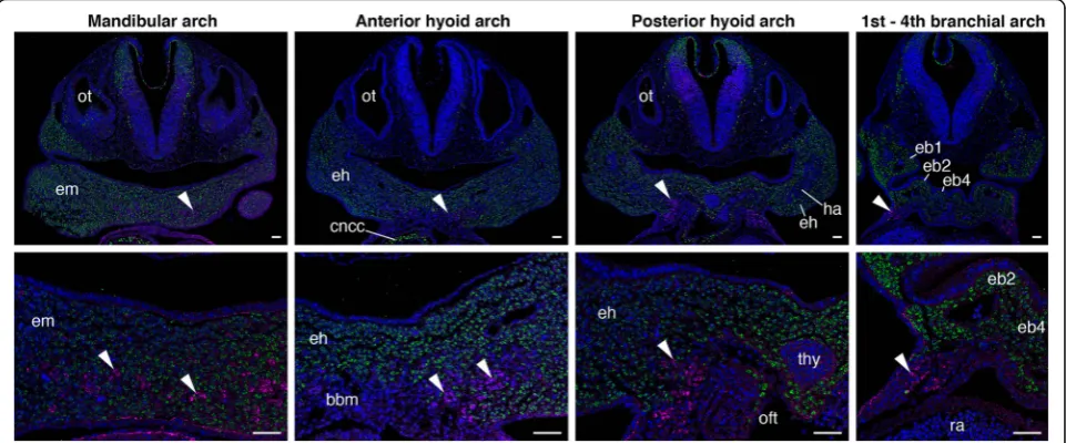
To visualize the distribution of HBM precursors and pharyngeal arches more precisely, we performed immu-nohistochemistry of PAX3 on Wnt1-Cre/R26R-H2B-EGFP mouse embryos, which rigorously mark the ecto-mesenchyme of pharyngeal arches with EGFP (Fig.3and Additional file 1: Figure S2). At the level of postotic pharyngeal arches (third to sixth arches), the HBM pre-cursors, immunostained with PAX3 antibody, were detected lateral to the EGFP-positive ectomesenchyme
and within the pericardial mesoderm (Fig. 3). This
indicates that the HBMs are not covered by the poster-iormost ectomesenchyme while traveling the posterior edge of the pharynx. The PAX3-positive cells extended from the pericardiac region to the ventromedial portion of the hyoid arch, and lay on both sides of the basibran-chial mesenchyme, which is not marked by EGFP anti-body. The precursors were laterally enclosed by EGFP-positive ectomesenchyme at the hyoid arch level, and completely surrounded by the ectomesenchyme at the floor of the mandibular arch (Fig.3). The HBM precursors thus passed through the posterior edge of the caudal phar-ynx, along the lateral side of the pericardium, ventral to branchial arches, and then inside the mandibular and hyoid arches in chicken and mouse embryos.
We next explored the developmental pathway of
HBMs in the cloudy catshark, S. torazame, to test
whether the trajectory of HBM precursors is shared in crown gnathostomes. We performed 3D reconstruction of shark embryos from the otic vesicle to heart levels (Additional file1: Figures S3a and S4). Shark mesoderm, which exhibits an epithelialized, not mesenchymal, state at early stages [43, 58], is a useful model for observing HBMs in gnathostomes.
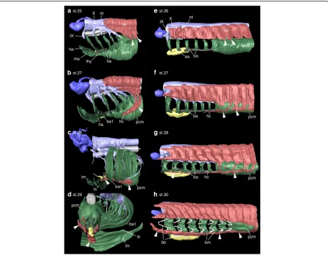
At stage 25, S. torazame embryos showed mesoderm
of seven pharyngeal arches located dorsal to the pericar-dium (Figs.4a,5a-c, and Additional file1: Figure S4a, e). This relative position of the pericardium ventral to the pharynx was also observed in the early stages of the em-bryos, resembling that of chicken and mouse embryos (Fig. 2 and Additional file 1: Figure S5). The rostral
somites formed ventral protrusions, forming a poster-iorly oriented arc behind the pharynx, which extended toward the pericardium, showing the initial migration of the HBMs, as described previously (Fig.4a) [40].
By stage 27, HBM precursors had descended around the posterior edge of the branchial arches to form the hypoglossal cord [27] on each side of the pericardium. The rostral tip of the cord had reached to the level of the fourth branchial arch (Fig.4b).
At stage 29, the HBM precursors extended medially at the level of the hyoid arch. Notably, its anterior part was embraced laterally and ventrally by the intermandibularis and interhyoideus muscles, while the posterior remained ventral to the branchial arches (Fig.4c, d). Crucially, this topographical relationship of HBMs and pharyngeal arches in this species perfectly coincided with that of chicken and mouse (Figs.2, 3and4). The HBM precur-sors branched out to form the coracohyoideus and cora-cobranchialis muscles on both sides of the ventral aorta and pericardium, and subsequently, the majority of the HBMs were found inside gill muscles and cartilages (interhyoideus and constrictor superficialis muscles, cer-atohyal, epibranchial, and ceratobranchial cartilages), as seen in the adult shark (Fig.4c, d, and Additional file1: Figure S4).
Development of HBMs in the lamprey
To compare the developmental trajectory of HBMs between cyclostomes and gnathostomes, we observed the development of the Arctic lamprey,L. camstchaticum
(Additional file1: Figures S3b and S6). The youngest lam-prey embryos examined in histological analysis were at Tahara’s stage 26 [39], when yolk granules were substan-tially reduced. Each of the pharyngeal arches possessed branchial mesoderm, which was located anterior to the pericardial mesoderm, immediately below the axial meso-derm (Figs.4e,5d–f, and Additional file1: Figure S6a, e). This topology of the pharyngeal mesoderm and pericar-dium was also observed in earlier stages of lamprey em-bryos (Additional file 1: Figure S7). Thus, the relative position of the pericardium posterior to the pharynx con-trasts with that in gnathostomes (Additional file1: Figures S5 and S7). Signs of HBM development in the lamprey were first observed on the ventrolateral edge of somites,
above the pericardial mesoderm (Fig.4eand Additional file 1: Figure S6a, e, j-k').
During stages 27 and 28, the HBM precursors on both sides extended ventrally between the ectoderm and the pericardial mesoderm as several streams of cell popula-tions (Fig. 4f and Additional file 1: Figure S6). These streams later converged ventroanteriorly to form the hypoglossal cord [27], which expands from the lateral side of the pericardium to the posterior branchial arches (Fig. 4f, g). Although the trajectory of these HBMs spreading on the lateral side of the pericardium is simi-lar to that in gnathostomes, the lamprey HBMs uniquely developed along the ventrolateral aspect of the branchial arches (no longer in the pericardial mesoderm as in
gnathostomes), differing clearly from those in gnathos-tomes (Figs.2,3and4).
By stage 30, HBM precursors developed further anteri-orly up to the otic level and resided ventrolaterally to the pharyngeal muscles and skeletons, including man-dibular and hyoid arches (Fig. 4g and Additional file 1: Figure S6). This relative position of HBMs and the phar-ynx is comparable with that in the adult lamprey.
Trajectory of the HBMs relative to pharyngeal arches To show the distribution of HBMs and pharyngeal arches in shark and lamprey embryos more clearly, we performed in situ hybridization using orthologous genes ofPax3and Dlx5, as well as Tbx1, which is expressed in the head mesoderm of pharyngeal arches, and Hox genes, which mark ectomesenchymal cells in the pharyngeal arches (Additional file1: Figure S1) [35,44,45,47,59].
At stage 27, S. torazame Pax3 marked HBM
precur-sors that extended posterior to the pharyngeal arches and arrived at the lateral side of the pericardium. Section
in situ hybridization clearly showed that the precursors did not intermix with theDlx5-,Hoxa2- and Tbx1-posi-tive branchial arches, and both pharyngeal arches and the pericardium were observed in the same transverse plane (Fig. 6a, d-f and Additional file 1: Figure S8a, b). The precursors passed through the pericardium laterally and reached to the second branchial arch level at stage 28 (Fig.6b, g-Iand Additional file1: Figure S8c, d). By stage 29,Pax3-positive cells were found ventral to the pharynx and the anterior tip of the expression at the hyoid arch level showed weak staining (Fig. 6c). In section in situ hybridization, thePax3expression was detected dorsome-dial to the Dlx5-positive ectomesenchyme and Tbx1 ex-pressions localized in the interhyoideus (Fig.6h–k). This indicates the medial extension of HBM precursors and the ventral covering of pharyngeal components at the hyoid arch level.
The above observations are consistent with results from chicken and mouse embryos, in which developing HBMs circumvent the postotic pharyngeal arches and travel in the
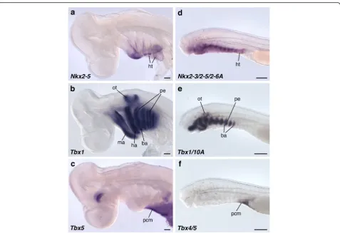
Fig. 5The relative position of the pericardium and pharyngeal arches.Nkx2–5(a),Tbx1(b), andTbx5(c) expressions inS.torazameat the stage 25 from the lateral view.Nkx2–3/2–5/2-6A(d),Tbx1/10A(e), andTbx4/5(f) expressions in the lamprey at the stage 26 from the lateral view.Nkx2–5
cephalic crest cell-free environment before reaching the hyoid arch. In contrast, lampreyPax3/7expression labeled HBM precursors extending from the lateral side of the peri-cardium to the posterior pharynx (Fig.7a–c, f, i, l). Lamprey DlxBandTbx1/10A were detected in the pharyngeal arch ectomesenchyme and mesoderm, respectively (Fig. 7a, b).
Analyses of histological sections after in situ hybridization revealed thatDlxB-, Hox2α-, Hox3α- and Tbx1/10A–posi-tive cells in the pharyngeal arches were observed dorsome-dial to the Pax3/7-positive cells, and pharyngeal arches and the pericardium were not in the same transverse plane (Fig. 7c-n and Additional file 1: Figure S8e-n). Thus, the
lamprey HBM precursors enter into the neural crest cell environment as soon as they pass through the pericardial domain located caudal to the entire pharynx.
Taken together, these results are consistent with our histological observations, indicating that shark HBM pre-cursors pass outside of the pericardium and expand inside the pharynx at the level between the hyoid and first bran-chial arches, whereas in lamprey HBM precursors remain outside the pericardium and pharyngeal arches. Thus, al-though the initial avoidance of the branchial region and the lateral migration to the pericardium are likely to be steps in a conserved developmental program in the entire vertebrate HBMs [9,24,27,30, 31], the trajectory of HBMs internal-ized at the hyoid level is a shared trait only for crown gnathostomes (Figs.2,3,4,6and7).
Discussion
In this study, we show that HBM precursors travel along the head trunk interface, namely, along the posterior
edge of the postotic pharyngeal arches and lateral to the pericardium in both crown gnathostomes and lampreys, while the topographical relationship of the pericardium to the pharynx and the trajectory of HBMs in the ventral pharynx differ substantially between the two groups. In the gnathostomes, the pericardium lies ventral to the pharyngeal arches, and the HBM precursors intrude in-side the hyoid and mandibular arches (Figs.2,3,4,6). In contrast, in the lamprey, the pericardium is located cau-dal to the pharynx, and the HBM precursors extend out-side of the pharyngeal muscles and skeletons (Figs.2,7). These observations provide unique insights into the evo-lution of the head–trunk interface as discussed below.
It has been reported that postotic pharyngeal arches ad-join occipital somites dorsally and the pericardium ven-trally, creating the head–trunk interface in the vertebrate body (Fig. 1b) [1]. Our study reveals that the pericardial mesoderm is ventral to the pharyngeal domain in gnathos-tomes, but caudal to the pharynx in lamprey (Figs. 4, 5,
Additional file1: Figures S5 and S7). This spatial difference in the pericardium implies that the ventral part of the head–trunk interface, especially the boundary between the pharynx and pericardium, is distinct in gnathostomes and lamprey (Fig. 8a, b), suggesting that the head trunk boundary may not be constant in all vertebrates. In addition, the position of the pericardium is also involved in the development of HBMs. We found that, in all ver-tebrate embryos observed here, the HBM precursors definitely passed through the lateral side of the pericar-dium (Figs.2,3,4,6and7). The migration of HBM pre-cursors lateral to the pericardium has also been observed in rays, salamanders, and mammals in previ-ous histological analyses [9,24,27,31,33].
These findings indicate that the pericardium is tightly coupled with the development of HBMs in vertebrates. This linkage suggests that the position of the gnathos-tome pericardium may permit the HBM precursors to circumvent branchial arches ventrally and project medi-ally into the hyoid arch to reach the inside of the mouth (Fig. 8). This trajectory is absent in the lamprey and in-conceivable in the embryos given the position of the lamprey pericardium, which does not attach to the man-dibular and hyoid arches (Figs.4,5,7and 8). Therefore, a heterotopic shift of pericardium relative to the pharynx may underlie a trajectory change of the HBMs.
The above discussion implies a morphological transition of the head–trunk interfacial domain from one state to another in vertebrate phylogeny. The prevalence of the pericardium, pharynx, and HBMs in extant vertebrates suggests the presence of those embryonic elements in a common ancestor of cyclostomes and gnathostomes, al-though the arrangement of ancestral elements remains unknown. In this regard, information from paleontology is important to speculate about the ancestral condition.
Some fossil jawless vertebrates, such as Achanarella, Anaspida, and Jamoytius, possess the posteriorly elon-gated branchial apparatus with a number of gill openings [60, 61], and trunk scales or myomeres dorsal to the branchial region [61–63], but lack apparent occipital and shoulder elements. These morphological features are shared by the interfacial domain of modern cyclostomes, although the hagfish likely represents a derived condi-tion in the hypoglossal nerve and HBMs as well [1,17].
It is difficult to speculate with confidence on the anat-omy of the head–trunk interface and HBMs in other fossil jawless vertebrates, such as Heterostracans, Galeaspids and Osteostracans, because data on their soft tissue morphology are very rare, insights into their internatl anatomy must be gleaned mainly from endo- and exoskel-etons [64]. Of these fossil species, Osteostracans have an ossified pericardium attached to the postbranchial wall, and the pericardium and posterior gills are positioned in the same longitudinal axis [60,64]. They also possess the
pectoral girdle, and possibly paired common cardinal veins (the ducts of Cuvier) just behind the postbranchial wall, suggesting a gnathostome-like condition of the head–trunk interface in this animal [54, 65, 66]. Thus, it may be conceivable that HBMs of Osteostracans are posi-tioned on the ventral aspect of the pharynx, separated from the dorsal muscle mass, and caudally attached to the pectoral girdle, or possibly to the ventral bridge of dermal bone located ventral to the pericardium [64]. The morph-ology of anterior HBMs and their position relative to the gill elements are difficult to assume, and further analyses of this fossil will be required. In contrast, placoderms dis-play muscle attachment sites for HBMs on the lower jaw and shoulder skeletons, which are comparable with those of extant gnathostomes [67].
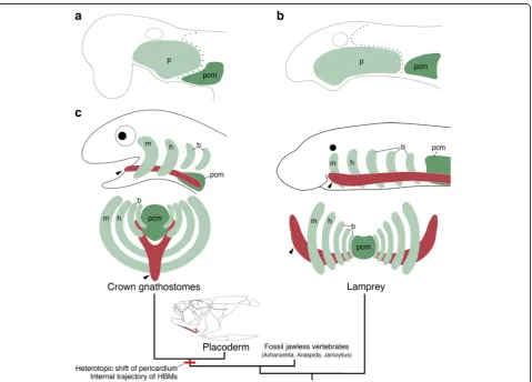
Given the current phylogenetic positions of the above-mentioned fossil vertebrates [63, 68], these observations suggest that the head–trunk interface found in the lamprey embryo may represent the ancestral condition, and a re-arrangement of preexisting embryonic components in the ancestral vertebrate may have occurred during the transi-tion from jawless to jawed vertebrates (Fig.8c). Thus, we hypothesize that a heterotopic shift of the pericardium to-gether with a gain of an internal trajectory of HBM precur-sors, was a key embryonic change for the evolution of the gnathostome HBMs, which function as jaw opening and tongue muscles (Fig. 8c) [7, 11, 13,14]. Furthermore, this rearrangement may have contributed to the establishment of the gnathostome head–trunk interface, which provides the basis for the development of the jawed vertebrate neck.
Conclusions
In our comparative embryological analyses, we found that the pericardium was posterior to the pharynx and HBM precursors extended outside of those elements in the lam-prey, whereas the pericardium was ventral to the pharynx and HBM precursors traveled outside of the pericardium and inside the hyoid and mandibular arches in crown gnathostomes. Based on current paleontological data on stem gnathostomes, we hypothesize that the embryonic arrangement of the pericardium, pharynx, and HBM pre-cursors likely underwent a transition from the lamprey condition to the crown gnathostome condition, which was a key embryonic change not only for the establishment of the head–trunk interface, but also for the evolution of the neck in jawed vertebrates.
Additional file
Additional file 1:Supplementary materials. (PDF 6266 kb)
Acknowledgements
Kuroda, W. Takagi, Y. Kageyama, and Y Nakai were engaged in sampling of lamprey and shark embryos. Drs. H. Nagashima and M. Takechi helped mouse and chicken sampling. Drs. H. Kiyonari, K. Inoue, S Nishikawa and W. Hsu helped transgenic mouse preparation. RIKEN Kobe Light Microscopy Facility supported the imaging experiments. Dr. Sansom kindly gave advice on fossils. We thank Drs. F. Sugahara, J. Pieretti and T. Onai for discussions and critical reading of the manuscript.
Funding
This work was supported in part by a Grant-in-Aid for Scientific Research (A) 15H02416, Grant-in-Aid for Scientific Research on Innovative Areas 17H06385, and a Naito Grant for the Promotion of Focused Research (The Naito Foundation) to S.K.
Availability of data and materials
All sequences generated in this study are publicly available in NCBI GenBank under the accession numbers LC331563-LC331566 and LC333760.
Authors’contributions
NA and SK designed the research. NA, JP-A, SH, and TH performed molecular experiments. NA, JP-A, and SK wrote the paper. All authors read and approved the manuscript.
Ethics approval
All animal experiments were carried out with the approval of the Institutional Animal Care and Use Committee of RIKEN, Kobe Branch.
Consent for publication
Not applicable.
Competing interests
The authors declare that they have no competing interests.
Publisher’s Note
Springer Nature remains neutral with regard to jurisdictional claims in published maps and institutional affiliations.
Author details
1Evolutionary Morphology Laboratory, RIKEN center for Developmental Biology, 2-2-3 Minatojima-minami, Chuo-ku, Kobe 650-0047, Japan. 2Department of Biology, Graduate School of Science, Kobe University, Kobe 657-8501, Japan.
Received: 28 October 2017 Accepted: 6 February 2018
References
1. Kuratani S. Spatial distribution of postotic crest cells defines the head/trunk interface of the vertebrate body: embryological interpretation of peripheral nerve morphology and evolution of the vertebrate head. Anat Embryol (Berl). 1997;195(1):1–13.
2. Ericsson R, Knight R, Johanson Z. Evolution and development of the vertebrate neck. J Anat. 2013;222(1):67–78.
3. Kuratani S. Evolutionary developmental studies of cyclostomes and the origin of the vertebrate neck. Develop Growth Differ. 2008;50(Suppl 1):S189–94. 4. Matsuoka T, Ahlberg PE, Kessaris N, Iannarelli P, Dennehy U, Richardson WD,
McMahon AP, Koentges G. Neural crest origins of the neck and shoulder. Nature. 2005;436(7049):347–55.
5. Nagashima H, Sugahara F, Watanabe K, Shibata M, Chiba A, Sato N. Developmental origin of the clavicle, and its implications for the evolution of the neck and the paired appendages in vertebrates. J Anat. 2016;229(4): 536–48.
6. Sefton EM, Bhullar BA, Mohaddes Z, Hanken J. Evolution of the head–trunk interface in tetrapod vertebrates. elife. 2016;5:e09972.
7. Bramble DM, Wake DB. Feeding mechanisms of lower Tetrapods. In: Hildebrand M, Bramble DM, Liem KF, Wake DB, editors. Functional vertebrate morphology. Cambridge: Harvard University Press; 1985. p. 230–61. 8. Deban SM, Wake DB, Roth G. Salamander with a ballistic tongue. Nature.
1997;389(6646):27–8.
9. Edgeworth FH. In: Edgeworth FH, editor. The cranial muscles of vertebrates. Cambridge: The University Press; 1935.
10. Fürbringer M. Ueber die spino-occipitalen Nerven der Selachier und Holocephalen ind ihre vergleichende Morphologie. Festschr für Carl Gegenbaur. 1897;3:349–788.
11. Hiiemae KM, Crompton AW. Mastication, food transport, and swallowing. In: Hildebrand M, Bramble DM, Liem KF, Wake DB, editors. Functional vertebrate morphology. Cambridge: Harvard University Press; 1985. p. 262–90. 12. Hiiemae KM, Palmer JB. Tongue movements in feeding and speech. Crit Rev
Oral Biol Med. 2003;14(6):413–29.
13. Lauder GV. Aquatic feeding in lower vertebrates. In: Hildebrand M, Bramble DM, Liem KF, Wake DB, editors. Functional vertebrate morphology. Cambridge: Harvard University Press; 1985. p. 210–29.
14. Liem KF. Ventilation. In: Hildebrand M, Bramble DM, Liem KF, Wake DB, editors. Functional vertebrate morphology. Cambridge: Harvard University Press; 1985. p. 185–209.
15. Neal HV, Rand H. Comparative anatomy. Philadelphia: The Blakiston Co.; 1936. 16. Kuratani S, Kuraku S, Murakami Y. Lamprey as an evo-devo model: lessons
from comparative embryology and molecular phylogenetics. Genesis. 2002; 34(3):175–83.
17. Oisi Y, Fujimoto S, Ota KG, Kuratani S. On the peculiar morphology and development of the hypoglossal, glossopharyngeal and vagus nerves and hypobranchial muscles in the hagfish. Zoological Lett. 2015;1:6. 18. Marinelli W. Vergleichende Anatomie und Morphologie der Wirbeltiere: 1.
Lampetra fluviatilis(L.) Wien: Franz Deuticke; 1954.
19. Marinelli W. Strenger A. Vergleichende Anatomie und Morphologie der Wirbeltiere: 2. Lieferung.Myxine glutinosa(L.). Wien: Franz Deuticke; 1956. 20. Kuratani SC, Kirby ML. Migration and distribution of circumpharyngeal crest
cells in the chick embryo. Formation of the circumpharyngeal ridge and E/ C8+ crest cells in the vertebrate head region. Anat Rec. 1992;234(2):263–80. 21. Köntges G, Lumsden A. Rhombencephalic neural crest segmentation is preserved throughout craniofacial ontogeny. Development. 1996;122(10): 3229–42.
22. Noden DM. The embryonic origins of avian cephalic and cervical muscles and associated connective tissues. Am J Anat. 1983;168(3):257–76. 23. Horigome N, Myojin M, Ueki T, Hirano S, Aizawa S, Kuratani S. Development
of cephalic neural crest cells in embryos of Lampetra japonica, with special reference to the evolution of the jaw. Dev Biol. 1999;207(2):287–308. 24. Bates MN. The early development of the hypoglossal musculature in the
cat. Am J Anat. 1948;83(3):329–55.
25. Couly GF, Coltey PM, Le Douarin NM. The triple origin of skull in higher vertebrates: a study in quail-chick chimeras. Development. 1993;117(2):409–29. 26. Huang R, Zhi Q, Izpisua-Belmonte JC, Christ B, Patel K. Origin and development
of the avian tongue muscles. Anat Embryol (Berl). 1999;200(2):137–52. 27. Hunter RM. The development of the anterior post-otic somites in the rabbit.
J Morphol. 1935;57(2):501–31.
28. Lours-Calet C, Alvares LE, El-Hanfy AS, Gandesha S, Walters EH, Sobreira DR, Wotton KR, Jorge EC, Lawson JA, Kelsey Lewis A, et al. Evolutionarily conserved morphogenetic movements at the vertebrate head–trunk interface coordinate the transport and assembly of hypopharyngeal structures. Dev Biol. 2014;390(2):231–46.
29. Mackenzie S, Walsh FS, Graham A. Migration of hypoglossal myoblast precursors. Dev Dyn. 1998;213(4):349–58.
30. Neal HV. Development of the hypoglossus musculature inPetromyzonand
Squalus. Anat Anz. 1897;13:441–63.
31. O'Rahilly R, Muller F. The early development of the hypoglossal nerve and occipital somites in staged human embryos. Am J Anat. 1984;169(3):237–57. 32. Piekarski N, Olsson L. Muscular derivatives of the cranialmost somites
revealed by long-term fate mapping in the Mexican axolotl (Ambystoma mexicanum). Evol Dev. 2007;9(6):566–78.
33. Platt JB. The development of the cartilaginous skull and of the Branchial and hypoglossal musculature in Necturus. Morphologisches Jahrbuch. 1897; 25:377–463.
34. Tajbakhsh S, Rocancourt D, Cossu G, Buckingham M. Redefining the genetic hierarchies controlling skeletal myogenesis: Pax-3 and Myf-5 act upstream of MyoD. Cell. 1997;89(1):127–38.
35. Kusakabe R, Kuraku S, Kuratani S. Expression and interaction of muscle-related genes in the lamprey imply the evolutionary scenario for vertebrate skeletal muscle, in association with the acquisition of the neck and fins. Dev Biol. 2011;350(1):217–27.
37. Danielian PS, Muccino D, Rowitch DH, Michael SK, McMahon AP. Modification of gene activity in mouse embryos in utero by a tamoxifen-inducible form of Cre recombinase. Curr Biol. 1998;8(24):1323–6. 38. Abe T, Kiyonari H, Shioi G, Inoue K, Nakao K, Aizawa S, Fujimori T.
Establishment of conditional reporter mouse lines at ROSA26 locus for live cell imaging. Genesis. 2011;49(7):579–90.
39. Tahara Y. Normal stages of development in the lamprey,Lampetra reissneri
(Dybowski). Zool Sci. 1988;5:109–18.
40. Ballard WW, Mellinger J, Lechenault H. A series of normal stages for development ofScyliorhinus canicula, the lesser spotted dogfish (Chondrichthyes: Scyliorhinidae). J Exp Zool. 1993;267(3):318–36. 41. Hamburger V, Hamilton HL. A series of normal stages in the development
of the chick embryo. J Morphol. 1951;88(1):49–92.
42. Theiler K. The house mouse. Atlas of embryonic development. New York: Springer-Verlag Berlin Heidelberg; 1989.
43. Adachi N, Kuratani S. Development of head and trunk mesoderm in the dogfish,Scyliorhinus torazame: I. Embryology and morphology of the head cavities and related structures. Evol Dev. 2012;14(3):234–56.
44. Adachi N, Takechi M, Hirai T, Kuratani S. Development of the head and trunk mesoderm in the dogfish,Scyliorhinus torazame: II. Comparison of gene expression between the head mesoderm and somites with reference to the origin of the vertebrate head. Evol Dev. 2012;14(3):257–76. 45. Kuraku S, Takio Y, Sugahara F, Takechi M, Kuratani S. Evolution of
oropharyngeal patterning mechanisms involving Dlx and endothelins in vertebrates. Dev Biol. 2010;341(1):315–23.
46. Mehta TK, Ravi V, Yamasaki S, Lee AP, Lian MM, Tay BH, Tohari S, Yanai S, Tay A, Brenner S, et al. Evidence for at least six Hox clusters in the Japanese lamprey (Lethenteron Japonicum). Proc Natl Acad Sci U S A. 2013;110(40):16044–9. 47. Takechi M, Adachi N, Hirai T, Kuratani S, Kuraku S. The Dlx genes as clues to
vertebrate genomics and craniofacial evolution. Semin Cell Dev Biol. 2013; 24(2):110–8.
48. Takio Y, Pasqualetti M, Kuraku S, Hirano S, Rijli FM, Kuratani S. Evolutionary biology: lampreyHoxgenes and the evolution of jaws. Nature. 2004; 429(6989):1. p following 262
49. Katoh K, Standley DM. MAFFT multiple sequence alignment software version 7: improvements in performance and usability. Mol Biol Evol. 2013; 30(4):772–80.
50. Edgar RC. MUSCLE: multiple sequence alignment with high accuracy and high throughput. Nucleic Acids Res. 2004;32(5):1792–7.
51. Kumar S, Stecher G, Tamura K. MEGA7: molecular evolutionary genetics analysis version 7.0 for bigger datasets. Mol Biol Evol. 2016;33(7):1870–4. 52. Capella-Gutierrez S, Silla-Martinez JM. Gabaldon T: trimAl: a tool for
automated alignment trimming in large-scale phylogenetic analyses. Bioinformatics. 2009;25(15):1972–3.
53. Ronquist F, Teslenko M, van der Mark P, Ayres DL, Darling A, Hohna S, Larget B, Liu L, Suchard MA, Huelsenbeck JP. MrBayes 3.2: efficient Bayesian phylogenetic inference and model choice across a large model space. Syst Biol. 2012;61(3):539–42.
54. Adachi N, Robinson M, Goolsbee A, Shubin NH. Regulatory evolution of Tbx5 and the origin of paired appendages. Proc Natl Acad Sci U S A. 2016; 113(36):10115–20.
55. Long S, Rebagliati M. Sensitive two-color whole-mount in situ hybridizations using digoxygenin- and dinitrophenol-labeled RNA probes. BioTechniques. 2002;32(3):494–6. 498 passim
56. Horst D, Ustanina S, Sergi C, Mikuz G, Juergens H, Braun T, et al. Comparative expression analysis of Pax3 and Pax7 during mouse myogenesis. Int J Dev Biol. 2006;50(1):47–54.
57. Depew MJ, Lufkin T, Rubenstein JL. Specification of jaw subdivisions by Dlx genes. Science. 2002;298(5592):381–5.
58. Balfour MF. A monograph on the Development of elasmobranch fishes. MacMillan and Co. London; 1878.
59. Tiecke E, Matsuura M, Kokubo N, Kuraku S, Kusakabe R, Kuratani S, Tanaka M. Identification and developmental expression of two Tbx1/10-related genes in the agnathan Lethenteron Japonicum. Dev Genes Evol. 2007;217(10):691–7. 60. Janvier P. Early specializations in the branchial apparatus of jawless
vertebrates: a consideration of gill number and size. In: Arratia G, MVH W, Cloutier R, editors. Recent Advances in the Origin and Early Radiation of Vertebrates. Munich: Verl. Dr. Friedrich Pfeil; 2004.
61. Janvier P, Arsenault M. The anatomy of Euphanerops longaevus Woodward, 1900, an anaspid-like jawless vertebrate from the Upper Devonian of Miguasha, Quebec, Canada. Geodiversitas. 2007;29(1):143–216.
62. Newman MJ. A new naked jawless vertebrate from the middle devonian of scotland. Palaeontology. 2002;45(5):933–41.
63. Sansom RS, Freedman KIM, Gabbott SE, Aldridge RJ, Purnell MA. Taphonomy and affinity of an enigmatic Silurian vertebrate, Jamoytius kerwoodi White. Palaeontology. 2010;53(6):1393–409.
64. Janvier P. Early Vertebrates. Oxford: Oxford Univ. Press; 1996. 65. Janvier P. Homologies and evolutionary transitions in early vertebrate
history. In: Anderson JS, Sues H-D, editors. Major Transitions in Vertebrate Evolution. Bloomington: Indiana University Press; 2007.
66. Janvier P, Percy LR, Potter IC. The arrangement of the heart chambers and associated blood vessels in the Devonian osteostracan Norselaspis glacialis. A reinterpretation based on recent studies of the circulatory system in lampreys. J Zool. 1991;223(4):567–76.
67. Johanson Z. Placoderm branchial and hypobranchial muscles and origins in jawed vertebrates. J Vertebr Paleontol. 2003;23(4):735–49.
68. Joseph N. Keating, Philip C. J. Donoghue. Histology and affinity of anaspids, and the early evolution of the vertebrate dermal skeleton. Proceedings of the Royal Society B: Biological Sciences. 2016;283(1826):20152917
• We accept pre-submission inquiries
• Our selector tool helps you to find the most relevant journal
• We provide round the clock customer support
• Convenient online submission
• Thorough peer review
• Inclusion in PubMed and all major indexing services
• Maximum visibility for your research
Submit your manuscript at www.biomedcentral.com/submit