R E V I E W
Open Access
Recent progress in the study of brown adipose
tissue
Xuan Yao
1,2, Shifang Shan
1,2, Ying Zhang
1,2and Hao Ying
1,2*Abstract
Brown adipose tissue in mammals plays a critical role in maintaining energy balance by thermogenesis, which means dissipating energy in the form of heat. It is held that in mammals, long-term surplus food intake results in energy storage in the form of triglyceride and may eventually lead to obesity. Stimulating energy-dissipating function of brown adipose tissue in human body may counteract fat accumulation. In order to utilize brown adipose tissue as a therapeutic target, the mechanisms underlying brown adipocyte differentiation and function should be better elucidated. Here we review the molecular mechanisms involved in brown adipose tissue development and thermogenesis, and share our thoughts on current challenges and possible future therapeutic approaches.
Keywords:brown adipose, differentiation, thermogenesis, obesity control
Introduction of brown adipose tissue and thermogenesis
Adipose depots in different parts of the body have unique micromorphology and molecular markers. According to their distinct physiological roles, adipose tissue in mam-mals is categorized into white adipose tissue (WAT) and brown adipose tissue (BAT) [1]. WAT functions to store energy in the form of triglyceride (TG)-containing intra-cellular droplets as well as to secrete a host of hormones that regulate overall energy balance. Unlike WAT, BAT regulates thermogenesis upon environmental stresses to maintain energy balance and protect the organism from hypothermia [2]. In addition, it has been shown that by activating BAT via short-term cold exposure, fatty acids are efficiently channeled into BAT due to a metabolic pro-gram that boosts TG-rich lipoproteins (TRL) uptake into BAT. In consequence, lipids clearance from plasma becomes more efficient, implicating that BAT might be a master regulator of TRL clearance and blood lipid abun-dance [3].
BAT thermogenesis is achieved by dissipating heat pro-duced from fatty acid oxidation. As little as 50 g of BAT could account for up to 20% of basal metabolic energy
expenditure of an adult human when maximally stimu-lated [4]. When animals are subjected to cold environment or ingest surplus energy, their sympathetic nervous system will be stimulated and catecholamine will be released. Catecholamine binds to theb3-Adrenoceptor on the plasma membrane of brown adipocyte and activates ade-nylyl cyclase which is able to accelerate the conversion of ATP to cyclic AMP (cAMP) [5]. cAMP is able to activate type 2 deiodinase (Dio2), an enzyme that converts thyroid hormone T4 to T3 in the brown adipocyte, resulting in enhanced local thyroid hormone signaling and increased energy. In addition, cAMP activates protein kinase A, which, in turn, phosphorylates and activates triacylglycerol lipase. Then the lipase accelerates the release of free fatty acid from triacylglycerol contained in the droplet of brown adipocyte, which acts both as the fuel of thermogenesis and as an activator of uncoupling protein 1 (UCP1), a key component of thermogenesis. UCP1 activation results in fast substrate oxidation with a low rate of ATP production. Thus, a large amount of energy is dissipated in the form of heat, which will be distributed throughout the body by cir-culation system [1,6].
Overview of recent breakthroughs in the study of BAT
It was previously believed that brown adipocyte and white adipocyte share a common ancestor in the course
* Correspondence: [email protected]
1Key Laboratory of Nutrition and Metabolism, Institute for Nutritional
Sciences, Shanghai Institutes for Biological Sciences, Chinese Academy of Sciences, Shanghai, China
Full list of author information is available at the end of the article
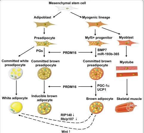
of adipogenesis. However, different opinion was rendered in 2007 when PR domain containing 16 (PRDM16) was discovered to be a master regulator of brown adipocyte differentiation [6]. Investigation of its specific role in brown adipocyte commitment led to the discovery that skeletal muscle and some depots of BAT share a common myogenic factor 5 (Myf5)-expressing progenitor [7]. Besides classic interscapular brown adi-pocytes, brown-like adipocytes (brite adipocytes) were found in WAT depots. When rats were submitted to cold or treated with b-adrenoceptor agonist, UCP1 could be detected in fat pads which had been considered as WAT. UCP1 was expressed in cells morphologically identical to typical brown adipocytes found in interscap-ular BAT [8]. More recent data suggested that there were inducible brown adipocytes in WAT when mice were exposed to cold environment [9]. These inducible brown adipocytes were not derived from Myf5-expres-sing myoblasts, and had a distinct gene expression pro-file in comparison with those adipocytes in interscapular BAT.
The mesenchymal stem cell is the precursor of brown adipocytes. BAT formation occurs in the early phase of embryonic development [10]. Once it was suggested that only small mammals and new born infants possess BAT, while human adults are practically devoid of functional BAT [1]. However, several research papers published in
New England Journal of Medicinein 2009 rebutted this assumption [11-14]. After analyzing thousands positron emission computed tomography/computed tomography (PET/CT) scans of adult subjects, researchers found that BAT was located in the neck and upper-chest regions. Moreover, BAT ratio was conversely correlated with body mass index (BMI) and this correlation was more signifi-cant among elderly [12]. Development of obesity is a result of prolonged positive energy balance. Most of surplus energy is stored in adipose tissue. Since BAT is essential in maintaining the balance of body fat and keeping indivi-duals from obesity, now BAT has been considered a pro-mising therapeutic target to combat obesity. We need to further delineate the mechanisms involved in brown adi-pose differentiation and thermogenesis, although they have been studied extensively (Figure 1).
Key players involved in brown adipose thermogenesis and differentiation
UCP1
As for human, UCP1 genetic polymorphisms were reported to be correlated with fat metabolism, obesity and diabetes [15-17]. These genetic studies underlie the role of UCP1 in maintaining energy balance. UCP1 is predomi-nantly expressed in brown adipocytes and responsible for the respiratory uncoupling during BAT thermogenesis [1]. Located on the inner membrane of mitochondria, UCP1
activity is regulated at multiple levels. There exist peroxi-some proliferator responsive element (PPRE) [18], thyroid hormone responsive element (TRE) [19], and retinoic acid responsive element (RARE) [20] in the upstream of UCP1 gene, which means these nuclear receptors and their ligands might directly regulate its transcription. In addi-tion, peroxisome proliferator-activated receptor gamma coactivator 1-alpha (PGC-1a), which is critical to mito-chondria genesis, is able to increase the transcriptional activity of peroxisome proliferator-activated receptorg (PPARg) and thyroid hormone receptor (TR) on the UCP-1 promoter [2UCP-1]. UCPUCP-1 activity can also be directly regu-lated by purine nucleotides and free fatty acids. Purine nucleotides bind from the cytosolic side of UCP1 and inhi-bit its activity [22]. Free fatty acids released in response to stimuli can replace the purine nucleotides and act as a UCP1 activator [23,24]. After the residual fatty acids are oxidized away, the mitochondria will return to coupled state.
Thyroid hormone and Dio2
Thyroid hormone is necessary for a full BAT thermo-genic response in cold adaptation. In the hypothyroid state, the response of BAT to sympathetic stimulation is diminished [25]. Mice deficient in TRaare cold intoler-ant and have impaired BAT thermogenic response to norepinephrine [26]. In addition, activation of thyroid hormone signaling is highly regulated by deiodination. Deiodination of minimally active thyroxine (T4) to the biologically active triiodothyronine (T3) is catalyzed by type I and type II deiodinases (Dio1 and Dio2) [27]. Dio2 expression has been shown to control intracellular T3 concentration. In response to sympathetic stimulation, increased cAMP stimulates T3 production by increasing Dio2 expression and activity, which leads to increased energy expenditure in BAT. Mice lacking Dio2 exhibit permanent BAT thermogenic defect, compromising ther-moregulation and the ability to dissipate excessive cal-ories from diet [28-30]. Interestingly, bile acids released from the gallbladder has been shown to increase energy expenditure in BAT by regulating local thyroid hormone production via G protein-coupled receptor TGR5-cAMP-Dio2 signaling pathway [31,32].
PGC-1a
increases in the course of brown adipocyte differentiation [34]. Moreover, PGC-1acan induce the expression of nuclear respiratory factor (NRF) and physically interact with NRF to coactivate the transcription. NRF is able to regulate a host of mitochondrial genes encoded in the cell nucleus, includingb-ATP synthase, cytochrome-c, cyto-chrome-c-oxidase subunit IV, and mitochondrial tran-scription factor A [35]. Cold exposure significantly stimulates PGC-1aexpression, which is mediated by pro-tein kinase A-cAMP response element-binding propro-tein (PKA-CREB) pathway [21]. Ectopic expression of PGC-1a in WAT leads to mitochondria genesis and UCP1 upregu-lation. Lack of PGC-1a in mice does not affect the
morphology of BAT but results in lower UCP1 level and intolerance to cold [36,37]. Correspondingly, loss of PGC-1ain cultured brown preadipocytes does not impede the maturation, but the matured adipocytes have impaired thermogenesis [34]. Thus, it can be concluded that PGC-1ais an important part of thermogenesis due to its indis-pensable role in mitochondria genesis while it has limited effect on brown adipocyte differentiation.
RIP140
Receptor-interacting protein 140 (RIP140), a nuclear receptor corepressor, can suppress nuclear receptor estrogen receptor (ER) and PPAR in the presence of their
Adipoblast
Myogenic lineage
Preadipocyte
Myf5+ progenitor
Committed white
preadipocyte
White adipocyte
Inducible brown
adipocyte
Committed brown
preadipocyte
Committed brown
preadipocyte
Brown adipocyte
Mesenchymal stem cell
BMP7
miR-193b-365
Myotube
PGC-1
D
UCP1
Skeletal muscle
PGs
RIP140
Ę
Rb/p107
Ę
PRDM16
Myoblast
PRDM16
Wnt
Ė
ligands [38]. It is more abundantly expressed in WAT than in BAT [39,40]. It was reported that RIP140 defi-ciency in cultured adipocytes led to increased energy expenditure and this phenomenon disappeared if RIP140 was re-expressed [41].In vivostudies show that RIP140 -/-mice consume a similar amount of food as control -/-mice, while their physical activity remains unaltered. The mice appear to be emaciated with a total fat tissue content dropped to 30% of that in control mice. However, in these mice, the fat cell number remains the same com-pared to the control group, but the volume decreases sig-nificantly. Moreover, the RIP140-/-mice are protected from high-fat diet-induced obesity. It is of particular interest that UCP1 expression in the WAT of these mice is 100 times higher than that in the WAT of wild type mice [39]. This finding suggests that RIP140 might be able to suppress brown adipocyte characteristics.
PRDM16
PRDM16, a 140-kDa protein, contains an N-terminal PR domain, ten zinc-fingers and other diverse sites that can mediate multiple protein-protein interaction. PRDM16 plays a critical role in determining brown adipocyte line-age commitment and differentiation. By comparing the transcriptome of BAT and WAT from mice, PRDM16 was for the first time recognized as a BAT-specific gene [6]. When PRDM16 is expressed in white fat cell progenitors, it can activate a robust brown fat phenotype including induction of PGC-1a, UCP1, and Dio2 expression and a remarkable increase in uncoupled respiration. Transgenic expression of PRDM16 in white fat depots stimulates the formation of brown fat cells. In contrast, knockdown of PRDM16 through shRNA expression in brown fat cells causes a near total loss of the brown characteristics. It is suggested that PRDM16 activates brown fat cell identity at least in part by simultaneously activating PGC-1a and PGC-1bthrough direct binding [6], and represses white adipocyte gene expression through forming complex with C-terminal-binding protein-1 (CtBP-1) and CtBP-2 [42].
A major breakthrough was achieved by a lineage tracing studyin vivowhich has revealed that brown adipocyte shares a mutual precursor, Myf5-expressing cell, with ske-letal muscle cell, but not white adipocyte as previously considered. Loss of PRDM16 from brown adipocyte pre-cursor promotes muscle differentiation, while ectopic expression of PRDM16 in myoblasts induces their differ-entiation into brown adipocytes [7]. Subsequent investiga-tion indicates that PRDM16 is able to form complex with CCAAT-enhancer-binding proteinb(C/EBPb) and this complex controls the cell fate switch from myoblastic pre-cursors to brown fat cells [43]. Forced expression of PRDM16 and C/EBP-bis sufficient to induce a fully func-tional brown fat program in naive fibroblastic cells. All the evidence above demonstrates that PRDM16 plays a critical
role in determining the differentiation fate of brown adi-pocyte. However, the specific upstream regulator of PRDM16 remains to be elucidated.
Recently, PRDM16 was also proved to be important in determining the thermogenic program of subcutaneous WAT [44]. Transgenic expression of PRDM16 in fat tissue robustly induces the development of brown-like adipo-cytes in subcutaneous, but not epididymal, adipose depots. PRDM16 transgenic mice display increased energy expen-diture. These findings indicate that PRDM16 is a key regu-lator for brown fat-like gene program and thermogenesis in subcutaneous adipose tissues.
Wnt
signaling [46]. Based on these results, we believe that the Wnt signaling pathway inhibits maturation of brown prea-dipocyte and suppresses the characters of mature brown adipocytes.
Pocket protein
Pocket proteins play key roles in cell cycle progression [48,49]. They can bind to E2F family and inhibit the tran-scription of target genes [50]. Pocket proteins include reti-noblastoma protein (pRb), p107 and p130. pRb has been shown to influence adipocyte differentiation. When embryonic stem cells of wild type and Rb-/- mice are induced to differentiate into adipocytes, UCP1 will be exclusively expressed in Rb-/-adipocytes. In addition, Rb -/-adipocytes have higher expression level of PGC-1a. Mouse embryonic fibroblast (MEF) is widely used to study the process of adipocyte differentiationin vitro. When MEFs are induced to differentiate into adipocytes, UCP1 is elevated only in Rb-/-MEF but not in wild type MEF, and the PGC-1alevel is much higher in the former one. In the mature adipocytes, the expression level of UCP1 and PGC-1ais comparable to that of wild type BAT. Observa-tion of the electron microscopic samples revealed that pRb-deficient adipocytes have more mitochondria than the control group [51]. In addition, Rb-/-adipocytes have higher levels of forkhead box protein C 2 (Foxc2) and reg-ulatory type I alpha (RIa) during differentiation as com-pared to the wild type. Further studies revealed that Foxc2 is indispensable to RIaactivation, whereas the activated RIais able to enhance cAMP sensitivity because RIahas very high affinity to cAMP. Enhanced cAMP sensitivity is critical in the early phase of adipocyte differentiation for the reason that it can activate CREB and induce the expression of PGC-1a, thus increases UCP1 expression level and mitochondria genesis. Alteration of p107, another member of the pocket proteins, can also affect the differentiation of adipocytes. As reported by Scime et al. in 2005, WAT of p107-/-mice are totally replaced by BAT [52]. Each adipocyte in WAT contains multilocular lipid droplets with PGC-1aand UCP1 expression levels similar to that in BAT. Additionally, pRb level decreases in WAT of p107-/-mice. To be noted, pRb is able to suppress the transcription of PGC-1a by binding to its promoter. Along this line of evidence, it can be concluded that PGC-1ais the target of p107 and pRb in preadipocytes.
Prostaglandins
Prostaglandins are important biological mediators derived from fatty acids. They are composed of twenty carbon molecules, five of which make up a five-member ring. Cyclooxygenase (COX) is the enzyme catalyzing the committed step in prostaglandin synthesis. At present, two COX isoenzymes, COX-1 and COX-2, have been well studied. They act in a similar fashion in the catalytic
reaction [53]. COX-1 is considered as a constitutive enzyme, being found in most mammalian cells. COX-2, on the other hand, is only detectable in specific tissues when inflammation happens. Being an inducible enzyme, COX-2 becomes abundant in activated macrophages and other cells at sites of inflammation [54,55]. Besides being critical mediators in inflammation, prostaglandins and COX are also found to play a role in maintaining energy balance. Mice heterozygous for COX-2 tend to be obese. Of great interest is the result reported by Vegiogoulos A et al. in 2010 that COX-2 is one of the targets of adrena-line stimulation in subcutaneous WAT and critical to the induction of brown adipocytes in WAT [9]. Treatment of prostaglandin, the product of COX-2 catalyzed reaction, is able to promote committed mesenchymal stem cells to differentiate into adipocytes with brown fat character. Moreover, overexpression of COX-2 in WAT of mice effectively inducesde novorecruitment of brown adipo-cytes, elevates the energy expenditure of mice and pro-tects the mice from high fat diet induced obesity. Before long, this conclusion was confirmed by Madsen A et al [56]. These data strongly indicate that COX-2 is critical for the UCP1 expression in brown adipocytes recruited in WAT.
BAT and Obesity Treatment
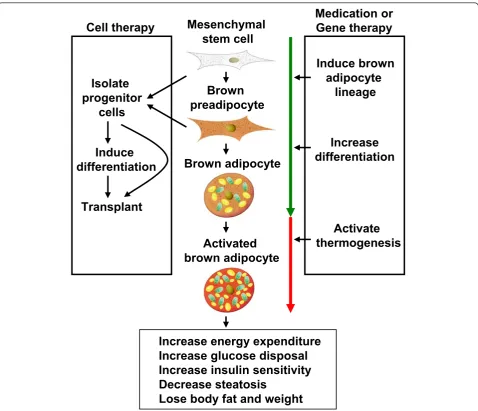
Obesity is defined not only as an excess of body weight, but also an increased adipose tissue accretion to the extent that health may be adversely affected. The anti-obesity medication phentermine and orlistat approved by Food and Drug Administration (FDA) are designed to suppress appetite and reduce fat absorption, respectively [57]. Unfortunately, phentermine has adverse effects including increased heart rate and elevated blood pressure, while orlistat may cause steatorrhea, fecal incontinence and fre-quent or urgent bowel movements. Due to the presence of BAT in adult humans, it is conceivable that weight loss can be achieved by increasing energy expenditure through activating BAT. Until now, two therapeutic strategies have been suggested in obesity control as shown in Figure 2 [58,59]. One is to stimulate the original BAT development and function by small molecules. The other is to trans-plant functional brown adipocytes induced from proper stem cells into obese patients. The second strategy can be also considered as BAT transplantation.
concerning human body weight suggests that there is a set point of body weight. Owning to the concurring effort of nerve and endocrine system, body weight will fluctuate in a narrow window. In other words, normal body weight can be regained even if short term energy intake (or expenditure) that leads to increased (or decreased) weight. However, under long term one-way stimulation, this“set point”can be resettled. This provides a possible therapy for the obese by treating them with BAT activat-ing drugs so as to lower their set point.
BAT transplantation becomes increasingly appealing due to the gradually perfection of stem cell technology. Adipose tissue transplantation has primarily been used for human reconstructive surgery. Now, transplantation of adipose tissue is being explored as a possible tool to promote the beneficial metabolic effects of subcutaneous WAT and BAT, as well as adipose-derived stem cells
[62]. Brown adipocytes at varied differentiation stages or the complete BAT can be transplanted. Cells that can be transplanted include stromal vascular fraction (SVF), pre-adipocytes and brown pre-adipocytes. Furthermore, confluent preadipocytes cultured in dishes, dedifferentiated primary adipocytes and SVF show better viability than the fully differentiated adipocytes and mature adipocytes. It is worth mentioning that the success rate of transplantation is partially determined by the degree of vascularization [63].
Problems and Perspectives
Rediscovery of BAT in human adults brought about the revival of BAT research as an anti-obesity therapy. Despite the growing number of research results reported, there are many unanswered questions on BAT development and thermogenesis, which may be better understood with
Brown
preadipocyte
Mesenchymal
stem cell
Brown adipocyte
Activated
brown adipocyte
Increase
differentiation
Activate
thermogenesis
Isolate
progenitor
cells
Induce
differentiation
Transplant
Medication or
Gene therapy
Cell therapy
Increase energy expenditure
Increase glucose disposal
Increase insulin sensitivity
Decrease steatosis
Lose body fat and weight
Induce brown
adipocyte
lineage
appropriate animal models. Animals expressing Cre recombinase under the control of UCP1 promoter have not yet been widely applied in the research of BAT. In addition, negligence of the temperature under which mice are raised may also mislead the data analysis. 30°C is the thermoneutral temperature for mice, at which mice main-tain their body temperature without adaptive nonshivering thermogenesis. However, mice are usually bred at 22°C, a temperature that UCP1 can be activated during BAT ther-mogenesis. As a result, behaviors exhibited by knockout mice under this circumstance may not solely reflect the gene function. In addition to environmental temperature, individual variance of hair density and physical activity, which influence body temperature and energy expendi-ture, will also incur error when explaining the phenotypes observed from mice.
Currently, most efforts are spent to delineate signaling pathways and transcriptional regulation. Emerging areas like RNA editing, alternative splicing, non-coding RNA and epigenetic modification are largely unexplored. During the preparation of this review, miR-193b-365, a brown-fat-enriched miRNA cluster, was identified as a key regulator of brown fat development [63]. Since the BAT develop-ment and function are highly orchestrated and complex processes, regulatory mechanisms involved deserve more attention.
Conclusions
With the help of modern technology, multiple studies con-clusively show that functional BAT exists in adult humans, and is inversely correlated with BMI, adipose tissue mass, glucose and insulin levels. Recently, studies were focused on illustrating which factors determine the unique feature of BAT. As summarized above, a great number of mole-cules are involved in the regulation of brown adipocyte differentiation and thermogenesis, be it in direct or indir-ect ways. Animal studies contribute a great deal for us to understand BAT development and function. However, a full understanding of BAT biology in humans will only be completed with clinical evidence. Taking advantage of the advancement of biomedical technology, we are expecting the next leap on BAT research.
List of abbreviations used
WAT: white adipose tissue; BAT: brown adipose tissue; TG: triglyceride; TGL: TG-rich lipoproteins; UCP1: uncoupling protein 1; cAMP: cyclic AMP; PRDM16: PR domain containing 16; Myf5: myogenic factor 5; PET/CT: positron emission computed tomography/computed tomography; BMI: body mass index; PGC-1α: Peroxisome proliferator-activated receptor gamma coactivator 1-alpha; PPAR: peroxisome proliferator-activated receptor; NRF: nuclear respiratory factor; CREB: cAMP response element-binding protein; RIP140: receptor-interacting protein 140; Dio2: type II deiodinase; ctBP: C-terminal-binding protein; C/EBP: CCAAT-enhancer-binding protein; pRb: retinoblastoma protein; MEF: mouse embryonic fibroblast; COX: cyclooxygenase; DNP: 2, 4-dinitrophenol; SVF: stromal vascular fraction.
Acknowledgements
We thank Dr. Caroline Kim and Dr. Changxue Lu for critical comments and reading our manuscript. This work was partially supported by grants from the One Hundred Talents Program of the Chinese Academy of Sciences, the Ministry of Science and Technology of China (973 Program 2009CB919000, 2010CB912500), the National Natural Science Foundation (30970587, 31070679), the Science and Technology Commission of Shanghai Municipality (10ZR1435000).
Author details
1Key Laboratory of Nutrition and Metabolism, Institute for Nutritional
Sciences, Shanghai Institutes for Biological Sciences, Chinese Academy of Sciences, Shanghai, China.2Graduate School of the Chinese Academy of Sciences, Chinese Academy of Sciences, Shanghai, China.
Authors’contributions
XY wrote the manuscript, SS, YZ, and HY revised the manuscript. All authors read and approved the final manuscript.
Competing interests
The authors declare that they have no competing interests.
Received: 5 August 2011 Accepted: 28 October 2011 Published: 28 October 2011
References
1. Cannon B, Nedergaard J:Brown adipose tissue: function and physiological significance.Physiol Rev2004,84:277-359.
2. Nedergaard J, Bengtsson T, Cannon B:New powers of brown fat: fighting the metabolic syndrome.Cell Metab2011,13:238-240.
3. Bartelt A, Bruns OT, Reimer R, Hohenberg H, Ittrich H, Peldschus K, Kaul MG, Tromsdorf UI, Weller H, Waurisch C,et al:Brown adipose tissue activity controls triglyceride clearance.Nat Med2011,17:200-205.
4. Rothwell NJ, Stock MJ:Luxuskonsumption, diet-induced thermogenesis and brown fat: the case in favour.Clin Sci (Lond)1983,64:19-23. 5. Collins S, Cao W, Robidoux J:Learning new tricks from old dogs:
beta-adrenergic receptors teach new lessons on firing up adipose tissue metabolism.Mol Endocrinol2004,18:2123-2131.
6. Seale P, Kajimura S, Yang W, Chin S, Rohas LM, Uldry M, Tavernier G, Langin D, Spiegelman BM:Transcriptional control of brown fat determination by PRDM16.Cell Metab2007,6:38-54. 7. Seale P, Bjork B, Yang W, Kajimura S, Chin S, Kuang S, Scime A,
Devarakonda S, Conroe HM, Erdjument-Bromage H,et al:PRDM16 controls a brown fat/skeletal muscle switch.Nature2008,454:961-967.
8. Cousin B, Cinti S, Morroni M, Raimbault S, Ricquier D, Penicaud L, Casteilla L:
Occurrence of Brown Adipocytes in Rat White Adipose-Tissue - Molecular and Morphological Characterization.Journal of Cell Science1992,103:931-942. 9. Vegiopoulos A, Muller-Decker K, Strzoda D, Schmitt I, Chichelnitskiy E,
Ostertag A, Berriel Diaz M, Rozman J, Hrabe de Angelis M, Nusing RM,et al:
Cyclooxygenase-2 controls energy homeostasis in mice by de novo recruitment of brown adipocytes.Science2010,328:1158-1161. 10. Houstek J, Vizek K, Pavelka S, Kopecky J, Krejcova E, Hermanska J,
Cermakova M:Type II iodothyronine 5’-deiodinase and uncoupling protein in brown adipose tissue of human newborns.J Clin Endocrinol Metab1993,77:382-387.
11. Frontini A, Cinti S:Distribution and development of brown adipocytes in the murine and human adipose organ.Cell Metab2010,11:253-256. 12. Cypess AM, Lehman S, Williams G, Tal I, Rodman D, Goldfine AB, Kuo FC,
Palmer EL, Tseng YH, Doria A,et al:Identification and importance of brown adipose tissue in adult humans.N Engl J Med2009,360:1509-1517. 13. van Marken Lichtenbelt WD, Vanhommerig JW, Smulders NM,
Drossaerts JM, Kemerink GJ, Bouvy ND, Schrauwen P, Teule GJ: Cold-activated brown adipose tissue in healthy men.N Engl J Med2009,
360:1500-1508.
14. Virtanen KA, Lidell ME, Orava J, Heglind M, Westergren R, Niemi T, Taittonen M, Laine J, Savisto NJ, Enerback S, Nuutila P:Functional brown adipose tissue in healthy adults.N Engl J Med2009,360:1518-1525. 15. Jia JJ, Tian YB, Cao ZH, Tao LL, Zhang X, Gao SZ, Ge CR, Lin QY, Jois M:The
16. Mori H, Okazawa H, Iwamoto K, Maeda E, Hashiramoto M, Kasuga M:A polymorphism in the 5’untranslated region and a Met229–> Leu variant in exon 5 of the human UCP1 gene are associated with susceptibility to type II diabetes mellitus.Diabetologia2001,44:373-376.
17. Oppert JM, Vohl MC, Chagnon M, Dionne FT, Cassard-Doulcier AM, Ricquier D, Perusse L, Bouchard C:DNA polymorphism in the uncoupling protein (UCP) gene and human body fat.Int J Obes Relat Metab Disord 1994,18:526-531.
18. Sears IB, MacGinnitie MA, Kovacs LG, Graves RA:Differentiation-dependent expression of the brown adipocyte uncoupling protein gene: Regulation by peroxisome proliferator-activated receptor gamma.Molecular and Cellular Biology1996,16:3410-3419.
19. Rabelo R, Schifman A, Rubio A, Sheng XY, Silva JE:Delineation of Thyroid Hormone-Responsive Sequences within a Critical Enhancer in the Rat Uncoupling Protein Gene.Endocrinology1995,136:1003-1013.
20. Larose M, CassardDoulcier AM, Fleury C, Serra F, Champigny O, Bouillaud F, Ricquier D:Essential cis-acting elements in rat uncoupling protein gene are in an enhancer containing a complex retinoic acid response domain.
Journal of Biological Chemistry1996,271:31533-31542.
21. Puigserver P, Wu Z, Park CW, Graves R, Wright M, Spiegelman BM:A cold-inducible coactivator of nuclear receptors linked to adaptive thermogenesis.Cell1998,92:829-839.
22. Nicholls DG:Hamster brown-adipose-tissue mitochondria. Purine nucleotide control of the ion conductance of the inner membrane, the nature of the nucleotide binding site.European Journal of Biochemistry 1976,62:223-228.
23. Rial E, Nicholls DG:A history of the first uncoupling protein, UCP1.
Journal of Bioenergetics and Biomembranes1999,31:399-406.
24. Locke RM, Rial E, Scott ID, Nicholls DG:Fatty-Acids as Acute Regulators of the Proton Conductance of Hamster Brown-Fat Mitochondria.European Journal of Biochemistry1982,129:373-380.
25. Silva JE:Thermogenic mechanisms and their hormonal regulation.
Physiological Reviews2006,86:435-464.
26. Marrif H, Schifman A, Stepanyan Z, Gillis MA, Calderone A, Weiss RE, Samarut J, Silva JE:Temperature homeostasis in transgenic mice lacking thyroid hormone receptor-alpha gene products.Endocrinology2005,
146:2872-2884.
27. Bianco AC:Minireview: cracking the metabolic code for thyroid hormone signaling.Endocrinology2011,152:3306-3311.
28. Castillo M, Hall JA, Correa-Medina M, Ueta C, Kang HW, Cohen DE, Bianco AC:Disruption of thyroid hormone activation in type 2 deiodinase knockout mice causes obesity with glucose intolerance and liver steatosis only at thermoneutrality.Diabetes2011,60:1082-1089. 29. Bianco AC, Silva JE:Intracellular conversion of thyroxine to
triiodothyronine is required for the optimal thermogenic function of brown adipose tissue.J Clin Invest1987,79:295-300.
30. de Jesus LA, Carvalho SD, Ribeiro MO, Schneider M, Kim SW, Harney JW, Larsen PR, Bianco AC:The type 2 iodothyronine deiodinase is essential for adaptive thermogenesis in brown adipose tissue.J Clin Invest2001,
108:1379-1385.
31. Thomas C, Auwerx J, Schoonjans K:Bile acids and the membrane bile acid receptor TGR5–connecting nutrition and metabolism.Thyroid2008,
18:167-174.
32. Houten SM, Watanabe M, Auwerx J:Endocrine functions of bile acids.
EMBO J2006,25:1419-1425.
33. Hansen JB, Kristiansen K:Regulatory circuits controlling white versus brown adipocyte differentiation.Biochem J2006,398:153-168. 34. Uldry M, Yang W, St-Pierre J, Lin J, Seale P, Spiegelman BM:
Complementary action of the PGC-1 coactivators in mitochondrial biogenesis and brown fat differentiation.Cell Metab2006,3:333-341. 35. Scarpulla RC:Transcriptional paradigms in mammalian mitochondrial
biogenesis and function.Physiological Reviews2008,88:611-638. 36. Leone TC, Lehman JJ, Finck BN, Schaeffer PJ, Wende AR, Boudina S,
Courtois M, Wozniak DF, Sambandam N, Bernal-Mizrachi C,et al: PGC-1alpha deficiency causes multi-system energy metabolic derangements: muscle dysfunction, abnormal weight control and hepatic steatosis.PLoS Biol2005,3:e101.
37. Lin J, Wu PH, Tarr PT, Lindenberg KS, St-Pierre J, Zhang CY, Mootha VK, Jager S, Vianna CR, Reznick RM,et al:Defects in adaptive energy metabolism with CNS-linked hyperactivity in PGC-1alpha null mice.Cell 2004,119:121-135.
38. Treuter E, Albrektsen T, Johansson L, Leers J, Gustafsson JA:A regulatory role for RIP140 in nuclear receptor activation.Mol Endocrinol1998,
12:864-881.
39. Leonardsson G, Steel JH, Christian M, Pocock V, Milligan S, Bell J, So PW, Medina-Gomez G, Vidal-Puig A, White R, Parker MG:Nuclear receptor corepressor RIP140 regulates fat accumulation.Proc Natl Acad Sci USA 2004,101:8437-8442.
40. Steel JH, White R, Parker MG:Role of the RIP140 corepressor in ovulation and adipose biology.J Endocrinol2005,185:1-9.
41. Christian M, Kiskinis E, Debevec D, Leonardsson G, White R, Parker MG:
RIP140-targeted repression of gene expression in adipocytes.Mol Cell Biol2005,25:9383-9391.
42. Spiegelman BM, Kajimura S, Seale P, Tomaru T, Erdjument-Bromage H, Cooper MP, Ruas JL, Chin S, Tempst P, Lazar MA:Regulation of the brown and white fat gene programs through a PRDM16/CtBP transcriptional complex.Genes & Development2008,22:1397-1409.
43. Spiegelman BM, Kajimura S, Seale P, Kubota K, Lunsford E, Frangioni JV, Gygi SP:Initiation of myoblast to brown fat switch by a PRDM16-C/EBP-beta transcriptional complex.Nature2009,460:1154-U1125.
44. Seale P, Conroe HM, Estall J, Kajimura S, Frontini A, Ishibashi J, Cohen P, Cinti S, Spiegelman BM:Prdm16 determines the thermogenic program of subcutaneous white adipose tissue in mice.J Clin Invest2011,121:96-105. 45. Logan CY, Nusse R:The Wnt signaling pathway in development and
disease.Annu Rev Cell Dev Biol2004,20:781-810.
46. Kang S, Bajnok L, Longo KA, Petersen RK, Hansen JB, Kristiansen K, MacDougald OA:Effects of Wnt signaling on brown adipocyte differentiation and metabolism mediated by PGC-1alpha.Mol Cell Biol 2005,25:1272-1282.
47. Longo KA, Wright WS, Kang S, Gerin I, Chiang SH, Lucas PC, Opp MR, MacDougald OA:Wnt10b inhibits development of white and brown adipose tissues.J Biol Chem2004,279:35503-35509.
48. Hatakeyama M, Weinberg RA:The role of RB in cell cycle control.Prog Cell Cycle Res1995,1:9-19.
49. Jacks T, Lipinski MM:The retinoblastoma gene family in differentiation and development.Oncogene1999,18:7873-7882.
50. Wells J, Boyd KE, Fry CJ, Bartley SM, Farnham PJ:Target gene specificity of E2F and pocket protein family members in living cells.Molecular and Cellular Biology2000,20:5797-5807.
51. Kristiansen K, Hansen JB, Jorgensen C, Petersen RK, Hallenborg P, De Matteis R, Boye HA, Petrovic N, Enerback S, Nedergaard J,et al:
Retinoblastoma protein functions as a molecular switch determining white versus brown adipocyte differentiation.Proceedings of the National Academy of Sciences of the United States of America2004,101:4112-4117. 52. Rudnicki MA, Scime A, Grenier G, Huh MS, Gillespie MA, Bevilacqua L,
Harper ME:Rb and p107 regulate preadipocyte differentiation into white versus brown fat through repression of PGC-1 alpha.Cell Metabolism 2005,2:283-295.
53. Smith WL, DeWitt DL, Garavito RM:Cyclooxygenases: structural, cellular, and molecular biology.Annu Rev Biochem2000,69:145-182.
54. Dubois RN, Abramson SB, Crofford L, Gupta RA, Simon LS, Van De Putte LBA, Lipsky PE:Cyclooxygenase in biology and disease.Faseb Journal1998,12:1063-1073.
55. Fitzpatrick FA:Cyclooxygenase enzymes: Regulation and function.Current Pharmaceutical Design2004,10:577-588.
56. Madsen L, Pedersen LM, Lillefosse HH, Fjaere E, Bronstad I, Hao Q, Petersen RK, Hallenborg P, Ma T, De Matteis R,et al:UCP1 Induction during Recruitment of Brown Adipocytes in White Adipose Tissue Is Dependent on Cyclooxygenase Activity.PLoS ONE2010,5.
57. Halpern A:Pharmacological Treatment of Obesity (Clinical Aspects).
Proceedings of the 13th International Congress of Endocrinology2008, 59-61. 58. Cypess AM, Kahn CR:Brown fat as a therapy for obesity and diabetes.
Curr Opin Endocrinol Diabetes Obes2010,17:143-149.
59. Kahn CR, Cypess AM:Brown fat as a therapy for obesity and diabetes.
Current Opinion in Endocrinology Diabetes and Obesity2010,17:143-149. 60. Bartlett J, Brunner M, Gough K:Deliberate poisoning with dinitrophenol
(DNP): an unlicensed weight loss pill.Emergency Medicine Journal2010,
27:159-160.
62. Kahn CR, Tran TT:Transplantation of adipose tissue and stem cells: role in metabolism and disease.Nature Reviews Endocrinology2010,6:195-213. 63. Van RL, Roncari DA:Complete differentiation in vivo of implanted
cultured adipocyte precursors from adult rats.Cell Tissue Res1982,
225:557-566.
doi:10.1186/2045-3701-1-35
Cite this article as:Yaoet al.:Recent progress in the study of brown adipose tissue.Cell & Bioscience20111:35.
Submit your next manuscript to BioMed Central and take full advantage of:
• Convenient online submission
• Thorough peer review
• No space constraints or color figure charges
• Immediate publication on acceptance
• Inclusion in PubMed, CAS, Scopus and Google Scholar
• Research which is freely available for redistribution