R E S E A R C H
Open Access
Modified halloysite nanotubes reduce the
toxic effects of zearalenone in gestating
sows on growth and muscle development
of their offsprings
Rui Gao
†, Qingwei Meng
†, Jianan Li, Min Liu, Yuanyuan Zhang, Chongpeng Bi and Anshan Shan
*Abstract
Background:Zearalenone (ZEN) is an estrogenic mycotoxin that is primarily produced by Fusarium fungi and has been proven to affect the reproductive capacity of many species to varying degrees. The present experiment was designed to study the maternal persistent effects of zearalenone toxicity in gestating sows on growth and muscle development of their offsprings, and the alleviation of zearalenone toxicity by modified halloysite nanotubes (MHNTs). Methods:Eighteen sows were fed with one of three dietary treatments that included the following: (1) a control diet, (2) a contaminated grain diet (with 50 % moldy corn, 2.77 mg/kg ZEN), and (3) a contaminated grain diet (with 50 % moldy corn, 2.76 mg/kg ZEN) + 1 % MHNTs. Each sow was exclusively fed its experimental diets from 35 to 70 d of gestation at a total of 2 kg daily. Muscle samples were collected from six piglets per treatment at birth, weaning and finishing.
Results:The results showed that feeding the sows with the ZEN-contaminated diets from 35 to 70 d of gestation decreased the ADG, ADFI and G:F of their offsprings (P< 0.05). The muscle fiber numbers in the newborn, weaning and growing-finishing pigs and the muscle fiber diameters at birth and weaning were also decreased by maternal ZEN exposure (P< 0.05). The expressions ofIGF-I,IGF-II,Myf-5andMstnat birth andIGF-II,Pax7,Myf-5andMyoD1at weaning were altered by feeding gestating sows with ZEN-contaminated diets (P< 0.05). The MHNTs reduced most of the ZEN-induced toxic effects: the ADG and ADFI on growth performance, the muscle fiber numbers at weaning and finishing and the muscle fiber diameters at weaning (P< 0.05). The expression levels ofIGF-IIandMstnin newborn piglets andIGF-IIandMyf-5in weaning piglets were also prevented by adding 1 % MHNTs (P< 0.05). Conclusions:The present study demonstrated that the offsprings of sows fed with ZEN-contaminated diets from 35 to 70 day of gestation exhibited weakening on growth performance, physiological changes in their muscle fibers and alterations of mRNA expression in their muscle tissues, and also indicated that MHNTs prevented most of the ZEN-induced weakening in the muscle tissues.
Keywords:Growth, MHNTs, Muscle development, Offsprings, Sows, Zearalenone
* Correspondence:[email protected] Rui Gao and Qingwei Meng are co-first authors †Equal contributors
Institute of Animal Nutrition, Northeast Agricultural University, Harbin 150030, P. R. China
Background
Fusarium graminearum is most frequently isolated from maize in temperate climates, and zearalenone production often occurs during the cold weather storage of high-moisture feeds carrying the mold [1]. Zearalenone (ZEN) is a mycotoxin that is produced primarily by fungi of the genus Fusarium in foods and feeds. ZEN has frequently been implicated in reproductive disorders in farm animals and occasionally in hyperoestrogenic syndromes in humans [2]. In farm animals, swine seem to be particularly sensitive to mycotoxins [3]. Meanwhile, maternal effects which play an important role on offspring’s growth and muscle devel-opment were proved by many previous researchers [4]. ZEN-related problems frequently occur in piglets under natural conditions and are by exposure in the uterus, the placental transfer from an exposed sow to her piglets and by ZEN stored in the sow during gestation and released via the suckling of the piglets [5].
There has been an increased interest in the application of halloysite nanotubes (HNTs) in order to find a way to detoxify contaminated feedstuffs or diets which is appro-priate for large quantities of raw material sources, inex-pensive, simple and results in products of stable quality. Halloysite is a type of aluminosilicate clay with a hollow nano-tubular structure and a set of characteristics that make it cheap, abundantly available, durable, highly mech-anically strong and biocompatible [6]. Around the world, halloysite nanotubes have been used as nanocomposites, nanocontainers [7] and new drug carriers in medicine [8, 9], but the use of HNTs in animals as adsorbents has not yet been reported.
Indeed, many studies of laboratory animals, farm ani-mals and humans have reported that exposure to the es-trogenic effects of ZEN causes relevant reproductive performance alterations [10, 11]. It has been reported that maternal zearalenone exposure causes fetal malformations and physiological alterations to sexual organs [12, 13]. However, few studies have examined the effects of zear-alenone on growth or muscle development, particularly in combination with maternal effects. These factors prompted us to begin a more extensive investigation of the possible effects of maternal zearalenone exposure on offspring growth and muscle development and to exploit a new adsorbent to mitigate the negative effects of zearalenone contamination.
Methods
Mold strain
Fusarium graminearum has previously been shown to produce ZEN in corn [14]. The fungus used in this ex-periment was purchased from the Agricultural Culture Collection of China (No. ACCC36249) and cultivated on potato dextrose agar (PDA, potato extract 0.4 %, glucose 2 % and agar 1.5 %, pH 5.6 ± 0.2). The experimental
culture media were obtained from Fluka (Bornem, Belgium) [15].
The corn used in this experiment was obtained from Xiang Fang Experimental Bases (Northeast Agricultural University, China) and milled in a hammer mill with a 40-mesh screen (Trapp-TRF model 90). The in vitro ZEN production was conducted according to the procedures outlined by Lıgia Martins [14]. The studies of mycotoxin production by Fusarium graminearum were performed in duplicate on trays containing 1,000 g of sterilized cracked corn following the addition of 400 mL of distilled water and adjusting the aw to 0.98 by water activity meter (HD-4, HuaKe Instrument & Meter Co. Ltd., Wuxi, China).
The corn contained no fungal infection or ZEN contam-ination. Autoclaved substrate was inoculated with 40 mL of the spore suspension according to the following pro-cedure: 100 mL of sterile distilled water were added to each slant of the 5-day-old culture, and the surface of the agar was gently scraped to produce a turbid suspension with 1 × 10¹⁴ spores/mL. One hundred milliliters of this suspension were added to the cracked corn. The inocu-lated flasks were stirred daily for the first 5 d. The culture conditions used in this experiment, 28 °C for 15 d followed by 12 °C with ZEN peaking on the 35th day of incubation, reflected the earlier work reported by Lıgia Martins [14]. To equalize the moisture contents of the samples, each sample was dried at 60 °C for 96 h and stored in a freezer at−20 °C until analysis [16].
Modification of the adsorbent
The adsorbent used in experiment was that described by Zhang et al. [17]. Halloysite nanotube powder (HNTs), was purchased and refined from Golden Sunshine Ceram-ics Co., Ltd. (Zhengzhou, China). The molecular formula of halloysite nanotube was Al2Si2O5(OH)4· nH2O, com-posed of 95 % kaolinite, trace amounts of quartz and natroalunite. The crystal diameter was 10 to 50 nm and the crystal length was less than 1 μm. The proportion of granule (<2μm) was 70 % in the original mineral.
The previously reported method of Jinhua was used to prepare the powder [7]. All solutions were prepared using distilled water. The powder was prepared as fol-lows: A water suspension solution (5 % in mass) was prepared by adding water to dry halloysite mineral. The suspension solution was intensively stirred for 2 h and sprayed to dry at 200 °C to obtain fine powder. Before use, dry halloysite powder was sieved to elimin-ate aggregelimin-ates.
mixed at a speed of 2,300 g with a reaction time of 10 min at 50 °C. When the reaction was complete, the suspensions were filtered, washed three times with deionised water, dried at 80 °C and crush 100 g of HNT to obtain particles that were less than 45μm in a beater mill at 7,400 g for 3 min. The made-up particles were added in the contaminated diets evenly to make the MHNTs diets.
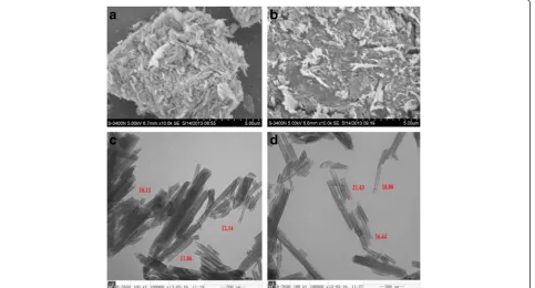
Figure 1 shows the images after electric microscope (EM). It can be seen that the samples consisted of cylin-drical tubes. After treatment with SKC, the dispersion of the halloysite (Fig. 1b) was increased compared to the untreated HNT powder (Fig. 1a). Moreover, the lumen of modified nanotubes shown in Fig. 1d was enlarged compared with untreated nanotubes (Fig. 1c). As shown in Fig. 1c and d, the external surface of the treated HNT was cruder than the natural halloysite [17].
Animals and experimental design
Eighteen pregnant Yorkshire sows were randomly divided into the following three treatment groups (6 per treat-ment): (1) control, (2) contaminated grains (with 50 % moldy corn); and (3) contaminated grains (with 50 % moldy corn) + 1 % modified HNTs (MHNTs). The doses of the MHNTs were selected based on the research of Jiang [19, 20]. The pregnant sows were housed in individ-ual stalls after 35 d of gestation (GDs). Each sow was fed
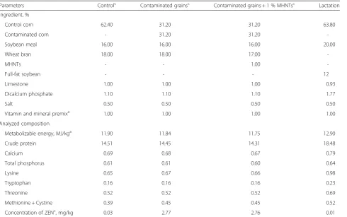
exclusively with the appropriate experimental diet from d 35 to d 70 of gestation at a total of 2 kg daily. All of the feedstuffs were subjected to post-processing analytical control. The feed compositions were compared using vali-dated analytical methods (National Standards of the Peo-ple’s Republic of China, GB/T 19540–2004). ZEN was the major contaminant and was present at 0.03 mg/kg in the control diet, 2.77 mg/kg in the contaminated diet, 2.76 mg/kg in the adding adsorbent contaminated diet. The concentration of deoxynivalenol (DON) which was also produced by Fusarium graminearum was 0.04 mg/kg in the contaminated diet, lower than the limits of national standards (1 mg/kg). Analyses of the corn and diets with GC-MS were performed to provide detailed characteriza-tions of the trichothecene mycotoxin patterns and re-vealed that the B-trichothecene mycotoxins, such as 15-acetyldeoxynivalenol, 3-acetyldeoxynivalenol, and niva-lenol, and A-trichothecene mycotoxin HT-2 toxins were lower than the detection limits [21]. At 7 d of age all pig-lets, the piglets received an iron injection, and the males were castrated. The piglets were weaned at 21 d of age and moved to post-weaning rooms with an ambient temperature of 27 °C. After weaning, the piglets had ad libitum access to the standard diets and water intake until finishing in individual pens. All experimental diets (Table 1) were formulated to meet or exceed the National Research Council nutrient requirements (2012) [22].
Sample collection
All of the animal experimental procedures were ap-proved by the Ethical and Animal Welfare Committee of Heilongjiang Province, China.
Six piglets in each group were slaughtered via an intraarterial injection of pentobarbital (200 mg/kg) for general anesthesia at birth and six more at weaning. At the end of the experiment, 18 growing-finishing pigs from the three treatments were transported to the abat-toir for slaughter. At birth, weaning and finishing, one pig was selected from each litter and slaughtered to col-lect samples.
After slaughter, longissimus muscle samples were quickly collected, frozen in liquid nitrogen, stored at −80 °C and analyzed for gene expression by RNA ex-traction, followed by quantitative reverse transcription PCR [11]. Portions of the longissimus muscle samples were fixed in 4 % paraformaldehyde in phosphate buf-fer (0.12 mol/L, pH 7.4) for histochemical examin-ation. The remaining organs were directly stored at −20 °C for analysis.
Growth performance
The growth performance was evaluated from weaning to finishing. All the pigs were weighed individually at weaning and slaughter. The feed intakes per pen were recorded after weaning. The average daily gain (ADG), average daily feed intake (ADFI) and gain/feed (G:F) values were calculated.
Histochemical examination
The longissimus muscle samples were embedded in par-affin and cut into 10-μm sections. The muscle sections were rehydrated via a series of incubations in xylene and ethanol solutions and then stained with hematoxylin and eosin for standard light microscopy. Ten fields were ran-domly selected to quantify the muscle fiber diameters. The majority of the muscle fibers were circular; thus, the diameters were easily measured. For the irregular muscle fibers, the maximum and minimum diameters of the muscle fiber circle were measured, and the average value of the maximum and minimum was regarded as the diameter of the fiber. The diameters of 10 muscle fiber per field were measured, and 100 muscle fibers per
Table 1Percentage composition of the diet
Parameters Controlc Contaminated grainsc Contaminated grains + 1 % MHNTsc Lactation
Ingredient, %
Control corn 62.40 31.20 31.20 63.80
Contaminated corn - 31.20 31.20
-Soybean meal 16.00 16.00 16.00 20.00
Wheat bran 18.00 18.00 17.00
-MHNTs - - 1.00
-Full-fat soybean - - - 12
Limestone 1.00 1.00 1.00 0.93
Dicalcium phosphate 1.10 1.10 1.10 1.77
Salt 0.50 0.50 0.50 0.50
Vitamin and mineral premixa 1.00 1.00 1.00 1.00
Analyzed composition
Metabolizable energy, MJ/kga 11.90 11.84 11.75 12.90
Crude protein 14.51 14.45 14.31 18.48
Calcium 0.69 0.68 0.67 0.79
Total phosphorus 0.61 0.61 0.60 0.64
Lysine 0.65 0.67 0.66 0.98
Tryptophan 0.16 0.16 0.16 0.23
Threonine 0.52 0.52 0.52 0.69
Methionine + Cystine 0.39 0.45 0.45 0.52
Concentration of ZENc, mg/kg 0.03 2.77 2.76 0.01
a
Provided the following per kilogram of diet: Cu, 18.2 mg; Zn, 126.0 mg; Se, 0.3 mg; Mn, 50.5 mg; Fe, 150.3 mg; I, 0.4 mg; vitamin A, 11, 050 IU; vitamin D, 2,310 IU; vitamin E, 62.8 IU; vitamin K, 2.6 mg; riboflavin, 5.8 mg; pantothenic acid, 20 mg; niacin, 25 mg; vitamin B12, 326μg; folate, 6.5 mg; pyridoxine, 1.8 mg; biotin, 350μg; and thiamin, 1.9 mg
b
Calculated values according to the Tables of Feed Composition and Nutritive Values in China [42] in this study c
Control: control diet; Contaminated grains: instead of 50 % moldy corn; Contaminated grains: instead of 50 % moldy corn + 1 % MHNTs;ZENZearalenone,MHNTs
sample were quantified using the Motic Images Plus 2.0 software. The muscle fiber diameters were measured in a blinded fashion. The averaged data were used for culations [4]. The percentages of muscle fibers were cal-culated based on the total fiber numbers per examined area, and this latter value for the control treatment was regarded as 100 % [23].
Quantitative real-time PCR
Total RNA was extracted from the muscles (30 mg of tis-sue) of the piglets using the Trizol reagent (E.Z.N.A. ® Total RNA Kit, Omega Bio-tek, Inc., United States). The RNA concentration was measured with a spectrophotometer at 260/280 nm. The quality of the RNA was estimated by de-tecting the number of bands by agarose gel electrophoresis.
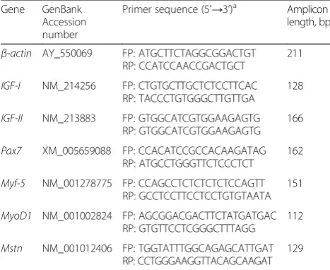
SYBR green I real-time polymerase chain reactions (RT-PCR) were used to measure the mRNA expression ofIGF-I,IGF-II,Pax7,Myf-5,MyoD1,Mstnand β-actin. First-strand cDNA was synthesized from 5 μg of total RNA (processed using DNase) using oligo (dT) primers and Superscript II reverse transcriptase according to the manufacturer’s instructions (Tiangen Biotech Co., Ltd, Beijing, China). Real-time PCR was performed in an ABI PRISM 7500 SDS thermal cycler (Applied Biosys-tems, Foster City, CA). Each sample was analyzed in triplicate. The primers used in the analyses are listed in Table 2. The reactions were performed with 2.0 μL of first-strand cDNA and 0.8 μL of sense and anti-sense primers in a final volume of 20μL as recommended by the SYBR real-time PCR kit (TaKaRa® BIO CATALOG, Da Lian, China). The RT-PCR conditions were as fol-lows: 1 cycle at 95 °C for 30 s, and 40 cycles at 95 °C for 5 s and 60 °C for 34 s. The relative expressions of the inflammatory cytokine mRNAs were determined with the 2-ΔCtmethod [24].
Statistical analyses
All data were analyzed with SPSS software (SPSS Inc., Chicago, IL, USA) and the results were expressed as LSMEANS, SEM and P-values. When treatment differ-ences were detected by ANOVA, the significance of the differences between the treatments was determined with Duncan’s multiple range tests. Significance was considered at the probability level ofP< 0.05.
Results
Growth performance
The growth performance of offsprings are presented in Table 3. The average daily gain and the average daily feed intake decreased in the corn contaminated with mold group compared with the control group (P< 0.05). The gain/feed was also decreased by maternal zearalenone exposure (P< 0.05).
The average daily gain raised by adding 1 % MHNTs to maternal diets (P< 0.05), but the level did not reach the ADG of control animals. The average daily feed in-take also raised following the addition of 1 % MHNTs to the diets (P< 0.05).
Muscle fiber diameters and numbers
The muscle fiber diameters and numbers of offsprings are presented in Table 4. The muscle fiber numbers in the newborn, weaning and growing-finishing pigs were de-creased in ZEN-treated group compared with the control group (P< 0.05). The moldy corn group exhibited the lower muscle fiber diameters at birth and weaning com-pared to the control group (P< 0.05). There were no dif-ferences in the muscle fiber diameters of finishing pigs between the three treatments.
Muscle fiber numbers and diameters at birth, though higher compared to moldy corn group, do not differ sig-nificantly when MHNTs were added to maternal diets. Nevertheless, following the addition of MHNTs to ma-ternal diets muscle fiber numbers increased at weaning and slaughter up to control animals levels (P< 0.05), and MHNTs reduced the damage in muscle fiber diameters at weaning (P< 0.05).
Table 2Primers used for quantitative real-time PCR
Gene GenBank Accession number
Primer sequence (5’→3’)a Amplicon length, bp
β-actin AY_550069 FP: ATGCTTCTAGGCGGACTGT
RP: CCATCCAACCGACTGCT
211
IGF-I NM_214256 FP: CTGTGCTTGCTCTCCTTCAC
RP: TACCCTGTGGGCTTGTTGA
128
IGF-II NM_213883 FP: GTGGCATCGTGGAAGAGTG
RP: GTGGCATCGTGGAAGAGTG 166
Pax7 XM_005659088 FP: CCACATCCGCCACAAGATAG RP: ATGCCTGGGTTCTCCCTCT
162
Myf-5 NM_001278775 FP: CCAGCCTCTCTCTCTCCAGTT
RP: GCCTCCTTCCTCCTGTGTAATA 151
MyoD1 NM_001002824 FP: AGCGGACGACTTCTATGATGAC
RP: GTGTTCCTCGGGCTTTAGG
112
Mstn NM_001012406 FP: TGGTATTTGGCAGAGCATTGAT RP: CCTGGGAAGGTTACAGCAAGAT
129
a
FPforward primer,RPreverse primer
Table 3Growth performance
Item Control 2.77 mg/kg ZEN 2.76 mg/kg ZEN+ 1 % MHNTs
ADG, kg/d 0.68 ± 0.01a 0.61 ± 0.01c 0.64 ± 0.01b
ADFI, kg/d 1.88 ± 0.03a 1.77 ± 0.03b 1.86 ± 0.03a
G:F 0.36 ± 0.01a 0.34 ± 0.01b 0.35 ± 0.01ab
Data are means ± SEM
ZENzearalenone
MHNTsmodified halloysite nanotubes
ADGaverage daily gain,ADFIaverage daily feed intake,G:Fgain/feed a, b, c
The mRNA expression in the longissimus muscle
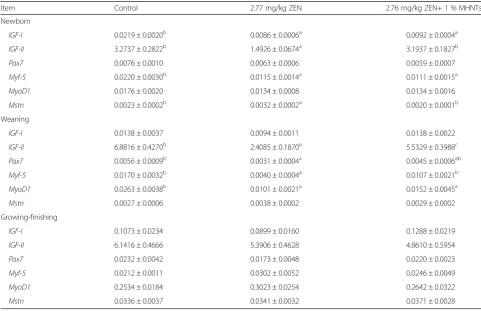
The mRNA expressions of IGF-I, IGF-II, Pax7, Myf-5,
MyoD1andMstnin the longissimus muscle are presented in Table 5. The mRNA expression ofIGF-Iwas decreased in the ZEN-contaminated group at birth (P< 0.05). No dif-ferences in the mRNA expression ofIGF-I were observed
between the ZEN-contaminated group and the control group at weaning. Maternal zearalenone exposure re-duced the expression ofIGF-II both at birth and wean-ing (P< 0.05). The results for Myf-5 and MyoD1 were similar: the expression of Myf-5 was reduced at birth and weaning (P< 0.05). The expression of MyoD1 was
Table 4Muscle fiber diameters and numbers
Item Control 2.77 mg/kg ZEN 2.76 mg/kg ZEN+ 1 % MHNTs
Newborn
Muscle fibre numbers 1.00 ± 0.02b 0.93 ± 0.02a 0.95 ± 0.02ab
Muscle fibre diameter 9.98 ± 0.42b 8.40 ± 0.33a 9.48 ± 0.40ab
Weaning
Muscle fibre numbers 1.00 ± 0.02b 0.93 ± 0.03a 1.01 ± 0.01b
Muscle fibre diameter 18.55 ± 0.86b 15.40 ± 0.79a 18.78 ± 0.96b
Growing-finishing
Muscle fibre numbers 1.00 ± 0.01b 0.97 ± 0.01a 1.00 ± 0.01b
Muscle fibre diameter 49.55 ± 1.63 49.12 ± 1.04 48.78 ± 1.64
Data are means ± SEM
ZENzearalenone
MHNTsmodified halloysite nanotubes a, b, c
means within a row with no common superscripts differ significantly (P< 0.05)
Table 5The mRNA expression in the longissimus muscle
Item Control 2.77 mg/kg ZEN 2.76 mg/kg ZEN+ 1 % MHNTs
Newborn
IGF-I 0.0219 ± 0.0020b 0.0086 ± 0.0006a 0.0092 ± 0.0004a
IGF-II 3.2737 ± 0.2822b 1.4926 ± 0.0674a 3.1937 ± 0.1827b
Pax7 0.0076 ± 0.0010 0.0063 ± 0.0006 0.0059 ± 0.0007
Myf-5 0.0220 ± 0.0030b 0.0115 ± 0.0014a 0.0111 ± 0.0015a
MyoD1 0.0176 ± 0.0020 0.0134 ± 0.0008 0.0134 ± 0.0016
Mstn 0.0023 ± 0.0002b 0.0032 ± 0.0002a 0.0020 ± 0.0001b
Weaning
IGF-I 0.0138 ± 0.0037 0.0094 ± 0.0011 0.0138 ± 0.0022
IGF-II 6.8816 ± 0.4270b 2.4085 ± 0.1870a 5.5329 ± 0.3988c
Pax7 0.0056 ± 0.0009b 0.0031 ± 0.0004a 0.0045 ± 0.0006ab
Myf-5 0.0170 ± 0.0032b 0.0040 ± 0.0004a 0.0107 ± 0.0021b
MyoD1 0.0263 ± 0.0038b 0.0101 ± 0.0021a 0.0152 ± 0.0045a
Mstn 0.0027 ± 0.0006 0.0038 ± 0.0002 0.0029 ± 0.0002
Growing-finishing
IGF-I 0.1073 ± 0.0234 0.0899 ± 0.0160 0.1288 ± 0.0219
IGF-II 6.1416 ± 0.4666 5.3906 ± 0.4628 4.8610 ± 0.5954
Pax7 0.0232 ± 0.0042 0.0173 ± 0.0048 0.0220 ± 0.0023
Myf-5 0.0212 ± 0.0011 0.0302 ± 0.0052 0.0246 ± 0.0049
MyoD1 0.2534 ± 0.0184 0.3023 ± 0.0254 0.2642 ± 0.0322
Mstn 0.0336 ± 0.0037 0.0341 ± 0.0032 0.0371 ± 0.0028
Data are means ± SEM
ZENzearalenone
MHNTsmodified halloysite nanotubes a, b, c
also reduced at weaning (P< 0.05), but not at birth (P> 0.05). The expression ofPax7was not affected by zearalenone at birth (P> 0.05) but reduced at weaning (P< 0.05). Maternal zearalenone exposure increased the expression ofMstnat birth (P< 0.05) but not at weaning (P> 0.05). However, there were no differences on the mRNA expressions ofIGF-I, IGF-II, Pax7,Myf-5,MyoD1 and Mstn in finishing pigs between ZEN-contaminated group and the control.
In this experiment, MHNTs reduced the toxic effects on the expressions ofIGF-IIandMstnin newborn piglets (P< 0.05), the expressions of IGF-IIand Myf-5 (P< 0.05) in weaning piglets. The results might show a tendency in reduction of toxic effects on expression of IGF-I,Pax7,
MyoD1 in weaning piglets and expressions of IGF-I and
Pax7 in growing-finishing pigs, but the mean values ob-tained do not differ statistically significantly from those obtained for animals fed the ZEN-contaminated diet.
Discussion
Because the nutritional levels of the three treatments during gestation were similar, and the other experimen-tal conditions were also identical, we hypothesize that the changes in the muscle fibers and mRNA expressions in the offsprings were predominantly caused by maternal ZEN exposure. The ameliorative effects of the treatment with MHNTs were considered to be the result of the addition of this adsorbent to the diet in our study.
The researches on growth performance and muscle development of zearalenone were lacking. Our previous research demonstrated that maternal zearalenone expos-ure in gestating sows decreased the average body weight at birth and weaning of piglets, and the average daily gain of weaning piglets were also reduced [25]. The pre-vious results were in accord with this experiment. Doll et al. [26] also observed that feed intake and growth rates were reduced in gilts fed diets with deoxynivalenol and zearalenone (DON, 8.6 mg/kg; ZEN, 1.2 mg/kg), but the toxin which took effects primarily were undefined. However, some researches had the different conclusions. Zearalenone had no effects on growth performance in prepubertal gilts [27]. Jiang et al. [19] observed gilts fed different amounts of dietary ZEN grew similarly with no differences in ADG and ADFI.
The growth and development of skeletal muscle in-cludes the increases in muscle fiber numbers (hyperpla-sia) and the enlargements in the volumes of the muscle fibers (hypertrophy). The number of muscle fibers is set a birth, and no further increases occur. The growth and development of muscle consists of only increases in the volumes of muscle fibers; thus, the fetal period plays a key in skeletal muscle development [28, 29]. In the em-bryo, a spot of muscle fibers (primary myofibers) begins to develop, and the majority of muscle fibers (secondary
myofibers) are formed in the fetal period. The primary myofibers of pigs are formed during the 38 d of early ges-tation. The secondary myofibers are formed from 46 to 95 d of gestation, and muscle fiber numbers do not increase after this period [30, 31]. Skeletal muscle lies at the bot-tom end of nutrient partitioning during development. As nutrient substances are always first assigned to the ner-vous system, organs and skeleton, skeletal muscle can eas-ily be affected by maternal nutrition fluctuations [4]. Therefore, such fluctuations can cause irreversible de-creases in the numbers of muscle fibers in the fetal period. In the present experiment, maternal ZEN exposure de-creased the muscle fiber numbers permanently, muscle fiber diameter were also reduced at birth and weaning compared with the control group. These results accorded with our previous research that the average body weight (BW) of fetuses at 70 d in pregnant, the litter birth weight, the average BW of piglet, and the born alive piglet BW at farrowing were all decreased by ZEN exposure [25]. In contrast to the current study, Kiessling et al. [32] reported that no significant changes in fiber number or diameter occur in male rats after prolonged zearalenone (1.25 or 3.75 mg/kg) feeding. ZEN has been found to be maternally toxic and fetotoxic but not teratogenic [33], and it seems that ZEN is unable to affect muscle fibers that have already formed in adult animals. Obviously, the fetuses were affected by maternal toxicity because the sows were fed contaminated diets from 35 to 70 d of gestation, which is the key period of secondary myofiber development. Dif-ferences in the muscle fiber diameters between the three treatments were not observed in the growing-finishing pigs, and these results indicate that the compensation abil-ities of the offsprings were able to partially eliminate the previous differences.
accorded with our results of muscle fiber deterioration. Zhang et al. [25] indicated that placenta weight and the apoptosis-related mRNA expression were altered in the ZEN treatment groups in the placenta and uterus of the sows and piglets. The pathologic uterine and placental changes could affect the functions of the organs and the transportation of nutrition to the fetuses, which may ex-plain the adverse effects on the fetuses [25]. We observed that the expressions of MyoD and Myf-5 were increased by ZEN-contaminated treatment at finishing, although these differences were not significant. It is possible that the offsprings in the control group had developed com-pletely, and the slower development of the offsprings in ZEN group had begun to be offset by the compensatory processes. IGFs play critical roles in skeletal muscle differ-entiation and growth, and previous studies have shown that IGFs not only stimulate myoblast proliferation but also promote myogenic differentiation, which are two mutually exclusive processes [38]. In the present study, the mRNA expression of IGF-I and IGF-II were both decreased in the ZEN-contaminated group at different periods. Previous research has shown that no changes on the expressions of IGF-I or IGF-II was observed by using a diet contaminated with fusarium (4.42 mg/kg DON, 0.048 mg/kg ZEN) from 35 to 70 d of gestation in neither sows or offsprings compared to a control diet [21]. Oliver et al. [27] observed that zearalenone (1.5 mg/ kg) does not alter skeletal muscle signaling in prepubertal gilts. The previous research seems to indicate that zearale-none might affect reproductive performance and thereby affect the development of the offsprings and that ZEN might not induce physiological changes in the muscle fibers or skeletal muscle signaling within a generation. However, our results proved that ZEN has a trans-generational toxicity that affected the muscle development of the offsprings.
With the use of an adsorbent modified with the surfac-tant SKC, the harmful effects of hydrophobic pesticides from a pollution source can be prevented by clay and soil [39], and the ZEN adsorption efficacies of HNTs and MHNTs have been shown in our previous research [17]. However, the inherent safety of halloysite should also be concerned. The results of Lai et al. [40] indicate that hal-loysite exhibits a high degree of biocompatibility charac-terized by an absence of cytotoxicity, in spite of elevated pro-inflammatory cytokine release. Halloysite nanotubes were also found safe forC. elegansat a concentration up to 1mg/mL which is about 1,000 times higher than the possible soil contamination concentrations, therefore its quickly growing industrial application is likely to be envir-onmentally safe [41]. In the present experiment, MHNTs alleviated the toxic effects of ZEN on muscle fiber diame-ters and numbers in piglets. MHNTs also increased the expressions ofIGF-I,IGF-II,Pax7,Myf-5andMyoD1and
reduced the expression of Mstn; these factors are closely related to the physiological changes that occur in muscle fiber. Although the MHNTs did not restore the partial in-dexes of the offsprings to the normal levels of the control group, an alleviative tendency was observed relative to the ZEN-treated group.
Conclusions
The present study demonstrated that the offsprings of sows fed with ZEN-contaminated diets from 35 to 70 d of gestation exhibited weakening on growth performance, physiological changes in their muscle fibers and alter-ations of mRNA expression in their muscle tissues. Our results also indicated that MHNTs prevented most of the ZEN-induced weakening on growth performance, physio-logical changes in the muscle fiber and the alterations of mRNA expression in the muscle tissues. MHNTs might be used as effective adsorbents in the feed during the pro-duction of sows to alleviate damage to the progeny.
Abbreviations
ZEN:Zearalenone; MHNTs: Modified halloysite nanotubes; ADG: Average daily gain; ADFI: Average daily feed intake; G:F: gain/feed.
Competing interests
The authors have declared that they have no competing interests.
Authors’contributions
AS and YZ conceived and designed the experimental plan. RG and QM performed the experiment, analyzed the data. RG drafted this manuscript, and ML made a revision of this manuscript. JL and CB participated in feeding pigs and collecting samples. All authors read and approved the final manuscript.
Acknowledgments
This work was supported by National Basic Research Program (2012CB124703), the China Agriculture Research System (CARS-36) and Program for Innovative Research Team of Universities in Heilongjiang Province (2012TD003).
Received: 27 October 2015 Accepted: 17 February 2016
References
1. Hollinger K, Ekperigin HE. Mycotoxicosis in food producing animals. Vet Clin North Am Food Anim Pract. 1999;15(1):133–65. x.
2. Zinedine A, Soriano JM, Molto JC, Manes J. Review on the toxicity, occurrence, metabolism, detoxification, regulations and intake of zearalenone: an oestrogenic mycotoxin. Food Chem Toxicol. 2007;45(1):1–18.
3. Kollarczik B, Gareis M, Hanelt M. In vitro transformation of the Fusarium mycotoxins deoxynivalenol and zearalenone by the normal gut microflora of pigs. Nat Toxins. 1994;2(3):105–10.
4. Zhu MJ, Ford SP, Means WJ, Hess BW, Nathanielsz PW, Du M. Maternal nutrient restriction affects properties of skeletal muscle in offspring. J Physiol. 2006;575(1):241–50.
5. Danicke S, Briissow KP, Goyarts T, Valenta H, Ueberschdr KH, Tiemann U. On the transfer of the Fusarium toxins deoxynivalenol (DON) and zearalenone (ZON) from the sow to the full-term piglet during the last third of gestation. Food Chem Toxicol. 2007;45(9):1565–74. doi:10.1016/fct.2007.02.016. 6. Lvov YM, Shchukin DG, Mohwald H, Price RR. Halloysite clay nanotubes for
controlled release of protective agents. ACS Nano. 2008;2(5):814–20. 7. Jinhua W, Xiang Z, Bing Z, Yafei Z, Rui Z, Jindun L, et al. Rapid adsorption of
Cr (VI) on modified halloysite nanotubes. Desalination. 2010;259(1):22–8. 8. Shi Y-F, Tian Z, Zhang Y, Shen H-B, Jia N-Q. Functionalized halloysite
9. Qi R, Guo R, Shen M, Cao X, Zhang L, Xu J, et al. Electrospun poly (lactic-co-glycolic acid)/halloysite nanotube composite nanofibers for drug encapsulation and sustained release. J Mater Chem. 2010;20(47):10622–9. 10. Etienne M, Jemmali M. Effects of zearalenone (F2) on estrous activity and
reproduction in gilts. J Anim Sci. 1982;55(1):1–10.
11. Zhang Y, Jia Z, Yin S, Shan A, Gao R, Qu Z, et al. Toxic effects of maternal zearalenone exposure on uterine capacity and fetal development in gestation rats. Reprod Sci. 2013. doi:10.1177/1933719113512533. 12. Becci PJ, Johnson WD, Hess FG, Gallo MA, Parent RA, Taylor JM. Combined
two-generation reproduction-teratogenesis study of zearalenone in the rat. J Appl Toxicol. 1982;2(4):201–6. doi:10.1002/jat.2550020406.
13. Schoevers EJ, Santos RR, Colenbrander B, Fink-Gremmels J, Roelen BA. Transgenerational toxicity of Zearalenone in pigs. Reprod Toxicol. 2012;34(1):110–9.
14. Lı́gia Martins M, Marina Martins H. Influence of water activity, temperature and incubation time on the simultaneous production of deoxynivalenol and zearalenone in corn (Zea mays) by Fusarium graminearum. Food Chem. 2002;79(3):315–8.
15. Demyttenaere JC, Moriña RM, De Kimpe N, Sandra P. Use of headspace solid-phase microextraction and headspace sorptive extraction for the detection of the volatile metabolites produced by toxigenic (Fusarium) species. J Chromatogr. 2004;1027(1):147–54.
16. Queiroz VAV, de Oliveira Alves GL, da Conceição RRP, Guimarães LJM, Mendes SM, de Aquino Ribeiro PE, et al. Occurrence of fumonisins and zearalenone in maize stored in family farm in Minas Gerais, Brazil. Food Control. 2012;28(1):83–6.
17. Zhang Y, Gao R, Liu M, Yan C, Shan A. Adsorption of modified halloysite nanotubes in vitro and the protective effect in rats exposed to zearalenone. Arch Anim Nutr. 2014;68(4):320–35.
18. Lemke SL, Mayura K, Reeves WR, Wang N, Fickey C, Phillips TD. Investigation of organophilic montmorillonite clay inclusion in zearalenone-contaminated diets using the mouse uterine weight bioassay. J Toxicol Environ Health A. 2001;62(4):243–58.
19. Jiang S, Yang Z, Yang W, Yao B, Zhao H, Liu F, et al. Effects of feeding purified zearalenone contaminated diets with or without clay enterosorbent on growth, nutrient availability, and genital organs in post-weaning female pigs. Asian-Aust J Anim Sci. 2010;23(1):74–81.
20. Jiang S, Yang Z, Yang W, Wang S, Liu F, Johnston L, et al. Effect of purified zearalenone with or without modified montmorillonite on nutrient availability, genital organs and serum hormones in post-weaning piglets. Livest Sci. 2012;144(1):110–8.
21. Tiemann U, Brüssow K-P, Dänicke S, Vanselow J. Feeding of pregnant sows with mycotoxin-contaminated diets and their non-effect on foetal and maternal hepatic transcription of genes of the insulin-like growth factor system. Food Addit Contam. 2008;25(11):1365–73.
22. NRC. Nutrient requirements of swine. Washington, DC: National Academic Press; 2012.
23. Greenwood PL, Slepetis RM, Bell AW, Hermanson JW. Intrauterine growth retardation is associated with reduced cell cycle activity, but not myofibre number, in ovine fetal muscle. Reprod Fertil Dev. 1999;11(5):281–91. 24. Bousquet L, Pruvost A, Guyot AC, Farinotti R, Mabondzo A. Combination of
Tenofovir and emtricitabine plus Efavirenz: in vitro modulation of ABC transporter and intracellular drug accumulation. Antimicrob Agents Chemother. 2009;53(3):896–902. doi:10.1128/aac.00733-08.
25. Zhang Y, Gao R, Liu M, Shi B, Shan A, Cheng B. Use of modified halloysite nanotubes in the feed reduces the toxic effects of zearalenone on sow reproduction and piglet development. Theriogenology. 2014;83(5):932–41. 26. Doll S, Danicke S, Ueberschar KH, Valenta H, Schnurrbusch U, Ganter M, et al. Effects of graded levels of Fusarium toxin contaminated maize in diets for female weaned piglets. Arch Anim Nutr-Arch Tierernahr. 2003;57(5):311–34. doi:10.1080/00039420310001607680.
27. Oliver W, Miles J, Diaz D, Dibner J, Rottinghaus G, Harrell R. Zearalenone enhances reproductive tract development, but does not alter skeletal muscle signaling in prepubertal gilts. Anim Feed Sci Technol. 2012;174(1):79–85.
28. Stickland N. A quantitative study of muscle development in the bovine foetus (Bos indicus). Anat Histol Embryol. 1978;7(3):193–205.
29. Du M, Zhu M. Fetal programming of skeletal muscle development. Appl Musc Biol Meat Sci. 2009:81–96
30. Wigmore P, Stickland N. Muscle development in large and small pig fetuses. J Anat. 1983;137(Pt 2):235.
31. Handel S, Stickland N. The effects of low birthweight on the ultrastructural development of two myofibre types in the pig. J Anat. 1987;150:129. 32. Kiessling KH. The effect of zearalenone on growth rate, organ weight and
muscle fibre composition in growing rats. Acta Pharmacol Toxicol. 1982;51(2):154–8.
33. Collins TF, Sprando RL, Black TN, Olejnik N, Eppley RM, Alam HZ, et al. Effects of zearalenone on in utero development in rats. Food Chem Toxicol. 2006;44(9):1455–65.
34. Maroto M, Reshef R, Münsterberg AE, Koester S, Goulding M, Lassar AB. Ectopic Pax-3 activates MyoD and Myf-5 expression in embryonic mesoderm and neural tissue. Cell. 1997;89(1):139–48.
35. Hyatt JPK, McCall GE, Kander EM, Zhong H, Roy RR, Huey KA. PAX3/7 expression coincides with MyoD during chronic skeletal muscle overload. Muscle Nerve. 2008;38(1):861–6.
36. Roth JF, Shikama N, Henzen C, Desbaillets I, Lutz W, Marino S, et al. Differential role of p300 and CBP acetyltransferase during myogenesis: p300 acts upstream of MyoD and Myf5. EMBO J. 2003;22(19):5186–96.
37. Ŕıos R, Carneiro I, Arce VM, Devesa J. Myostatin is an inhibitor of myogenic differentiation. Am J Physiol-Cell Physiol. 2002;282(5):C993–C9.
38. Duan C, Ren H, Gao S. Insulin-like growth factors (IGFs), IGF receptors, and IGF-binding proteins: roles in skeletal muscle growth and differentiation. Gen Comp Endocrinol. 2010;167(3):344–51.
39. Sanchez-Martin M, Rodriguez-Cruz M, Andrades M, Sanchez-Camazano M. Efficiency of different clay minerals modified with a cationic surfactant in the adsorption of pesticides: influence of clay type and pesticide hydrophobicity. Appl Clay Sci. 2006;31(3):216–28.
40. Lai X, Agarwal M, Lvov YM, Pachpande C, Varahramyan K, Witzmann FA. Proteomic profiling of halloysite clay nanotube exposure in intestinal cell co‐culture. J Appl Toxicol. 2013;33(11):1316–29.
41. Fakhrullina GI, Akhatova FS, Lvov YM, Fakhrullin RF. Toxicity of halloysite clay nanotubes in vivo: a Caenorhabditis elegans study. Environ Sci: Nano. 2015;2(1):54–9.
42. Xiong B, Pang Z, Luo Q. Tables of feed composition and nutritive values in China. Chin Feed. 2008;22:35-35.
• We accept pre-submission inquiries
• Our selector tool helps you to find the most relevant journal
• We provide round the clock customer support
• Convenient online submission
• Thorough peer review
• Inclusion in PubMed and all major indexing services
• Maximum visibility for your research
Submit your manuscript at www.biomedcentral.com/submit