Computational Analysis of
Functional Sites in Proteins
Duncan Milburn
Biomolecular Structure and Modelling Unit
Department of Biochemistry and Molecular Biology
University College London
A thesis submitted to the University of London
for the degree of Doctor of Philosophy
ProQuest Number: 10014384
All rights reserved
INFORMATION TO ALL USERS
The quality of this reproduction is dependent upon the quality of the copy submitted.
In the unlikely event that the author did not send a complete manuscript and there are missing pages, these will be noted. Also, if material had to be removed,
a note will indicate the deletion.
uest.
ProQuest 10014384
Published by ProQuest LLC(2016). Copyright of the Dissertation is held by the Author.
All rights reserved.
This work is protected against unauthorized copying under Title 17, United States Code. Microform Edition © ProQuest LLC.
ProQuest LLC
789 East Eisenhower Parkway P.O. Box 1346
Abstract
Enzymes are the basis of life, fulfilling a wide spectrum of functional roles in all cells.
Primary sequences of many enzymes are publicly available, and for some of these their
three dimensional structure is known. This thesis presents a computational approach
to help identify relatives of an enzyme sequence by enhancing conventional analysis
techniques with the addition of structural information. The Web-based tool. Sequences
Annotated by Structure (SAS), uses the FASTA algorithm to find potential relatives
with known structures whose sequences are then coloured according to a range of phys
ical and chemical properties such as secondary structure, active site and molecular
interactions. On inspection, the results can be used to assign homologous relationships
between sequences and allow functional inferences to be made, thus making the jump
from sequence^structure —^function.
Two families of enzymes both utilising aspartic acids in their catalysis have been stud ied in more detail - the polymerase family and the aspartic protease family. In each
case, SAS has been used alongside many structural tools including SSAP and TESS, shedding new light onto the relationships between family members. The polymeraises
are a diverse family of enzymes whose function is to replicate or repair sections of DNA
or RNA. Despite little similarity between their sequences, detailed structural analysis led to the identification of a conserved region (called the ‘palm’) found in members
of all four functional classes. The investigation into the sequences and structures of
the aspartic protease family of enzymes showed that despite great functional diversity, there is some structural similarity between all observed members. In both studies, a
relationship has been found between known structures and an industrially relevant pro
tein sequence.
A method for constructing active site templates from structures was applied to both
families. The active site of the polymerase family was located in the ‘palm’ region of
the enzyme and while essentially the same for all examples, different templates were
necessary to represent unrelated members of the family efficiently using both TESS and
SPASM programs. The active site of the aspartic protease family, situated between two
domains in the structure, is much more rigid and a single TESS template was found to
represent the site very well. However, both active site motifs were found to be highly
sensitive and specific when compared to all of the structures in the Protein Data Bank.
Acknowledgements
I would like to take this opportunity to thank my supervisor Janet Thornton
for her confidence and enthusiasm, and for giving me the motivation I needed,
especially whilst writing up. In addition, I am indebted to Roman Laskowski for
his guidance, especially in the early days, and for being a perpetual source of
information and solutions. I am also grateful to my industrial supervisor Neera
Borkakoti for her guidance and valuable industrial perspective. On the financial
side, I acknowledge my sponsors the BBSRC and Roche Discovery Welwyn for
funding my research.
I would like to thank my good friends and colleagues Phil Scordis, Julian Selley, Denise Henriques, Christine Mason, Maria Karm irantzou and Terri Attwood for
being there for eats, drinks and the many constructive chats inside and out of UCL.
Thanks also go to all members of the BSM Unit th a t I’ve had the pleasure of working with, in particular Craig Porter for the enzyme work, James Bray for
all the CORA help, Adrian Shepherd for comments and discussions and Duncan
McKenzie for computer support.
Finally, special thanks to Mum, Dad and Darren, whose patience and endless
Contents
A bstract 2
Acknowledgem ents 3
1 Introduction 19
1.1 Organisation of genetic m a te r ia l... 19
1.2 Protein sequence information ... 22
1.2.1 Analysis of protein sequences ... 24
1.3 Protein structure in f o r m a tio n ... 25
1.3.1 Functional te m p la te s ... 27
1.4 This w o r k ... 29
2 A nnotating protein sequences w ith structural inform ation 30 2.1 In tro d u c tio n ... 30
2.2 SAS fu n c tio n a lity ... 31
2.3 Structural in fo r m a tio n ... 32
2.3.1 Molecular i n te r a c tio n s ... 32
2.3.2 O ther structural in fo rm atio n ... 40
2.4 Adding the Web interface ... 40
2.4.1 Compiling a library of sequences... 41
2.4.3 A nnotating matches ... 45
2.5 Examples of use ... 49
2.5.1 CASP2 target T 3 1 ... 49
2.5.2 Fold recognition benchmarking target I C P T ... 51
2.5.3 a-lactalbum in and ly so z y m e ... 52
2.6 Summary ... 52
3 Polym erase families: sequence, structure and relationships 55 3.1 In tro d u c tio n ... 55
3.2 D ata t r a w lin g ... 56
3.2.1 Searching sequence so u rc e s... 56
3.2.2 Searching structure s o u r c e ... 56
3.3 Functional analysis of the p o ly m e ra s e s ... 57
3.3.1 Functional classification t r e e ... 59
3.3.2 Functional domains in the p o ly m e ra se s ... 60
3.4 Sequence A n aly sis... 61
3.4.1 Clustering the polymerase sequences ... 61
3.4.2 Sequence Identity M a trix ... 63
3.4.3 Database sequence m o t i f s ... 64
3.4.4 Literature sequence m o t i f s ... 66
3.5 Structural A n a ly s is ... 71
3.5.1 Outline of the polymerase s t r u c t u r e s ... 71
3.5.2 Definition of the polymerase sub-dom ains... 71
3.5.3 Topology of the polymerase s t r u c t u r e s ... 76
3.6 Pictures of the representative structures ... 80
3.7 Structural a lig n m e n t... 80
3.8 Complexes and the catalytic s i t e ... 86
3.8.2 Interactions in the complexes ... 92
3.9 S u m m a r y ... 94
3.10 Analysis of viral polym erases... 95
3.10.1 Analysis of the viral polymerase s e q u e n c e s ... 95
3.10.2 Analysis of the poliovirus polymerase structure ... 96
3.10.3 Implications for drug d e s i g n ...103
3.11 New crystal s tr u c tu r e ...103
4 3D tem p lates o f enzym e active sites: polym erases 108 4.1 In tro d u c tio n ... 108
4.2 Site in fo rm a tio n ...108
4.3 TESS ... 109
4.4 The polymerase active s i t e ...112
4.5 Methodology of tem plate d eterm in a tio n ... 113
4.6 Definition of the tem plate a to m s ... 116
4.6.1 DNA polymerase I ... 119
4.6.2 Reverse tra n s c r ip ta s e ... 120
4.6.3 DNA polymerase a ... 122
4.6.4 DNA polymerase P ...122
4.7 TESS results for the polymerase t e m p la te s ...125
4.7.1 DNA polymerase I ...125
4.7.2 Reverse tra n s c r ip ta s e ... 126
4.7.3 DNA polymerase a ...126
4.7.4 DNA polymerase / 3 ...129
4.8 Summary - TESS and the polymerase te m p la te s ... 129
4.9 SPASM ... 132
4.10 SPASM results ... 133
4.10.2 Reverse tra n s c rip ta s e ...134
4.10.3 DNA polymerase a ... 134
4.10.4 DNA polymerase P ... 137
4.10.5 Substituted t e m p l a t e ...137
4.11 Summary - SPASM and the polymerase te m p la te s ...140
4.12 Measuring the performance of the t e m p l a t e s ...143
4.13 Summary ...146
5 A spartic proteases: sequences, structures and 3D tem p late 148 5.1 In tro d u c tio n ... 148
5.2 Sequence analysis... ...149
5.2.1 Sequence g a th e r in g ... 149
5.2.2 Elucidating the sequence f a m il i e s ... 150
5.2.2.1 Non-viral fam ilies... 152
5.2.2.2 Viral f a m ilie s ...155
5.2.3 Comparison of viral and non-viral s e q u e n c e s ...156
5.2.4 Sequence m o tif s ...157
5.3 Structural a n a l y s i s ... 158
5.4 C atalytic s i t e ... 162
5.5 TESS t e m p l a t e ...166
5.5.1 The 14-atom t e m p l a t e ...167
5.5.2 The di-carboxylate and C^ tem plate ...169
5.5.3 The Ca t e m p l a t e ... 170
5.5.4 The Oxygen-only te m p la te ... 170
5.5.5 The oxygen and C« tem plate ...171
5.5.6 S ta tistic s ...172
5.6 A p p lic a tio n ... 173
5.7 S u m m a r y ...180
List of Figures
1.1 The process of protein s y n th e s is ... 21
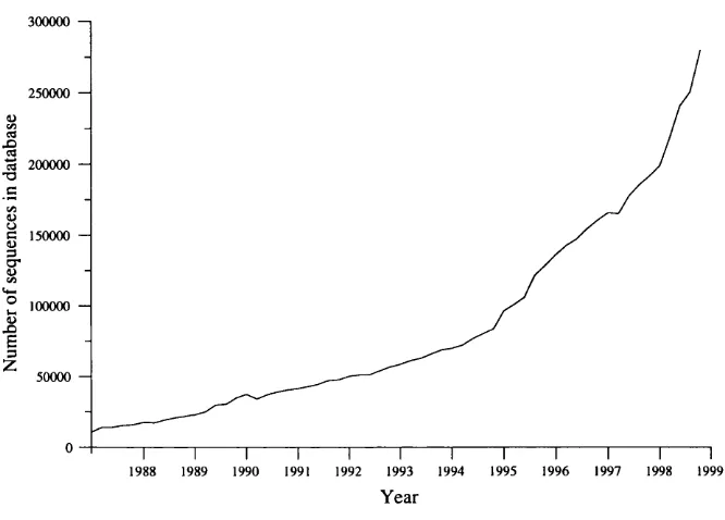
1.2 Growth of the OWL sequence database... 23
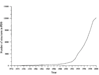
1.3 Growth of the Protein D ata Bank (PD B )... 26
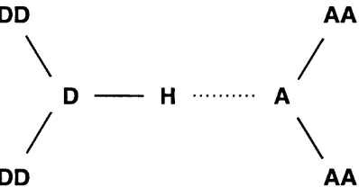
2.1 Determ ination of potential hydrogen bonds in protein structures using HBPlus... 34
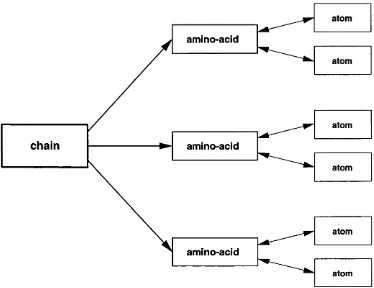
2.2 Schematic of the inheritance model used by Grow... 37
2.3 The processes involved in generating SAS o u tp u t... 43
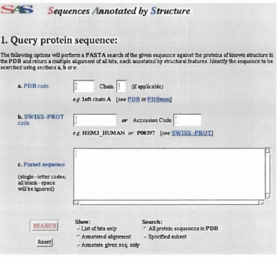
2.4 A screen dump of the SAS query page... 44
2.5 A sample output of SAS, as displayed in the browser window, using PDB code 7HVP chain ‘A ’ as query. Note th a t there are two frames (see te x t)... 47
2.6 The colouring of the different annotation types... 48
2.7 SAS alignment showing CASP2 target 31 and elastase (PDB code 3EST). Note the two of the four active site residues are conserved (alignment positions 75 and 132). Green graphics represent the predicted secondary structure for the ta rg e t... 50
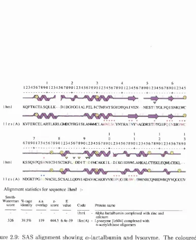
2.9 SAS alignment showing a-lactalbum in and lysozyme. The coloured
residues show metal and ligand binding regions (respectively).
Note th a t the alignment numbers do not correlate to the num
bers in the respective PDB files as in the text. Alignment
positions (IH M L/ILZS) - 35=Thr33/G lu35; 53=Glu49/Asp53;
91=Asp87/Asp91... 53
2.10 RasMol representation of the 3D structures of a-lactalbum in {left)
and human lysozyme {right). The green areas indicate amino-acids
involved in m etal and sugar binding respectively... 54
3.1 Functional classification of the polymerase s t r u c t u r e s ... 60
3.2 Functional domains in the polymerase structures. Domains present
in the sequence but not the structure are showed by dotted shapes. 62
3.3 The shape of a DNA polymerase I from Escherichia coli rendered
by the Ribbons program. The “thum b” is the helical regional on the right, the “fingers” the larger helical region on the left and the
“palm” is the central region with both helical and sheet character. 72
3.4 DOMPLOT schematics for the three DNA polymerase I structures
from Bacillus stearothermophilus (IB D P), Escherichia coli (IKFS)
and Thermus aquaticus (ITAQ). The thum b sub-domain is shown
in green, the palm in red and the fingers in blue... 74
3.5 DOMPLOT schematics for the representative structures. The
sub-domains are coloured as in figure 3.4. The PROSITE signatures
found in the proteins are shown by a red line under the appropriate region... 75
3.6 TOPS diagrams for the three DNA polymerase I {top left - IBDP;
top right - IKFS; bottom left - ITAQ) and the T7 DNA polymerase
{bottom right - 1T7P). a-helices are shown as blue circles, while
/5-strands are shown as red triangles... 77
3.7 TOPS diagrams of the reverse transcriptases {top left - IRTH; top
right - IMML), DNA polymerase a {bottom left - IWAJ) and DNA
3.8 TO PS diagrams of the palm sub-domains of the five representative
structures. The dotted areas illustrate the common topologies in
IKFS, 1T7P and IR T H ... 79
3.9 RasMol pictures of DNA polymerase I {top left - IK FS), T7 DNA
polymerase {top right - 1T7P) and T7 RNA polymerase {bottom
-4RNP). The domains are coloured as in the DOM PLOT schematics. 81
3.10 RasMol pictures of the reverse transcriptases {top left - IRTH; top
right - IMML), DNA polymerase a {bottom left - IWAJ) and DNA
polymerase P {bottom right - IB P Y )... 82
3.11 The position of the sequence motifs in the structure of DNA poly
merase I (PDB code IK FS). Sequence m otif ‘A ’ is shown in red, ‘B’ in green and ‘C ’ in blue... 83
3.12 Sequences A nnotated by Structure (SAS) illustration of the CORA structural alignment of the representatives (less DNA polymerase
P structure, PDB code IBPY ). a-Helical regions in the sequences
are shown in red, while /5-strand regions are shown in blue. The
two (three) acidic residues are shown in cyan conserved at positions
155, 241 and 242 in the alignm ent... 87
3.13 Ligplot illustration of interactions between the catalytic aspartate
residues, the magnesium ions and the nucleoside triphosphate in
DNA polymerase ^ (PDB code IB P Y )... 90
3.14 DOMPLOT schematics of the polymerase representatives showing
the active site residues and sequence motifs (coloured lines). The
sequence motifs are those defined in table 3.7... 91
3.15 SAS illustration of DNA polymerase /5 structure IBPY. The pur
ple graphics above the sequence represent the secondary structure.
Residues in the sequence are coloured according to the number
of contacts (hydrogen bonds and non-bonding) made between the
protein and the DNA molecule. Black = 0, purple = 1, blue = 2,
green = 3, orange = 4 and red = 5 or more. Green dots below the
structure graphics show where protein-DNA contacts occur, while
red dots show protein-ligand contacts and blue dots show protein-
metal contacts. The green triangles indicate catalytic site residues
3.16 Evolutionary tree of the polymerase structures. Like colours indi
cate homology in at least the palm sub-domain. Red - family 1;
green - family 2; blue - family 3... 94
3.17 CINEMA representation of the RNA-polymerase motifs A through
D. In each motif, the sequences are as follows {from top down) :
HIV-1 reverse transcriptase, Poliovirus polymerase. Hepatitis C
virus polymerase (NS5b), Influenza virus polymerase... 97
3.18 DOM PLOT schematics of family 1 (see section 3.9) polymerases
including the preliminary poliovirus structure... 98
3.19 TO PS diagram of the poliovirus polymerase, showing the flngers {top left), thum b {top right) and palm {bottom) sub-domains. . . . 99
3.20 RasMol picture of the shape of the poliovirus polymerase structure.
As in previous flgures, the palm, flngers and thumb sub-domains
are shown in red, blue and green respectively...100
3.21 CORA alignment of the palm sub-domains of the poliovirus poly
merase and HIV-1 reverse transcriptase annotated using SAS. . . 101
3.22 The palm sub-domains of the poliovirus polymerase {blues) and
HIV-1 reverse transcriptase {reds) overlayed using the SAS/CORA
alignment and displayed using RasMol. Sequence motifs ‘A ’ and
‘C ’ are shown in bolder colours {blue, red), and the catalytic as
partic acid residues are highlighted {cyan, magenta.) ...102
3.23 SAS/CORA alignment of HIV-1 reverse transcriptase and po liovirus polymerase. The Hepatitis C and Influenza A polymerase
sequences have been added, and their PHD-predicted secondary
structure shown in green, in contrast to th a t of the known struc
tures shown in m agenta... 104
3.24 DOM PLOT schematic of the Hepatitis C virus polymerase structure. 105
3.25 RasMol representation of the shape of the Hepatitis C virus poly
merase structure. As in previous flgures, the palm, flngers and
thum b sub-domains are shown in red, cyan and green respectively. 106
3.26 SAS/CORA alignment of Hepatitis C virus polymerase with po
4.1 The process of both compiling and searching the TESS hash table. 110
4.2 Ball and stick representation of the Ser-His-Asp catalytic triad
tem plate as used by T E S S ... 112
4.3 General form of the polymerase reaction...113
4.4 The three aspartic acid residues in the polymerase active site as
seen in reverse transcriptase (PDB code IRTH). The Asp in red is
residue ‘A ’, green is ‘B’ and blue is ‘C ’...114
4.5 The polymerase active sites superimposed. Spheres coloured green
are the O ^i/O d oxygen atoms and the blue are Oj2/Oe2 oxygen
atom s... 117
4.6 The DNA polymerase I active sites superimposed on the site of
representative structure, PDB code IRTH. The spheres in green
are the O ji/O a oxygen atoms while the spheres in blue are the
0 ^2/O f2 oxygen atom s... 120
4.7 The atoms constituting the polymerase tem plates - top left - DNA
polymerase I; top right - reverse transcriptase; bottom left - DNA
polymerase a and bottom right - DNA polymerase ^...121
4.8 The reverse transcriptase active sites superimposed on the site of
representative structure, PDB code IRTH. The spheres in green
are the O ji oxygen atoms while the spheres in blue are the 0 ^ 2
oxygen atom s... 122
4.9 The DNA polymerase a active sites superimposed on the site of
representative structure, PDB code IW AJ. The spheres in green
are the O^i oxygen atoms while the spheres in blue are the 0 ^ 2
oxygen atom s... 123
4.10 Distribution of RMS values from fitting of other DNA polymerase
P sites onto th a t of the representative structure, PDB code IBPB.
Light coloured bars represent structures with N T P bound (includ
ing IB PB ), while darker bars represent structures without NTP
4.11 The DNA polymerase ^ active sites superimposed on the site of
representative structure, PDB code IBPB. The spheres in green
are the O^i oxygen atoms while the spheres in blue are the Oj2
oxygen atom s...125
4.12 Distribution of the matches found when searching the PDB hash
table w ith the DNA polymerase I tem plate. The light bars repre
sent true matches (DNA polymerase I family members) while the
dark bars are false m a tc h e s... 127
4.13 Matches found when searching the PDB TESS database with
the reverse transcriptase template. The light bars represent true
matches while the dark bar is a false m atch...128
4.14 Matches found when searching the PDB TESS database with
the DNA polymerase a template. The light bars represent true
matches (DNA polymerase a) while the dark bars are false matches 130
4.15 Matches found when searching the PDB TESS database with
the DNA polymerase /? template. The green bars represent true
matches (DNA polymerase while the red bars are false matches. 131
4.16 Distribution of RMS deviation values obtained from searching the SPASM library with the DNA polymerase I tem plate. Light bars
represent true matches, while dark bars indicate false matches. . . 135
4.17 Distribution of RMS deviation values obtained from searching the SPASM library with the reverse transcriptase tem plate. Light bars
represent true matches, while dark bars indicate false matches. . . 136
4.18 Distribution of RMS deviation values obtained from searching the
SPASM library with the DNA polymerase a tem plate. Light bars
represent true matches, while dark bars indicate false matches. . . 138
4.19 Distribution of RMS deviation values obtained from searching the
SPASM library with the DNA polymerase j3 tem plate. Light bars
4.20 D istribution of RMS deviation values obtained from searching the
PDB TESS database with the DNA polymerase I tem plate, al
lowing Glu to be substituted with Asp. Light bars represent DNA
polymerase I matches, dark bars reverse transcriptase matches and
hatched bars false m atches... 141
4.21 D istribution of RMS deviation values obtained from searching the
SPASM library with the DNA polymerase I tem plate allowing Glu
to substituted with Asp. Light bars represent DNA polymerase I
matches, dark bars reverse transcriptase matches and hatched bars
false m atches...142
5.1 Diagram illustrating the grouping of the types of aspartic proteases
in the PDB. The non-viral enzymes cluster by several attributes. . 150
5.2 Phylip plot of the phylogeny of the non-viral aspartic protease
sequences... 154
5.3 Phylip plot of the phylogeny of the viral aspartic protease sequences. 156
5.4 DOM PLOT illustration of the domain organisation of the five rep
resentative structures. The first lobe (by sequence) is shown in red, the second in green. The two chains of the aspartic protease
from HIV-2 are coloured like the non-viral proteins for comparison. 159
5.5 RasMol representation of the 4 types of aspartic protease structures - top left - pepsin; top right - penicillopepsin; bottom left - cardosin
A and bottom right - HIV-2...161
5.6 SAS representation of the CORA alignment of the 4 types of as
partic protease structures (includes saccharopepsin also)... 163
5.7 The two sequential units of amino acids which constitute the cat
alytic site are close in three-dimensional space. The lobes are
coloured in pale red and pale green, while the catalytic site residues
in th a t lobe are highlighted in a stronger hue. (Example: pepsin,
PDB code IP SN )... 164
5.8 The six amino acids which cluster together to form the active site
of the aspartic proteases (Example: pepsin, PDB code IPSN ). The
amino acids are red - D32; green - T33; blue - 034; magenta - D215;
5.9 The superimposition of all-atoms in the active sites of the sequence
representative structures (19 in all). Atoms are coloured red for
oxygen, blue for nitrogen and grey for carbon... 168
5.10 The five templates used for TESS searches against all structures
in the PDB. Top left - 14-atom; top centre - dicarb-[-Ca; top right
- Ca, bottom left - oxygen only and bottom right - OCA... 173
5.11 Distribution of matches obtained while searching the PDB using
the 14-atom tem plate...174
5.12 D istribution of matches obtained while searching the PDB using
the dicarboxylate and Ca tem plate...175
5.13 Distribution of matches obtained while searching the PDB using
the Cq tem plate... 176
5.14 D istribution of matches obtained while searching the PDB using
the Oxygen-only tem plate...177
5.15 D istribution of matches obtained while searching the PDB using
the oxygen and Ca tem plate...178
5.16 Distribution of matches obtained while searching the PDB using the oxygen and Ca template. The true matches have been sepa
rated into viral and non-viral species... 179
5.17 Alignment of BAE2_HUMAN with the three non-viral aspartic
acid sequences in addition to the closest match, saccharopepsin
(PDB code 2JXR), annotated by secondary structure (predicted
for BAE2 HUMAN) - 6/we=strand; red=helix; cyan=a,ctive site. 181
5.18 Alignment of BACE HUMAN with the three non-viral aspartic
acid sequences in addition to the closest match, saccharopepsin
(PDB code 2JXR), annotated by secondary structure (predicted
for BACE HUMAN) - blue=strand; red=helix; cyan=active site. 182
5.19 Modified aspartic protease tree to incorporate the aspartic pro
List of Tables
1.1 The sizes and number of chromosomes of several organisms. . . . 20
3.1 Polymerase structures in the PDB. ‘(f)’ in the ‘Length’ column
indicates th a t the structure is a fragment of the full protein . . . . 58
3.2 sequence identity m atrix of the c lu s te r s ... 64
3.3 PROSITE motifs in the representative structures ... 65
3.4 PRINTS motifs in the polymerase a structure from Bacteriophage
Rb 69 ... 66
3.5 Motifs in DNA polymerase I sequences. Columns shown in red
highlight conserved residue positions. M otif 3 corresponds to lit erature m otif ‘A ’, m otif 4 to ‘B ’ and m otif 5 to ‘C’... 67
3.6 Conserved motifs in RNA-dependent polymerases. Columns in red
highlight conserved residue positions. Viral RNA polymerases have no equivalent m o tif‘E ’. Motifs ‘A’ and ‘C ’ correspond to literature
motifs ‘A ’ and ‘C ’. * indicates position in the complete SWISS-
PRO T protein... 68
3.7 Conserved motifs in all polymerases. Strictly conserved residues
positions are highlighted in red. motifs ‘A ’ and ‘C ’ are common
to all polymerases, while motif ‘B ’ is specific to DNA and RNA
polymerases. Note the negative position of m otif ‘B ’ with respect
3.8 Consensus motifs if the polymerases. The consensus motifs as re
viewed by [Joyce & Steitz, 1995] are shown in the upper section
with strictly conserved residues shown in red. In the lower section,
residues th a t are conserved in the structural examples are shown
in green... 70
3.9 Ranges of sequence defining the sub-domains of the polymerase
structures as assigned by OATH... 71
3.10 Sequence ranges of sub-domain assignments for the structural rep
resentatives... 73
3.11 SSAP Score m atrix for the palm sub-domains of the representative
structures. T. aquaticus DNA polymerase I structure (PDB code
ITAQ) is included since it shows greater SSAP scores with the
reverse transcriptases... 84
3.12 SSAP Score m atrix for the fingers sub-domains of the representa
tive structures... 85
3.13 SSAP Score m atrix for the thumbs sub-domains of the representa
tive structures. Note there is no thum b sub-domain in the reverse transcriptase structure IM M L... 85
3.14 Structures of polymerases involved in c o m p le x e s... 88
3.15 Catalytic site residues in the representative s tr u c tu r e s ... 89
3.16 Sequence identity m atrix of the pairwise alignment of the viral
polymerase sequences, including HIV-1 reverse transcriptase. The
total number of amino-acids constituting the aligned region is
shown in p a r e n th e s e s ... 96
3.17 SSAP scores of the palm sub-domains of the poliovirus polymerase
(IRDR), E. coli DNA polymerase I (IKFS) and HIV-1 reverse
transcriptase (IRTH ). The percentage overlap in the structural
alignment is shown in parentheses... 99
3.18 SSAP scores of the palm sub-domains of the Hepatitis C virus poly
merase (IQUV), poliovirus polymerase (IRDR) and HIV-1 reverse
transcriptase (IRTH ). The percentage overlap in the structural
4.1 M atrix of RMS values (in
A)
from all-atom fitting of the three conserved amino-acids in the polymerase active site. The PDBcodes for the polymerases are as follows : RT - IRTH; DNA-I -
IKFS; T7 DNA - 1T7P; T7 RNA - ICEZ; DNA (3 - IZQY; DNA
a - IW A J... 117
4.2 Breakdown of the PDB by polymerase family and non-polymerase
structures... 144
4.3 The specificity, sensitivity and M atthews correlation coefficient of
each tem plate calculated from the results of TESS searches against
the PDB hash table... 145
4.4 The specificity, sensitivity and M atthews correlation coefficient
of each tem plate calculated from the results of SPASM searches
against the PDB SPASM library... 146
4.5 Summary of the results of using TESS and SPASM vs PDB. . . . 147
5.1 The breakdown of all the aspartic protease sequences in the PDB by Enzyme Classification (E.G.) number. E=eukaryotic; V =viral;
F=fungal and P = p la n t... 151
5.2 Sequence identity m atrix of the eukaryotic aspartic protease se
quences... 153
5.3 Sequence identity m atrix of the fungal aspartic protease sequences. 153
5.4 Sequence identity m atrix of the non-viral aspartic protease sequences. 154
5.5 Sequence identity m atrix of the viral aspartic protease sequences. 155
5.6 Sequence identity m atrix of the 5 types of aspartic protease. . . . 157
5.7 SSAP score m atrix of the 5 types of aspartic protease (upper fig
ures) and overlap m atrix (lower figures)... 162
5.8 The different combinations of amino acids in the catalytic site. . . 165
Chapter 1
Introduction
Biochemistry plays a crucial role in the life sciences, expressing the characteristics
and reactions of a living system. Many great advances have been made in recent years, as the focus of much research turns to not only understanding ourselves but
to an array of living organisms. The breadth of the subject can only be illustrated by the very scale of systems th at are studied - from whole cellular organisms,
down to the smallest interactions on an atomic level. The field is dominated by molecular biology, the physicochemical organisation of an organism. Here lies a
number of inter-related disciplines, from the sequencing of DNA sequences of an
organism, to the interpretation and understanding of this information, through to the proteins th a t are thus synthesised and how they work and interact.
1.1
Organisation of genetic material
The genetic m aterial of prokaryotes and eukaryotes is formed by DNA molecules,
while in viruses it can be formed by DNA or RNA molecules. In cellular organ
isms, this m aterial is organised into chromosomes. In prokaryotes, the entire
genome is carried on a single chromosome with circular DNA. In eukaryotes, the
genome consists of a number of chromosomes, each consisting of linear DNA. The
number of chromosomes is not necessarily linked to the overall size of the genomic
DNA, measured in the number of bases (the constituent units of DNA) th a t are
paired in sequence in the double-stranded, double-helix molecule. Several exam
chromosome four million base pairs long, while yeast Saccharomyces cerevisiae,
fruit fly Drosophila melanogaster and human genomes consist of multiple chro
mosomes with the to tal number of base pairs running from millions to billions.
Organism # of base pairs # of chromosomes
E. coli 4 X 10® 1
S. cerevisiae 1 .4 X 10^ 16
D. melanogaster 1 .7 X 10» 4
Human > 3 X 10^ 2 3
Table 1.1: The sizes and number of chromosomes of several organisms.
Genetic information is encoded in the DNA by variation of the nucleotides
adenine (A), guanine (G), thymine (T) and cytosine (C) in the sequence of the
molecule, providing an ‘alphabet’ consisting of four letters. In double-stranded DNA, the second strand complements the first because of hydrogen bonding be
tween the bases whereby adenine pairs with thymine and guanine with cytosine. The sequence of bases in a DNA molecule is commonly elucidated by a process
called dideoxy sequencing (or Sanger sequencing after its developer [Sanger et
al, 1977]). This m ethod uses DNA polymerase I to copy a sequence of single
stranded DNA using radioactively or fiuorescently tagged deoxyribonucleosides
with the addition of a dideoxy analogue. The dideoxy analogue causes prem ature term ination of the copying on incorporation since it lacks the 3’-hydroxyl group
necessary to form a 3’-5’ phosphodiester bond and thus extend the sequence.
The fragments th a t are sequenced by this method are of different lengths and can
be analysed by electrophoresis where the smallest fragments travel further along
the gel than the larger ones. Autoradiography or fluorescence detection directly reveals the sequence of bases in the molecule.
In recent years, much work has focussed on sequencing the entire genomes of sim
ple organisms such as E. coli and S. cerevisiae which have since been completed.
Indeed, to date the genomes of many organisms have been sequenced including
those from archaea, bacteria, eukaryota as well as those of mitochondria and
viruses. These goals, and the experiences gained from achieving them, are in
strum ental to the current global effort to sequence the human genome, which
although vast is expected to be completed soon. The main data depositories
for DNA sequences are those in the International Nucleotide Sequence Database
ular Biology Laboratory, EMBL [Baker et ai, 2000], and the DNA D ata Bank
of Japan, DDBJ [Tateno et al, 2000]. At present, they contain nearly 3 billion
bases of d ata from over 47000 species, although their content grows at an almost
exponential rate.
Much information can be gained from this data, however it doesn’t give any
insight into how biological systems function. It is therefore necessary to consider
how this genetic information is expressed to give functional moieties. This process
involves the units of heredity called genes, which are regions of sequence in a
chromosome which encode a functional product, a protein. Figure 1.1 shows
a much simplified schematic of how a protein is synthesised from its genetic
blueprint.
transcription transiation
DNA --- ► RNA --- ► protein
Figure 1.1: The process of protein synthesis
The first step in the process is th a t of transcription, where an RNA molecule is synthesised from the DNA tem plate for each gene th a t is to be expressed. This
RNA molecule is called messenger RNA (mRNA) because it is the tem plate for
the synthesis of the protein. The second step involves the translation of the infor
m ation contained in the mRNA molecule into a protein sequence. Experiments found th a t each triplet of nucleotides in the mRNA (and thus DNA) coded for
a particular amino-acid and is therefore called a codon. Although there are only
20 amino-acids observed in studies of proteins, each position of the triplet can be
1 of 4 different nucleotides, giving a to tal of 64 combinations. This genetic code
was also found to be degenerate, in th a t each amino-acid can be represented by
more than one triplet such th a t m utations could occur in the DNA which would
still preserve the sequence of the synthesised protein. O ther significant findings
were codons th a t signalled to other molecules in the complex protein synthesis
mechanism, such as those for initiation and term ination of the protein synthesis.
In an ideal world, the protein sequence encoded by a gene could be translated
directly from the DNA in a linear fashion. This is indeed the case for prokary
otes, where little (if any) information other than the genes are contained within
th e chromosome. However, for eukaryotes it is not as simple, since many genes have been found to be discontinuous with their coding regions (called exons) be
effect in the protein synthesis process in th a t transcription of DNA yields the
primary transcript, which then undergoes splicing to link the exons together to
form mRNA. Many exons have been found to encode discrete structural and func
tional units in protein structures and an elegant hypothesis follows from this by
suggesting th a t new proteins evolved through the ‘shuffling’ of exons in genes,
thus affecting the arrangement of these units. It is widely accepted th a t introns
introduce a large amount of non-coding sequence into eukaryotic genomes. For
example, despite being in excess of 3 billion bases long, the human genome con
tains a disproportionate number of genes possibly totalling as little as 2% of the
whole. Yet the reasons why the introns exist, what they contain and what they
do remains a mystery.
1.2
Protein sequence information
The use of recombinant DNA technologies to express genes of interest followed by
their interpretation has yielded a large amount of protein sequence information. However, it is also possible to analyse a given protein to elucidate the sequence
of amino-acids from which it is constructed. This m ethod uses a ‘divide and conquer’ strategy, where the protein is divided up into smaller segments with
each segment being analysed by an autom ated Edm an degradation process to
reveal its sequence. The sequences of all the segments are then pieced together by considering segments of the same protein divided at different positions, and
comparing the overlapping sections between the two results to fit the segments
together correctly.
By a combination of the two complementary approaches the sequence of a
given protein can be found. The ability to do this is very significant, because
not only is a protein the product of a gene, but it is the functional unit within
a biological system - proteins are responsible for fulfilling the m ajority of tasks
in keeping an organism alive. Only analysis of these proteins can shed light onto
how biological systems work. In a genomic context, the entire mechanism of an
organism is defined by the proteins which arej described by its genes.
The m ain resources for protein sequences are GenBank and TrEMBL (Translated
EM BL) which contain the translations of the coding regions of their respective
databases such as SWISS-PROT [Bairoch & Apweiler, 2000], Protein Informa
tion Resource, P IR [Barker et al, 2000], and a composite database sourcing se
quences from SWISS-PROT, GenBank, PIR and NRL-3D called OWL [Bleasby
et ai, 1994]. Each of these provide annotations such as the classification and
organism of the sequence, references and known characteristics of the protein
such as function and any sites of biological importance. The sequence databases
have seen very rapid growth in the number of sequences they contain, with OWL
(utilising redundancy) doubling about every 18 months as shown in figure 1.2.
OWL contains nearly 300000 non-identical sequences at the tim e of writing.
300000 -1
250000
-200000
C 150000
-° 100000
i
— I--- 1--- 1--- 1--- 1--- 1--- 1--- 1--- 1 I I I
1988 1989 1990 1991 1992 1993 1994 1995 1996 1997 1998 1999 Year
Figure 1.2: Growth of the OWL sequence database.
The wealth of protein sequences in the databases not only facilitates analysis of
the relationships within a protein family from different species (an evolutionary
angle), but also allows the assignment of the function of a new protein sequence
when a similar sequence (whose function is known) can be found. In itself, this
approach conveys no functional information since the assignment is based on
inference from the m atching sequences.
Secondary sequence databases build on this idea by detailing regions of sequence
(in multiple alignments) th a t are conserved across a collection of examples. PRO-
SITE [Hofmann et ai, 1999] is an example of a such a database, containing 1386
individual ‘p attern s’ which describe different conserved sites which usually carry
sions which can be used to search a large number of sequences in a short period
of time. However, PRO SITE patterns are short and are limited by their descrip
tion of a site only and retain no other information about the proteins which they
represent. This is addressed by the PRINTS [Attwood & Beck, 1994, Attwood
et al, 2000] database which contains 1360 ‘fingerprints’. Each fingerprint is a
signature consisting of m ultiple patterns which conserve much more information
than the site alone (such as conserved elements of protein secondary structure)
and are therefore more descriptive. More complex still are profile methods such
as PFam [Bateman et ai, 1999] which use Hidden Markov models [Krogh et al,
1994] to retain even more information and generate a complete profile from a mul
tiple alignment of sequences which is used for searching databases and matching
unknown sequences with known ones with a great deal of success.
1.2.1
A nalysis o f protein sequences
In addition to the secondary sequence databases with their search facilities, there
are other methods th a t can be employed to find related sequences. These meth ods contrast with the p attern methods since they do not search using a regular
expression but compare a query sequence with a whole database, and are espe cially useful when looking for homologues, the products of divergent evolution,
to build up an evolutionary picture.
The two most widely used tools for sequence comparison are FASTA [Pearson &
Lipman, 1988] and BLAST [Altschul et ai, 1990]. Both use optimised dynamic
programming algorithms based on work by Needleman and Wunch [Needleman
& Wunsch, 1970] and Smith and W aterman [Smith & W aterman, 1981], sacrific
ing searching all of the possible positions in sequence space for speed. In their
operation, the two programs perform slightly different in th a t FASTA is particu
larly efficient at finding global alignments between sequences whereas BLAST, by
virtue of its name (Basic Local Alignment Search Tool), is optimised for finding
similarities at the local level (yet the sequences may be very different at a global
level). More recently, BLAST has been improved to allow gapped local align
ments and position-specific iterations, the latter being a pseudo-profile approach.
PSI-BLAST [Altschul et a i, 1997] can construct its own profile for local align
ments which can be used to search a database and the matches used to further
on subsequent iterations. In principle at least, this is the same strategy as in
PFam without resorting to Hidden Markov Models which are slower due to their
complexity.
Alignments of multiple sequences can be produced using the CLUSTALX tool
[Thompson et ai, 1997] which focuses on regions th a t are conserved in all the
sequences provided. Despite using tricks learned from pairwise comparison, multi
ple alignments are much more difficult to obtain and often need m anual assistance
to be of much use. Once this is effected, the relationships th a t can be highlighted
are often more striking than pairwise methods and are used as the basis for the
m ultiple patterns in PRINTS. Methods employing Hidden Markov models also use
m ultiple alignments as a seed alignment, which is enhanced iteratively depending
on the variety of sequences th a t the algorithm is presented with (a better model
can be constructed when a dataset has many members). HMMER [Eddy, 1998] is
an example of such a tool, which not only allows construction of a custom model but also allows database searching using it, often yielding superior results over
simpler methods particularly when searching for more distant relatives.
1.3
Protein structure iuforruatiou
Sequence information is limited, however, in th a t it can give no indication of w hat fold the protein adopts in three-dimensional (3D) space or insights into the
mechanism of the biochemical system th a t it is involved in. If the step beyond
evolutionary relationships and functional identification by inference is to be made,
it is necessary to look at the structures of proteins.
P rotein structures are determined by techniques such as X-ray crystallography
or NMR spectroscopy. The resulting coordinates specify the positions of each
atom of the protein in three-dimensional space. The prim ary resource for this
information is the Protein D ata Bank or PDB [Berman et a/., 2000] which cur
rently contains over 12000 structures. Like the sequence databases, the PDB has
experienced rapid growth as techniques have improved in recent years, as shown
in figure 1.3.
Over the past fifteen years an extensive complement of software has been devel
15000- 1
12000
-9000
-I
o"o
I
6000
-3000
-1972 1974 1976 1978 1980 1982 1984 1986 1988 1990 1992 1994 1996 1998 2000 Year
Figure 1.3: Growth of the Protein D ata Bank (PDB).
to develop a m ethod of assessing the quality and validity of such d a ta result
ing in the PROCHECK program [Morris et a/., 1992, Laskowski et a/., 1993] ,
with later work focusing on such topics as a consensus approach to domain as
signment [Jones et ai, 1998], autom ated secondary and supersecondary structure
assignment - PRO M O TIF [Hutchinson & Thornton, 1996], characterisation of
atomic interactions - HBPLUS [McDonald & Thornton, 1994], and more. Using
these tools alone, it is possible to look at a protein structure at different scales
from tertiary structure down to atoms.
Much of this information is very helpful in highlighting more distant relationships
between protein sequences when sequence analysis has proved inconclusive. For example, knowledge of a pattern of amino-acids responsible for binding in sig
nalling or reaction pathways would prove invaluable in assigning the identity of
a sequence with unknown function possessing the same pattern of amino-acids.
However, residue-based 3D functional information has not been used much for
function assignment to date [Overington et al., 1990].
Methods for structural comparison parallel (in concept at least) those used for
sequence comparison. Early attem pts using rigid-body superposition were not
only com putationally expensive but had difficulties in dealing with insertions and
In recent years improved methods have been developed to tackle this problem
using dynamic program m ing algorithms to exploit larger features of the local
environment such as secondary structural elements. SSAP [Taylor & Orengo,
1989], for example, uses double dynamic programming which not only compares
the structural environments th a t are defined by vectors between atoms, but
also identifies residue equivalences. This results in a relative insensitivity to
insertions and deletions and a greater ability to identify more remote similarities,
yet does not depend on the conservation of specific amino-acids. The ability to
identify relationships at the fold level is the premise for the CATH [Orengo et
ai, 1999] and SCOP [Lo Conte et al, 2000] resources which classify proteins
into families by structural similarity. As with sequence analysis, the function
of a query protein can be inferred only by comparison with proteins of known function.
1.3.1
Functional tem plates
Methods for assigning the function to a new protein by inference from a homolo gous protein with known function are now well established. However, a problem
lies with these m ethods in th a t the sequence or structure of the protein under investigation must bear some global similarity (obvious in the case of sequence,
or more remote if analysing structures) to one previously characterised. Many
new sequence families are being discovered by the genome sequencing efforts and it is inevitable th a t the structures of these will need to be determined before any
conclusions can be drawn about their mechanism of function. This may well give
rise to a situation where there are structures solved th a t have unknown function
and are not similar to other known structures. An effective method of functional
analysis of these structures will therefore prove invaluable, and this has led to
the idea of ‘functional m otifs’ in 3D, which have been likened with PROSITE
patterns and PRINTS signatures.
Several types of functional site have so far been studied in the literature, espe
cially metal binding sites [d u sk er, 1991] and anion binding sites [Chakrabarti,
1993, Copley & Barton, 1994]. The first attem pts to develop these functional
site motifs into functional ‘tem plates’ used conventional database software [Is
lam & Sternberg, 1989] to allow crude searches of a database of structures for
ine proteases. Using this technique proved to be very slow, so tools have been
developed specifically with searching 3D structures more quickly in mind.
The first of such tools is a program called ASSAM [Artymiuk et al., 1994]. The
side-chain of each amino-acid is represented by two ‘pseudo-atoms’ which are
situated near th e start and end of the functional part of the side-chain. For
example, the sta rt (S) pseudo-atom for glutamic acid is situated on the C.y of the
side-chain, while the end (E) pseudo-atom is situated midway between the two
carboxylate oxygen atoms. In this manner, the connection of the pseudo-atoms is
a vector and allows direct comparison between amino-acids with similar functional
groups, such as glutam ic acid and aspartic acid, resulting in a description of a
property of given amino-acids - in this case, th a t of an acidic residue. Using this
method, two side-chains can be related by the vector between the S and E pseudo
atoms in each side-chain, and the distances between the S and E pseudo-atoms in one with the other. This yields four distances - S-S, S-E, E-S and E-E - which
are used to define a m atrix representing this relationship or motif. Increasing the
number of side-chains represented by the motif, to represent an active site for example, increases the size of the m atrix. This m atrix is known as the subgraph
for use in the subgraph-isomorphism algorithm used to search a much larger graph representing a protein structure, with each side-chain within also represented by
S and E pseudo-atoms. Testing the approach with the serine protease catalytic
triad illustrated th a t the program was very effective, yet suggested th a t greater precision could be obtained by utilising a greater number of pseudo-atoms or
using the atom positions themselves.
This more specific approach was presented in the TESS (Template Search and
Superposition) program [Wallace et a/., 1996, Wallace et a i, 1997]. TESS used
the actual coordinates of the atoms of the constituent amino-acids in the motif
as a basis of a tem plate. A database of structures represented in the same way
can be compiled and a geometric hashing algorithm used to search the database
with a query tem plate. W ith this approach, not only were atoms belonging to
amino-acids allowed in a motif, but any hetero-atoms such as metal ions could be
included. More recently, the SPASM (Spatial Arrangements of Side-chains and
Main-chain) program [Kleywegt, 1999] has enhanced the pseudo-atom approach
by representing the side-chain by a single pseudo-atom at the centre of gravity of
the side-chain atoms and incorporating the main-chain Cq in its motifs. By using
(such as TESS) yet sacrifices some sensitivity.
Regardless of which approach is taken, it is clear th a t for functional identification
to be possible in the future, a library of these 3D functional motifs will need to be
constructed to facilitate scanning a new structure for the occurrence of any such
motifs. In structural genomics, where the wealth of sequences being generated
by genome projects will lead to a plethora of interesting and im portant structure
determ ination targets, quick autom ated procedures will be necessary to discover
more about their function [Skolnick et a/., 2000].
1.4
This work
The work described in this thesis tackles several issues. The first is the creation of a tool for the annotation of linear protein sequences with information derived
from protein structures.The large amount of structural analyses performed at UCL
in recent years provides a good basis for implementing key structural features.
However, the m ajority of this work is concerned with enzymes, proteins th a t catal
yse a wide range of im portant biochemical reactions. The above tool will be used to illustrate the results of sequence and structure analyses of two specific fami
lies - the polymerases and the aspartic proteases - to highlight the evolutionary relationship between proteins th a t share a common function. Protein structural
motifs which encapsulate their functional cores will be derived for each of these
large families and used to explore the variation of the active site geometry within
each family. The sensitivity and specificity of these 3D functional tem plates is
Chapter 2
Annotating protein sequences with
structural information
2.1
Introduction
Understanding the function of a protein is fundamental to biochemistry. Yet
with the vastness of the field and the complexity of the systems th a t lie within it, exploring all conceivable pathways to identify the function of a given protein
experimentally would be an immense task. This is highlighted by the numerous
genome sequence projects in progress around the world, which are producing an ever expanding wealth of genome sequence information which awaits analysis.
Assigning a function to a protein is often more involved than simply identifying
a related sequence by comparison and inferring the function of the query se
quence from th a t of its relative. This is the premise of current sequence analysis
techniques. It is becoming increasingly clear th a t related proteins often perform
different functions and therefore uncovering molecular details of the mechanism
of a protein’s function is im portant, and this necessitates looking at the protein
in much greater detail. Indeed, a great deal has been learned by obtaining and
analysing 3D structures of proteins in recent years.
The aim of work presented in this chapter was to develop a tool which would facil
itate the overlaying of key structural features (derived from structural analyses)
onto not ju st one, but a collection of aligned sequences. The relationship thus
ing current approaches of sequence analysis in helping to interpret the genome
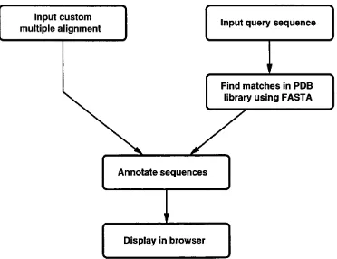
information. Sequences A nnotated by Structure (SAS) is the result of this work, aiming to bridge th e gap between sequence and structural analyses. The sources
of the structural analyses and the methods used to employ them will be discussed.
2.2
SAS functionality
The rationale behind SAS was to provide a simple, Web-based tool th a t would
allow researchers with modest computing skills to utilise, what would otherwise
be, more specialised techniques. W ith such an approach, SAS therefore needs to
autom ate many tasks while presenting the results in a clear, informative manner.
SAS was developed with R.A. Laskowski and J.M. Thornton to address the pro tein sequence annotation issue. In SAS the annotations are represented by colour
ing the individual amino-acids in a protein sequence according to a particular
attribute. The net result is th a t, at its simplest, SAS is able to annotate a sin gle protein sequence for those who are only interested in a lone protein. Over
and above this, SAS can annotate a batch of sequences th a t have been aligned. This is fundamentally more novel since it allows browsing of structural features
of a whole family of related proteins, highlighting trends and differences between members.
As a starting point, SAS takes a specified query sequence. This can be any pro
tein sequence, and is not dependent on structural information being present (if
the structure was available, other techniques would be employed - CATH [Orengo
et a/., 1999], SCOP [Lo Conte et a l, 2000]). This is the premise for research after
all, since the power of annotating potentially related sequences with structural information becomes more apparent when the relationship between the query se
quence and any (FASTA [Pearson & Lipman, 1988] suggested) matches is weak.
The identity, and possibly function, are then inferred from sequences (whose
structures are known) which m atch critical regions of sequence well. It is the
features within these regions th a t SAS can highlight well, and give greater confi
dence in assigning a relationship between protein sequences than would otherwise
2.3
Structural information
The prim ary source of protein structure coordinate data is the Protein D ata Bank
or PDB [Berman et a l, 2000]. Protein structures (PDB files) can be analysed
in many different ways to highlight different properties. The first step in this
development was to decide which of these many properties would be the most
useful.
Amongst the most desirable attributes for annotation are the molecular interac
tions between atom s in the protein, nucleic acids, metal ions and ligands. Other
desirable features include active site residues (when known), secondary structural
elements, and domain information.
2.3.1 M olecular interactions
Interactions between atom s are the key to the function of any protein, be it in a signalling role or in a catalytic one. This molecular recognition forms the
basis of all biological functions, and so it is crucial to understand how the protein
interacts with other molecules in order elucidate how it functions. A good example of this is in the pharm aceutical industry, where recent application of computers
to biochemical problems have resulted in the (broad) fields of drug design and bioinformatics. In the case of drug design, the characteristics of a de novo drug
must complement those of the active site or binding site for which the drug is
targeted. In bioinformatics however, there is a target to improve methods of
establishing sequence relationships, which this work is intended to enhance.
Moreover, it is the non-covalent interactions between certain atom types th at
are responsible for the structural integrity, and thus stability, of macromolecu-
lar structures as a whole, not ju st protein structures. Perhaps the best known
examples of these interactions are the main-chain hydrogen bonds found in the
a-helical and /3-sheet secondary structure elements in proteins, and the base-pairing
in double-helical DNA.
There are différent types of molecular interactions which require classification, as
described
below:-• Covalent bonds - a strong bond between two atoms where the bond is con
• Ionic complexes - a strong ‘bond’ between two atoms where the interaction
is evaluated as the attraction force existing between the charged members
(one positively, the other negatively)
• Hydrogen bonds - a weak interaction between two atoms opposing partial
charges, resulting in a electrostatic attraction (commonly between and
c / - )
• Nearest-neighbour contacts - a very weak interaction between two (nor
mally) hydrophobic atom s (for example, carbon atoms)
W ith this classification, it is possible to represent a large proportion of the atomic
interactions th a t are observed in protein structures and their complexes. It is
therefore necessary to be able to recognise these interactions in the d ata repre
senting any given structure.
Previous work by McDonald & Thornton [McDonald & Thornton, 1994] has gone
a long way to achieving this with a program called HBPlus. HBPlus can be used
to provide data from a PDB file corresponding to potential contacts of three of the four types in the above classification - ionic complexes, hydrogen bonds and
nearest-neighbour contacts. The final type of interaction, the covalent bond, can
be implied by using the standard set of rules for bonding in amino acids, and can be readily extracted from a PDB file (non-standard covalent bonds are listed in
CONECT fields in the data). In the case of ionic complexes and nearest-neighbour
contacts, a potential interaction between two atoms is assigned to them when the
(sterically unhindered) distance between them is below a pre-defined cut-off. This
cut-off is set slightly higher than the sum of the van der Waals radii [Chothia,
1976] of the two atoms, where the sum of these van der Waals radii is defined
as being the closest possible approach of two non-covalently bound atoms. In
practice, the cut-off used is 3.9Â, which is slightly higher than the sum of the van
der Waals radii for two carbon atoms (2 x 1.87Â = 3.74Â).
Hydrogen bonds require a different approach involving two stages. This is nec
essary because structures obtained using X-ray crystallography (accounting for a
large proportion of high-resolution protein structures) do not generally show the
positions of hydrogen atoms in the structure. The reason for this is th at hydrogen
atom s diffract X-rays very weakly, and therefore are difficult to identify in the
these hydrogen atom s in the structure. To describe how HBPlus does this, it is
necessary to define a hydrogen bond.
A hydrogen bond is an interaction between two electro-negative atoms, a donor
and an acceptor - for example, nitrogen and oxygen. A hydrogen atom is cova
lently bound to the donor and is situated between the donor and the acceptor.
In such a case, the donor can a ttra ct the electron from the hydrogen atom orbital
towards itself. This results in the hydrogen becoming partially positively charged,
and is thus attracted to the partial negative charge of the acceptor (possessing at
least one lone-pair of electrons). The resulting favourable electrostatic energy is
largely responsible for the exceptional stability of secondary structural elements
in protein structures and the double-helix in DNA where large hydrogen bonding
networks are present.
HBPlus calculates the position of the hydrogen atoms attached to potential donor
atoms by analysis of the positions of the atoms with which the donor is bound.
T hat is to say, atoms th a t are covalently bound to the donor will be positioned according to certain geometries, dictated by the hybridisation of the donor atom -
either trigonal planar for sp^ hybridisation and tetrahedral for sp^ hybridisation. The hydrogen atom occupies the position which is not already filled, as obtained
from the protein structure coordinates (assuming th a t the atoms involved do
indeed fit this geometry).
Then, the potential of a hydrogen bond is assessed according to geometries of
the donating atom, the hydrogen atom and the accepting atom (and the atoms
covalently bound to these), as shown in figure 2.1.
DD
AA
\
/
D --- H
A
/
\
DD
AA
Figure 2.1: Determination of potential hydrogen bonds in protein structures using HBPlus.
The atoms DD and AA are the donor and acceptor antecedents respectively, with
