3 5 0 0
'?Reorganisation of Sensorimotor Function in
Children with Brain Disease
Victoria Holloway
Radiology and Physics Unit Institute of Child Health and Great Ormond Street Hospital
U n iv e r s ity o f L o n d o n
Submitted for the Degree o f Doctor of Philosophy
ProQuest Number: U643025
All rights reserved
INFORMATION TO ALL USERS
The quality of this reproduction is dependent upon the quality of the copy submitted.
In the unlikely event that the author did not send a complete manuscript and there are missing pages, these will be noted. Also, if material had to be removed,
a note will indicate the deletion.
uest.
ProQuest U643025
Published by ProQuest LLC(2015). Copyright of the Dissertation is held by the Author.
All rights reserved.
This work is protected against unauthorized copying under Title 17, United States Code. Microform Edition © ProQuest LLC.
ProQuest LLC
789 East Eisenhower Parkway P.O. Box 1346
Abstract
Introduction: In this study, paradigms were developed for the investigation of sensorimotor function in children using functional MRI (fMRI), somatosensory evoked
potential (SEP) recordings and behavioural measures. These techniques were applied
both to normal controls subjects and to children with brain disease. A major aim was to
investigate the remarkable recovery of function that can take place following brain injury
sustained early in life.
Methods: Three fMRI paradigms were developed, namely active movement of the
hand, passive flexion/extension movement of the fingers and median nerve stimulation.
In addition, SEPs of functional cortical responses to stimulation of the median nerve
were recorded at high temporal resolution. Finally, the extent of residual or recovered
sensory and motor hand function was assessed using behavioural tests, including grip
strength and double simultaneous stimulation. In one set of investigations, all three
techniques were applied to children following hemispherectomy or children following
vascular damage to the middle cerebral artery territory, to examine the pattern of residual
sensorimotor function following brain injury. In a second study, fMRI was carried out
in pre-surgical paediatric patients for mapping of the sensorimotor cortex in preparation
for surgical resection of lesions in the vicinity of this cortical region.
Results and D iscussion: fMRI was successful in locating the hand cortical
sensorimotor area in 11 out of 12 paediatric patients pre-operatively, and was of value to
the neurosurgeon in helping to delineate the boundaries of subsequent cortical resection.
In patients following stroke and hemispherectomy, a combination of fMRI, SEP and
behavioural techniques provided evidence for inter-hemispheric reorganisation of
sensorimotor function through ipsilateral sensorimotor pathways, and also suggested an
increase in the involvement of ipsilateral secondary sensorimotor areas. The data also
indicate that cortical sensorimotor reorganisation and functional recovery can be seen in
patient both with congenital disease and with late-onset acquired disease, suggesting that
factors additional to age at injury may influence the degree of residual function resulting
from cerebral reorganisation.
Informed consent was obtained for all patients and controls, and the study was approved by
the Great Ormond Street Hospital for Children/Institute of Child Health Research Ethics
Contents
CHAPTER 1: GENERAL INTRO DUCTIO N...1
1.1 In t r o d u c t io n...1
1.2 Th ese n so r ya n dm otorsy st e m s - briefh isto ricalb a c k g r o u n d...1
1.3 Th em o t o rs y s t e m... 3
1.3.1 The primary motor cortex... 3
1.3.1.1 Somatotopy within the primary motor cortex... 4
1.3.1.2 Anatomical location of the motor hand area... 6
1.3.1.3 Are individual muscles or movements represented differently in the primary motor cortex?... 6
1.3.2 Association cortical areas o f the motor system... 8
1.3.3 Subcortical motor structures and pathways... 10
1.3.4 The motor pathway from brain to muscle... 11
1.3.4.1 The ipsilateral corticospinal pathway in the normal individual... 15
1.3.5 Interhemispheric pathway - the role o f the corpus callosum... 17
1.4 Th eso m a t o se n so r ys y s t e m... 18
1.4.1 The primary somatosensory area...18
1.4.2 Association cortical areas o f the somatosensory system...21
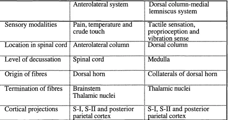
1.4.3 The somatosensory pathway from touch to brain...2 2 1.4.3.1 The dorsal column-medial lemniscus system ... 24
1.4.3.2 The anterolateral system... 25
1.4.3.3 The ipsilateral sensory pathway in the normal individual... 27
1.5 Re o r g a n isa t io no fsenso rim o to rf u n c h o nina n im a l sa n dm a n... 2 8 1.6 Aim so fth iss t u d y... 33
CHAPTER 2: FUNCTIONAL MAGNETIC RESONANCE IMAGING -PRINCIPLES AND FUNCTIONAL SENSORIMOTOR S T U D IE S ...34
2.1 In t r o d u c t io n... 34
2 .2 Principleso ffu n c t io n a lm a g n eticr e so n a n c eim a g in g... 3 4 2.2.1 Sensitivity...3 6 2.2.2 Spatial Resolution... 37
2.2.3 Temporal Resolution...3 9 2 .3 In v e st ig a t io n so fsenso rim o to rf u n c t io n...41
2.3.1 Activation paradigms to investigate sensory and motor function...4 2 2.3.1.1 Complex versus simple motor tasks involving the hand... 42
2.3.1.2 Passive movement...44
2.3.1.3 Electrical stimulation of a peripheral nerve... 45
CHAPTER 3: FUNCTIONAL MAGNETIC RESONANCE IMAGING
-METHODOLOGY... 51
3.1 In t r o d u c t io n... 51
3 .2 Po s t-pr o c essin ga n da n a l y s iso ffM R I d a t a... 51
3.2.1 Statistical Parametric Mapping... 52
3.2.1.1 Realignment... 52
3.2.1.2 Spatial normalisation... 53
3.2.1.3 Spatial smoothing...53
3.2.1.4 Statistical analysis...54
3.2.1.5 Statistical inferences...56
3.2.2 Analysis o f percentage signal change... 57
3 .3 Acquisitionm eth o do lo g ya n ddevelo pm en to fa noptim alp a r a d ig m... 61
3.3.1 Two-dimensional versus three-dimensional EPl sequences...61
3.3.1.1 M ethods... 63
3.3.1.2 Results... 63
3.3.1.3 Conclusions... 65
3.3.2 Fine tuning o f the protocol...65
3.3.2.1 Frequency of task/rest alternations... 65
3.3.2.1.1 M ethods... 66
3.3.2.1.2 R esults... 67
3.3.2.1.3 Conclusions... 68
3.3.2.2 Achievement of an MR ‘steady state’ ...68
3.3.2.3 Total number of scans to acquire per experiment...69
3.3.2.4 Origins of image motion artefacts...69
3 .4 Su m m a r yo fp a r a d ig m... 7 2 3 .5 Repea ta bilityo fsensorim otorfM R I ac tiv a tio n sb e t w e ena n dw ithins e s s io n s. .. 7 3 3.5.1 Methods...74
3.5.2 Results... 75
3.5.3 Conclusions...77
CHAPTER 4: SOMATOSENSORY EVOKED POTENTIAL RECORDING - METHODOLOGY... 78
4 .1 In t r o d u c t io n...7 8 4.1.1 Somatosensory evoked potentials...78
4.1.2 Electrical SEPs... 79
4.1.2.1 Early latency components of the electrical cortical SEP...79
4.1.2.2 Muscle afferent contribution to the eSEP... 80
4.1.3.1 Early latency vSEPs...84
4.1.4 Aims o f this study...84
4.2 Me t h o d s... 85
4.2.1 Stim uli... 85
4.2.2 SEP Recordings...86
4 .3 Re s u l t s...88
4 .4 Di s c u s s i o n... 9 2 4.4.1 Summary o f individual components...94
4.5 Co n c l u s i o n s... 96
CHAPTER 5: BEHAVIOURAL MEASURES OF SENSORIMOTOR FUNCTION - M ETHODOLOGY...9 7 5.1 In t r o d u c t io n... 97
5 .2 Me t h o d s...9 8 5.2.1 Sensory Tests...9 8 5.2.1.1 Joint position sen se...98
5.2.1.2 Double simultaneous stimulation - hand...98
5 .2 .2 Motor Tests...9 9 5.2.2.1 Tapping... 99
5.2.2.2 Force Production... 99
5.2.2.3 Peg sorting... 100
5.2.2.4 Mirror movements... 101
5.2.2.5 Handedness...101
5.3 Ge n e r a lCOMMENTS...103
CHAPTER 6: ADULT AND CHILD CONTROL D A T A ...104
6.1 In t r o d u c t io n... 104
6.1.1 Aims o f this chapter... 104
6.2 Me t h o d s...104
6.2.1 Behavioural Tests... 104
6.2.2 fM R I... 105
6.2.2.1 Active versus passive hand movement...105
6.2.2.2 Left versus right hand movement in controls with left or right hand dominance... 106
6.2.2.3 fMRI with electrical stimulation...106
6.2.3 SEP recordings... 107
6.3 Re s u l t s... 108
6.3.1 Adult Controls... 108
6.3.1.1.1 Active versus passive movement... 108
6.3.1.1.2 Left versus right hand active movement... 110
6.3.1.1.3 fMRI with electrical stimulation...112
6.3.1.2 SE P s...113
6.3.1.2.1 Comparison between left and right median nerve electrical stimulation... 113
6.3.1.2.2 Comparison between left and right vibrational stimulation... 116
63.2 Child controls... 118
6.3.2.1 Behavioural T e sts... 119
6.3.2.1.1 Handedness...119
6.3.2.1.2 Motor tests... 119
6.3.2.1.3 Sensory te s ts ... 120
6.3.2.2 fM R I... 121
6.3.2.3 SE P s... 122
6.3.2.3.1 Comparison between left and right median nerve electrical stimulation... 122
6.3.2.3.2 Comparison between left and right index and middle finger vibrational stimulation... 124
6.3.3 Summary o f adult and child control data... 125
6.4 Di s c u s s i o n... 125
6.4.1 Ipsilateral primary sensorimotor cortex responses detected in normal controls using fM R I... 127
6.4.2 Ipsilateral responses in the region o f the primary sensorimotor cortex in normal controls using SEP recordings... 128
CHAPTER 7: REORGANISATION OF SENSORIMOTOR FUNCTION IN PATIENTS WITH HEM ISPHERECTOM Y... 132
7.1 iN T O O D U cnoN ...132
7 .7 .7 Aims o f this study... 132
7 .7 .2 Hemispherectomy... 132
7.1.3 Sensory and motor functional outcome after hemispherectomy... 133
7 .1 .3 .1 Contralesional residual motor function after hemispherectomy...13 4 7 .1 .3 .2 Contralesional residual sensory function after hemispherectomy 135 7 .1 .3 .3 Mirror movements...135
7 .1 .3 .4 Do sensorimotor deficits occur in the side of the body ipsilateral to the hemispherectomy?... 137
7.1.4 Ipsilateral pathways in functional reorganisation in hemispherectomised patients... 138
7.1.4.2 fMRI studies... 139
7.1.5 Factors affecting functional outcome after hemispherectomy... 140
7 .2 Me t h o d s...141
7.2.1 Patients... 141
7.2.2 Behavioural measures... 141
7.2.3 SEP recordings... 142
7.2.4 Functional M R I... 143
7.2.5 Statistical Analysis... 1 4 4 7 .3 Re s u l t s...145
7.3.1 Summary o f patient population... 145
7.3.2 Behavioural results... 147
7.3.3 SEP results... 149
7.3.3.1 Electrical SEPs... 150
7.3.3.1.1 Patients with Congenital Disease... 150
7.3.3.1.2 Patients with Acquired Disease... 152
7.3.3.1.3 Summary of SEP responses following electrical stimulation... 155
7.3.3.2 Vibrational S E P s...156
7.3.3.2.1 Patients with Congenital Disease... 156
7.3.3.2.2 Patients with Acquired Disease...157
7.3.3.2.3 Summary of SEP responses following vibrational stimulation 158 7.3.3.3 Summary of electrical and vibrational SEPs in patients with congenital and acquired disease... 159
7.3.4 fM R I results... 160
7.3.4.1 Active movement of the normal hand (ipsilateral to surgery)... 160
7.3.4.2 Passive movement... 161
7.3.4.2.1 Patients with congenital disease...161
1.3.4.2.2 Patients with acquired disease...162
7.3.4.3 Electrical stimulation... 165
7.3.5 Comparison between findings from different techniques within individuals. 165 7.4 Di s c u s s i o n...168
7.4.1 Reorganisation o f sensorimotor function... 168
7.4.2 Factors affecting brain reorganisation... 172
7.4.3 Methodological issues... 173
7.5 Co n c l u s i o n s... 175
CHAPTER 8: REORGANISATION OF SENSORIMOTOR FUNCTION IN CHILDREN FOLLOWING VASCULAR DAMAGE TO THE MIDDLE CEREBRAL ARTERY TERRITORY...176
8.1.1 Aims o f this study... 176
8.1.2 The terminology o f stroke... 176
8.1.3 Reorganisation o f sensorimotor function following stroke... 177
8.1.3.1 SEP studies...179
8.1.3.2 fMRI studies... 181
8.1.3.3 Behavioural te sts... 183
8.2 Me t h o d s...185
8.2.1 Behavioural tests... 185
8.2.2 S E P s... 186
8.2.3 fM R I... 187
8.2.4 Statistical Analysis... 187
8.3 Re s u l t s... 188
8.3.1 Behavioural tests... 189
8.3.2 SEP results... 191
8.3.2.1 Summary of SEP findings... 196
8.3.3 fM R I results... 197
8.3.3.1 Patient 1 (SB)...198
8.3.3.2 Patient 3 (S F)...199
8.3.3.3 Patient 7 (A H )...200
8.3.3.4 Patient 9 (H C )...201
8.3.3.5 Patient 10 (L B )...202
8.3.3.6 Summary of fMRI findings... 203
8.3.4 Overall summary o f patient results... 203
8.4 Di s c u s s i o n... 206
8.4.1 Reorganisation o f sensorimotor function... 206
8.4.2 Factors influencing functional recovery and brain reorganisation following infarction... 208
8.4.3 Specific issues pertinent to the SEP results... 211
8.5 Conclusions... 212
CHAPTER 9; PRE-SURGICAL FMRI INVESTIGATIONS IN CHILDREN WITH INTRACTABLE EPILEPSY ...2 1 3 9.1 In t r o d u c t io n... 213
9.1.1 Aims o f this study...214
9.2 Me t h o d s... 214
9.2.1 fM R I...214
9.2.2 Invasive studies...215
9.2.2.2 SEPs recorded during invasive monitoring... 216
9.2.2.3 Localisation of points intraoperatively on the MR scan...216
9.3 Re s u l t s...220
9.3.1 Summary o f patients...220
9.3.2 3-D MRI reconstructions o f the brain... 223
9.3.3 Results o f individual patients...223
9.3.3.1 Patient 1 ... 224
9.3.3.2 Patient 2 ...227
9.3.3.3 Patient 4 ... 231
9.3.3.4 Patient 5... 231
9.3.3.5 Patient 7... 232
9.3.3.6 Patient 9... 233
9.3.2.7 Patient 11... 235
9.3.2.S Patient 12... 236
9.4 Di s c u s s i o n... 237
9.4.1 The use o f fM R I in pre-surgical mapping o f the sensorimotor cortex in children...237
9.4.2 Comparison o f fM RI with other functional brain mapping techniques...238
9.4.3 Methodological issues... 238
9.4.4 Reorganisation o f sensorimotor function... 242
9.5 Co n c l u s i o n s... 245
CHAPTER 10: GENERAL DISCUSSION...2 4 6 10.1 P a t i e n t GROUPS...246
10.2 T h e REORGANISATION OF s e n s o r im o t o r FUNCTION... 246
10.2.1 Factors affecting brain reorganisation and recovery o f sensorimotor fun ctio n... 250
10.3 Meth o d o lo g ic a l Co n s id e r a t io n s...253
10.4 Fu t u r e St u d ie s...257
10.5 S u m m a r y OF C o n c l u s i o n s... 260
List of Figures
Figure 1.1
Figure 1.2
Figure 1.3
Figure 1.4
Figure 1.5
Figure 1.6
Figure 1.7
.. 2 ..5 ,12 ,19 ,20 ,22 ,24 Figure 2 .1 :... 35
Figure 3.1 Figure 3.2 Figure 3.3 Figure 3.4 Figure 3.5 Figure 3.6 Figure 3.7 Figure 3.8 Figure 3.9 Figure 3.10:...71 Figure 3.11:...72
Figure 3.12:... 75
Figure 3.13:...75
Figure 3.14:...76
.52 ,58 ,59 60 62 64 64 67 68 Figure 4.1 Figure 4.2 Figure 4.3 87 89 89 Figure 6 .1 :... 106
Figure 6 .2 :...107
Figure 6 .3 :...109
Figure 6 .4 :...109
Figure 6 .5 :...109
Figure 6.6... 111
Figure 6 .7 :...I l l Figure 6 .8 :...112
Figure 6.10: Figure 6.11: Figure 6.12: Figure 6.13: Figure 6.14: Figure 6.15: Figure 6.16: Figure 6.17: Figure 6.18: Figure 6.19: 114 114 116 117 121 122 123 123 124 125 Figure 7.1 Figure 7.2 Figure 7.3 Figure 7.4 Figure 7.5 Figure 7.6 Figure 7.7 Figure 7.8 Figure 7.9 143 150 151 151 152 152 153 153 154
Figure 7.10:... 154
Figure 7.11:... 155
Figure 7.12:... 156
Figure 7.13:... 157
Figure 7.14:... 157
Figure 7.15:... 158
Figure 7.16:... 158
Figure 7.17:... 162
Figure 7.18:... 163
Figure 7.19:... 164
Figure 7.20:... 165
Figure 8.1:
Figure 8.2:
Figure 8.3:
Figure 8.4:
Figure 8.5:
Figure 8 .9:... 202
Figure 9.1 Figure 9.2 Figure 9.3 Figure 9.4 Figure 9.5 Figure 9.6 Figure 9.7 Figure 9.8 Figure 9.9 219 224 225 225 226 227 227 228 229 Figure 9.10:...229
Figure 9.11:...230
Figure 9.12:... 231
Figure 9.13:...232
Figure 9.14:...233
Figure 9 .1 5 ...234
Figure 9.16:... 235
List of tables
Table 1.1:...27
Table 4.1 Table 4.2 Table 4.3 Table 4.4 ,90 ,90 ,90 ,91 Table 6.1 Table 6.2 Table 6.3 Table 6.4 Table 6.5 Table 6.6 Table 6.7 110 115 115 117 118 119 120 Table 7.1 Table 7.2 Table 7.3 Table 7.4 Table 7.5 Table 7.6 Table 7.7 146 146 147 148 159 160 166
Table 8.1:...189
Table 8.2 Table 8.3 Table 8.4 Table 8.5 Table 8.6 Table 8.7
Table 8.8
Table 8.9 190 191 192 196 196 198 204 204
Table 8.10:... 205
Acknowledgements
There have been a great number of people who have helped me in the process of this
thesis. My gratitude goes to David Gadian, Alan Connelly and Faraneh Vargha-Khadem
for their supervision, advice and discussion throughout the acquisition and processing of
data, and thesis completion. Alan, thankyou for joining my supervisory team, and for
your patience in teaching me MR, on-the-spot discussions of the data, and continued
friendship.^
This thesis would not have been possible without the contribution from two groups of
people. Firstly, the many patients and their families who were prepared to give their time
for this research. I am also extremely grateful to all of the volunteers who have
gratuitously given large amounts of their time and for being so patient, especially those
who lay in the scanner for over 2 hours and were still willing to come back for more!
There have been other people who have helped out a great deal in the data collection for
this thesis. I am very grateful to Alki Liasis, Stewart Boyd and Tony To well for teaching
me how to carry out SEP recordings. A particular thankyou to Alki for your friendship
and help, especially with the more difficult patients! Many thanks to William Harkness for providing the pre-surgical patients and for helping with financial support during the
latter stages of my PhD. I am grateful to Martin King for helping me with the statistical
analysis in this thesis. Thankyou to David Porter for making SPM seem logical and
helping to get me set-up with fMRI, and to Chloe Hutton for giving me a copy of her
SPM display program. Also, thankyou for the varied and important contributions to this research from Elizabeth Issacs, John Howseman, Sally Dowsett, Helen Smith, Carol
Young and John Verness.
The friendship of many other people in MR2 and the neuropsychology department have
also helped me keep going, in particular Kling Chong, Alex Hogan, Fernando
Calamante, Claire Salmond, and Brigitte Vollmer. Thankyou for your advice, coffee
time and listening to my moans! Other fellow-PhD students have also given me massive
support. Thankyou to Karyn-Ann Marshall for nagging me to keep going! Also Rachel
and Neil Menon, who’s friendship has been too valuable to write down. Thankyou for
always being willing to volunteer for my sometimes wacky experiments and still giving
me the privilege of being Joshua’s God-mother!
Finally, a BIG thankyou to my family, particularly my mum and dad, Liz, Ian and
Charlotte who have continued to show interest, given advice and support, and fed me
Chapter 1: General Introduction
Chapter 1: General Introduction
1.1 Introduction
The purpose of the work described in this thesis is to examine the cortical representation
of hand sensorimotor function in children who have suffered brain damage affecting the
somatosensory or motor system. In particular, a primary aim of this study is to
investigate the role of functional reorganisation in mediating sensorimotor recovery after
brain damage. To this end, investigations have been performed using functional MRI
(fMRI), somatosensory evoked potentials (SEPs), and behavioural measures, in three
groups of patients: namely, patients who have undergone hemispherectomy, patients
who have suffered a stroke, and patients who were candidates for resective surgery in
the region of the sensorimotor areas.
This introductory chapter presents an overview of the literature on the somatosensory
and motor systems in animals and man. Particular attention is given to the primary
cortical representation of somatosensory and motor function and the pathways that
connect these primary central areas to the peripheral systems. In Sections 1.3 and 1.4,
the motor and somatosensory systems are discussed separately, although, as is
evidenced by the literature review, the inter-dependence of these systems is well
established. Emphasis is placed on the upper limb organisation (especially the hand),
with a mention of other body parts only where appropriate. In Section 1.5, a review of
brain plasticity and the mechanisms of reorganisation of somatosensory and motor
function is provided. Section 1.6 details the aims of the research described in this thesis.
1.2 The sensory and motor system s - brief historical background
Partly due to an overreaction to the concept of phrenology, scientific opinion in the mid-
1800s held that the entire cerebral cortex functions as one indivisible unit. Then in the
late 1860s to early 1870s, Hughlings Jackson examined a number of patients with focal
motor seizures and proposed that motor functions might be localised to particular areas
of the cortex (Jackson, 1870; 1873). At the same time, several groups were
investigating the cortex in animals and man, and discovered that discrete regions of the
brain control movement in contralateral body parts (for example Friesch & Hitzig, 1870;
Chapter 1: General Introduction
num bers are assigned to different regions o f the cerebral cortex based on m icroscopic
patterns o f nerve cell bodies (cytoarchitectonies) (B rodm ann, 1909). These areas were
found to correspond with patterns of connections and separable brain functions (Figure
1.1). Several further studies found that m otor effects are elicited m ost readily, and with
the low est stimulation intensities, from the pre-central gyrus (G runebaum &
Sherrington, 1903; C am pbell, 1905; Leyton & Sherrington, 1917). This region is now
referred to as the prim ary m otor cortex, corresponding to B rodm ann’s area 4.
The ‘sensory cortex’ was first identified by D usser de Barenne (1916). He show ed that
application of strychnine to a small area o f the m onkey’s postcentral gyrus resulted in
scratching of the skin in a particular area. He was thus able to use this technique to map
the sensory cortex o f the m onkey. M uch of our present-day understanding o f the
organisation o f the som atosensory cortex stem s from evoked potential studies in animal
m odels, first described by W oolsey et al. (1942). They discovered that the post-central
cortex was activated by tactile somatic stimuli (B rodm ann’s areas 1,2, 3a and 3b)
(Figure 1.1). These areas were driven alm ost exclusively from the contralateral side o f
the body (sim ilar in organisation to the m otor system pathw ay). A lthough it is now
alm ost universally accepted that the m otor cortex is within the pre-central gyrus and the
sensory cortex in the post-central gyrus, this is to some degree sim plistic since there is
m uch evidence to show an overlap in the anatomical pathw ays and functions o f these
two cortical areas (Uem atsu et al. 1992a& b; Nii et al. 1996; for review see C anedo,
1997).
Chapter 1: General Introduction
1.3 The motor system
The motor system can be divided into three major anatomical sections: the primary and
associated motor cortical areas, the subcortical motor structures and the pathways from
the brain to the muscles. Although these sections are described individually below, it is
important to remember that in practice they generally act in unison to complete a
functional motor cortex output. It is now accepted that there is a hierarchical organisation
within the motor system, first recognised by Jackson (1873). Jackson argued that the
most automatic responses are organised at the level of the spinal cord, whereas more
complicated motor behaviours are organised by successively higher centres. This
hypothesis has been supported by much work subsequently, indicating that the motor
system consists of separate neural circuits that are linked, namely the spinal cord, the
brainstem and reticular formation, the motor cortex and associated cortical areas.
1.3.1 The primary motor cortex
As mentioned previously, the primary motor area is identified as the region of the brain
with the lowest threshold for eliciting motor responses on electrical stimulation. This
area in the frontal lobe (Brodmann’s area 4) occupies the precentral gyrus on the medial
(paracentral) and lateral aspects of the cerebral hemisphere. The motor area is thicker
(4.5 mm) than cortex elsewhere, and is classified as agranular heterotypical cortex
because it contains a large number of pyramidal (Betz) cells, a feature unique to the
motor area (Betz, 1874). These giant neurones are almost always situated in layer 5
which can obscure the basic six-layer pattern of the neocortex (Coulter et al. 1976;
Murray & Coulter, 1981; Toyoshima & Sakai, 1982).
Since its discovery, the primary motor area has been thought to be involved purely in the
execution of movement (Roland et al. 1980; Colebatch et al. 1991; Grafton et al. 1992b;
Jenkins et al. 1997). Several recent reports, however, have suggested a more complex
role of the primary motor area in generating movement. It is thought to play a decisive
role in the co-ordination of movement and posture. This is possible because it has
access, both directly and indirectly through collaterals, to structures governing the eye,
head, trunk, and limb musculature. Changes have also been found to occur in the
primary motor cortex in man relating to the learning and processing of complex
sequences, mental rotation, planning muscle activity and movement preparation (for
Chapter 1: General Introduction
al. 1998b), which were processes previously thought to be controlled by associated
motor areas. The precise role and functional organisation of this area is still under
investigation and is further reviewed in Section 2.3.1.1 of Chapter 2, which describes
cortical plasticity following simple and complex movement tasks.
In order for inferences to be made regarding the reorganisation of the primary
sensorimotor area following brain damage, firstly an understanding is required of the
location of the primary motor cortex in the normal human brain. The following three
sections concentrate on the representation of the body, and in particular the hand, in the
primary motor cortex. Firstly a brief overview is given of how the body is represented
in the motor cortex, followed by a section detailing the localisation of hand motor
function. Finally, a brief review of several hypotheses is given for how the primary
motor cortex represents muscles or movements.
1.3.1.1 Somatotopy within the primary motor cortex
The excitation of neurones in the motor area manifests itself primarily in the movement
of the contralateral musculature of the body. A somatotopic representation of the primary
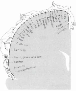
motor cortex was first demonstrated using electrical stimulation in patients by Penfield &
Boldrey (1937). Subsequently, a more detailed illustration of the motor and sensory
‘homunculus’ (meaning ‘little man’, a map of the human cortical representation, relating
to actual brain areas identified at surgery) was drawn either side of the central sulcus by
Penfield & Rasmussen (1950) (see Figure 1.2 for the motor homunculus). These
studies illustrated two important principles; firstly, the body representation in humans is
arranged in an orderly fashion within the precentral gyrus, and secondly, the muscle
groups used in movements that require fine control (for example in facial expressions
and the hands) are given disproportionately large representation (see below). This
ordered mapping of the body surface on to central neural structures is called
‘somatotopy’. However, there is still debate as to the accuracy of this reported
organisation (for review see Woolsey et al. 1979). Despite the popularity of this pictorial
view of sensory and motor cortical representation shown in Figure 1.2, many scientists
have criticised Penfield’s method and naïveté (for example Schott, 1993). Many studies
have demonstrated much intersubject variability as well as plasticity in the diseased
brain. In addition, cortical electrical stimulation and recent functional imaging techniques
have shown the activation of neurones beneath the surface of the brain. None of these
Chapter 1: General Introduction
(although it must be noted that the later functional imaging studies were obviously not
available to him).
: % I Y'7
\ ^ ~ 0 ' , v e f f,-,
I gu m s, a n d jaw
Tongu*
Figure 1.2: Map depicting the different body parts represented in the m otor cortex, reproduced from (Penfield & B oldrey, 1937). The large portions of the m otor area assigned to parts o f the body in which m ovem ents are delicate and precise, reflect the many cortical neurones required for such m ovem ents.
For the hum an prim ary m otor cortex ( Ml ) in particular, positron em ission tom ography
(PET) im aging has proved to be a useful technique capable of dem onstrating som e of the
characteristics of som atotopy in the cortex (Colebatch et al. 1991; G rafton et al. 1991;
1993). Other techniques for exam ining som atotopy of the m otor cortex include
m agnetoenchephalography (M EG) (Cheyne et al. 1991), scalp-recorded evoked
potentials (Bischert & Deecke, 1986) and transcranial magnetic stim ulation (TM S)
(Cohen & Hallett, 1988; W asserm ann et al. 1992). M any o f these studies have also
suggested an overlapping of within-lim b functional areas, and m any do not completely
agree with the classical hom unculus. These observations have been corroborated by
more recent fM RI studies reporting separate but overlapping representations of
individual fingers, elbow and toe m ovem ents in individual hum an subjects (Rao et al.
1995; K leinschm idt et al. 1997). One explanation for the m erging o f functional
activation sites for different m ovem ents could be that there is an overlap o f muscle
groups required to carry out the tasks (for review see Schieber, 1990). In fact, while the
‘cartoons’ depicting the M 1 hom unculus derived from electrostim ulation studies feature
som atotopic organisation even within the hand area, this is less clear from the original
Chapter 1: General Introduction
1.3.1.2 Anatomical location of the motor hand area
Numerous studies have identified the location of the hand motor and sensory area in a
specific segment of the pre- and postcentral gyrus (for example Penfield & Boldrey,
1937). This motor hand area has a characteristic shape: it is knob-like, most often
having the form of an inverted omega, a ‘bayonet-shaped’ hook or a horizontal epsilon
when examined in the axial plane, and it appears as a posteriorly directed hook when
viewed in the sagittal plane (Salamon et al. 1991; Talairach & Tournoux, 1993; Yousry
et al. 1997). It has been nicknamed the precentral ‘knob’ or ‘hook’ or ‘knuckle’ by
many neuroscientists (Naidich & Brightbill, 1995; Yousry et al. 1997). The knob
projects to the middle genu of the central sulcus, the middle being the posterior convex
in contrast to the superior and inferior genu that are anteriorly convex (first described by
Testut, 1911). It has also been identified as the ‘upper genu’, in contrast to the inferior
genu which corresponds to the projection area of the face over the external aspect of the
hemisphere (Rumeau et al. 1994). The pre-central knob can be regarded either as an
‘insulization’ (Salamon et al. 1991), or as a protrusion of the precentral gyrus towards
the central sulcus (Yousry et al. 1997). This knob can also be located on the cortical
surface by the neurosurgeon; however its configuration is less characteristic than it is at
deeper levels. It appears as the structure opposite to the intersection of the superior
frontal gyrus with the precentral sulcus (Kido et al. 1980; Yousry et al. 1997). The
identification of such a landmark clearly increases the accuracy of locating the precentral
gyrus. Other landmarks for locating the precentral gyrus have also been adopted, such
as the anterior ascending and horizontal rami of the sylvian fissure (Ebeling et al. 1989;
Naidich et al. 1995) and the ramus marginalis of the cingulate gyrus (Naidich &
Brightbill, 1995). However, with all methods large inter-rater variability exists (Sobel et
al. 1993).
1.3.1.3 Are individual muscles or movements represented differently in the primary
motor cortex?
The discovery of a somatotopic map raised a new question of whether local areas in the
motor cortex represent individual muscles or rather elementary movements involving the
co-ordinated activity of several muscles. Many studies have been carried out in an
attempt to answer this question, but there have been conflicting findings (for a review
see Schieber, 1990). In humans, lesions to the motor cortex or corticospinal tract
Chapter 1: General Introduction
movements, despite retaining the ability to flex and extend all the fingers together
(Twitchell, 1951; Penfield & Jasper, 1954). On the basis of these studies and of animal
studies (Passingham et al. 1983), the motor cortex appears to be necessary for
individuation, but how does it impart this ability?
Using cortical stimulation in monkeys, apes and humans, mapping evidence has
accumulated to demonstrate cortical representation of individual muscles (Penfield &
Boldrey, 1937; Woolsey et al. 1979; Cheney & Fetz, 1985). There has been growing
evidence, however, to suggest that although the dominant projection from a local area of
cortex may be to a single distal muscle, a single output neurone in the cortex often has
many collaterals that may influence several muscles, for example within the hand (Fetz,
Cheney, 1980; Cheney et al. 1985; Buys et al. 1986; for review see Lemon, 1988). In
addition, the cortical regions representing different muscles appears to overlap
extensively (Strick, 1988; Nudo et al. 1992; Schieber & Hibbard, 1993; Sanes et al.
1995; Volkmann et al. 1998; Hlustik et al. 1999). It appears likely that there is a
convergence from different cortical points on to a single pool of motor neurones, not a
direct one-to-one relationship, which may be irrespective of a detailed somatotopic
relationship. Accordingly, it is possible that a single finger can be activated from
different locations in the motor cortex, depending upon the complex patterns of
movement in which it is involved (Donoghue et al. 1992). Furthermore, Galea &
Darian-Smith (1994), and subsequently Kawashima et al. (1994) and Geyer et al.
(1996), proposed that rather than one homunculus in the primary motor cortex in man
there may be two or three, so for example finger movements can indeed be obtained by
stirriulating several areas of the motor strip.
It has also been suggested that the motor cortex is specialised not only for individual
muscle movements represented somatotopically but in addition discrete finger, hand and
arm movements (Humphrey, 1986; Schieber, 1995; Kleinschmidt et al. 1997). For
example, the thumb may move alone, in conjunction with the index finger, or with the
whole hand as in a grasp. All of these actions involve the thumb motor cortex region,
but various non-thumb movements (such as joint stabilising) must also occur to
complete each of the tasks listed. Rao et al. (1995) and Sanes et al. (1995), using fMRI,
both found that a large expanse of the human precentral gyrus exhibits activation during
individuated finger and wrist movements, and suggested that the neurones in this area
form a distributed and co-operated network that can simultaneously and optimally
control collections of arm muscles. More specifically, Schieber (1995) reported that for
‘movements of different fingers, a given muscle could act as an agonist, antagonist, or
Chapter 1: General Introduction
cortex, beyond just the area of a single digit. There are, however, problems with this
view. Firstly, cortical stimulation of the hand area may produce isolated movement of
the thumb, but an isolated movement of any other digit is evoked only rarely (Penfield &
Boldrey, 1937; Woolsey et al. 1952; 1979). Secondly, these studies showed extensive
overlapping of the regions for each finger. In addition, if movements of the different
fingers were represented in a lateromedial somatotopy (as depicted by Penfield &
Rasmussen, 1950), one might expect occasionally to see human patients who have
cortical lesions with impaired thumb movements but not movements of the little finger,
or vice versa. No such cases have been reported to date.
Another view is that the entire hand cortical area participates in producing a discrete
movement of any given finger. An integrated mosaic of efferent neurones within the
motor cortex hand area may project to particular muscles (Asanuma & Rosen, 1972;
Gould et al. 1986). There is evidence from a number of different studies in support of
this model of the organisation of the primary motor cortex (Humphrey, 1986; Fetz,
Cheney, 1987; Lemon et al. 1987). Schieber & Hibbard (1993) conjectured that rather
than being specified by a somatotopic map, each finger movement appears to be
specified by a neuronal population distributed throughout the motor cortex hand area.
The model would be consistent with the failure of cortical lesions to impair isolated
digits, and would also be consistent with the difficulty in evoking discrete movements of
single fingers in cortical stimulation studies and with the extensive overlapping
functional cortical areas and diverging muscular projections of the cortico-motomeuronal
cells.
There has been no definitive evidence as yet to exclude any of these hypotheses or even
to suggest the likelihood of one being prominent over any of the others. Many groups
hypothesise that the various ideas mentioned above should not be considered mutually
exclusive, as each may pertain to particular movements of particular digits and/or the
degree of mechanical independence of the digits involved.
1.3.2 Association cortical areas of the motor system
Other cortical areas have been shown to be linked to the primary motor cortex, so
comprising a large neural circuit within the brain and contributing substantially to motor
functional outputs. These are the supplementary motor area (SMA), situated on the
medial portion of the superior frontal gyrus (Brodmann’s area 6), the premotor area,
Chapter 1: General Introduction
postcentral somatosensory area (Brodmann’s areas 1, 2, 3, 5, and 7), and the second
somatosensory cortex (S-II) (area 43). However the anatomical boundaries, and even
the nomenclature, of these structures are not precisely defined (for review see Donoghue
& Sanes, 1994). All of these areas are thought to contribute directly to the fibres of the
descending pyramidal system (Murray & Coulter, 1981; Martino & Strick, 1987;
Hutchins et al. 1988; Dum & Strick, 1991). It has been discovered that these and other
components of the motor system (such as the subcortical areas and within the spinal
cord) contain individual somatotopic maps (Muakkassa & Strick, 1979; Hutchins et al.
1988; Dum & Strick, 1991; Lim et al. 1994). The SMA and premotor area receive
projections from many other cortical areas, including the parietal lobe, thalamus,
cerebellum, basal ganglia, sensory and visual areas (among others, Matelli et al. 1986;
Wiesendanger, 1986; Hutchins et al. 1988; Mushiake et al. 1991). However, each has
different projections from some or all of these structures, indicating that they all have
unique functions in the final specification of movement.
Approximately 20% of corticospinal fibres have been found to originate from the
primary somatosensory cortex (S-I) postcentrally (Brodmann’s areas 3a, 3b, 2, and 1),
S-II (area 43) and the posterior parietal cortex (areas 5 and 7) and project to the dorsal
horn (Jane et al. 1967; Coulter et al. 1976; Kuypers, 1981; Porter & Lemon, 1993).
This is in line with cytoarchitectonie evidence that pyramidal cells can be found in the
post- as well as precentral gyrus (Brodmann, 1909; Soso & Fetz, 1980; Jennings et al.
1983). The postcentral gyrus thus contributes to the formation of the pyramidal tract and
may therefore be additionally activated by the efferent part of the motor task. Similarly,
sensory responses can be elicited by stimulating the motor area (for example Powell &
Mountcastle, 1959). In addition, the role of the left parietal cortex has been shown to be
associated with motor attention (Rushworth et al. 1997). Due to the apparent
overlapping functions and projection systems between the primary sensory and motor
cortex they are usually considered as an integrated region of ‘sensorimotor cortex’
surrounding the central sulcus.
In brief, the SMA is generally thought to play an important role in the programming of
internally remembered complex sequences of movement (Roland et al. 1980;
Passingham, 1987; Jenkins et al. 1994; Catalan et al. 1998), and also in the preparation
of movement and in motor planning (Fox et al. 1985; Grafton et al. 1992a; Rao et al.
1993; Richter et al. 1997). The premotor cortex is thought to be involved in ‘externally
generated’ movement (see for example Passingham, 1989; 1993; Jenkins et al. 1994;
Sadatoet al. 1996a; Catalan et al. 1998). However, the literature demonstrates that the
Chapter 1: General Introduction
multiple roles in the control and production of movements, possibly explaining the huge
contradictions in the literature (for review see Passingham, 1993; 1996; Tanji & Shima,
1996).
Additional motor areas have also been identified on the medial and lateral cortical surface
(for example Luppino et al. 1991; Shima et al. 1991; Vogt et al. 1992). These are the
anterior cingulate gyrus (Grafton et al. 1993), the frontal eye fields (Mann et al. 1988),
and pre-frontal cortex (di Pellegrino & Wise, 1991; di Pellegrino & Wise, 1993; Jenkins
et al. 1994).
1.3.3 Subcortical motor stmctures and pathways
Fibres of the pyramidal system give off collateral branches as they traverse the internal
capsule and brain stem. These branches terminate in a number of subcortical structures,
namely the corpus striatum of the basal ganglia, red nucleus, pontine nuclei, inferior
olivary nucleus, cerebellum and the reticular formation (Phillips & Porter, 1977).
Perhaps the most researched of these subcortical structures are the cerebellum and the
basal ganglia. It is thought that the cerebellum acts as a ‘comparator’, to adjust the
actions of both the brain stem motor structures and the motor cortex (for example
Parsons et al. 1997; Jueptner & Weiller, 1998). It receives and compares descending
control signals responsible for the intended motor response (i.e. its planning and
execution) with sensory signals resulting from the consequences of ongoing motor
actions (Jueptner et al. 1996; Xue et al. 1997). The output of the cerebellum is delivered
both to the periphery and back to the cerebral cortex (particularly the primary motor and
premotor areas (Schell & Strick, 1984)), so that any discrepancies between the intended
and actual motor actions can be corrected. The cerebellum is also thought to have a direct
involvement in motor execution (Shibasaki et al. 1993), co-ordination of movement
(Llinas & Welsh, 1993), initiation and execution of planned movement (Krams et al.
1998), motor learning (Friston et al. 1992; Jenkins et al. 1994; Sadato et al. 1996a), and
sensory processing (Fox et al. 1985). The connections of the cerebellum are ordered
such that each cerebellar hemisphere is concerned with ipsilateral muscles; however it is
also thought to have a role in the control of bilateral limb movements (Ellermann et al.
1993; Fox et al. 1985).
The basal ganglia consists of the corpus striatum (subdivided into the caudate, putamen
and globus pallidus, the latter two areas collectively termed the lentiform nucleus),
Chapter 1: General Introduction
territory (Lehericy et al. 1999) and is the focus of the majority of inputs into the basal
ganglia from the primary motor and sensory areas, premotor and supplementary motor
cortices (Alexander & Crutcher, 1990). Some of the roles that have been postulated for
the function of the basal ganglia are the selection, control and preparation of specific
motor programs, starting and practising of sequential actions, inhibition of unwanted
movements and the promotion of motor learning and planning (Brooks, 1995; Bucher et
al. 1995; Boecker et al. 1998; Krams et al. 1998). Bilateral activation in the basal
ganglia may occur with unilateral motor tasks; however it was more significant on the
contralateral side (Hallett, 1993). The involvement of the cerebellum and basal ganglia in
motor action and other functions is still very much undetermined (for reviews see Leiner
et al. 1993; Brown & Marsden, 1998; Jueptner & Weiller, 1998).
1.3.4 The motor pathway from brain to muscle
The simplest functional stmcture in the motor hierarchy is the spinal cord. It is
responsible for organising the most automatic and stereotyped responses to stimuli in
addition to transmitting signals from higher cortical areas to carry out more skilful motor
outputs. Two pathways run within the cord, the corticospinal tract (or so-called
pyramidal tract as it mainly traverses the pyramid of the medulla and terminates in the
spinal cord), and the corticobulbar tract (which has the same relationship to motor
neurones of the cranial nerves as the corticospinal tract to motor neurones of the spinal
cord). Both tracts come under the heading of the pyramidal motor system. In this
section, and for the purpose of this thesis, emphasis will be on the organisation of the
corticospinal tract in the transmission of motor information.
The corticospinal tract comprises of motor neurones within the spinal cord, and is the
major cortically-derived descending pathway (see Figure 1.3 for overview; Canedo,
1997). A large portion of cortical neuronal fibres forming the origin of the tract are from
the primary motor area. In the cat and monkey, this area contributes more fibres to the
corticospinal tract than any other region (Porter & Lemon, 1993), ranging from 31%
(Russell & DeMyer, 1961) to 51% (Toyoshima & Sakai, 1982). In the human,
approximately 40-80% of the pyramidal fibres originate in the precentral gyrus (Jane et
al. 1967; Kuypers & Brinkman, 1970; Kuypers, 1981). The remainder of the fibres in
the corticospinal pathway are projection fibres arising from association areas within the
Chapter 1: General Introduction
The corticospinal tract exists in m ost m am m als, but it reaches its largest size and
im portance in hum ans where much o f its function deals with voluntary control o f the
upper and lower lim bs, in addition to m odifying sensory im pulses to regulate ascending
inform ation (A rm and, 1984). The tract allow s the m otor cortex to play a crucial role in
the co-ordination of m ovem ent and posture via direct and collateral branches to
structures governing eye, head, neck, trunk and limb m usculature (C anedo, 1997). N o
other neurones in the brain besides the pyramidal tract cells have such w ide access to
other structures w ithin the central nervous system . The follow ing description o f the
m otor pathw ay applies m ainly to hum ans; how ever, in principle this pathw ay is sim ilar
in m ost higher and low er prim ates.
Figure 1.3: The contralateral pyram idal m otor system (taken from B arr & K iernan, 1993). Refer to text for description. Structures labelled o f particular interest are the m otor cortex (1), the internal capsule (4), the cerebral peduncle (8), the pons (10), the ventral horn o f the spinal cord (17) and the ventral corticospinal tract decussating in the spinal cord (19). The lateral corticospinal tract is seen to decussate at the level o f the pyram ids (13). The ipsilateral corticospinal pathw ay is not indicated on this diagram .
T he tract em anates from the cortical areas m entioned above, traversing the corona radiata
and narrow ing down to enter the posterior limb o f the internal capsule (running betw een
the lentiform nucleus and thalam us). There is a discrete som atotopic organisation in the
internal capsule, as with all areas o f the m otor system . Fibres arising from the medial
frontal lobe (for exam ple from the SM A) pass through the anterior lim b, those arising
Chapter 1: General Introduction
from the premotor cortex descend through the genu and anterior portion of the posterior
part of the internal capsule, and finally those fibres originating from the primary motor
cortex itself pass through the middle third of the posterior limb of the internal capsule
(Fries et al. 1993). The descending pathway gives off branches to the ventral lateral and
centre median areas of the thalamus (Parent & Hazrati, 1995). The tract then continues
through the middle three-fifths of the basis pedunculi within the midbrain and is broken
up into many fasiculi (individual neuronal tracts) by clusters of pontine cells as it crosses
to the ventral portion of the pons. The fasiculi coalesce in the ‘pyramids’ on the anterior
surface of the medulla to form the descending compact corticospinal tract. In general it is
taken that, within the brainstem, fibres for the upper part of the body tend to be medial
and those for the lower half lateral, although this is known to lack precision because of
the intermingling of fibres concerned with different parts of the body.
In 1981, Kuypers noted in primates that the descending pathways could be divided into
two major groups according to the location of their terminations in the spinal cord, and
this corresponded to a systematic difference in their functional roles (Kuypers, 1981).
One group is located in the medial part of the ventral horn (in the anterior funiculus of
the spinal cord) and the other much larger group lies more laterally (in the lateral
funiculus of the spinal cord) (Kuypers, 1981). The latter group, which largely crosses
the midline at the caudal level of the medulla, constitutes the ‘lateral corticospinal tract’
(described below). The group within the ventral horn is called the ‘ventral corticospinal
tract’, most of the fibres of which do not decussate until they reach the spinal cord.
These two corticospinal tracts were also subsequently delineated in humans (Nathan et
al. 1990). The two motor neurone groups connect to different muscles; the lateral group
project to distal muscles such as those of the digits, and those innervating the axial
muscles lie in the medial part of the ventral horn (Ghez, 1991). In this section,
concentration is placed on the lateral corticospinal tract; the ventral corticospinal tract is
further mentioned only in Section 1.3.4.1.
It is at the caudal end of the medulla (at its junction with the spinal cord) where a large
number of the corticospinal fibres cross the midline, in what is known as the
‘decussation of the pyramids’, to form the lateral corticospinal tract. The portion of
crossing fibres in the medulla is approximately 75% to 85%, although it varies between
individuals (for review see Nyberg-Hansen & Rinvik, 1963). The decussation consists
of coarse bundles, with a portion of the fibres from pyramids on both sides of the
medulla crossing the midline. Once the fibres have crossed over, the axons regroup to
form direct pathways projecting down to the spinal cord grey matter. Because of this
Chapter 1: General Introduction
usage is incorrect, however, as some fibres leave the medullary pyramids to terminate in
brain stem nuclei such as the dorsal column nuclei. Within the spinal cord, such
descending corticospinal fibres lie in the dorsal part of the lateral white column of the
spinal cord and appear to be represented by longitudinal columns of motomeurons that
innervate a given muscle (Romanes, 1951; Sterling & Kuypers, 1967). Davidoff (1990)
suggested that the lateral corticospinal tract may vary considerably in size from person to
person.
The lateral corticospinal tract descends within the spinal cord, giving off many
collaterals, with only between 5% and 20% synapsing directly with motor cells
(Kuypers, 1981; Donoghue & Sanes, 1994; Canedo, 1997), most synapsing with
intercalating neurones which in turn synapse with alpha motor neurones and some
gamma motor neurones (Preston & Whitlock, 1961). This presence of direct
connections from the cortex to the motor neurones allows the ability to control individual
muscles or movements independently from one another. This important function is
termed th e ‘fractionation of movement’ (Phillips & Porter, 1977; Porter, 1985). Further
evidence for this control of movement comes from several investigations in monkeys. In
the study, it was shown that the animals’ development into making fine finger
movements over the first 7-8 months parallels the development of the
corticomotomeuronal connections (Lawrence & Hopkins, 1976). In two subsequent
studies, corticomotomeuronal cells showed a greater increase in activity when monkeys
performed a precision grip as opposed to the less fractionated movement of a power grip
(Muir & Lemon, 1983; Buys et al. 1986). In addition, sectioning of the corticospinal
tract or ablation of the motor cortex always results in deficits of fine finger movement in
animals (Lawrence & Kuypers, 1968a; Hepp-Reymond et al. 1974; Passingham et al.
1983) and man (Penfield & Jasper, 1954; LaPlane et al. 1977). Although a patient or
animal with such a lesion may retain the ability to flex and extend all the fingers
together, the ability to move fingers individually remains permanently deficient. For a
more detailed review of the cortical representation of movements see Section 1.3.1.3.
Although most of the corticospinal fibres decussate in the lower medulla to form the
contralateral lateral corticospinal tract, 25% remain uncrossed (Nyberg-Hansen &
Rinvik, 1963; Yakovlev & Rakic, 1966; Brodai, 1981; Davidoff, 1990). The origin and
termination of these descending ipsilateral fibres are described in detail below (Section
1.3.4.1).
In addition to the direct projection of the corticospinal pathways to the spinal motor
Chapter 1: General Introduction
parts of the brain stem. The most well understood are the vestibulospinal (originating in
the lateral vestibular nucleus) and the reticulospinal (originating in the reticular
formation) tracts, which are largely concerned with postural adjustments of movement
and balance (Kuypers, 1981; Holstege, 1996). In addition, cortical fibres are also
thought to maintain indirect connections with the spinal cord via terminations within the
superior collicus and tegmentum. These connections are important in co-ordinating head
and eye movements. All of these tracts have ipsilateral projections and are also described
in Section 1.3.4.1. Although to date most information about these tracts has been
derived from research in animals, it seems probable that the reticulospinal tracts mediate
control over most movements that do not require manual dexterity or the maintenance of
balance, and the vestibulospinal pathway appears to be essential for highly skilled
accomplishments of motor co-ordination, such as that required in a gymnast.
1.3.4.1 The ipsilateral corticospinal pathway in the normal individual
There has been much debate over the last few decades as to the origin of ipsilateral
sensorimotor responses to unilateral movements (Roland et al. 1980; Kim et al.
1993a&b; Salmelin et al. 1995). On the basis of anatomical data and mapping
techniques, evidence has been collected for the presence of ipsilateral sensory and motor
pathways in the normal central nervous system that may account for these responses (for
example Nakahama, 1958; Korvenoja et al. 1995; Noachtar et al. 1997). Several
groups, however, argue for other afferent pathways mediating ipsilateral responses,
such as transcallosal fibres, subcortical pathways, other cortical links such as through
the second somatosensory cortex (S-II), or simply volume conduction of the potentials
generated in the contralateral hemisphere (Larson et al. 1966; Williamson et al. 1970;
Tamura, 1972; Aleev & Varezhkin, 1978; Kuksova & Sumskii, 1983; Kakigi, 1986). In
this section, the normal anatomical ipsilateral pathway is summarised; its functional
application in the diseased brain is described later in Section 1.3.
In addition to the contralateral lateral corticospinal tract described in Section 1.3.4
above, ipsilateral lateral and ventral corticospinal tracts have also been identified in the
motor system of man (Nathan & Smith, 1955; Nathan et al. 1990; for review see Jones
et al. 1989). The pathways have also been found in the monkey (Glees & Cole, 1952;
Liu & Chambers, 1964; Semmes & Mishkin, 1965). Both tracts are smaller than the
crossed lateral corticospinal tract. Of the 25% uncrossed fibres in man, it has been found
that 15% are in the ventral ipsilateral corticospinal tract (Nyberg-Hansen & Rinvik,
(Nyberg-Chapter 1: General Introduction
Hansen & Rinvik, 1963; Brodai, 1981; Toyoshima & Sakai, 1982). In contrast,
however, Nathan & Smith (1973) suggested that the lateral corticospinal tract carries the
majority of ipsilateral fibres. The various tracts mentioned are thought to terminate in
different areas in the spinal cord. Several studies have reported that the majority of
axons of the ipsilateral ventral tract decussate in the spinal cord and thus terminate
contralaterally in the ventral horn to innervate the contralateral side (Figure 1.3) (Liu &
Chambers, 1964). The remaining ventral tract axons, however, terminate predominantly
in the ipsilateral ventral horn. In contrast, all of the ipsilateral lateral corticospinal tract
remains uncrossed and terminates in the ipsilateral dorsal horn, ventral horn and
intermediate zone (Glees & Cole, 1952; Liu & Chambers, 1964).
The cortical origin of these ipsilateral corticospinal fibres has been investigated (Liu &
Chambers, 1964). It was demonstrated that both ipsilateral and contralateral lateral
corticospinal tracts arose from the precentral area and part of the postcentral area. In
contrast, the ipsilateral ventral tract was found to originate only from the precentral
gyrus. The suggestion of ipsilateral tracts originating precentrally has recently been
supported by Toro et al. (1994) and Wassermann et al. (1994), and also in fMRI studies
demonstrating ipsilateral activation in the precentral area (for example Singh et al.
1998a). One study suggested that uncrossed corticospinal fibres terminating on spinal
neurones controlling axial and proximal muscles originated mainly from area 6, anterior
to the primary motor cortex (Weisendanger, 1981). Other studies have demonstrated a
much wider cortical representation of ipsilateral motor projection fibres; however, the
majority were found to arise from area 4 (Toyoshima & Sakai, 1982; Ghez, 1991; Ghez
& Gordon, 1995). It was initially thought that ipsilateral descending projections only
terminated on motor neurones innervating proximal limb muscles, as well as
contributing to the bilateral cortical control of the axial musculature either directly
through corticospinal pathways or indirectly via cortico-reticulospinal pathways (Liu &
Chambers, 1964; Brinkman & Kuypers, 1973). Several studies have now shown
ipsilateral cortical representation patterns in the precentral, premotor and SMA for distal
parts of the upper limb (for example Tanji et al. 1988; Benecke et al. 1991; Wassermann
et al. 1991; 1994). However these neurones formed only a small proportion of active
neurones during hand movements. The results of these human studies suggest that
ipsilateral projections from the primary motor cortex to the upper limb muscles exist but
are considerably weaker than contralateral projections. The proximal muscles are mainly
targeted, with distal muscles receiving only weak ipsilateral projections.
Certain brainstem pathways have also been found to contain a proportion of ipsilateral
Chapter 1: General Introduction
(predominantly the rubrospinal pathway) projects to the distal portion of the contralateral
limb. The rubrospinal tract originates in the red nucleus and projects down to the dorsal
part of the lateral column of the spinal cord. In contrast, the ventromedial brainstem
pathways project bilaterally to axial and proximal muscles (Ghez, 1991). The
ventromedial pathways consist of the vestibulospinal, reticulospinal and the tectospinal
pathways (refer also to Section 1.3.4). In particular, the reticulospinal pathway
originates predominantly in the premotor cortex (a small number of projections also arise
from the primary motor cortex) which sends projections down to the reticular formation
which in turn gives rise to a bilaterally organised reticulospinal tract (Lawrence &
Kuypers, 1968b; Brodai, 1969; Benecke et al. 1991). This pathway is not thought to
provide control of fractionated individual finger movements but does provide basic
action patterns of the contralateral and ipsilateral trunk and the limbs.
The organisation of the descending motor pathways described above suggest that
hemidecortication or focal sensorimotor lesions will have different consequences for
distal and proximal movements. In principle, the spinal motomeurones innervating distal
muscles contralateral to the side of the removed hemisphere will lose a considerable
amount of input, whereas the control of proximal muscles is largely maintained through
brainstem pathways and ipsilateral cortical connections both with the spinal cord and
ventromedial brainstem systems. It follows that proximal muscles ipsilateral to the side
of surgery or lesion may be only slightly affected but distal muscles may show a more
pronounced deficit (for example Jones et al. 1989; Dijkerman, 1996).
1.3.5 Interhemispheric pathway - the role of the corpus callosum
An additional source of connections into and out of the motor system comes from the
corpus callosum, which relays information from one hemisphere to another. Most
regions of the sensory and motor cortex are directly connected between hemispheres,
although it was originally thought that a major exception to this rule is the distal portion
of the limbs. These regions (namely the hand and foot areas) were thought by many to
be functionally disconnected from each other in older children and adults, and to act
totally independently (for review see Ghez, 1985). However, recent evidence from
clinical studies of patients with complete commissurotomies or agenesis of the corpus
callosum has suggested that the corpus callosum may play a role in bimanual motor co
ordination and the pre-motor control of movement, and may also inhibit the opposite
hemisphere from interfering when a simple unimanual movement is required (Geffen et