R E V I E W
Ultrasonography and computed tomography
in patients with right lower quadrant pain:
Dif
fi
cult cases of appendicitis
Adrienne van Randen1,2
Wytze Laméris1,2
Marja A Boermeester2
Julien BCM Puylaert3
Jaap Stoker1
1Department of Radiology; 2Department of Surgery, Academic
Medical Center, University of Amsterdam, Amsterdam, The Netherlands; 3Department
of Radiology, Westeinde Hospital, The Hague, The Netherlands
Correspondence: Adrienne van Randen Academic Medical Center, Meibergdreef 9 suite G1-227, 1105 AZ Amsterdam, The Netherlands
Tel +31 20 566 2630 Fax +31 20 566 9119 Email a.vanranden@amc.uva.nl
Abstract: Acute appendicitis is a common cause of acute abdomen and both computed tomography (CT) and ultrasonography (US) are used in the diagnostic work-up of these patients. In general, imaging has high accuracy in diagnosing acute appendicitis. Although the imaging features of appendicitis are well known, in some patients fi ndings are less conclusive. This pictorial essay will give an overview of diffi cult US and CT cases of patients suspected of acute appendicitis. Keywords: acute appendicitis, ultrasonography, CT
Introduction
Acute abdominal pain, and in particular right lower quadrant (RLQ) pain, is a common patient presentation at the emergency department (ED), usually requiring immediate diagnostic work-up and care. Acute appendicitis is a common cause for RLQ pain, although many other diagnoses should be considered. Ultrasonography (US) and computed tomography (CT) play an important role for a quick and accurate diagnostic work-up.1 With this pictorial essay we aim to give insight into diffi cult US and CT
cases of patients suspected of appendicitis.
Acute appendicitis
Lifetime risk of developing acute appendicitis is 9% for males and 7% for females.2
Symptoms generally start with nondescriptive visceral pain in the periumbilical region and anorexia followed by nausea and vomiting. When the disease progresses, typical migration of the pain to the RLQ occurs because of more localized peritoneal infl am-mation. Patients with a clear-cut physical history can be diagnosed clinically and may not require imaging.3 However, diagnosing acute appendicitis may be not that simple,
as negative appendectomy rates of 20% to 30% have been reported for patients who did not undergo additional imaging after clinical assessment.4,5 However, additional
usage of US and CT can reduce the negative appendectomy rate signifi cantly to 6%.6
Therefore, imaging should be used to confi rm or reject the diagnosis appendicitis, and in the latter situation propose an alternative diagnosis.
Imaging techniques
US with graded compression is a widely accepted technique for evaluation of the appendix.7 The CT technique for evaluating an acute abdomen, with a special interest in
the RLQ, involves a CT of the complete abdomen after intravenous contrast medium. High accuracy (98%) has been reported for focused appendiceal CT with oral and rectal contrast medium,8 but the major disadvantage is the possibility of missing an
alternative diagnosis outside the volume imaged or an infl amed appendix at an unusual site (Figure 1). In general, appendicitis can be diagnosed without oral, rectal, or
Reports in Medical Imaging downloaded from https://www.dovepress.com/ by 118.70.13.36 on 27-Aug-2020
van Randen et al
intravenous contrast medium.9 However, intravenous contrast
medium facilitates diagnosing alternative diagnoses and may result in a higher level of confi dence, especially when fat interfaces are (almost) absent (slim patients) or obliterated (areas of infi ltration). Reconstructed images by multiplanar reformation (MPR) can be of additional help in equivocal CT scans and increases the radiologist level of confi dence (Figure 2).10 Plain X-ray does not play a role in the work-up
of patients suspected with appendicitis.11
In current literature CT has higher accuracy compared to US. In a recent meta-analysis of head-to-head compara-tive studies of US and CT, a summary sensitivity of 91% was found for CT and of 78% for US. Specifi city was 90% for CT and 89% for US.1 In another study by Daly and
col-leagues, 13% (176/1397) of the CT scans performed for appendicitis were interpreted as equivocal. Of these patients with an equivocal CT 30% (n = 53) had appendicitis.12 The
sensitivity decreased from 83% to 64% if the equivocal scans were interpreted as positive, and the specifi city was lowered from 99.8% to 94% if the equivocal scans were interpreted as negative.
In our opinion, US should be considered the fi rst modality of choice in female, young, and slender patients, not only because of radiation dose, but also because CT scans of appendicitis in slender patients are more diffi cult to interpret due to absence of delineating fat. Vice versa, CT is generally better in obese patients because of the delineating fat planes. In very obese patients, the extensive amount of fat causes noise on CT, and will therefore not simplify interpretation of CT images (Figure 3).
Imaging
fi
ndings of acute
appendicitis
At US the noninfl amed appendix appears as a compressible, tubular blind-ending structure with a maximum diameter of 6 mm, without adjacent fat infl ammation (Figure 4). In appendicitis the appendiceal diameter is increased, the appendiceal wall is thickened and the surrounding fat is
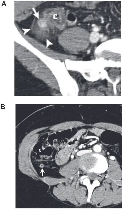
Figure 2 A 39-year-old female with a classical clinical presentation of appendicitis. The appendix could not be visualized on ultrasonography, because of disturbing (bowel) air (not shown). sagittal reformatted computed tomography image after iv contrast medium clearly shows a retrocecal infl amed appendix (arrow); C: cecum. Appendicitis was confi rmed at surgery and histopathology.
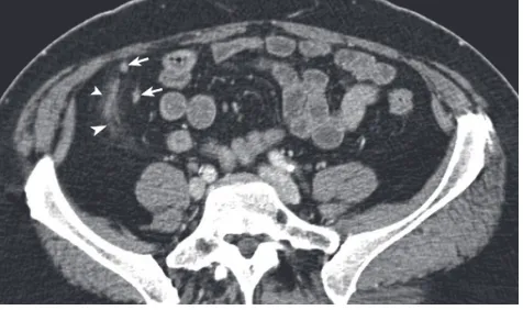
Figure 1 A) 20-year-old male suspected for acute appendicitis. Ultrasonography demonstrates in this sagittal oblique image an infl amed appendix (arrow) in the right upper quadrant with surrounding fat infl ammation (arrowheads) adjacent to the liver and right kidney. B) To guide the operating surgeon, the appendix was marked on the patient’s skin. The marked point represents the McBurney point.
A
B
Reports in Medical Imaging downloaded from https://www.dovepress.com/ by 118.70.13.36 on 27-Aug-2020
Diffi cult cases of acute appendicitis
hyperechoic, and both the appendix and surrounding fat are noncompressible (Figure 5). Appendicitis can only be excluded when the appendix is completely visualized, including the appendiceal tip, and has a normal appearance (Figure 6).
At CT, a noninfl amed appendix appears as a tubular blind-ending structure, usually with a diameter of less than 6 mm, and often containing air. Features of an infl amed appendix at CT are a tubular, blind-ending structure with a diame-ter ⬎6 mm which is surrounded by infl amed fat (Figure 7). In both US and CT, an appendicolith can be identifi ed in many individuals with and without appendicitis, thus the presence of an appendicolith is not pathognomonic for appendicitis
(Figure 4). Also in both, complications can be visible, such as perforation and abscesses.
Dif
fi
cult cases of US or CT
US or CT images are diffi cult to interpret when only one or two of the above mentioned imaging features can be identifi ed. The solitary fi nding of a thickened appendix, an appendicolith, or fat infi ltration adjacent to the appendix are not conclusive signs for the diagnosis acute appendicitis.12
These fi ndings may represent normal fi ndings or may refl ect early stages of disease or a reactive response to another disease in the RLQ. If the treating physician suspects an early presentation of appendicitis, a ‘wait-and-see’ policy is
Figure 3 A 37-year-old obese female with a body mass index of 42.7 with histological proven appendicitis. CT after iv contrast medium showed a thickened appendix (arrow) and mild adjacent fat infi ltration. C: Cecum.
Figure 4 A 23-year-old male with right lower quadrant pain. Ultrasonography (US) shows a nonthickened appendix (arrows) without surrounding infi ltration and multiple appendicoliths (curved arrows) and therefore not an US (and fi nal) diagnosis of appendicitis.
Figure 5 A 49-year-old female suspected of acute appendicitis. Ultrasonography (US) demonstrates a noncompressible, thickened appendix (arrow) surrounded by infl amed mesenteric fat (arrowheads); a clear US diagnosis of acute appendicitis which was confi rmed at surgery.
Figure 6 A 43-year-old male with a surgical and histopathological proven infl ammation of the appendiceal tip. Ultrasonography images show a normal proximal diameter of the appendix (arrow), but a distal diameter of 8.3 mm (arrowheads).
Reports in Medical Imaging downloaded from https://www.dovepress.com/ by 118.70.13.36 on 27-Aug-2020
van Randen et al
justifi ed, and the patient maybe asked to return the next day for a second clinical evaluation.
No classical imaging presentation
Single imaging
fi
nding
Patients with only a thickened appendix and no clinical or laboratory signs of appendicitis are treated conservatively at our center, because symptoms may resolve spontaneously, and may be clinical irrelevant (Figure 8). Patients with mild fat infl ammation adjacent to a nonthickened or nondistended appendix do not have acute appendicitis (Figure 9). In these patients often another diagnosis can be made, while the diagnosis of nonspecifi c abdominal pain (NSAP) is made when no other clinical and radiological fi ndings are present. Visualization of a normal appendix is crucial for disregarding the diagnosis acute appendicitis.
Multiple imaging
fi
ndings
If patients with RLQ pain have multiple, although subtle, infl ammatory appendiceal changes or infl ammatory changes adjacent to the appendix, the diagnosis appendicitis must be made. If the appendix is thickened and (subtle) fat infi ltra-tion is present, acute appendicitis is likely. Appendicitis is also likely when the appendix is somewhat thickened with an enhancing wall and fat infi ltration (Figures 10, 11). When a thickened appendix is the sole feature, then the diagno-sis acute appendicitis is not likely. The only exception is a perforated appendix in which the appendix may not be thickened, but adjacent infl ammatory changes are usually extensive in this situation (Figure 12). In patients with mild
Figure 7 A 49-year-old male suspected for appendicitis. Computed tomography (CT) after iv contrast medium shows a thickened appendix (arrow) with a thickened appendiceal wall, infi ltration of the adjacent fat (arrowheads) and a thickened fat plane (curved arrow). Appendicitis was proven at surgery and histopathology.
Figure 8 A 75-year-old male suspected for acute appendicitis. A) Computed tomography (CT) after iv contrast showed a fl uid fi lled thickened appendix of 10 mm diameter (arrow) without infl ammation of the adjacent mesenteric fat, more proximal to the cecal orifi ce the appendiceal lumen was fi lled with air (not shown) B) An appendicolith was present at the appendiceal orifi ce (arrow) with cecal wall thicken-ing (arrowheads). On re-examination a few hours later the patient had appetite and pain had decreased. Therefore the patient was discharged home the next day. After more than two years of follow-up, the patient had no recurrent episode of acute right lower quadrant (RLQ) pain. Therefore the fi nal diagnosis of nonspecifi c abdominal pain (NSAP) was made.
A
B
Figure 9 A 60-year-old female with right lower quadrant (RLQ) pain for three days with a fi nal diagnosis of nonspecifi c abdominal pain after clinical follow-up (up to two years). Computed tomography (CT) after intravenous contrast shows moderate streaky fat infi ltration (arrowheads) in the RLQ, but the appendix (arrows) is not enlarged and contains air (not shown).
Reports in Medical Imaging downloaded from https://www.dovepress.com/ by 118.70.13.36 on 27-Aug-2020
Diffi cult cases of acute appendicitis
infl ammatory changes in the RLQ and without visualization of the appendix on US and CT, no conclusive diagnosis can be made and management should be based on clinical fi ndings and laboratory fi ndings only and laparoscopy can be considered. However, when the infl ammatory changes are extensive (possibly abscesses) the appendix may not be discernable as the fat planes are obliterated (Figure 13). In this situation the diagnosis appendicitis becomes quite likely when no other fi ndings (eg, normal terminal ileum visualized) are present, especially in young men.
Differentiation between terminal ileitis and appendicitis can be diffi cult when the continuity or blind-ending of an infl amed tubular structure is not visualized. However when a normal-appearing ileum and ileocecal region are visualized, the diagnosis appendicitis becomes very likely (Figure 14).
Secondary appendicitis
The appendix may become thickened by an adjacent infl am-matory condition, such as diverticulitis, a gynecological cause or an infl ammation in the ileocecal area (Figure 15). Identifi cation of the primary cause of infl ammation is often possible, with the infl ammatory response at the primary site being more extensive than adjacent to the appendix.
Atypical location
A challenge in clinical diagnosing acute appendicitis arises in patients with atypical history and fi ndings at examination because of atypical location of the appendix (Figure 1). In case of a retrocecal appendix, US may have diffi culty in diagnosing appendicitis as the appendix is obscured by an
Figure 10 A 38-year-old male with peri-umbilical pain for 16 hours, nausea without vomiting and a low grade temperature was clinically suspected for acute appendicitis. Computed tomography (CT) after intravenous contrast shows a thickened appendix (12 mm) with an appendicolith (arrow) and subtle fat infi ltration (arrowhead). Acute appendicitis was confi rmed at surgery and histopathology.
Figure 11 A 31-year-old male with right lower quadrant (RLQ) pain for two days, nausea without vomiting, and rebound tenderness without guarding, was clinically suspected of acute appendicitis. This computed tomography (CT) image after intravenous (and oral) contrast shows a curved appendix of 9-mm diameter with wall enhancement (arrows) and subtle adjacent fat infi ltration (arrowheads). Acute appendicitis was proven by surgery and histopathology.
Figure 12 A 55-year-old female clinically suspected for appendicitis shows an perfo-rated appendix (arrow) on this axial computed tomography (CT) image after iv contrast medium. Peri-appendiceal free air is seen (arrows). Also adjacent fat infl ammation, with a thickened fat plane is visible (arrowheads). Surgery and histopathology both proved perforated appendicitis in this patient.
Figure 13 A 44-year-old male with right lower quadrant (RLQ) pain suspected for acute appendicitis. The appendix could not be visualized on computed tomography (CT). This coronal CT image after iv contrast medium demonstrates an abscess (A) with adjacent fat infi ltration (arrowhead) located at the suspected site for the appendix and closely related to the cecum (C) and Ileum (I) The abscess was drained percutaneously and no appendectomy was performed.
Reports in Medical Imaging downloaded from https://www.dovepress.com/ by 118.70.13.36 on 27-Aug-2020
van Randen et al
air-fi lled cecum or ascending colon. At CT visualization is not hampered by appendix localization, provided that no focused appendiceal CT of the lower quadrants has been performed, where only part of the abdomen is visualized.
Perforated appendix
Imaging has limitations in identifying a perforated appendix, although discontinuity of the wall and fl uid adjacent to the appendix is suggestive for the diagnosis.13 In patients with
frank fat infi ltration, thickened appendiceal wall, but no distended appendiceal lumen, a perforated appendix should be considered (Figures 12, 15).
Mimics of appendicitis
Alternative diagnoses mimicking acute appendicitis include right sided diverticulitis (Figure 16), infl ammatory bowel disease, epiploic appendagitis, and gynecological causes in women in the reproductive age (Figure 17). Evaluation of spe-cifi c features of these mimickers are beyond the scope of this pictorial essay, but are well described in the literature.14,15
Summary
The diagnosis of appendicitis can be readily made at US and CT, if multiple imaging features of acute appendicitis are present. In patients with only single or subtle imaging fi ndings such as a thickened appendix without fat infi ltration, or merely an appendicolith or fat infi ltration adjacent to the appendix, the diagnosis of acute appendicitis becomes unlikely and another cause for their complaints must be sought for. To substanti-ate these clinical observations, a prospective study should be performed evaluating features of appendicitis at imaging.
Figure 14 A 33-year-old male with right lower quadrant (RLQ) for one day which migrated from the peri-umbilical region to the RLQ. Computed tomography (CT) after intravenous contrast showed an abnormal tubular structure in the RLQ which could not be visualized completely (arrow). Adjacent to this tubular structure extensive fat infi ltration was seen (arrowheads). Furthermore a normal ileum could be identifi ed (not shown). The diagnosis of acute appendicitis was very likely in this male patient, although the blind end of the tubular structure was not visualized. Appendicitis was proven by surgery and histopathology.
Figure 15 A 27-year-old female with right lower quadrant (RLQ) pain for three days, clinically suspected for a gynecological disorder or acute appendicitis. Computed tomography (CT) after intravenous contrast showed an appendicolith (arrow), extensive fat infi ltration (arrowheads) suggestive of a perforated appendix, although the appendix could not be clearly visualized. Perforated appendicitis was proven at surgery and histopathology.
A
Figure 16 A 25-year-old female with acute right lower quadrant (RLQ) pain. A) A right-sided colon diverticula (arrow) is visible with adjacent fat infi ltration (arrowheads), fecolith, wall enhancement on this axial computed tomography (CT) image after i.v. contrast medium; B) Somewhat lower, a nondistended appendix with thickening, containing air, is visible. The appendiceal wall is secondarily infl amed in this patient with right sided diverticulitis. C: Cecum.
B
Reports in Medical Imaging downloaded from https://www.dovepress.com/ by 118.70.13.36 on 27-Aug-2020
Diffi cult cases of acute appendicitis
Disclosure
The authors declare no confl icts of interest in this work.
References
1. van Randen A, Bipat S, Zwinderman AH, Ubbink DT, Stoker J, Boermeester MA. Acute appendicitis: Meta-analysis of diagnostic performance of CT and graded compression US related to prevalence of disease. Radiology. 2008;249(1):97–106.
2. Addiss DG, Shaffer N, Fowler BS, Tauxe RV. The epidemiology of appendicitis and appendectomy in the United States. Am J Epidemiol. 1990;132:910–925.
3. Andersson RE. Meta-analysis of the clinical and laboratory diagnosis of appendicitis. Br J Surg. 2004;91:28–37.
4. Jones PF. Suspected acute appendicitis: trends in management over 30 years. Br J Surg. 2001;88:1570–1577.
5. Raman SS, Osuagwu FC, Kadell B, Cryer H, Sayre J, Lu DS. Effect of CT on false positive diagnosis of appendicitis and perforation. N Engl J Med. 2008;358:972–973.
6. Florence M, Flum DR, Jurkovich GJ, et al. Negative appendectomy and imaging accuracy in the Washington State Surgical Care and Outcomes Assessment Program. Ann Surg. 2008;248:557–563.
7. Puylaert JB, Rutgers PH, Lalisang RI, et al. A prospective study of ultrasonography in the diagnosis of appendicitis. N Engl J Med. 1987;317:666–669.
8. Rao PM, Rhea JT, Novelline RA, Mostafavi AA, McCabe CJ. Effect of computed tomography of the appendix on treatment of patients and use of hospital resources. N Engl J Med. 1998;338:141–146. 9. Lane MJ, Katz DS, Ross BA, Clautice-Engle TL, Mindelzun RE,
Jeffrey RB, Jr. Unenhanced helical CT for suspected acute appendicitis. AJR Am J Roentgenol. 1997;168(2):405–409.
10. Paulson EK, Harris JP, Jaffe TA, Haugan PA, Nelson RC. Acute appen-dicitis: added diagnostic value of coronal reformations from isotropic voxels at multi-detector row CT. Radiology. 2005;235:879–885. 11. Rao PM, Rhea JT, Rao JA, Conn AK. Plain abdominal radiography in
clinically suspected appendicitis: diagnostic yield, resource use, and comparison with CT. Am J Emerg Med. 1999;17:325–328.
12. Daly CP, Cohan RH, Francis IR, Caoili EM, Ellis JH, Nan B. Incidence of acute appendicitis in patients with equivocal CT fi ndings. AJR Am J Roentgenol. 2005;184:1813–1820.
13. Bixby SD, Lucey BC, Soto JA, Theysohn JM, Ozonoff A, Varghese JC. Perforated versus nonperforated acute appendicitis: accuracy of multi-detector CT detection. Radiology. 2006;241:780–786.
14. Yu J, Fulcher AS, Turner MA, Halvorsen RA. Helical CT evaluation of acute right lower quadrant pain: part II, uncommon mimics of appendicitis. AJR Am J Roentgenol. 2005;184:1143–1149.
15. Yu J, Fulcher AS, Turner MA, Halvorsen RA. Helical CT evaluation of acute right lower quadrant pain: part I, common mimics of appendicitis. AJR Am J Roentgenol. 2005;184:1136–1142.
Figure 17 A 42-year-old female with acute abdominal pain suspected for acute appendicitis or diverticulitis. The coronal reformatted image shows a normal appen-dix (arrow) and a located fl uid collection with wall enhancement suggestive for an (ovarian) abscess (A), which was in concordance with the fi nal diagnosis of tubo-ovarian abscess. The mesenteric fat infl ammation is visible just cranial of the fl uid collection (arrowhead) C: cecum; I: ileum.
Reports in Medical Imaging downloaded from https://www.dovepress.com/ by 118.70.13.36 on 27-Aug-2020
Reports in Medical Imaging downloaded from https://www.dovepress.com/ by 118.70.13.36 on 27-Aug-2020