IJSRR, 8(1) Jan. – Mar., 2019 Page 1121
Research article
Available online www.ijsrr.org ISSN: 2279–0543
International Journal of Scientific Research and Reviews
HGSIL on Pap smear
: Good indication for direct LEEP procedure
SangeetaRai
*1and Kalpana Singh
2Department of Obstetrics and Gynaecology, Institute of Medical Sciences ,Banaras Hindu University, H.No - Manish Bhawan,B-1/5-6, Ravindrapuri Extension, Assi Varanasi,
221005,Uttarpradesh.,Ph- 09621591247, E- mail : drsangeeta1977@yahoo.co.in Medical officer , UEHCC, Institute of Medical Sciences ,Banaras Hindu University.
Mail id -kalpana11dec@yahoo.com
ABSTRACT
Objective: Our aim was to evaluate the effectiveness and accuracy of LEEP procedure inpatients with high-grade dysplasia on cytology without prior colposcopically directed biopsy.
Study Design: It was a prospective study for a period of 1year where LEEP was performed on all the patients for high-grade squamous intraepithelial lesion (HGSIL) on Papanicolaou (Pap) smear without a prior cervical biopsy. Colposcopy was done before LEEP and scoring was done based on Reid’s colposcopic index. After informed consent LEEP was performed. Specimen sent for histopathology. Histology findings were correlated and data analysed taking specimen of LEEP for histology as gold standard. Results: Out of 81 patients undergoing LEEP, 44 patients (54.3%) had cervical intraepithelial neoplasia (CIN) grade 2 or greater. 18 (41%) of these 44 patients with high-grade dysplasia on histopathology had a normal or low-high-grade lesion on colposcopy. There were 20 patients with CIN 1 to CIN 1-2 and 17 patient with normal findings. Conclusion: LEEP is better decision in patients with high grade lesion on cytology without prior colposcopic biopsy in resource constrained countries.
KEYWORDS
High-grade squamous intraepithelial lesion;
colposcopy;
loop electrosurgical excision procedure;
dysplasia
Loop electrosurgical excision
*Correspondence address
DrSangeetaRai,
Associate Professor, Department of Obstetrics and Gynaecology, Institute of Medical Sciences.
Banaras Hindu University, H.No- Manish Bhawan,B-1/5-6,
Ravindrapuri Extension, Assi Varanasi, 221005, Uttarpradesh.,Ph- 09621591247,
IJSRR, 8(1) Jan. – Mar., 2019 Page 1122
INTRODUCTION
Cervical cancer is the commonest malignancy found amongst Indian women and the third
most common cancer in the world. Over 5,00,000 new cases of invasive cervical cancer are
diagnosed annually worldwide.
There are many cervical cancer screening programmes with treatment options available
depending upon the resources and infrastructure. We usually screen with pap smear at first visit. The
results of the cytology is used to decide the treatment plan accordingly. In cytology report of CIN or
higher, colposcopic evaluation and directed biopsy is done usually.
LEEP is the ideal treatment for of choice in patients with histopathology report of CIN 2 or
more on colposcopic biopsy. We know that the colposcopic evaluation needsexpertise and training.
Besides, the accuracy of the colposcopic findings are still less reliable in correlation to histology .
Colposcopic evaluation is still very much dependent on the operator. It needs better
understanding of the procedure. Studies has shown that there is considerable subjective variations
and many factors like prior knowledge of the cytology report can change the accuracy of the
diagnosis.A new scoring system of Reid is developed to increase the accuracy of the colposcopic
assessment and histological diagnosis.
But still in developing countries resources are required for colposcopy machine and training
the doctors .Almost all thecolposcopic examination is done by gynecologist who lacks necessary
skill in performing this procedure.These factors are hurdle in implementation and accuracy of
colposcopic evaluation. This has prompted to evaluate the alternative strategy of perfoming LEEP in
HGSIL on cytology without prior colposcopic biopsy.
LEEP is the ideal treatment in patients with high grade lesions because it offers the tissue
specimenfor histopathology. LEEP excise whole of the transformation zone, the site for all the HPV
infection and precancerous changes. If the margins of the LEEP specimen are free from CIN, it
indicates successful surgery. The standard indication of LEEP are positive endocervical curettage,
discrepancy between cytology and histopathology results.
METHODS AND MATERIALS
This is a prospective study done for a period of 2 year in the department of Obstetrics and
gynecology at Sir Sunderlal Hospital, Varanasi from December 2015 to 2017. 81 subjects were
included in our study who came positive for HGSIL in cytology. The cytology smear was reported
by single trained pathologist in our hospital. All the subjects were informed about the study. General
and gynecological examination was done. Colposcopy was performed and reid’s scoring done. This
IJSRR, 8(1) Jan. – Mar., 2019 Page 1123 performed in the same sitting. The results of the ECC was used as a predictor of repeated or
persistent cervical intraepithelial pathological process (CIN). LEEP specimen was sent for histology.
Patients was discharged next day with proper advice. Follow up was done after 10 days with the
histology reports. The histology reports were taken as gold standard.
RESULT
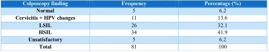
The distribution of colposcopic findings is given in Table 1. In our study, 26 and 34 among
81 subjects were reported to be LGSIL and HGSIL respectively. Despite HGSIL on Pap smear
before the LEEP, 16 patients had no pathologic findings and in 5 patients, colposcopic evaluation
was unsatisfactory. Table 2 shows the study of study samples according to the histology reports of
LEEP. 17(20.9%) patients has normal HPE findings, 24(24.8%) has HPE report of CIN 1 to CIN1-2
and 44 (54.3%) has HPE report of CIN 2 or more.One patient had a microinvasive squamous cell
cancer.
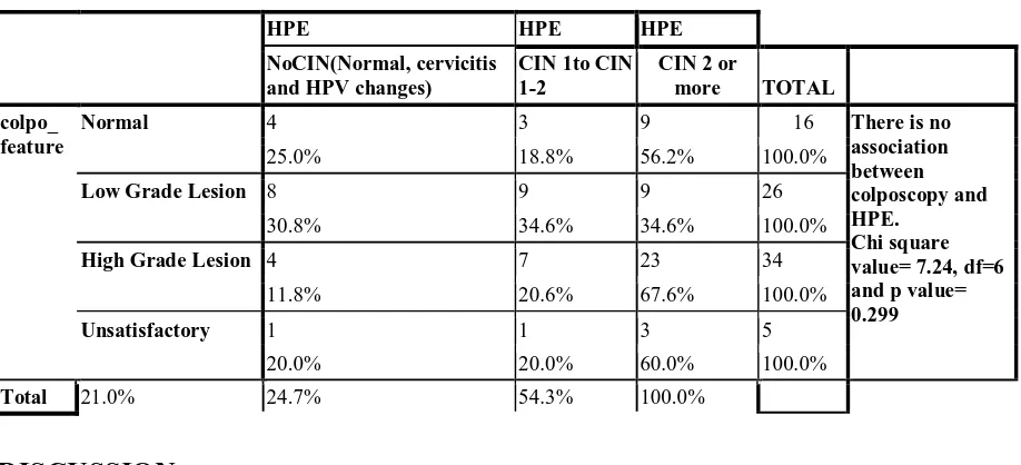
In Table 3, colposcopic findings are correlated with the histological diagnosis. Of 81 patients
undergoing LEEP, 44 patients (54.3%) had cervical intraepithelial neoplasia (CIN) grade 2 or
greater. 18 (41%) of these 44 patients with histologically proved high-grade dysplasia had a normal
or low-grade colposcopic examination. There were 20 patients with CIN 1 to CIN 1-2 and 17 patient
with normal findings. Thus, there is significant number of patients 18(22.22%) who are saved by
doing the direct LEEP instead of prior colposcopy which showed normal or low grade changes. The
results has shown that there is no association between colposcopy and HPE (p value 0.299).
Table 1: Distribution of Study Subjects according to the colposcopy finding.
Colposcopy finding Frequency Percentage (%)
Normal 5 6.2
Cervicitis + HPV changes 11 13.6
LSIL 26 32.1
HSIL 34 41.9
Unsatisfactory 5 6.2
Total 81 100
Table 2 : Distribution of study samples according to biopsy result of LEEP Specimen:
Biopsy Finding Frequency Percentage (%)
Normal 5 6.2
Cervicitis 12 14.8
CIN 1 to CIN1-2 20 24.8
CIN 2 or more 44 54.3
IJSRR, 8(1) Jan. – Mar., 2019 Page 1124 Table 3: Correlation between colposcopic finding and biopsy result of LEEP Specimen:
HPE HPE HPE
NoCIN(Normal, cervicitis and HPV changes)
CIN 1to CIN 1-2
CIN 2 or
more TOTAL
colpo_ feature
Normal 4 3 9 16 There is no
association between colposcopy and HPE.
Chi square value= 7.24, df=6 and p value= 0.299
25.0% 18.8% 56.2% 100.0%
Low Grade Lesion 8 9 9 26
30.8% 34.6% 34.6% 100.0%
High Grade Lesion 4 7 23 34
11.8% 20.6% 67.6% 100.0%
Unsatisfactory 1 1 3 5
20.0% 20.0% 60.0% 100.0%
Total 21.0% 24.7% 54.3% 100.0%
DISCUSSION
In our study,colposcopic findings are correlated with the histology findings. Among 34
patients with high grade lesion in colposcopy, 23 (63.8%) had histological report of high grade
lesion. Among 42 patients with normal or low grade lesion in colposcopy, 18 (42.9%) had
histologically proven high grade abnormality. Thus, women with negative colposcopy remain at
significant risk for subsequent detection of CIN 2 or higher. The insufficiencies of colposcopy have
been widely documented. 3Study by Pretorius showed that the ability to detect the abnormal areas
with colposcopy is not consistent with CIN. 37.1% of the CIN 2 lesions or worse were diagnosed
from biopsies of normalareas at colposcopy.10 Increasing the number of biopsies taken each time
will increase sensitivity for the detection of high-grade disease11 but it requires multiple biopsies
which will decrease the patient compliance due to increased discomfort.12.Thus the performance and
accuracy of colposcopic findings are based on the training, experience and skill of the operator.A
total of 63 patients underwent ECC immediately after loop excision, of which 56 (88.9%) patients
were found to have benign endocervical tissue, whereas 2 had HGSIL. There were no specific
clinical predictors of a high-grade ECC such as age, size of the dysplastic lesion, or adequacy of the
colposcopic examination.
CONCLUSION
The diagnosis and treatment of cervical abnormality at one setting visit is a good option in
IJSRR, 8(1) Jan. – Mar., 2019 Page 1125 patient by saving time, money and discomfort due to multiple visits as done in standard screening
and treatment protocol for CIN. Besides it overcome the problem of need for skilled
colposcopist.This also decreases the patients lost to follow up and coming with the frank cancer later
on. Howeverdirect LEEP has also got its disadvantages by overtreatment in a normal case with its
related morbidity. In our study 20.9% has normal HPE report who underwent unnecessary LEEP.
But the percentage is relatively low as compared to other studies. Hence we observe that our study
strategy has got advantages which outweigh the risk of overtreatment in our settings. Though we
acknowledge the constraints of this study, small sample size and no control group, we believe our
results support the use of LEEP on a see-and-treat basis.
CONFLICT OF INTEREST
: NilREFERENCES
1. Sankaranarayanan R, Budukh AM, Rajkumar R. Effective screening programmes for cervical
cancer in low- and middle-income developing countries. Bull World Health Organ 2001; 79:
954-62.
2. Parkin DM, Bray FI, Devesa SS. Cancer burden in the year 2000. The global picture. Eur J
Cancer 2001; (37 Suppl 8): S4-66.
3. Jeronimo J, Schiffman M. Colposcopy at a crossroads.ObstetGynec2006;195:349Y53.
4. Etherington I, Luesley D, Shafi M, Dunn J, Hiller L,Jordan J. Observer variability among
colposcopists from the West Midlands region. Br J ObstetGynaecol 1997;104:1380Y4.
5. Reid R, Scalzi P. Genital warts and cervical cancer. VII. An improved colposcopic index for
differentiating benign papillomaviral infections from high-grade cervical intraepithelial
neoplasia. Am J ObstetGynecol 1985;153:611Y8.
6. Walboomers JM, Jacobs MV, Manos MM, et al. Human papillomavirus is a necessary cause
of invasive cervical cancer worldwide. J Pathol Sep1999;189(1):12–9. [PubMed: 10451482]
7. Munoz N, Bosch FX, de Sanjose S, et al. Epidemiologic classification of human
papillomavirusgenotypes associated with cervical cancer. N Engl J Med Feb
62003;348(6):518–27. [PubMed:12571259]
8. Schiffman MH, Bauer HM, Hoover RN, et al. Epidemiologic evidence showing that human
papillomavirus infection causes most cervical intraepithelial neoplasia. J Natl Cancer Inst
June161993;85(12):958–64. [PubMed: 8388478]
9. Kreimer AR, Guido RS, Solomon D, et al. Human papillomavirus testing following loop
IJSRR, 8(1) Jan. – Mar., 2019 Page 1126 intraepithelial neoplasia grade2 or 3 disease. Cancer Epidemiol Biomarkers Prev
May2006;15(5):908–14. [PubMed: 16702369]
10.Pretorius RG, Zhang W, Belinson JL, Huang M, Wu L,Zhang X, et al. Colposcopically
directed biopsy, random cervical biopsy, and endocervical curettage in the diagnosis of
cervical intraepithelial neoplasia II or worse. ObstetGynecol 2004;191:430Y4.
11.Gage JC, Hanson VW, Abbey K, Dippery S, Gardner S,Kubota J, et al. Number of cervical
biopsies and sensitivity of colposcopy. Am J ObstetGynecol 2006;108:264Y72.
12.The TOMBOLA (Trial of Management of Borderline and Other Low-grade Abnormal
smears) Group. After-effects reported by women following colposcopy, cervical biopsies and
LLETZ: results from the TOMBOLA trial. BJOG 2009;116:1506Y14.
13.Stafl A, Mattingly RF, Colposcopic diagnosis of cervical neoplasia, ObstetGynecol, 1973;