EFFECTS OF HYPOTHYROIDISM ON
CELL SIGNALLING IN THE
DEVELOPING RAT BRAIN
By
Feng Chiao Lilian Leung
A thesis submitted for the degree o f Doctor o f Philosophy in the Faculty o f Science at the
University o f London.
March 1995
Institute o f Neurology
Department o f Neurochemistry Queen Square
ProQuest Number: 10016771
All rights reserved
INFORMATION TO ALL USERS
The quality of this reproduction is dependent upon the quality of the copy submitted. In the unlikely event that the author did not send a complete manuscript and there are missing pages, these will be noted. Also, if material had to be removed,
a note will indicate the deletion.
uest.
ProQuest 10016771
Published by ProQuest LLC(2016). Copyright of the Dissertation is held by the Author. All rights reserved.
This work is protected against unauthorized copying under Title 17, United States Code. Microform Edition © ProQuest LLC.
ProQuest LLC
789 East Eisenhower Parkway P.O. Box 1346
ABSTRACT
Rat pups bom o f dams maintained on a propylthiouracil/low iodine diet regime were used
to assess the effects o f hypothyroidism on signalling components in the developing brain.
The success o f the animal model o f perinatal hypothyroidism was confirmed following
measurements o f free T3 and T4 plasma levels. Alterations in development and behaviour
noted in hypothyroid pups were similar to those observed previously by other groups.
Plasma metabolite concentrations indicated that the impairments in body and brain
development noted in the hypothyroid state, were not the result o f serious under-nutrition.
Crude and synaptosomal membranes were isolated from forebrain and hindbrain regions
o f euthyroid and hypothyroid animals at various ages. Changes in a-subunit abundance
o f Gq, Gq, Gjl and Gj2 were measured via quantitative immunoblotting.
No differences in (%-subunit levels o f euthyroid and hypothyroid cmde membranes were
seen in either brain region. Compared to euthyroid animals, between 10 and 20 days
postpartum a number o f significant alterations were detected in synaptosomal membranes
isolated from forebrains o f hypothyroid pups; with the exception o f G{la, all o f these changes were that o f subunit up-regulation. By contrast, the hindbrain showed very few
hypothyroid-induced changes; the only significant effects were seen in G jla at day 1 0
(total hindbrain) and day 25 (medulla oblongata). Hypothyroidism also altered the normal
developmental profiles o f all four o:-subunits in both forebrain and hindbrain regions.
Synaptosomal membranes o f 15 day old pups were also used to assess hypothyroid-
activation/inhibition o f adenylyl cyclase by GTP were observed in hypothyroid forebrain
and hindbrain membranes in the presence o f forskolin. Furthermore, a significant decrease
in specific cyclase activity was observed in hypothyroid forebrain membranes. By
contrast, in the hindbrain, a significant increase in cyclase activity was evident.
Together, the changes observed may contribute to some o f the neurological defects seen
ACKNOWLEDGEMENTS
I would like to thank my two supervisors Professor E. Dave Saggerson and Professor John B. Clark for giving me the opportunity o f doing this Ph.D. and for their advice and support throughout the duration o f the project.
My thanks also go to the people, past and present, in Lab 302 (UCL), the Dept o f Biochemistry (UCL) and in the Dept, o f Neurochemistry (ION), whom I have had the good fortune to know and befriend. I am grateful for all their inputs both stimulatory and inhibitory, i.e. advice, humour, alcohol etc. I apologise for not naming you all in person, but you know who you are. I am particularly grateful to June Smalley for her help with the more elaborate diagrams in this thesis and to the members o f the Denny Brown Laboratory for their assistance.
I am indebted to the Wellcome Trust Foundation for their generous funding o f this project and for the workshops and meetings which were extremely helpful over the three years.
Finally I would like to extend my gratitude to my family and friends for their continual moral support and numerous attempts to understand what it was exactly I was doing for my Ph.D.
This thesis is dedicated to my Mum, Dad and brother Gabriel.
To the one person without whom all this would not be possible, who persevered (eventually) through good times and bad, toiled, sweated, cursed and agonized over numerous hours. It was worth it.
CONTENTS
Page
Title P a g e ... 1
Abstract ... 2
A cknow ledgem ents...4
List o f Figures and T a b le s ... 10
A bbreviations... 13
CHAPTER 1 INTRODUCTION 1.1 The B r a in ... 15
1.2 Anatomy o f the Human B r a i n ... 15
1.2.1 Medulla Oblongata and P o n s ... 16
1.2.2 C e re b e llu m ... 16
1.2.3 Midbrain ... 17
1.2.4 Hypothalamus and T h a la m u s ... 17
1.2.5 Basal G a n g lia ... 18
1.2.6 Cerebral C o r te x ... 18
1.3 Development o f the Human Brain ... 20
1.4 Cells ... 21
1.4.1 N e u r o n s ... 21
1.4.2 G l i a ... 24
1.5 Synapses ... 25
1.6 The T h y r o id ... 27
1.6.1 Historical A s p e c t... 27
1.6.2 Structure o f the T h y r o id ... 27
1.7 Thyroid H o rm o n e s... 28
1.7.1 Thyroid Hormone Synthesis ... 29
1.7.2 Secretion o f the Thyroid Hormones ... 30
1.7.3 Thyroid Hormones in the Serum ... 30
1.8 Regulation o f Thyroid F u n c tio n ... 31
1.8.1 Thyroid Stimulating Hormone ( T S H ) ... 31
CHAPTER 1 INTRODUCTION Continued
9 Action o f Thyroid H o rm o n es... 34
9.1 Nuclear Action ... 35
9.2 Extranuclear A c t i o n ... 35
10 R e c e p to rs ... 37
11 Clinical Aspects o f Thyroid H o rm o n e s ... 37
11.1 H ypothyroidism ... 38
1.11.2 Hyperthyroidism ... 40
1.12 Thyroid Hormones and the Developing Brain ... 41
1.12.1 Neurological Development in Rats and H u m a n s ... 41
1.12.2 Stages in Brain Development in Relation to Thyroid H o rm o n e s... 42
1.12.3 Thyroid Hormone Effects on Brain D e v e lo p m e n t... 44
1.12.4 Morphological Effects ... 45
1.12.5 Biochemical E f f e c ts ... 48
1.12.6 Electrophysiological Effects ... 50
1.12.7 Behavioural E f f e c t s ... 51
1.13 Maternal Hypothyroidism and E f f e c ts ... 51
1.14 Transmembrane Signalling ... 54
1.15 G-Proteins ... 55
1.15.1 Structure and P r o p e r tie s ... 55
1.15.2 Mechanism o f G-Protein A c tio n ... 58
1.16 a-Subunits ... 61
1.16.1 G3O !... 62
1.16.2 G j o : ... 63
1.16.3 G ^ a... 65
1.16.4 GqO:... 65
1.16.5 Other G a S u b u n its... 6 6 1.17 G(3y S u b u n its ... 67
1.18 Effector S y s te m s ... 69
1,18.1 Adenylyl C y c la se ... 69
CHAPTER 1 INTRODUCTION Continued
1.18.2 Phospholipase C ... 74
1.18.3 Ion C h an n e ls... 76
1.19 R e c e p to rs ... 78
1.20 G-Proteins and Pathological S t a t e s ... 80
1.21 Aims o f P r o je c t... 82
CHAPTER 2 MATERIALS AND METHODS 2.1 Materials ... 85
2.2 A n im a ls ... 87
2.2.1 Induction o f H ypothyroidism ... 87
2.2.2 M o th e r s ... 87
2.2.3 Pups ... 8 8 2.3 Dissection Procedure ... 8 8 2.4 Preparation o f Synaptosomal, Mitochondrial and Myelin Fractions from Rat Brain ... 90
2.4.1 Adults ... 90
2.4.2 Pups ... 92
2.5 Preparation o f Crude M e m b ra n e s... 94
2.6 Collection o f Plasma ... 95
2.7 Hormone and Metabolite Measurements o f P l a s m a ... 95
2.7.1 Free Triiodothyronine (Tg) ... 95
2.7.2 Free Thyroxine (T4) ... 96
2.7.3 3-H ydroxybutyrate... 96
2.7.4 L a c ta te ... 96
2.7.5 Non-Esterified Fatty Acids ... 97
2.7.6 Glucose ... 97
2.8 Estimation o f P r o te i n ... 97
2.9 Quantitative Immunoblotting o f G-Protein o:-S u b u n its... 98
2.9.1 Polyacrylamide Gel Electrophoresis ... 98
2.9.2 M in i-G e ls... 100
CHAPTER 2 MATERIALS AND METHOD Continued
2.9.4 Western B lo ttin g ... 101
2.9.5 Immunological Detection o f Western Blots ... 101
2.9.6 Quantitation o f G-Protein o :-S u b u n its... 102
2.10 A n tis e r a ... 105
2.11 lodination o f Immunoglobulins ... 108
2.11.1 lodination Procedure ... 108
2.11.2 Validation o f lodination P ro c e d u re ... 109
2.12 Spectrophotometric Enzyme A s s a y s ... 110
2.12.1 Acetylcholinesterase (EC 3 .1 .1 .7 )... 110
2.12.2 2% 3 '-Cyclic Nucleotide 3 '-Phosphohydrolase (EC 3.1.4.37) ... 110
2.12.3 Succinate Dehydrogenase (EC 1.3.99.1) ... I l l 2.12.4 Calculation o f Enzyme Activity ... 112
2.13 Assay o f Adenylyl C y c la s e ... 112
2.13.1 Adenylyl Cyclase Assay P ro ce d u re ... 112
2.14 cAMP A s s a y ... 115
2.14.1 Principle o f A s s a y ... 115
2.14.2 cAMP P ro c e d u re ... 116
2.15 Trial ELISA P ro ce d u re ... 118
2.16 Statistical A n a ly sis... 119
CHAPTER 3 RESULTS AND DISCUSSION: HYPOTHYROIDISM AND THE DEVELOPING RAT 3.1 Hypothyroidism and D e v e lo p m e n t... 121
3.2 Effects o f Hypothyroidism on the Developing Rat ... 122
3.3 Effects o f Hypothyroidism on the Developing B r a i n ... 129
3.4 Plasma Thyroid Hormone and Metabolite Measurements o f Developing Euthyroid and Hypothyroid R a t s ... 132
3.5 Plasma Thyroid Hormone and Metabolite Measurements o f Developing Euthyroid and Hypothyroid D a m s ... 142
CHAPTER 4 R E SU L T S AND D IS C U S S IO N : E F F E C T S OF HYPOTHYROIDISM ON G-PROTEIN AND ADENYLYL CYCLASE IN THE DEVELOPING BRAIN
4.1 Effects o f Hypothyroidism on G-Protein and Adenylyl
Cyclase in the Developing Brain ... 150
4.2 Preparation o f Subcellular Fractions from Developing A n im a ls... 151
4.3 Effects o f Hypothyroidism on the Developmental Profile o f Brain Synaptosomal Membrane G-Protein oj-subunits ... 156
4.3.1 Gq/iia ... 159
4.3.2 G ^o !... 163
4.3.3 G ila ... 165
4.3.4 G;2a ... 172
4.3.5 G-Protein D is c u s s io n ... 177
4.4 Effects o f Hypothyroidism on Adenylyl Cyclase Activity in Brain Synaptosomal Membranes o f 15 Day Old P u p s ... 181
4.4.1 Effects o f GTP on Basal Adenylyl Cyclase Activities in Forebrain and Hindbrain Synaptosomal Membranes ... 182
4.4.2 Effects o f GTP on Forskolin-Stimulated Adenylyl Cyclase Activities in Forebrain Synaptosomal Membranes in the Presence and Absence o f NaCl ... 185
4.4.3 Effects o f GTP on Forskolin-Stimulated Adenylyl Cyclase Activities in Hindbrain Synaptosomal Membranes in the Presence and Absence o f NaCl ... 188
4.4.4 Adenylyl Cyclase D iscu ssio n ... 191
CHAPTER 5 GENERAL D IS C U SSIO N ... 195
LIST OF FIGURES AND TABLES
Page
CHAPTER 1
Figure 1.1 Longitudinal Section o f the Primate B r a i n ... 19
Figure 1.2 Thyroid Hormone Structures ... 29
Figure 1.3 Regulation o f Thyroid F u n c tio n ... 33
Figure 1.4 Brain Neurological Development in Relation to the Thyroid Hormones in Rat and H u m a n ... 43
Figure 1.5 G-Protein Mediated Transmembrane Signalling ... 59
Figure 1.6 Regulation o f Mammalian Adenylyl Cyclase ... 70
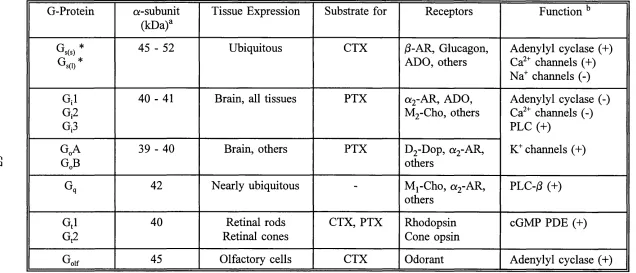
Table 1.1 Summary o f G-Protein P ro p e rtie s ... 57
Table 1.2 Properties o f Adenylyl C y c la s e s ... 72
CHAPTER Figure 2.1 Figure 2.2 Figure 2.3 Figure 2.4 Figure 2.5 Figure 2.6 Diagrammatical Representation o f Dissection Procedure for Rat Brain ... 89
Synaptosomal Membrane Preparation... 93
Quantitation o f G-Protein CK-Subunits with Respect to Amount o f protein Loaded... 103
Immunoblots o f Synaptosomal Membrane G-Protein «-Subunits Visualised using Specific Polyclonal Antisera... 106
Time Course o f Basal and Forskolin-Stimulated Adenylyl Cyclase Activity in Brain Synaptosomal membranes o f 15 Day-Old Pups... 113
CHAPTER 3 Page
Figure 3.1 Body Weights o f Euthyroid and Hypothyroid Pups
During Postnatal Development... 123
Figure 3.2 Euthyroid and Hypothyroid Pups at 1 Day P o stp artu m ... 124
Figure 3.3 Euthyroid and Hypothyroid Pups at 15 Day P o stp a rtu m ... 126
Figure 3.4 Euthyroid and Hypothyroid Pups at 25 Day P o stp a rtu m ... 127
Figure 3.5 Forebrain Weights o f Euthyroid and Hypothyroid Pups at Various Stages o f Development ... 130
Figure 3.6 Hindbrain Weights o f Euthyroid and Hypothyroid Pups at Various Stages o f Development ... 131
Figure 3.7 Plasma T3 Levels o f Euthyroid and Hypothyroid Rats at 1, 10, 15, 20 and 25 Days Postpartum ... 134
Figure 3.8 Plasma T4 Levels o f Euthyroid and Hypothyroid Rats at 1, 10, 15, 20 and 25 Days Postpartum ... 135
Figure 3.9 Plasma 3-Hydroxybutyrate Levels o f Euthyroid and Hypothyroid Rats at 1, 10, 15, 20 and 25 Days Postpartum . . . . 137
Figure 3.10 Plasma Glucose Levels o f Euthyroid and Hypothyroid Rats at 1, 10, 15, 20 and 25 Days Postpartum ... 138
Figure 3.11 Plasma Lactate Levels o f Euthyroid and Hypothyroid Rats at 1, 10, 15, 20 and 25 Days Postpartum ... 140
Figure 3.12 Plasma NEFA Levels o f Euthyroid and Hypothyroid Rats at 1, 10, 15, 20 and 25 Days Postpartum ... 141
Figure 3.13 Body Weights of Euthyroid and Hypothyroid Dams During and After Pregnancy... 146
Table 3.1 Plasma Thyroid Hormone and Metabolite Levels o f Euthyroid and Hypothyroid D a m s ... 143
Page
Figure 4.2 Relative Abundance o f GqO: in Synaptosomal Membranes
from Rat Pup Whole Hindbrain and Hindbrain R e g io n s ... 161 Figure 4.3 Effects o f Hypothyroidism on the Relative Abundance o f G^a
in Synaptosomal Membranes from Rat Pup Forebrain... 164 Figure 4.4 Relative Abundance o f GqO; in Synaptosomal Membranes
from Rat Pup Whole Hindbrain and Hindbrain R e g io n s ... 166 Figure 4.5 Effects o f Hypothyroidism on the Relative Abundance o f G jla
in Synaptosomal Membranes from Rat Pup Forebrain... 169 Figure 4.6 Relative Abundance o f G jla in Synaptosomal Membranes
from Rat Pup Whole Hindbrain and Hindbrain R e g io n s ... 170 Figure 4.7 Effects o f Hypothyroidism on the Relative Abundance o f G;2o:
in Synaptosomal Membranes from Rat Pup Forebrain... 173 Figure 4.8 Relative Abundance o f Gj2o: in Synaptosomal Membranes
from Rat Pup Whole Hindbrain and Hindbrain R e g io n s ... 175 Figure 4.9 Effects o f GTP upon Basal and Adenylyl Cyclase
Activity in Brain Synaptosomal Membranes from
15 Day-Old Pups ... 183 Figure 4.10 Effects o f GTP upon Forskolin-Stimulated Adenylyl Cyclase
Activity in Forebrain Synaptosomal Membranes from
15 Day-Old Pups in the Presence and Absence o f NaCl ... 186 Figure 4.11 Effects o f GTP upon F orskolin- Stimulated Adenylyl Cyclase
Activity in Hindbrain Synaptosomal Membranes from
15 Day-Old Pups in the Presence and Absence o f NaCl ... 189
Table 4.1 Distribution o f Marker Enzymes in Subcellular Fractions
from Rat Forebrain at Various Stages o f D ev elo p m ent... 152 Table 4.2 % Distribution o f Marker Enzymes in Subcellular Fractions
from Rat Forebrain at Various Stages o f D evelopm ent... 155 Table 4.3 Effect o f Hypothyroidism on G-Protein ce-subunit Levels
ABBREVIATIONS
HR Hormone-Receptor complex
CNS Central nervous system
CTX Cholera toxin
DAG Diacylglycerol
D IT Di-iodotyrosine
EGL External granular layer
IP3 Inositol 1,4,5-trisphosphate
M IT Mono-iodotyrosine
PAGE Polyacrylamide gel electrophoresis
PIP2 Phosphatidylinositol 4,5-bisphosphate
PKC Protein kinase C
PLC Phosphatidylinositol specific phospholipase C
PTU 6 -n-propylthiouracil
PTX Pertussis toxin
SDS Sodium dodecyl sulphate
T3 Triiodothyronine
CHAPTER 1
INTRODUCTION
1.1 THE BRAIN
The brain to all intents and purposes bears a striking resemblance to a cauliflower with
a thick rope suspended beneath it. However this mass o f specialised cells acts as a control
centre for the rest o f the body and has the daunting responsibility o f collecting and
processing information as well as co-ordinating and regulating the body’s movements
and activities. The brain governs our emotions; shapes our thoughts, hopes, dreams and
gives rise to our imagination which makes us all so unique from one another. It
determines the type o f person we are.
The brain is complex in structure and organisation. It is divided into many regions, each
possessing its own role in the overall function o f the brain.
1.2 ANATOMY OF THE HUMAN BRAIN
It is convenient to divide the brain into three regions: forebrain, midbrain and hindbrain.
The hindbrain emerges from the spinal cord and encompasses the medulla oblongata,
cerebellum and pons. The midbrain denotes the top o f the brain stem and is situated
above the pons in the human. The forebrain is the most prominent part o f the brain and
can be subdivided into two gross areas; cerebrum (basal ganglia and cortex) and
diencephalon (thalamus and hypothalamus). The brain stem, which lies above the spinal
cord, acts as a bridge allowing messages to pass between the brain and the rest o f the
body. The anatomical and functional aspects o f the brain may be found in the majority
o f anatomy and physiology text books available (e.g. Carpenter, 1990; Dowling, 1992a;
1.2.1 MEDULLA OBLONGATA AND PONS
The medulla and pons are the most primitive parts o f the brain. The medulla, situated
rostrally to the brain stem, contains numerous ascending and descending nerve tracts.
Discrete nuclei (clusters o f neurons) with specific functions including balance, control o f
head and facial features, and regulation o f body functions (e.g. heart rate, respiration,
gastrointestinal functions) are also housed in this region o f the brain. The medulla is
capable o f exerting wide spread influences on virtually all parts o f the brain by virtue o f
the vast axonal extensions o f a separate group o f neurons known as the reticula formation.
The pons, resembling a thick bulge, is a portion o f the brain stem just superior to the
medulla oblongata. The pons as its name suggests (bridge), connects the medulla to the
more developed areas o f the brain. Discrete nuclei in the anterior portion o f the pons
relay information from the cerebrum to the cerebellum. Nuclei present in the posterior
section are associated with cranial nerves. Like the medulla, the pons also contains
ascending and descending nerve tracts. The pons is involved in control o f cardiac
functions and houses the sleep and respiratory centres.
1.2.2 CEREBELLUM
Attached to the medulla and pons is the cerebellum (little brain), a highly regulated
structure whose main function is to co-ordinate and integrate motor activity. It receives
input from the cerebral cortex concerning initiation o f voluntary movement. The
cerebellum integrates the information and relays it to the descending motor system along
with sensory inputs to ensure smooth overall movement. Lesions in the cerebellum result
1.2.3 M IDBRAIN
Situated just superior to the pons, the midbrain denotes the top o f the brain stem and is
a point o f integration o f various impulses. The roof o f the midbrain consists o f four
mounds {colliculi) collectively termed corpora quadrigemia. There are two superior and two inferior colliculi. The latter are involved in hearing and are an integral part o f the
auditory pathways whereas the former are concerned with visual reflexes and receive input
from the eyes, inferior colliculi, skin and cerebrum. The midbrain also plays a role in motor neuron stimulation responsible for eye and head turning.
1.2.4 H Y PO TH A LAM U S AND THALAM US
W ithin the forebrain is "buried" the|thalamus (from the Greek word for inside chamber)
and the hypothalamus (under).The former acts as a relay station for the majority o f
information that passes to and from the brain. The hypothalamus contains several small
nuclei and nerve tracts with functions that include regulation o f olfactory reflexes, eating,
drinking and sexual activity. A stalk protruding from the floor o f the hypothalamus
connects it to the pituitary gland. Consequently the hypothalamus is crucial in endocrine
system regulation as it controls hormone secretion from the pituitary. Functions as diverse
as metabolism, reproduction, autonomic (heart rate) muscle control and temperature
regulation are all influenced by the hypothalamus. Moreover, pituitary hormones possess
the ability to effect hormone release from endocrine glands elsewhere in the body (e.g.
thyroid, adrenal), constituting yet another level o f regulation by the hypothalamus. The
thalamus, resembling something similar to a yo-yo, consists o f numerous prominent nuclei
w hich modify and relay sensory and motor activity information to the cerebral cortex.
Auditory, visual and sensory impulses congregate in this particular area o f the brain.
1.2.5 BASAL GANGLIA
The basal ganglia are five prominent nuclei positioned around the thalamus concerned
with movement (initiation and execution) and posture. They receive input from the cortex
and emit signals to the thalamus. Lesions lead to movement dysfunction characterised by
tremor, repetitive and slow movements. Parkinson’s disease and Huntington’s chorea are
both results o f impaired basal ganglia function.
1.2.6 CEREBRAL CORTEX
The outer layer o f the cerebrum is called the cortex (shell). For humans and many other
mammals, the cerebral cortex houses the majority o f the higher nervous systems.
Functions localised in this particular brain region include skilled movement, sensation,
consciousness, memory and intelligence to name but a few. Cortical functions are
arranged topographically on the cerebral cortex. The cortex itself invaginates many times
to form grooves called gyri, resulting in a much greater surface area. In addition the cortex has evolved into two separate sections termed hemi-spheres which can be further
subdivided into four lobes. Neurons in the cortex lie close to the surface. Specific neural
functions are carried out by the lobes. The frontal lobes are involved primarily with
movement and olfaction. The parietal lobes deal with somatic sensation, the occipital
lobes with vision, and the temporal lobes with hearing and memory. The lobes receive
information from the thalamus and initiate specific movements. Also situated within the
cerebrum beneath the temporal lobes, are two hippocampi, concerned with long term
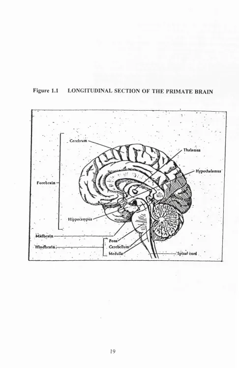
Figure 1.1 LONGITUDINAL SECTION OF TH E PRIM ATE BRAIN
TtuUmas
Forcbrsin
one is removed. Each cerebral hemisphere controls and receives inputs from the opposite
side o f the body. In the majority o f people, the left side o f the brain is dominant
controlling speech and analytical skills; the right side deals with spatial and musical
abilities.
Figure 1.1 depicts the locations o f the major regions o f the primate brain.
1.3 DEVELOPMENT OF THE HUMAN BRAIN
In the human, the first signs o f the brain appear over the first three weeks o f gestation.
A single cell formed at conception gives rise to an embryo, and it is from a layer o f
tissues (neural plate) on the dorsal surface o f the embryo that the central nervous system
(CNS) develops. The lateral sides o f the plate form neural crests which move forward
and fuse together creating the neural tube. The expanded anterior portion o f the tube goes
on to form the brain. Constrictions in this region serve to separate the future fore-, mid-
and hindbrain. The narrower posterior end o f the neural tube gives rise to the future
spinal cord. Alongside and separate from the neural tube lie crest cells from which the
majority o f the peripheral nervous system will develop.
Once the tube has formed, cell proliferation begins. What starts o ff as a single layer soon
develops into a multi-layer o f cells. Cell division takes place in specialised areas known
as germinal zones located in the inner surface o f the neural tube. Spongioblasts give rise
to glia whereas neuroblasts are the precursors o f neurons. As the cells migrate they form
temporary contacts with one another but once at their final destination, differentiate into
specific cells with precise properties and connections. Further development involves
proliferate after migration, specifically those found in the basal ganglia and granule cell
o f the cerebellar cortex.
By five weeks gestation in the human, the major regions o f the brain (forebrain, midbrain
and hindbrain) are recognisable albeit in a primitive form. The forebrain divides into the
telencephalon (cerebrum) and the diencephalon. The midbrain remains as a single
structure whilst the hindbrain gives rise to the pons, cerebellum and medulla oblongata.
More detailed descriptions o f brain ontogeny can be found in Lund, 1978; Carpenter and
Sutin, 1983; Naula and Fiertag, 1986; Dowling, 1992a.
1.4 CELLS
Interactions between nerve cells underlie much o f the brains functions and
accomplishments allowing the animal to leam, behave, remember and create. They
communicate chemically by means o f electrical signals relayed by neurons. There are two
major types o f cell in the brain: neurons and glia.
1.4.1 NEURONS
Neurons or nerve cells, are responsible for processing and receiving signals from sensory
organs and other neurons, integrating that information and transmitting it. In addition to
the same intracellular components o f other cells, neurons possess unique features such as
distinct cell shape and specialised structures known as synapses. There are about 10^^
neurons in the human brain each composed o f three parts:
(1) cell body - contains the nucleus, enzymes and components essential for the cell’s
survival;
appearance and are the sites where the neuron receives signals from other cells;
(3) axon - an extension o f the cell body and the means by which signals travel to and
from the cell body to other cells o f the brain.
The axon may be branched and is frequently myelinated along its course increasing its
efficiency as a conducting unit for electrical impulses/ signals (Palay and Chan-Palay,
1977). Neurons communicate with each other by conducting these electrical signals and
are able to do so for long distances without loss o f signal strength. Neurons can be
excitatory, inhibitory and modulatory in effect, or motor, sensory and secretory in
function. They can also be classified according to the number o f processes they have e.g.
unipolar (one axon), bi-polar (two axons), multi-polar (several axons). Neurons differ
from other cells in the body in that once differentiated, possess a limited ability to
regenerate following injury due to the high degree o f differentiation.
As mentioned earlier, neurons can come in various forms and sizes. Each part o f the
brain is made up o f certain types o f neurons pertaining to that region’s particular function.
In other words, neurons in the cerebellum are different from those located in the cerebral
cortex. Neurons can be divided into two broad classes: Golgi type I and Golgi type II
(Dowling, 1992b).
Golgi type I neurons from one part o f the brain can carry information to another part o f
the brain, spinal cord or an effector organ (e.g. muscle). They possess dendrites which
receive information from other cells and effectors, and long axons which branch near the
terminus forming synapses with other neurons or effectors. Golgi type I neurons possess
nodes o f Ranvier, points at which they are not myelinated along their axon, for rapid relay
o f information.
these neurons are confined to one area o f the brain and are involved in local interactions
between nerve cells. They are often called association neurons and unlike Golgi type I
cells, both axons and dendrites are capable o f transmitting information. Golgi type II are
believed to be an important part o f subtle neuronal interaction.
Neurons within a particular brain region may be classified even further. The cerebellum
is made up o f five different neuronal cell types whereas in the cerebral cortex, two major
types have been identified. Microscopic studies have revealed neurons and nerve terminals
in areas o f the brain such as cerebral cortex and cerebellum, are arranged in distinct
layers. In the cerebral cortex, six layers can be distinguished labelled I-VI starting from
the outermost layer. Pyramidal cell bodies predominate in layers II, III, V and VI
whereas stellate cells though present in all layers predominate in layer IV. The relative
abundance o f the different neuronal cell types differ from region to region and reflect the
various functions o f that region in terms o f input and output requirements.
The cerebellum in contrast, consists o f three principal layers: molecular (outermost) layer;
Purkinje cell (intermediate) layer and the granule (inner) cell layer. Purkinje cells o f the
cerebellum serve a similar role to that o f pyramidal cells located in the cerebral cortex.
The granule cell layer consists o f densely packed small neurons whose axons penetrate the
molecular layer and form contacts with Purkinje cell dendrites (parallel fibres). The
Purkinje cell layer aside from Purkinje cells and parallel fibres, consists o f basket cells and
stellate cells. Stellate cells and basket cells are inhibitory intemeurons. They along with
Golgi cells, can be found in the granule layer and contain y-amino butyric acid (GABA)
or some other inhibitory neurotransmitter. Stellate and basket cells located in the
molecular and Purkinje cell layers respectively, both receive inputs from the granule cell
whereas the axons o f basket cells synapse around the initial portion o f Purkinje axons,
thus enabling effective inhibition o f the Purkinje cell layer if so required. Inputs to the
cerebellar cortex are termed mossy and climbing fibres. The former terminate on granule
cells whilst the latter terminate on groups o f Purkinje cells whose activity they control
(Strange, 1992).
1.4.2 GLIA
In 1846, Virchow came across a tissue that was not neuronal in nature. It possessed no
synaptic connections and occupied all remaining space in the brain not already taken up
by nerve cells. He named the tissue glia meaning glue, pertaining to its first assigned
function, to hold the neuron network together. Though they do not appear to participate
in the integration and processing o f information, glia are ten times more abundant than
neurons and can be divided into two categories: macroglia (astrocytes and
oligodendrocytes) and microglia (similar to macrophages). Despite the extent o f their
numbers, their exact functions are uncertain but they appear to have several roles from
protection o f the brain against electrical/ionic environment changes, to the uptake and
metabolism o f released neurotransmitters or excess chemicals, e.g. K^. In addition, they
participate in CNS repair and provide structural and metabolic support for the vast array
o f neurons. During the early stages o f the developing mammal, glia are required for
guiding neuronal migration in certain regions o f the brain (Hatten, 1990). Astrocytes are
believed to possess connective and supportive functions as well as being involved in repair
as they are seen to proliferate subsequent to trauma. They have also been assigned a
putative role in transport within the blood brain barrier system and a firmer one in the
production o f myelin. The majority o f cell axons are encased in myelin which acts as an
insulator and also serves to increase the rate at which nerve impulses can travel along the
cell with respect to an unmyelinated cell. Oligodendrocytes are distinguishable under
electron microscopy from neurons and found to predominate in white matter, the colour
characteristic to the presence o f myelin. It can take several oligodendrocytes to encase
an axon with myelin depending on the size o f the axon. As mentioned earlier, the myelin
sheath is not continuous. Unmyelinated points along the axon, known as nodes o f
Ranvier, allow signals to jump along the length o f the axon resulting in rapid
communication and relay of signals. Oligodendrocytes possess poor regenerative
properties due in part to their slow mitotic rates. CNS diseases which target myelin result
first in the capitulation o f these vulnerable cells. The interaction between neurons and glia
is an important one especially during the development, functioning and maintenance of
the nervous system (Palayand Chan-Palay, 1977).
1.5 SYNAPSES
Brain function relies on the relay o f information between cells. Axons and dendrites
emerging from different or the same neurons communicate at specialised sites.
Sherrington in 1897 first proposed a name for these contact points, from the Greek word
for connection, synapses. A typical neuron can have anything from 1000 to 10,000
synapses and is capable o f receiving information from 1000 other neurons. Briefly, the
arrival o f the nerve impulses at the synapse cause vesicles to fuse with the cell o f the pre-
synaptic membrane, releasing the contents into the intervening gap (synaptic cleft) that
separates the axon from the neighbouring neuron (Heuser and Reese, 1977).
relaying the signal to the next neuron. There are many types o f neurotransmitters:
acetylcholine; catecholamines (adrenaline, dopamine) and amino acids (glutamate, G ABA,
5 -hydroxytryptamine). The function o f neurotransmitters in the brain is an important one,
controlling the numerous actions o f the body from memory to movement.
Neurotransmitters can also act like hormones; by initiating a chain o f events via G-protein
mediated signalling pathways, they are
j
capable o f regulating a vast array o f metabolicand physiological processes in the body.
There are two different types o f synapses: chemical and electrical (Dowling, 1992b).
Chemical synapses are distinguishable by their pre-synaptic vesicles which cluster near
sites o f neurotransmitter release. Two classes o f chemical synapses have been identified.
Type I synapses are excitatory in nature and found mainly on dendrites. They possess
spherical synaptic vesicles and widened synaptic clefts between the pre- and post- synaptic
membranes. Type II chemical synapses are inhibitory and predominate in cell bodies.
Like type I synapses, they are distinguishable by virtue o f their flattened vesicles and
narrow synaptic clefts. Type I vesicles contain excitatory neurotransmitters such
glutamate and acetylcholine whereas type II contain inhibitory neurotransmitters which
include G ABA and glycine.
The most common synapses are those present between axon terminals and dendrites which
are usually excitatory in nature. Inhibitory synapses occur between axon terminals and
cell bodies.
Electrical synapses allow direct continuity between the interior o f connecting cells thereby
permitting ions and small molecules (<1.2 kDa) to pass from one cell to another.
Electrical changes in one cell can be transmitted to the adjacent cell almost instantaneously
restricted to one direction.
1.6 THE THYROID
1.6.1 HISTORICAL ASPECT
The importance o f thyroid hormones for normal growth, development and maintenance
o f the brain and body has been established for well over a century. It was Fagge in 1871
who first proposed the link between cretinism and thyroid dysfunction, whilst Coindet
discovered the benefits o f iodine administration to goitrous patients following the success
o f eating seaweed which is rich in iodine. The thyroid gland was confirmed to contain
iodine in 1896 by Bauman, and in 1914 Kendall purified an iodine containing substance
from extracts o f thyroid tissue, later found to be thyroxine (T4). Some forty years later
in 1952, Pitt-Rivers isolated the more biologically active thyroid element, triiodothyronine
(T3). Numerous extensive studies over the years have revealed that a wide variety o f
biological and biochemical activities are dependent, either directly or indirectly, on thyroid
hormone during all stages o f life.
1.6.2 STRUCTURE OF THE THYROID
The thyroid gland, situated in the anterior neck and caudal to the larynx, consists o f two
pear shaped lobes. Thomas Wharton in 1656 first described and named the organ
pertaining to its appearance (Greek for shield-like). In the new-born human, it weighs
about 1.5 g, increasing with size and age to about 20 g in the adult. It is one o f the most
vascular organs gram for gram with a flow rate o f 5 ml/g/min, and is served by four main
arteries and a rich lymphatic supply. The role o f the thyroid is to concentrate iodide from
upon a constant supply o f dietary iodide and when this element is scarce, the gland
enlarges in response to demands for the gland to trap more iodide. This visible swelling
in the neck is called a goitre. The cells o f the thyroid responsible for production and
secretion o f hormone are arranged into structures called follicles and known as follicular
cells. A single layer o f follicular cells surrounds a mass o f colloidal material composed
o f hormone stores incorporated into a protein called thyroglobulin. Microvilli project
from the apical end o f each cell into the colloid. Depending on conditions such as iodine
availability, physiology and pathological stimulations, follicle diameters vary from 1 00 -
1000 /xm diameter, though on average they are 200-300 /xm. The smallest follicles are
the more active, with 20 to 50 follicles forming lobules encased in a fine mesh o f
connective tissue. The internal structures o f follicles are typical o f any cell involved in
protein formation and secretion, i.e. mitochondria, rough E.R., Golgi Apparatus. In
humans, follicular cells are able to trap iodine by the 1 2th week o f gestation with hormone
production occurring soon after (reviewed in McDougall, 1992).
1.7 THYROID HORMONES
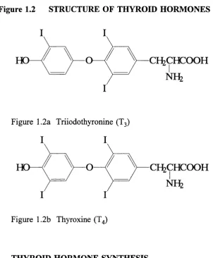
Together, triiodothyronine (Tg) and thyroxine (T4) constitute the thyroid hormones. The
former is more important in terms o f biological activity; the latter, less active, is present
at higher concentrations and considered a precursor (or prohormone) o f T3. Both are
iodothyronines, incorporating two iodinated phenyl rings joined together by an ether link,
Figure 1.2 STRUCTURE OF THYROID HORMONES
HD
CÎ^CHCOOH
Figure 1.2a Triiodothyronine (T3)
I
I
HCF
OCFtCHCOOH
NFk
1.7.1
Figure 1.2b Thyroxine (T4)
THYROID HORMONE SYNTHESIS
Iodide in the bloodstream is actively transported into the follicular cells against a chemical
and electrical gradient. The trapped iodide is organised in the colloid which involves
the iodide combining with tyrosine previously incorporated into thyroglobulin molecules.
Thyroglobulin is the main constituent o f the colloid and acts as a repository for thyroid
hormones and their precursors. Prior to organification, the iodide is first oxidised by the
action o f thyroid peroxidase in the presence o f hydrogen peroxide (H2O2) at the interface
o f the cell and colloid (Taurog, 1970). O f the 120 tyrosines per thyroglobulin, thirty are
iodinated and six to eight available for thyroid hormone synthesis. Mono-iodotyrosine
respectively. Two DIT molecules couple to form T4 whereas one DIT together with a
MIT produce T3. The coupling step occurs via the same peroxidase involved in the
organification procedure. At times o f sufficient iodine in the diet there are 7-10 MIT, 5-10
DIT, 2 T4 and 1 T3 for every thyroglobulin molecule (Van Herle et al., 1979). A more
extensive account o f hormone synthesis can be found in McDougall (1992).
1.7.2 SECRETION OF THYROID HORMONES
Before secretion o f T3 and T4, thyroglobulin is first hydrolysed by lysosomes to release
the active hormones along with MIT, DIT and amino acids. T3 and T4 enter the
circulation; MIT and DIT are deiodinated. The iodide is reused with a small fraction lost
from the thyroid (Dunn and Dunn, 1982a; Dunn and Dunn, 1982b).
1.7.3 THYROID HORMONES IN THE SERUM
T3 and T4 travel through the serum reversibly bound to carrier proteins. A minute
proportion o f hormone is free/unbound. Bound hormone is metabolically inert and acts
like a hormone store as well as a buffer, providing a means for maintaining the amount
o f free hormone. In the serum, 3 out o f 1000 T3 and 3 out o f 10,000 T4 are free which
means that 99.97% o f T4 and 99.7% o f T3 are bound. As a result o f binding there is no
loss o f hormone via the urine except in cases o f extreme proteinuria. Three types o f
carrier proteins carry thyroid hormone in the serum; thyroid binding globulin (TBG),
thyroid binding prealbumin (TBPA) and albumin.
Although TBG is present at the lowest concentration o f the three (1-2 mg/dl) it possesses
the highest affinity for T3 /T4, carrying 70% o f both hormones. A single chain
TBPA is also synthesized in the liver and hastwo binding sies for T4 molecules though
only one site is ever occupied. TBPA carries 10-20% o f T4 and no or very little T3.
Albumin, with the weakest affinity for hormone, is present in the highest quantity
transporting approximately 10% and 40% o f T4 and T3 respectively.
Levels o f these proteins are affected by a number o f conditions (both psychological and
pathological) and any deviation o f affinity for the hormones results in change o f total
thyroid hormone. Free hormones enter the cell and dictate thyroid function. An increase
in thyroid binding protein, for example, indicates a fall in T4. This is recognised at the
level o f the pituitary and T4 levels are adjusted accordingly (reviewed in Robbins and
Bartalena, 1986; McDougall, 1992).
1.8 REGULATION OF THYROID FUNCTION
1.8.1 THYROID STIMULATING HORMONE (TSH)
Thyroid stimulating hormone (TSH) or thyrotrophin, is produced and secreted by specific
cells (called thyrotrophes) in the anterior pituitary. TSH regulates the formation and
release o f T3 and T4.Levels o f free T3 and T4 act as negative feedback controls for TSH
secretion. Thyrotrophe nuclei possess T3/T4 receptors; the affinity for T3 being 10-20
times greater than that for T4. The cells are rich in 5’deiodinase II, responsible for the
conversion o f T4 to T3 (Larsen, 1982). Approximately 50% o f the T3 in the cell arises
as a consequence o f 5’deiodinase II action and it is therefore intra-thyrotrophe T3 and not
serum T3 that acts as a regulator. High T3 levels in the mouse lead to a decrease in the
mRNA’s o f TSH subunits. Normally, receptor occupancy by T3 is 50%, but when this
stores, with some o f the newly synthesized hormone released within the hour (O ’Riordan
et a l , 1988).
Certain factors such as somatostatin and dopamine, inhibit TSH production and
consequently its release. Oestrogen increases the levels o f TSH by augmenting the
regulatory action o f TRH (thyrotrophin release hormone). The time immediately
following birth sees a sudden rise in TSH level (which subsides after 24 to 48 hours) and
precedes an increase in total and free thyroid hormone levels. The rise in thyroid
hormone is believed to protect the newborn from the new, cold environment by
stimulating an increase in hormones involved in heat production. TSH release is itself
under control, by thyrotrophin release hormone (TRH) secretion from the hypothalamus.
1.8.2 THYROTROPHIN RELEASE HORMONE (TRH)
The existence o f a hypothalamic factor governing anterior pituitary function was proposed
over 40 years ago by Green and Harris (1947). Lesions in the hypothalamus led to organ
failure whilst electrical stimulation o f specific sites resulted in the release o f hormone
from the pituitary. In 1968, TRH was isolated, characterised, synthesized and shown to
act both under in vivo and in vitro conditions (review in McDougall, 1992). As mentioned earlier, TRH controls the release o f thyroid stimulating hormone from the
pituitary.
Guillemin (1978) obtained Im g pure TRH from the hypothalami o f 5 million sheep. TRH
is a tri-peptide (propylglutamyl-histidyl-prolinamine) and made in the hypothalamic cells.
Other sites o f synthesis include other areas o f the brain, as well as the spinal cord and
pancreas. After synthesis it is transported, via the blood stream, to the anterior pituitary
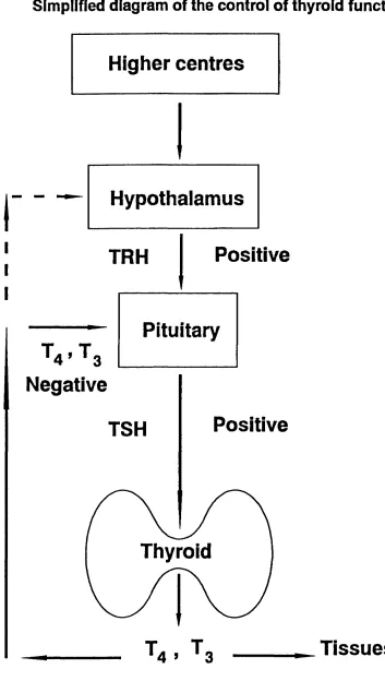
Figure 1.3
Simplified diagram of the control of thyroid function
Higher centres
Hypothalamus
Positive
TRH
Pituitary
Negative
Positive
TSH
Thyroid
production. The mechanism proposed suggests the involvement o f IP3 signalling
following receptor binding, resulting in a rise o f intracellular Ca^^ which stimulates a
cascade o f events culminating in TSH-containing vesicles discharging their contents into
the circulation (Kolesnick and Gershengorn, 1985). TRH has a very short half life of
around 5 minutes and is excreted in the urine. TRH is present in other areas o f the body
and brain where it is thought to function as a neuromodulator.
In summary, the function o f the thyroid gland is governed by a number o f factors secreted
by various organs. Thyroid hormone production is under the control o f TSH secreted
from the pituitary which in turn is regulated by TRH produced by the hypothalamus. The
hypothalamus is under less defined control from higher centres in the brain. In addition,
the thyroid gland itself is able to autoregulate hormone synthesis and release, though this
is less important than TSH control (Figure 1.3).
1.9 ACTION OF THYROID HORMONES
Thyroid hormones are involved in a vast number o f developmental and physiological
events. For many years, the general consensus o f opinion maintained that the mode o f
thyroid hormone action was confined to the nucleus only. This view has been revised
following the discovery o f thyroid hormone binding sites at locations external to the
nucleus in a variety o f cell types. As a result thyroid hormones have been assigned an
extranuclear role which though minor in comparison, is o f no less importance in the
1.9.1 NUCLEAR ACTION
The nuclear action o f thyroid hormones is similar to that observed with steroid hormones.
This is hardly surprising given the homology these two exhibit in their receptors. Thyroid
hormone action is mediated via nuclear receptors which in turn regulate the transcription
o f specific mRNA in target cells. Virtually all o f the physiological effects o f thyroid
hormones are believed to occur in this manner.
Briefly, after entering the cell, thyroid hormone binds to receptors present in the nucleus.
Two genes encode thyroid receptor proteins; c-erb A(3 and c-erb A a found on chromosome 3 and 17 respectively. They possess the same affinity for thyroid hormones.
50% o f the receptor sites are occupied under normal conditions with 80% containing T3.
Binding initiates modulation o f gene expression generating positive or negative effects on
transcription. Consequently, it is usually several hours before a biological response is
observed. The genes affected encode a range o f biologically important components
ranging from enzymes to hormones and receptors (Baxter and Eberhardt, 1979; Samuels
et al., 1982). More extensive accounts detailing the nuclear action o f thyroid hormones can be found in numerous reviews (Oppenheimer et al., 1987; Samuels et al., 1989; Brent
et al., 1991; De Groot, 1991).
1.9.2 EXTRANUCLEAR ACTION
The majority o f extranuclear binding studies have been focused on the plasma membranes
o f several tissues including human placenta (Alderson et al., 1985), rat synapses (Mashio et al., 1983), and rat and human erythrocytes (De Mendoza et al., 1977; Holm and Jacquemin, 1979). Two separate sites, differing in , have been identified. Both play
as a result, in the ability o f the cell to respond to the hormone (Krenning and Doctor,
1986).
Thyroid hormones stimulate glucose transport in cells within minutes, a time course not
consistent with a nuclear effect involving transcription. Instead plasma membrane sites
are thought to be directly involved. Furthermore, interaction with the plasma membrane
may be the only route possible for anuclear erythrocytes to respond to thyroid hormones.
Sterling et ah (1977) demonstrated the presence o f thyroid hormones in the inner mitochondrial membrane and linked them with immediate rises seen in O2 consumption,
again implicating a direct effect o f thyroid hormones. Mowbray and co-workers have
shown that the lowered A D P/0 ratio observed in hypothyroid mitochondria could be
completely restored within 15 minutes after administration o f a near physiological dose
o f T3 (Crespo-Armas and Mowbray, 1987). Subsequent studies by the same group
suuggest that mitochondrial processes under rapid thryoid hormone control may be
mediated by ADP-ribosylation (Thomas and Mowbray, 1987; Hardy and Mowbray, 1992).
The cytosolic binding sites have been assigned putative roles o f storage and supply to
organelles such as the nucleus (Francon et ah, 1985). Thyroid hormones can also regulate enzyme activity in a manner which excludes nuclear receptor involvement. One example
is the effect o f thyroid hormones on deiodinase II action leading to the possibility o f
similar modes o f action in other biological processes (Silva and Leonard, 1985).
During recent years the information on extranuclear roles o f thyroid hormones have
grown. Only a few have been mentioned here, a more detailed account can be found in
1.10 RECEPTORS
Both nuclear and cytosolic binding sites for T3 and T4 have been identified in the brain
and other tissues.
The cytosolic site is about 70 kDa with a greater affinity for T4. Its suggested roles
include that o f transporter and subunit o f the nuclear receptor involved with chromatin
(Baxter and Eberhardt, 1979). The nuclear receptor is 50-70 kDa and is associated with
DNA and chromatin (Oppenheimer et al., 1974, Eberhardt et al., 1978).
In the rat embryo, nuclear T3 binding sites are present in the brain at 14 days gestation.
The number increases 3-fold between day 14 and 15 gestation before remaining constant
up until birth. After birth, T3 sites rise again peaking after a week before falling to adult
levels (Schwartz and Oppenheimer, 1978). The nuclear binding capacity in the neonate
is double that o f the adult. As with other situations, the number, distribution and density
vary according to brain region (Coulombe et ah, 1981).
Bernal and Pekoven found high affinity T3 nuclear receptor concentrations dropped after
10 weeks gestation but rose 10-fold by week 16 gestation in the human feotus (Bernal and
Pekonen, 1984).
Current knowledge o f thyroid hormone levels and receptors in both the developing and
adult states is reviewed recently by Puymirat (1992).
1.11 CLINICAL ASPECTS OF THYROID HORMONES
As previously mentioned, thyroid hormones govern numerous biological and metabolic
events in virtually all parts o f the body throughout all stages o f life. Consequently any
imbalance will give rise to a variety o f disorders linked to these events, the degree o f
The two extremes o f abnormal thyroid status are hypothyroidism and hyperthyroidism.
The former arises as a result o f deficient amounts o f thyroid hormone or insufficient
action o f thyroid hormone to meet the body’s requirements; the latter is caused by over
activity o f the thyroid.
1.11.1 HYPOTHYROIDISM
Thyroid hormone deficiency is usually caused by a disease o f the thyroid (primary
hypothyroidism). However, it can also arise from lack o f stimulation at the level o f the
pituitary or hypothalamus (secondary and tertiary hypothyroidism respectively). Severe
hypothyroidism involving skin disorder is known as myxoedema.
Autoimmune disorders, medication, radiotherapy and iodine deficiency are all causes o f
hypothyroidism. 1% o f the adult population are afflicted with the disease with women
being 5-10 times more susceptible (McDougall, 1992). Generally, the symptoms mirror
that o f reduced function. They include sluggish/tired behaviour, weight gain, cold
intolerance, depression and reduced brain activity. The effects are easily reversed with
hormone replacement. If left untreated, myxoedema coma can result culminating in death.
Causes o f hypothyroidism in children include dysgensis o f the thyroid, iodine deficiency
and inborn enzyme defects in, for example, organification, thyroid hormone action and
iodine trapping (Hutchinson, 1980).
In neonates the most extreme case o f hypothyroidism leads to cretinism and the effects
are irreversible due to the vulnerable state o f the developing brain at this time. Three
different forms o f cretinism have been identified on basis o f their etiology.
Congenital hypothyroidism (also known as neonatal/sporadic cretinism) results when the
abnormal thyroid development or exposure to goitrogens and /or antithyroid drugs
(Malvaux, 1981; Foley, 1983). This form o f cretinism may also result as a consequence
o f TSH or TRH shortage and inborn disorders. Severe impairment o f neurological
development, mental retardation, poor co-ordination and balance, abnormal motor
movements, speech defects, tremor and spasticity are all indicative o f the disease. It
affects 1 out o f every 4000 births with girls twice as likely to acquire it. I f treated within
6 weeks, some recovery o f intelligence quotient (I.Q.) occurs thus assuaging the effect on
mental capabilities (MacFaul and Grant, 1977).
Endemic cretinism ensues from dietary deficiency o f iodine inflicting profound mental
deficiency along with irreversible abnormalities ranging from neuromuscular disorders to
deaf mutism (McDougall, 1992). It is still prevalent in a number o f countries like Brazil,
China and Pakistan. There are two classes o f the disease: hypothyroid/ myxoedematous
cretinism and neurological cretinism. The former is caused by severe hypothyroidism
during the very early stages o f life (Konig, 1981). The clinical symptoms are similar to
that o f congenital hypothyroidism and can be avoided if mothers are given iodine
replacement by the 20th week o f gestation. The neurological form differs in terms o f
clinical manifestation in that it is due to iodine deficiency in utero. It is averted only if the mother is treated in the very early stages o f pregnancy, ideally before conception as
seen in the rat and sheep animal models (Hetzel and Hay, 1979). In addition to the
symptoms seen in the other forms o f neonatal cretinism, deaf mutism and spastic diplegia
are also observed (Fierro-Benitez et al., 1974; Querido et al., 1978). As many as a billion people live in iodine deficient areas and nearly all suffer from some degree o f goitre. The
offspring o f these women are the most at risk from endemic cretinism with 1 0%
o f every 5000 births in the Western countries.
Disorders resulting from hypothyroidism (and hyperthyroidism) in the later periods o f life
are reversible with appropriate hormone treatment. This is not the case during the
developmental period as previously mentioned. Although treatment can help alleviate
some o f the effects, they cannot fully correct the damage already done. In order for
irreversible damage to be avoided,! there is growing evidence that therapy should begin
in the first trimester o f pregnancy as the thyroid state o f the mother is just as important
as that o f the growing feotus (Pharoah et a l, 1971; Porterfield and Hendrich, 1993).
1.11.2 HYPERTHYROIDISM
Hyperthyroidism is attributed to over activity o f the thyroid gland. In cases where there
are toxic levels o f thyroid hormone, the condition is known as thyrotoxicosis. Treatment
involves the administration of antithyroid drugs such as PTU or carbimozole, radio-iodine
or removal o f the gland (thyroidectomy). As for hypothyroidism, there are many causes
and forms o f the disease (McDougall, 1992). The most common is Graves’ disease and
is the consequence o f an autoimmune disorder. Briefly, antibodies to the TSH receptor
(TRAb) mimick and prolong the action o f TSH resulting in over-production and over
secretion o f thyroid hormones (Kriss et al., 1964). Many o f the symptoms are akin to that o f elevated basal metabolic rate and hence generally the reverse o f those observed in
hypothyroidism. Common signs are nervousness, weight loss despite no change or
increase in calorific intake, heat intolerance, fatigue and goitre. The majority o f cases o f
hyperthyroidism in pregnancy and children are usally a result o f Graves’ disease. If
treated at an early enough stage with antithyroid drugs, the effects are reversible in
to the neonate in the form o f malformation and spontaneous abortion by the mother.
Furthermore, the possible transfer o f the antibodies from the mother to the neonate via the
placenta during pregnancy, increases the neonate’s chances o f developing Graves’ disease
(Momotani et a l, 1984).
1.12 THYROID HORMONES AND THE DEVELOPING BRAIN
The majority o f data gathered on the role o f thyroid hormones during brain development
was obtained from studies carried out on neonatal rats though an appreciable amount o f
data have also come from sheep and mice.
The value o f the data obtained from these studies however, is frequently debated in
relation to its significance regarding human situations. Most o f the information obtained
from these experiments would be unavailable from humans, and the rat remains as a good
model providing equivalent human and rodent developmental stages are compared.
1.12.1 NEUROLOGICAL DEVELOPMENT IN RATS AND HUMANS
Compared to the human, the rat brain is immature in terms o f development at birth. At
birth, the rat brain is at the same developmental stage as the human brain at 5 to 6 months
gestation Eayrs (1968). At 10 days post partum (after birth), the rat brain is equivalent
to the human brain at birth (Bass et al., 1977). In other words, some developmental events that occur in utero (before birth) in the human, occur after birth in the rat. This facilitates manipulation o f the environment around the developing rat brain during this
important period in life. An important matter to consider is that though the same
sequence o f events takes place in every region, the time that this occurs differs from one
The cerebellar cortex (hindbrain) develops later then the cerebrum (forebrain) with nearly
80% o f the neurogenesis in the rat occurring in the former after birth (Balazs and Richter,
1973). In contrast, by birth rat cerebral neurogenesis is more or less over; the majority
occurring between day 12 gestation and birth (Balazs, 1973; Berry, 1974; Stein et al.,
1989). In the human, cerebral neurogenesis is virtually complete after the second month
o f pregnancy (Zamenhof and Van Marthens, 1971).
In the rat gliogenesis begins at birth, continuing into adult life (Berry, 1974) with the
same event occurring in utero in humans (Dobbing and Sands, 1973).
1.12.2 STAGES IN BRAIN DEVELOPMENT IN RELATION TO THYROID
HORMONES
Hamburgh et al. (1971) and workers alikened thyroid hormone function to "time clocks" in the developing nervous system, governing the precise timing o f events that occur.
Brain development can be separated into distinct stages when viewed in relation to thyroid
hormone influence.
Stage I embraces the first 10 to 12 weeks gestation in the human and the first 17 days
gestation in rat. It is the period o f development which occurs before feotal thyroid
hormone synthesis and therefore the only source o f thyroid hormone during this time is
maternal in origin. Recent evidence indicates that thyroid hormones have an impact on
early embryogenesis (see later). At this time cerebral neurogenesis takes place along with
a degree o f neuronal migration.
Stage II picks up from the end o f stage I and terminates at birth in both rat and human.
During this time, thyroid hormone is synthesized and released by the feotus with some
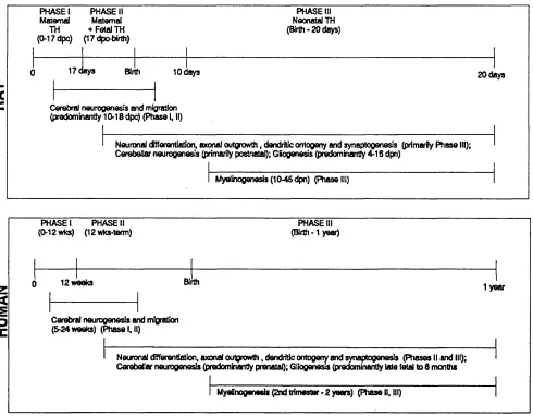
Figure 1.4
Brain Neurological Development In Relation to Thyroid Hormones In Rat and Human
4^ U)
5
t rPHASE I PHASE II Maternal Maternal
TH + Fetal TH (0-17 dpc) (17 dpo-birth)
PHASE III Neonatal TH (Birth- 2 0 days)
1 7 d a y s Birth to d a y s 2 0 days
Cerebral neurogenesis and migration (predominantly 10-18 dpc) (Phase I, II)
Neuronal (ffierentiaüon, axonal outgnowth, dendritic ontogeny and synaptogenesis (primarily P hase III); Cerebellar neurogenesis (primarily postnatal); Gliogenesis (predominantly 4-16 dpn)
M yelinogenesis(1 0 -4 5 dpn) (Phase!
z
<
PHASE I PHASE II (0-12 wks) (12 wks-term)
PHASE III (Birth -1 yeaO
12 w eeks Birth
1 year
Cerebral neurogenesis and migration (5-24 weeks) (Phase I, II)
Neuronal différentiation, axonal outgrowth. dendritic ontogeny and synaptogenesis (P h ases II and III); Cerebellar neurogenesis (predomhantly prenatal); Gliogenesis (predominantly late fetal to 6 months