R E S E A R C H
Open Access
Screening with magnetic resonance
imaging, mammography and ultrasound in
women at average and intermediate risk of
breast cancer
Tomasz Huzarski
1†, Barbara Górecka-Szyld
2,3†, Jowita Huzarska
1, Gra
ż
yna Psut-Muszy
ń
ska
2,3, Gra
ż
yna Wilk
2,
Robert Sibilski
4, Cezary Cybulski
1, Beata Kozak-Klonowska
5, Monika Sio
ł
ek
5, Ewa Kilar
6, Dorota Czudowska
7,
Hanna Janiszewska
8, Dariusz Godlewski
9, Andrzej Mackiewicz
10, Joanna Jarkiewicz-Tretyn
11, Jadwiga Szabo-Moskal
12,
Jacek Gronwald
1, Jan Lubi
ń
ski
1, Steven A. Narod
13*and the Polish Hereditary Breast Cancer Study Group
Abstract
Background:The addition of MRI to mammography and ultrasound for breast cancer screening has been shown to improve screening sensitivity for high risk women, but there is little data to date for women at average or intermediate risk.
Methods:Two thousand nine hundred and ninety-five women, aged 40 to 65 years with no previous history of breast cancer were enrolled in a screening program, which consisted of two rounds of MRI, ultrasound and mammography, one year apart. Three hundred and fifty-six women had aCHEK2mutation, 370 women had a first-degree relative with breast cancer (and noCHEK2mutation) and 2269 women had neither risk factor. Subjects were followed for breast cancer for three years from the second screening examination.
Results:Twenty-seven invasive epithelial cancers, one angiosarcoma and six cases of DCIS were identified over the four-year period. Of the 27 invasive cancers, 20 were screen-detected, 2 were interval cancers, and five cancers were identified in the second or third follow-up year (i.e., after the end of the screening period). For invasive cancer, the sensitivity of MRI was 86%, the sensitivity of ultrasound was 59% and the sensitivity of mammography was 50%. The number of biopsies incurred by MRI (n= 156) was greater than the number incurred by mammography (n= 35) or ultrasound (n= 57). Of the 19 invasive cancers detected by MRI, 17 (89%) were also detected by ultrasound or mammography.
Conclusions: In terms of sensitivity, MRI is slightly better than the combination of mammography and ultrasound for screening of women at average or intermediate risk of breast cancer. However, because of additional costs incurred by MRI screening, and the small gain in sensitivity, MRI screening is probably not warranted outside of high-risk populations.
Keywords:Breast cancer,CHEK2, Magnetic resonance imaging, Mammography, Ultrasound, Screening
* Correspondence:steven.narod@wchospital.ca
†Equal contributors
13Women’s College Research Institute, Women’s College Hospital and the
University of Toronto, 76 Grenville Street, 6th Floor, Toronto, ON M5S 1B2, Canada
Full list of author information is available at the end of the article
Background
Several studies have demonstrated that annual magnetic resonance imaging (MRI) is a more sensitive means of screening for breast cancer than annual mammography for women at high risk of cancer [1–10]. These studies have focused on women at high risk of cancer due to a
BRCA1 orBRCA2 mutation, or at moderate risk due to a family history. It is not clear if MRI should be used to screen women at average or intermediate risk. It is pos-sible that MRI performs differently in women with and without BRCA mutations because of the differences in cancer risk and also potentially because of the difference in the natural history of cancers in the two groups. Most cancers in non-carriers or in CHEK2 carriers are ER-positive [11] whereas cancers in BRCA1 carriers are usually triple-negative [12] and these are relatively ag-gressive in their natural history [13]. Women with a truncating CHEK2 mutation have a breast cancer risk that is about three fold higher than average and women with both a family history and a CHEK2 muta-tion have a five-fold increased risk [14]. Missense mu-tations in CHEK2 confer a 1.4 fold increase in breast cancer risk [14].
There are many relevant issues relating to expanding MRI screening to average - and moderate-risk popula-tions, including the costs of screening, technical consid-erations including the availability of MRI-guided biopsy and the possibility of over-diagnosis. It is important to establish whether or not the gain in sensitivity obtained with MRI screening in terms of cancers detected over and above mammography (or a combination of mam-mography and ultrasound) justifies the expense. Ultim-ately the value of MRI screening should be determined by demonstrating an improvement in breast cancer mor-tality among women undergoing regular MRI screening.
We conducted a multicenter observational study in Poland wherein we followed 2995 women (2269 at average risk and 726 at moderately elevated risk) who were screened with three screening modalities (MRI, mam-mography, ultrasound) on two occasions one year apart and then were followed for an additional three years. We estimate the risks of prevalent cancers, incident cancers and interval cancers in the cohort.
Methods
Study population
The study group consisted of women at average risk of cancer and women with a moderately increased risk of cancer due to an inherited CHEK2 mutation or a first-degree relative with breast cancer. To identifyCHEK2 -posi-tive patients, the records of the hereditary cancer clinic of the Pomeranian Medical University were reviewed. Women who were between the ages of 40 and 65 and who had no previous history of breast cancer or of another cancer and
were known to carry a CHEK2 mutation (truncating or missense) were invited to participate. Through this process, 496 CHEK2 carriers were invited, of whom 356 (72%) agreed to participate. The remaining study participants were recruited through a newspaper campaign conducted in different regions of Poland. Women with and without a family history of breast cancer were eligible to participate. Through this means, the study cohort was expanded to include an additional 2639 women at average risk, in-cluding 370 with a family history of breast cancer and 2269 with no family history of breast cancer. The distri-bution of patients was as follows (Szczecin 758; Olsztyn 604; Zielona Góra 463; Bydgoszcz 100; Toruń 111; Kielce 418; Poznań 142;Świdnica 239; Legnica 160). A blood sample was taken from each woman for DNA extraction. Genetic testing was done for four CHEK2
mutations (I157T, IVS2 + 1G > A, 1100delC and del5395) and for three BRCA1 mutations that are founder muta-tions in Poland (5382insC, C61G and 4153delA) [14]. We included those with a CHEK2 mutations but women with aBRCA1mutation were excluded.
Study protocol
Women were screened twice, one year apart and were followed for three more years for incident breast cancers. Women were included in the study if they had all three examinations (MRI and ultrasound and mammography) at the first round. The ultrasound examination and mammography either took place the day before the MRI examination or on the same day. In some cases the radiologist had access to the MRI results at the time the ultrasound was conducted, but this was un-common. Clinical breast examination was performed by the radiologist on a routine basis, but was not evaluated formally in this analysis. Ethics approval was obtained by the institutional review board of the Pomeranian Medical University and by all participating institutions. All partici-pants provided written informed consent.
suggestive of malignancy [14]. If any imaging modality was scored as BI-RADS 5, a biopsy was performed. If the modality was scored as BI-RADS 0 or 4, then a biopsy was not automatically recommended, but was done at the discretion of the treating physician and radiologist, based on the interpretation of all three screening modalities and the physical examination. In some cases, one or more screening test was repeated. In most cases, core biopsies and excisional biopsies were performed under ultrasound or stereotactic guid-ance. For BI-RADS 5 lesions that were only visualized on MRI-only, an excisional biopsy or quadrantectomy was performed. MRI-guided biopsy was not available. The biopsy specimens were reviewed by pathologists with expertise in breast cancer. Details of cancers detected were collected from hospital medical records and pathology reports. Cancers were divided into ductal carcinoma in situ (DCIS) and invasive cancer. There was one non-epithelial cancer detected (angiosarcoma). Cancers detected on the first screening round were categorized asprevalentcancers. Cancers detected on the second screening round were categorized as incident
cancers. Cancers detected between screening rounds or within one year of the last screen were categorized as
intervalcancers. Cancers detected in the second or third year post-screening were categorized as post-screening
cancers. These cancers were not considered in the evalu-ation of screening parameters (e. g. sensitivity) but were included in order to estimate the incidence rate of cancers in the cohort.
Follow-up
Women were contacted by telephone or by mail annu-ally to inquire if they had been diagnosed with breast cancer in the three-year period following the second screening round. For 80% of the women who entered the study, follow-up was at least two years and for 76% of the women follow-up was three years after the second screening round. No screening MRI was done in the follow-up period. In some cases, the woman may have had screening mammography in the follow-up period outside of the study, but these details were not collected. Further, details on clinical breast examinations were not collected. For those women who developed cancer, de-tails of the cancer were retrieved from medical records.
Women with a BI-RADS 4 abnormality detected on MRI were followed for four to five years from the time of the abnormal MRI (until July 2015) in order to interpret the clinical impact of having a BI-RADS 4 abnormality without a biopsy on subsequent cancer risk. Follow-up was by telephone interview to the patients herself and inquiring about new diagnoses of breast cancer.
Sensitivity and positive predictive value
We collected information on both invasive cancers and in situ cancers. Sensitivity was calculated for invasive epithelial cancers only. The sensitivity of a given modality was defined as the number of biopsy-proven cancers detected by that modality, divided by the total number of cases detected by all modalities, plus the interval cancers. The positive predictive value was the ratio of the number of biopsy-proven cancers detected by a given modality (BI-RADS 0,4 or 5) divided by the number of biopsies per-formed that were occasioned by a positive screening test by the same modality. In the cost analysis, the costs used were the actual prices which were negotiated between the Pomeranian Medical University, with the Polish Ministry of Science and Euromedic. The cost analysis is restricted to the costs of screening only (and does not include the costs of biopsies or subsequent clinical care).
Results
Patients
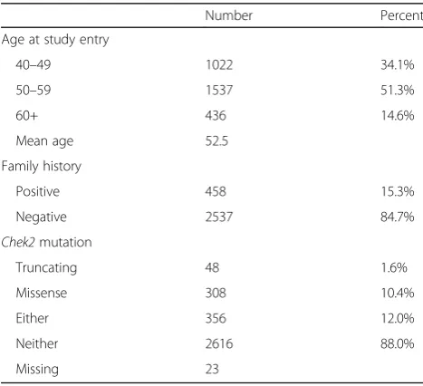
A total of 2995 women completed the first round of screening (Table 1). The mean age at entry was 52.5 years (range 40 to 65 years). Of these women, 458 (15%) had a first-degree relative with breast cancer and 356 (12%) had a CHEK2 mutation (49 truncationg and 307 missense). 51 women had both a first-degree relative with breast cancer and a CHEK2 mutation. Over the four year study period, breast cancer was diagnosed in 34 women, including six cases of ductal carcinoma in situ, one angiosarcoma and 27 invasive breast cancers. The following analyses are limited to the 27 invasive breast cancers.
In the first round of screening, 13 invasive cancers were detected (prevalence 0.4% or 430 per 100,000)
Table 1Characteristics of 2995 study patients
Number Percent
Age at study entry
40–49 1022 34.1%
50–59 1537 51.3%
60+ 436 14.6%
Mean age 52.5
Family history
Positive 458 15.3%
Negative 2537 84.7%
Chek2mutation
Truncating 48 1.6%
Missense 308 10.4%
Either 356 12.0%
Neither 2616 88.0%
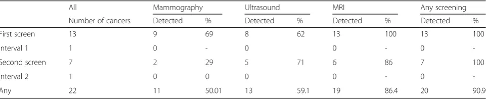
(Table 2). Six of the 13 cancers were palpable, five of 13 were above 2cm in size and three of 13 were node- posi-tive. After one year, the 2982 remaining women were in-vited for a second screen. Of these, 2565 women (86%) participated in the second round of screening and 417 women (14%) did not return. Of the 2565 women who returned, 2327 women had a second MRI (91%), 2391 had a second ultrasound (93%) and 2329 had a second mammography (91%). One interval cancer was detected between the first and second screening round. This was a 3 cm, node-positive cancer detected in a 50 year old woman. There were seven invasive cancers detected at the second screening round (incidence 300 per 100,000 per year) one of seven was palpable, one of seven was above 2cm in size and two of seven were node-positive.
Of the 2565 women who participated in the second screening round, 2335 women were followed for an add-itional two years and 2273 women were followed for an additional three years through telephone interview and a mailed questionnaire. In the first year after the screening period (year three) there was only one invasive cancer diagnosed, in a 52 year old woman. It was 3.0 cm in size and node negative. She had been screened with MRI/ ultrasound/mammography eight months prior to diag-nosis. In the second year post screening, four additional cancers were diagnosed; on average these were 1.7 cm in size and one was node positive. In the third year after screening there was only one breast cancer diagnosed in the left breast of a 53-year old woman. She also had a breast cancer detected in the first round of screening in the right breast. The second (contralateral) cancer was detected by the woman herself and mammography was negative. The cancer was 2.2 cm in size and was node-positive. In total, after the second screen, six cancers detected among 2335 women contributing 6643 person-years of follow-up (incidence 86 per 100,000 per year).
Screening parameters
Sensitivity
Of the 22 invasive cancers detected during the screening period (first screen to one year after second screen) 20 were detected by one of the screening modalities (eight were palpable and 14 were non-palpable) and two were interval cancers. The highest sensitivity was seen with
MRI (86%), followed by ultrasound (59%) and mammog-raphy (50%). Eighteen of the 22 cancers were detected by either mammography or ultrasound and the sensitivity of mammography and ultrasound in combination (77%) was only slightly inferior to that of MRI (86%). This combin-ation identified one cancer that was missed by the MRI. There were two cancers that were identified by the MRI that were not identified by any other screening modality. These were diagnosed in a 51-year old and a 66-year old woman and were 0.8 cm and 1.1 cm respectively. Both cancers were node-negative, non-palpable, ER-positive and HER2-negative.
Positive predictive value
In the entire study, 571 women (19.1% of total) had an abnormal examination by at least one screening modality (BI-RADS 0, 4 or 5). Two hundred and four women (36%) underwent a total of 216 biopsies, including all women with a BI-RADS 5 anomaly, 139 women with a BI-RADS 4 anomaly and 65 women with a BI-RADS 0 anomaly. These 204 women represent 6.8% of the entire study population. On 26 occasions (12.0%) the biopsy revealed cancer (21 invasive and four DCIS, one angiosarcoma) and on 190 occasions (88.0%) the biopsy revealed either normal tissue or benign disease. Overall, the positive predictive value was 12.1% for MRI, was 14.2% for ultrasound and was 31.4% for mammography. There were 216 biopsies performed. There were 156 biopsies performed in women with an abnormal MRI, followed by 57 with an abnormal ultrasound and 35 women with an abnormal mammogram.
Breast cancers
Detection rates
Overall, 22 invasive breast cancers were detected during the screening period (20 screen-detected and two inter-val cancers). Two of the women had a positive family history of breast cancer and five women had a CHEK2 mutation (one case had both) (Table 3). Assuming a baseline risk of that of women between the age of 40 and 50 without a mutation and without a family history (0.26%), the relative risk associated with aCHEK2 muta-tion was 5.4 (95% CI 1.1 to 2.8) and that associated with a family history of breast cancer (first-degree relative)
Table 2Numbers of invasive breast cancers detected by different screening modalities during the screening period
All Mammography Ultrasound MRI Any screening
Number of cancers Detected % Detected % Detected % Detected %
First screen 13 9 69 8 62 13 100 13 100
Interval 1 1 0 - 0 0 - 0
-Second screen 7 2 29 5 71 6 86 7 100
Interval 2 1 0 0 0 0 - 0
was 1.7 (95%CI 0.2 to 12). The detection rate was 0.43% at the first screening round (prevalent cancers cancers) and was 0.27% at the second screening round (incident cancers).
Tumor characteristics
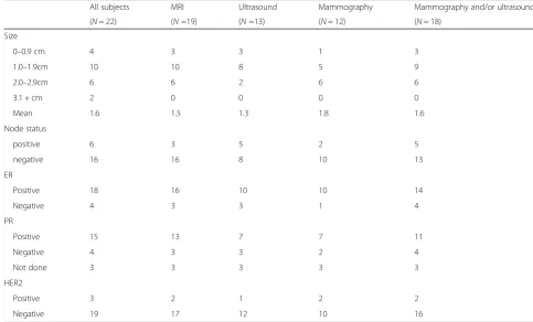
The mean size of the 22 invasive cancers was 1.6 cm (range, 0.5 to 3.0 cm). The mean sizes of prevalent and incident invasive tumors were 1.7 cm and 1.2 cm re-spectively. Three out of 13 prevalent cancers were node-positive and two of seven incident cancers were node-positive (Table 4).
Long term follow-up of women with abnormal MRI (BI-RADS 4)
In our study, a biopsy was performed on all patients with an MRI BI-RADS 5 lesion, but not on all patients with MRI BI-RADS 4 lesion. This was either for a tech-nical reason or due to patient preference or because of the radiologist’s interpretation of the other two imaging studies done concurrently. Of the 192 women who had a BI-RADS 4 (suspicious) lesion detected on MRI, 87 women had a biopsy and 11 breast cancers were found (12.6%). The 76 women who had a negative biopsy were followed for 4.0 years on average from the date of the Table 3Proportions of subjects with invasive breast cancer identified, by risk group
Number of women screened
Cancers detected
Rate of detection
Sensitivities of different screening modalities
Mammography Ultrasound M and US MRI
Family history positive 458 2 0.4% 50% 50% 100% 100%
CHEK2mutation 353 5 1.4% 60% 80% 100% 100%
No risk factors 2269 16 0.7% 53% 56% 75% 81%
40–49 1022 5 0.5% 60% 80% 100% 100%
50–59 1537 11 0.7% 64% 45% 71% 82%
60–65 436 6 1.4% 40% 67% 83% 830%
All 2995 22 0.7% 57% 59% 82% 86%
Mmammography USultrasound
Table 4Characteristics of invasive breast cancers detected during screening period
All subjects MRI Ultrasound Mammography Mammography and/or ultrasound
(N= 22) (N=19) (N=13) (N= 12) (N= 18)
Size
0–0.9 cm 4 3 3 1 3
1.0–1.9cm 10 10 8 5 9
2.0–2.9cm 6 6 2 6 6
3.1 + cm 2 0 0 0 0
Mean 1.6 1.5 1.3 1.8 1.6
Node status
positive 6 3 5 2 5
negative 16 16 8 10 13
ER
Positive 18 16 10 10 14
Negative 4 3 3 1 4
PR
Positive 15 13 7 7 11
Negative 4 3 3 2 4
Not done 3 3 3 3 3
HER2
Positive 3 2 1 2 2
abnormal MRI (BI-RADS 4) and two cases of invasive cancer were reported. There were also 105 women who had an abnormal MRI and who did not have a biopsy; these women were followed for an average of 4.3 years and two women were diagnosed with breast cancer. One was diagnosed with an invasive 4 cm node-positive can-cer 55 months from the abnormal MRI. The other was diagnosed with a breast cancer in the breast contralat-eral the MRI abnormality, eight months after the initial abnormal MRI. The tumor was 5.0 cm and the patients was node-positive. In summary, of 192 women in the study who had a BI-RADS 4 abnormality, 11 breast can-cers were detected at the time of screening and three cancers were diagnosed in the four year period following the abnormal MRI (one of these was diagnosed within the four year study period). Of the 151 women with an MRI BIRADS 4 abnormality and no other screening abnormality, four breast cancers were found, two at the time of the MRI and two thereafter. Of these, two were less than two centimeters and three were node negative.
Conclusion
In this study of 2995 women at average or intermediate risk of breast cancer, we found the combination of mammography and ultrasound to be nearly equivalent to annual MRI. Of the 22 women who were diagnosed with invasive breast cancer over the two-year screening period, 19 were visualized with MRI and 17 were visual-ized with ultrasound and/or mammography. We report a high overall sensitivity (91%) and a very low proportion of interval cancers (9%)–both are indicators of an effective screening program. The average size of the breast cancers was small (1.6 cm) and the incident cancers were smaller, on average (1.2 cm) than were the prevalent cancers (1.7 cm). Fourteen of the 22 invasive cancers were detected in the cohort at a non-palpable stage and no cancer was detected solely by physical examination. This was an observational study and the management of the patients found to have an abnormal lesion was at the discretion of the treating physicians.
We also identified six cases of DCIS. There is no con-sensus regarding the value of identifying Stage 0 breast cancers in the context of screening and in order to guard against over-interpretation, we have separated these from the invasive cancers.
In this study, there is little evidence to support the position that annual MRI screening should be recom-mended for women from the average/moderate risk population who are being screened with annual mam-mography. Two cancers were identified through MRI screening that were missed by the other screening mo-dalities. To some extent, the small number of cancers detected through MRI alone (n= 2) might be a reflection of the reluctance of the radiologist or surgeon to biopsy
a patient with an abnormal lesion detected on MRI (BI-RADS 4) when there was no corresponding lesion detected on mammography or ultrasound. However, among the 105 women with a BI-RADS 4 abnormality who did not have a biopsy, only one cancer was detected clinically in the three year period following the positive MRI test and this was detected in the contralateral breast. The other cancer was diagnosed 55 months post-MRI and the length of time elapsed suggests that this was an unrelated (new) event. These observations suggest that the reluctance to biopsy these women initially did not result in the failure to diagnosis many cancers that would have been clinic-ally apparent in the three year interval following the second MRI examination. The lack of incident cancers in this group of 105 women in the three years follow-ing the abnormal MRI abnormal result suggests that there were some false positive MRI tests or that some cancers may be have been due to over-diagnosis. Over-diagnosis is defined as a cancer that presents on screening but would not otherwise become apparent in a patient’s lifetime. Miller et al estimate in the Canadian NBSS study [15] that approximately one-half of non-palpable breast cancers detected solely by mammog-raphy are examples of over-diagnosis and there is no reason to suppose the proportion to be less among MRI-detected cancers (which on average are smaller) should be less than this.
It is important that the results of screening studies be interpreted in the context of the level of risk of the par-ticipating subjects. Studies of women at high risk (i.e.,
founder mutations comprise the majority of BRCA muta-tions in Poland. It is possible that there are other mutation carriers in this cohort but given the rarity of non-founder mutations in the Polish population and the fact that study subjects were unaffected, we expect the number of these to be very low.
In the present study 25 cancers (DCIS and invasive) were found through 216 biopsies (positive fraction 11.6%). In an early study of women with a BRCA1 or BRCA2 mutation, 33 biopsies revealed seven cancers (21.2%) [17]. This difference is expected; given the lower preva-lence of cancer in the average risk women compared to high risk women, the positive predicive value will be lower in the former group.
In a recent report from the UK, (the FH01 study), the sensitivity of mammography alone was reported to be 77% for screening of 6710 young women at intermediate risk [17]. They estimated a mortality reduction of ap-proximately 20% associated with mammography. In that study, 21% of cancers were interval cancers (compared to 5% in the present study), 32% of cancers were node-positive (compared to 24% in the present study) and 30% were above 2 cm (compared to 33% in the present study). To some extent, these differences may be due to different age distributions. All patients in the FH01 study were under 50, compared to 34% of the subjects in our study.
In the ACRIN study, 2662 women at high risk under-went 7443 mammogram and ultrasound screening tests over two years (three annual screens). Thirty three can-cers were detected by mammography alone, 32 were de-tected by ultrasound alone, 26 were dede-tected by both modalities and nine were not detected. The overall detection rate was 17 per 1000 woman-years. At the end of screening 612 women had an MRI and nine more cancers were detected (1.5%). The authors concluded that the routine use of MRI was not justified in this intermediate risk population.
The data presented here do not support adding MRI screening to routine mammography and ultrasound for women at average risk at this time. The addition of MRI to the combination of ultrasound and mammography in this cohort of 2995 women led to the identification of two additional cancers. The resources expended to iden-tify these two cancers were 5319 MRI examinations and 64 biopsies. This is equivalent to 3.7 cancers detected per 10,000 MRI examinations. Assuming a cost of 155 dollars per MRI (the actual reimbursed cost), this is equivalent to 400,000 dollars per additional cancer detected in Poland, but is expected to be much higher than this in North America or western Europe. In this study, the cost of the ultrasound and mammography were 40 dollars per exam-ination. Based on these prices, the total costs of screening 10,000 women would be 400,000 dollars for a combination
of mammography and ultrasound and 1,550,000 using MRI alone. Our study is based on a relatively small number of detected cancers, and we recommend that further research be done in this area. It is important that the value of screening MRI ultimately be judged on studies with cancer mortality as the endpoint, but to date, there are no published studies with mortaity as the endpoint. It is also important to consider that there are potential harms associated with screening, particularly in low and average risk populations, such as increased numbers of confirmatory tests, anxiety, surgical morbidity and (in the case of over-diagnosis) unnecessary sugeries. Given the very low risk of cancer we observed in the follow-up period of women with BIRADS 4 lesion who did not undergo a biopsy, futher studies which consider the potential harms of overdiagnosis associated with MRI screening are warranted. Given the high cost of MRI examinations, until supporting evidence is avail-able, we concur with other investigators [17] that the addition of MRI to mammography and ultrasound for screening women at intermediate risk of breast cancer is not appropriate.
Abbreviations
BI-RADS:Breast imaging reporting and data system; DCIS: Ductal carcinoma in situ; MRI: Magnetic resonance imaging
Acknowledgements
We would like to thank K Wałęsa, I Chodzyńska, E Putresza and L Kubera Nowakowska for help with database management.
Other Members of the Polish Hereditary Breast Cancer Study Group: T Byrski, T Dębniak, A Tołoczko-Grabarek, O Oszurek, M Michalak, H Rogowska-Droś, E Szatkowska, R Kulus, W Kwiecień, L Szyiński, I Winiarska, P Kasprzak, L Tomiak-Rówińska, M Kosterska-Spalska, T Dróżdż, P Sosnowski.
Funding
This study was supported by a grant from the Polish Ministry of Science, Grant number PBZ-MNiSW-05/I/2007/02.
Availability of data and materials
The datasets generated during and/or analysed during the current study are not publicly available due to privacy and confidentiality but are available from the corresponding author on reasonable request.
Authors’contributions
TH, SAN and JL were responsible for revising the manuscript critically for intellectual content. SAN and JL were responsible for analysis and interpretation of data and drafting the manuscript. SAN and JL were responsible for conception and design. All authors were responsible for data acquisition and patients recruitment and review of the medical records. All authors have given final approval of the version to be published and agree to be accountable for all aspects of the work in ensuring that questions related to the accuracy or integrity of any part of the work are appropriately investigated and resolved.
Competing interests
The authors declare that they have no competing interests.
Consent for publication Not applicable.
Ethics approval and consent to participate
Author details
1Department of Genetics and Pathology, International Hereditary Cancer
Center, Pomeranian Medical University, Szczecin, Poland.2Department of
Radiology, Pomeranian Medical University, Szczecin, Poland.3Euro-Medic Diagnostics Poland Ltd, Szczecin, Poland.4Oncology Diagnostic Center,
Zielona Góra, Poland.5Regional Oncology Center, Kielce, Poland. 6Department of Oncology, District Specialist Hospital,Świdnica, Poland. 7
Oncology Diagnostic Center, Legnica, Poland.8Department of Clinical Genetics, Collegium Medicum, Nicolaus Copernicus University, Bydgoszcz, Poland.9Prophylactic and Epidemiology Cancer Center, Poznań, Poland. 10Department of Cancer Immunology, Poznan University of Medical Sciences,
Greater Poland Cancer Centre, Poznań, Poland.11District Specialist Hospital, Toruń, Poland.12Department of Radiology, Regional Oncology Hospital,
Bydgoszcz, Poland.13Women’s College Research Institute, Women’s College
Hospital and the University of Toronto, 76 Grenville Street, 6th Floor, Toronto, ON M5S 1B2, Canada.
Received: 11 November 2016 Accepted: 11 February 2017
References
1. Warner E, Plewes DB, Hill KA, et al. Surveillance of BRCA1 and BRCA2 mutation carriers with magnetic resonance imaging, ultrasound, mammography, and clinical breast examination. JAMA. 2004;292:1317–25.
2. Lehman C, Blume JD, Weatherall P, et al. Screening women at high risk for breast cancer with mammography and magnetic resonance imaging. Cancer. 2005;103:1898–905.
3. Kriege M, Brekelmans CTM, Boetes C, et al. Efficacy of MRI and mammography for breast-cancer screening in women with a familial or genetic predisposition. N Engl J Med. 2004;351:427–37.
4. Kuhl CK, Schrading S, Leutner CC, et al. Mammography, breast ultrasound, and magnetic resonance imaging for surveillance of women at high familial risk for breast cancer. J Clin Oncol. 2005;23:8469–76.
5. Leach MO, Boggis GRM, Dixon AK, et al. Screening with magnetic resonance imaging and mammography of a UK population at high familial risk of breast cancer: a prospective multicenter cohort study (MARIBS). Lancet. 2005;365:1769–78.
6. Lehman CD, Isaacs C, Schall MD, et al. Cancer yield of mammography, MR, and US in high-risk women: prospective multi-institution breast cancer screening study. Radiology. 2007;244:381–8.
7. Sardanelli F, Podo F, D’Agnolo G, et al. Multicenter comparative multimodality surveillance of women at genetic-familial high risk for breast cancer (HIBCRIT study): interim results. Radiology. 2007;242:698–715.
8. Hagen AI, Kvistad KA, Maehle L, et al. Sensitivity of MRI versus conventional screening in the diagnosis of BRCA-associated breast cancer in a national prospective series. Breast. 2007;16:367–74.
9. Rijnsburger AJ, Obdeijn I, Kaas R, et al. BRCA1-associated breast cancers present differently from BRCA2-associated and familial cases; long-term follow-up of the Dutch MRISC screening study. J Clin Oncol. 2010;28:5265–73.
10. Cybulski C, Huzarski T, Byrski T, Gronwald J, Debniak T, Jakubowska A, Górski B, Wokołorczyk D, MasojćB, Narod SA, Lubiński J. Estrogen receptor status in CHEK2-positive breast cancers: implications for chemoprevention. Clin Genet. 2009;75:72–8.
11. Foulkes WD, Stefansson IM, Chappuis PO, Bégin LR, Goffin JR, Wong N, Trudel M, Akslen LA. Germline BRCA1 mutations and a basal epithelial phenotype in breast cancer. J Natl Cancer Inst. 2003;95:1482–5. 12. Dent R, Trudeau M, Pritchard KI, Hanna WM, Kahn HK, Sawka CA,
Lickley LA, Rawlinson E, Sun P, Narod SA. Triple-negative breast cancer: clinical features and patterns of recurrence. Clin Cancer Res. 2007;13(15 Pt 1):4429–34.
13. Cybulski C, Wokołorczyk D, Jakubowska A, Huzarski T, Byrski T, Gronwald J, MasojćB, Deebniak T, Górski B, Blecharz P, Narod SA, Lubiński J. Risk of breast cancer in women with a CHEK2 mutation with and without a family history of breast cancer. J Clin Oncol. 2011;29(28):3747–52.
14. Górski B, Jakubowska A, Huzarski T, et al. A high proportion of founder BRCA1 mutations in Polish breast cancer families. Int J Cancer. 2004;110(5):683–6.
15. Orel SG, Kay N, Reynolds C, Sullivan DC. BI-RADS categorization as a predictor of malignancy. Radiology. 1999;211:845–50.
16. Miller AB, Wall C, Baines CJ, Sun P, To T, Narod SA. Twenty five year follow-up for breast cancer incidence and mortality of the Canadian National Breast Screening Study: randomised screening trial. BMJ. 2014;348:g366.
17. Passaperuma K, Warner E, Causer PA, et al. Long-term results of screening with MRI in carriers of BRCA mutations. Br J Cancer. 2012;107:24–30.
• We accept pre-submission inquiries
• Our selector tool helps you to find the most relevant journal
• We provide round the clock customer support
• Convenient online submission
• Thorough peer review
• Inclusion in PubMed and all major indexing services
• Maximum visibility for your research
Submit your manuscript at www.biomedcentral.com/submit