R E S E A R C H A R T I C L E
Open Access
Association of radiation risk in the second
and third generations with polymorphisms
in the genes
CYP1A1, CYP2E1, GSTP1
and
changes in the thyroid
Meruyert Massabayeva
1*, Nailya Chaizhunusova
1, Nurlan Aukenov
2, Tolkyn Bulegenov
1, Bakytbek Apsalikov
1,
Aigerim Shapihanova
1and Yersin Zhunussov
1Abstract
Background:To study the association of radiation risk in the 2nd–3rd generations with polymorphisms in the genesCYP1A1, CYP2E1, GSTP1and changes in the thyroid.
Methods:5 polymorphic gene variants (rs1048943, rs4646421, rs2070676, rs3813867, rs1695) were studied in 399 people living in the East Kazakhstan region in this research. 248 people of the 2nd - 3rd generation lived in the territory with radiation exposure in Abai, Borodulikha areas, and 151 people the comparison group lived in Kurchum district without radiation exposure comparable in sex and age with control group.
Results:The results show that there is a significant association of rs1048943 in exposed and unexposed groups (p< 0.003), and the absence of association of rs4646421, rs2070676, rs3813867, rs1695 in the studied groups. The mean value of thyroxine in carriers of the AG + GG genotype of rs4646421 is significantly lower than in AA genotype carriers (p= 0.04); no significant changes were found in genotypes’distribution with thyroid-stimulating hormone and anti-thyroid peroxidase indicators. Significant changes were in levels of anti-thyroid peroxidase between exposed and unexposed groups (p= 0.007). The thyroxine - thyroid-stimulating hormone levels were not significantly different in exposed and unexposed groups (p> 0.3).
Conclusions:This study demonstrated the association of rs1048943 polymorphism with living in the radiation zone in the 2nd and 3rd generations for the first time. Thyroxine levels decrease was identified in the 2nd and 3rd generation residents of the exposed area, as well as a significant increase of anti-thyroid peroxidase occurs in individuals of the 2nd and 3rd generation living in areas with radiation exposure.
Keywords:Radiation risk, Second-third generations,CYP1А1,CYP2Е1,GSTP1genes polymorphisms
Introduction
The impact of environmental factors affecting the health of the population is an urgent problem at the present time. An intense anthropogenic impact on the environ-ment leads to the emergence of environenviron-mentally caused human diseases, which can lead to dysfunctions of various body systems. One of the unfavorable factors include ionizing radiation (Markabayeva et al., 2018). Many works have been published recently on the
medical consequences of long-term radiation effect on the health of directly exposed people and their descen-dants which allowed to establish a significant increase in somatic pathology, changes in the immune status, gen-etic effects of radiation-induced factors (Tanaka et al.,
2000; Mudie et al., 2007; Kolyado et al., 2019). Hence, (Tang and Loganovsky, 2018) stated that exposure to low doses of ionizing radiation can affect health, depend-ing on the genetic background, gender, age and source of radiation.
East Kazakhstan region (Kazakhstan) applies to the territories with radiation burden (Grosche et al., 2015).
© The Author(s). 2019Open AccessThis article is distributed under the terms of the Creative Commons Attribution 4.0 International License (http://creativecommons.org/licenses/by/4.0/), which permits unrestricted use, distribution, and reproduction in any medium, provided you give appropriate credit to the original author(s) and the source, provide a link to the Creative Commons license, and indicate if changes were made. The Creative Commons Public Domain Dedication waiver (http://creativecommons.org/publicdomain/zero/1.0/) applies to the data made available in this article, unless otherwise stated. * Correspondence:[email protected]
1Medical University Semey, Semey, Kazakhstan
The population living in this area had been exposed to various doses of radiation for a long time (Simon & Bouville, 2002). The risk assessment of the effect of radi-ation exposure on individual organs or the body as a whole is based on an in-depth study of mechanisms of molecular and biochemical changes in the body. It has been established that exposure to radiation depending on the dose and age increases the risk of developing thyroid cancer (Yamashita et al., 2017). Survivors of the atomic bombing in Hiroshima and Nagasaki had a higher risk of radiation-induced thyroid cancer in children and adoles-cents (Furukawa et al., 2013). Genetic predisposition to thyroid diseases should be distinguished from hereditary genetic pathologies because its basis is the genetic poly-morphism inherent in humans, but not the mutations of certain genes (Saenko & Rogounovitch,2018).
Genes encoding enzymes 1st and 2nd phase of the process of biotransformation of foreign chemicals, car-cinogens, environmental pollutants and other xenobi-otics include CYP1A1, CYP2E1, GSTP1 (Nourozi et al.,
2018). Mutations in the structure of these genes lead to an increase in the concentration of intermediate toxic metabolites in the body and the accumulation of free radicals (Caccamo et al.,2013).
This study was conducted to investigate the associ-ation of radiassoci-ation risk with polymorphisms in the genes SUR1A1, SUR2E1, GSTP1 and changes in the thyroid gland in individuals of 2nd - 3rd generations exposed to low doses of radiation and parents/ grandparents who lived during the nuclear tests.
Methods Subjects
A total of 399 people living in the East Kazakhstan re-gion (EKR) took part in the study. The contingent of surveyed persons included 248 people living in areas with adverse environmental factors, such as radiation exposure (Karaul village, Abay district and Borodulikha village, Borodulikha district, East Kazakhstan), as well as 151 people were not exposed to adverse environmental factors (Kurchum village, Kurchum district, EKR). These areas were included in the study due to different radi-ation doses as a consequence of the localizradi-ation of areas at different distances from the Semipalatinsk Test Site (STS). The State Scientific Automated Medical Register (SSAMR) serves as a mechanism for assessing the indi-vidual dose of radiation received by a person while living in the territories adjacent to the STS between 1949 and 1990. Persons aged from 27 to 50 years living in the territory of Abay district of East Kazakhstan received individual dose loads in the range from 0.06 to 17 cSv due to the activities of the STS. For the population of Borodulikha district of East Kazakhstan, this range was from 0.05 to 15 cSv. Kurchum district of East
Kazakhstan is environmentally safe. According to Article 6 of the 2nd chapter of the Law of the Republic of Kazakhstan of December 18, 1992 N 1787-XII“On social protection of citizens who suffered from nuclear tests at the Semipalatinsk nuclear test site”, Abay district belongs to the zone of maximum radiation risk, and Borodulikha district belongs to the zone of increased radiation risk. In turn, Kurchum district which is not mentioned in this law is chosen as a zone for the comparison group. The popu-lation of the above-mentioned territories were taken from the database of the State Scientific Automated Medical Register (SSAMR) of the population of Kazakhstan exposed to radiation (Grosche et al.,2015).
Criteria for inclusion: age 18–60 years (2– 3rd gener-ation with exposure), legally confirmed residence of par-ents (grandparpar-ents) in the territory of the influence of Semipalatinsk Test Site during nuclear weapon tests (in accordance with the database of the SSAMR of the population of Kazakhstan exposed to radiation); and persons permanently residing in the territory of the Kurchum district, EKR. Exclusion criteria: organic CNS damage, somatic diseases in the stage of decompensa-tion, the presence of genetic diseases.
Genotyping and thyroid hormone levels analyses
Laboratory measurements of thyroid hormones were carried out in the laboratory In Vivo, Semey, Kazakhstan by Chemiluminescent enzyme immunoassay (CLIA) method on the equipment Architect i2000SR (Abbott Laboratories, IL, USA) using commercial diagnostic kits (Abbott Laboratories, USA) in accordance with the manufacturer’s instructions. Indicators of the reference ranges for thyroxine are (T4) – 9-22 pmol/L; thyroid stimulating hormone (TSH)– 0.4–4.0 mIU/L; antithy-roid peroxidase (anti-TPO)-0–35 IU/mL.
For genetic research, peripheral blood taken into vac-uum tubes with K2/K3 EDTA of subjects was used. Isolation of genomic DNA from blood samples (100μl) was performed using ready-made commercial kits Gene-JET Mini kit (Thermo Scientific, Vilnius, Lietuva), care-fully in accordance with the manufacturer’s instructions. DNA concentration was assessed using fluorometer Qubit 4 (Thermo Scientific, Waltham, MA, USA). The isolated DNA was frozen and stored at−20 °C.
followed by 48 cycles at 95 °C for 10 s and 60 °C for 40 s for all SNPs.
Statistical analyses
Statistical analysis was performed using IBM SPSS Statistics Version 21 (International Business Machines Corp., Armonk, NY, USA).
Comparison of genotypic distribution frequencies between groups was performed using χ2 criterion (Pearson’s chi-squared test). Continuous variables were assessed for normality using Shapiro–Wilk test, as well as the Mann-Whitney or Kruskal - Wallis quantitative test was utilized for one-dimensional analysis. Logistic regression tests were used to determine the independent influence of radiological history; thyroid parameters were used as dependent variable. However, the influence of gender and age was not taken into account in the logis-tic regression. Differences between samples were consid-ered statistically significant at p< 0.05. Deviations of genotype frequencies of each polymorphism from the Hardy-Weinberg equilibrium were estimated byχ2−test.
Results
The demographic characteristics of the subjects are pre-sented in Table 1. A total of 399 people of which 248 people with radiological history (2 – 3rd generations with exposure) and 151 persons the control group with-out radiological history. Among exposed people only 19.5% were male and 80.5% female, similar data was among unexposed people: 21% were male and 79% were female. The studied groups are comparable by sex and age.
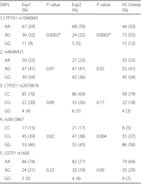
Table 2 shows the genotype frequency of polymor-phisms in the genes CYP1A1 (rs1048943, rs4646421), CYP2E1 (rs2070676, rs3813867), GSTP1 (rs1695) in the exposed (Abay and Borodulikha districts) and unexposed (Kurchum district) groups. Significant difference in the distribution of rs1048943 polymorphism genotype in the exposed and unexposed groups (p< 0.003) (Table2). It should be noted that significant differences were both in the comparison of the exposed Abai district with the unexposed Kurchum district, and in the comparison of Borodulikha district with the latter. There were no
statistically significant differences in the distribution of genotypes of polymorphisms rs4646421, rs2070676, rs3813867 and rs1695 (p> 0.004). No deviations from the Hardy-Weinberg equilibrium were found in any group (p> 0.05).
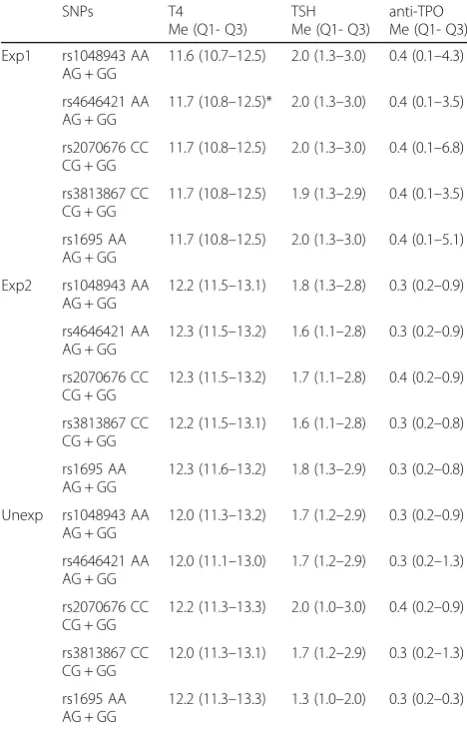
Table 3 demonstrates the association between the genotypes of CYP1A1 (rs1048943, rs46421), CYP2E1 (rs2070676, rs3813867), GSTP1 (rs1695) and thyroid parameters (thyroxine, thyroid-stimulating hormone, anti-thyroid peroxidase) in the studied populations. Since few of them had the genotype AG and GG in the polymorphisms rs1048943, rs46421, rs1695 we com-bined them AG + GG, we also comcom-bined the genotypes CG + GG of polymorphisms rs2070676 and rs3813867. The average value of T4 in carriers of АА (11.7), AG + GG (8.9) polymorphism rs4646421 was significantly lower in residents of Abay district than in residents of the control Kurchum district АА (12.0), AA+AG (11.8) (p= 0.04); however, there were no significant changes in the distribution of genotypes with TSH and anti-TPO levels. We did not find a statistically significant associ-ation between the polymorphisms rs1048943, rs2070676, rs3813867, rs1695 and parameters of the thyroid gland (p> 0.05) for any comparison groups.
Table 1Demographic Characteristics According to Exposed and Unexposed Groups (N= 399)
Exposed (n= 248) Unexposed (n= 151) P
value
Exp1 (n= 118) Exp2 (n= 130)
Males,n(%) 42 (28) 15 (11) 32 (21) 0.974
Age (years) 18–60 36.28 ± 7.3
18–58 38.0 ± 7.0
18–58 38.0 ± 7.0
1.0
Exp1: Abay District, East Kazakhstan Exp2: Borodulikha District, East Kazakhstan Unexp: Kurchum District, East Kazakhstan
Table 2Genotype Frequencies of Polymorphism CYP1А1, CYP2E1, GSTP1 genes in the studied groups
SNPs Exp1 (%)
Pvalue Exp2 (%)
Pvalue VS Unexp (%)
1.CYP1А1rs1048943
AA 67 (59) 68 (70) 44 (33)
AG 36 (32) 0.0002* 24 (25) 0.0002* 73 (55)
GG 11 (9) 5 (5) 15 (12)
2. rs4646421
AA 29 (25) 27 (23) 33 (25)
AG 47 (41) 0.97 47 (41) 0.92 55 (41)
GG 39 (34) 42 (36) 45 (34)
3.CYP2Е1rs2070676
CC 85 (76) 86 (69) 99 (79)
CG 22 (20) 0.89 33 (26) 0.17 22 (18)
GG 4 (4) 6 (5) 4 (3)
4. rs3813867
CC 17 (15) 21 (17) 8 (5)
CG 45 (39) 0.02 47 (38) 0.004 55 (37)
GG 53 (46) 55 (45) 86 (58)
5.GSTP1rs1695
AA 84 (74) 82 (77) 79 (64)
AG 24 (21) 0.23 20 (19) 0.09 35 (29)
GG 5 (5) 4 (4) 9 (7)
Table 4 presents logistic regression data to analyze the association between radiological history and thy-roid parameters. Significant changes were identified in anti-thyroid peroxidase levels between exposed and unexposed groups (p= 0.007). Indicators of thy-roxine, thyroid-stimulating hormone were not signifi-cantly different in exposed and unexposed groups (p> 0.3).
Discussion
Hereditary genetic traits are known to determine a person’s sensitivity to toxic and chemical substances (Ricarda Thier et al.,2003). The polymorphisms of genes encoding enzymes involved in the biotransformation of toxins and chemicals include cytochrome P-450 (CYP), glutathione S - transferase (GST) (Satyender Singh et al.,
2011). In the present study, we found an association be-tween rs1048943 polymorphism of CYP1A1 gene with living in areas with radiation exposure in 2nd and 3rd generations, and there was no association of polymor-phisms rs4646421, rs2070676, rs3813867 and GSTP1 rs1695 with living in areas with radiation exposure.
In this study, we found an association between a poly-morphism of the gene CYP1A1 rs46421 and hormonal imbalance of the thyroid gland, a decrease in the level of T4 in carriers of the genotype АА in residents with radiation exposure of 2nd and 3rd generations of the Abai district of EKR, which belongs to the territory of maximum radiation exposure (Grosche et al., 2015). However, there were no significant changes in TSH and anti-TPO in Abai district. It is noteworthy that the aver-age value of T4 and TSH coincides with the reference ranges (T4–9-22 pmol/L, TSH – 0.4-4.0 mIU/L) in the groups with and without radiation exposure, but the average value of anti- TPO in the group with radiation exposure is much higher than the reference ranges (anti-TPO–0-35 IU/mL). The thyroid gland is known to be a radiation-sensitive organ (Suzuki S. et al.,2019). The risk of developing thyroid cancer also depends on the dose and has not been reduced for many decades (Williams et al.,2004; Tronko et al., 2017). It is worth noting that the EKR does not belong to iodine-deficient regions (Hamada et al. 2003), which in turn reduces the risk of thyroid disease, in contrast to the radiation exposure that is present in this study and can play a crucial role in the imbalance of thyroid hormones. Previous studies in-dicate that searching for new SNPs that may contribute to a genetic predisposition to thyroid disease is feasible synergistically with environmental studies (Saenko and Rogounovitch,2018).
This is the first cross-sectional study of the search for a potential association of radiation exposure in individ-uals of the 2nd and 3rd generation with polymorphisms in the genes СYР1А1 (rs1048943, rs4646421), CYP2E1 (rs2070676, rs3813867), GSTP1 (rs2070676, rs3813867) and changes in the thyroid gland in residents of EKR.
Limitations
The study has some limitations. We recognize that we rely only on the value of T4, TTG, and anti-TPO, not in-cluding a more advanced thyroid study. In the future, we are planning a more detailed study of this issue with a large number of samples and a more thorough study of
Table 3The Association between Genotypes and Thyroid Hormone Parameters in the Studied Population
SNPs T4
Me (Q1- Q3)
TSH Me (Q1- Q3)
anti-TPO Me (Q1- Q3)
Exp1 rs1048943 AA AG + GG
11.6 (10.7–12.5) 2.0 (1.3–3.0) 0.4 (0.1–4.3)
rs4646421 AA AG + GG
11.7 (10.8–12.5)* 2.0 (1.3–3.0) 0.4 (0.1–3.5)
rs2070676 CC CG + GG
11.7 (10.8–12.5) 2.0 (1.3–3.0) 0.4 (0.1–6.8)
rs3813867 CC CG + GG
11.7 (10.8–12.5) 1.9 (1.3–2.9) 0.4 (0.1–3.5)
rs1695 AA AG + GG
11.7 (10.8–12.5) 2.0 (1.3–3.0) 0.4 (0.1–5.1)
Exp2 rs1048943 AA AG + GG
12.2 (11.5–13.1) 1.8 (1.3–2.8) 0.3 (0.2–0.9)
rs4646421 AA AG + GG
12.3 (11.5–13.2) 1.6 (1.1–2.8) 0.3 (0.2–0.9)
rs2070676 CC CG + GG
12.3 (11.5–13.2) 1.7 (1.1–2.8) 0.4 (0.2–0.9)
rs3813867 CC CG + GG
12.2 (11.5–13.1) 1.6 (1.1–2.8) 0.3 (0.2–0.8)
rs1695 AA AG + GG
12.3 (11.6–13.2) 1.8 (1.3–2.9) 0.3 (0.2–0.8)
Unexp rs1048943 AA AG + GG
12.0 (11.3–13.2) 1.7 (1.2–2.9) 0.3 (0.2–0.9)
rs4646421 AA AG + GG
12.0 (11.1–13.0) 1.7 (1.2–2.9) 0.3 (0.2–1.3)
rs2070676 CC CG + GG
12.2 (11.3–13.3) 2.0 (1.0–3.0) 0.4 (0.2–0.9)
rs3813867 CC CG + GG
12.0 (11.3–13.1) 1.7 (1.2–2.9) 0.3 (0.2–1.3)
rs1695 AA AG + GG
12.2 (11.3–13.3) 1.3 (1.0–2.0) 0.3 (0.2–0.3)
*p< 0.05
Table 4Associative Analysis of the Radiological History and Parameters of the Thyroid Using the Linear Regression Model
Dependent variable
Radiological History
Вeta Se Pvalue
T4 0.000 0.000 0.314
TSH 0.007 0.057 0.909
anti-TPO 0.440 0.164 0.007*
the thyroid gland. We also plan to continue this study with other genetic polymorphisms of enzymes involved in the metabolism of biotransformation of toxins and chemicals.
Conclusions
For the first time the study revealed the association of rs1048943 polymorphism with living in the radiation zone in the 2nd and 3rd generations. Thyroxine levels decrease was found in the 2nd and 3rd generation resi-dents of the exposed area, as well as a significant increase of anti-thyroid peroxidase occurs in individuals of the 2nd and 3rd generation living in areas with radi-ation exposure.
Abbreviations
anti-TPO:anti-thyroid peroxidase; TSH: Thyroid-stimulating hormone; Т4: Thyroxine
Acknowledgements
We express our deep gratitude to Yerkezhan Tokbergen, University of California, Riverside for the valuable advice and comments on the article. We also would like to show our special gratitude to Karaganda Medical University, Karaganda, Kazakhstan for assistance in genotyping.
Authors’contributions
NC, NA, TB, and YeZh developed the study and reviewed the manuscript. MM performed basic experiments and analyses and wrote the manuscript. BA wrote the manuscript and reviewed the literature. ASh prepared the data for analysis, conducted a literature review. All authors read and approved the final manuscript.
Funding
The research was carried out within the financing of the Ministry of the
Republic of Kazakhstan“Development of scientific and methodological
bases for minimizing the ecological risk, health care, social protection and health of the population of ecologically unfavorable Kazakhstan territories”N of registration 49017/The Ministry of Healthcare of the Republic of Kazakhstan.
Availability of data and materials
The datasets used and/or analyzed during the current study are available from the corresponding author on reasonable request.
Ethics approval and consent to participate
Informed consent to participate in the study was obtained from all participants in the study in accordance with the Protocol of the Ethical Committee of the Medical University of Semey and the requirements of the World Medical Association of Helsinki Declaration.
Consent for publication Not applicable.
Competing interests
The authors declare that they have no competing interests.
Author details
1Medical University Semey, Semey, Kazakhstan.2Department of Health and Human Resources, Ministry of Health of the Republic of Kazakhstan, Nur-Sultan, Kazakhstan.
Received: 20 August 2019 Accepted: 30 October 2019
References
Caccamo D, Cesareo E, Mariani S, Raskovic D, Ientile R, Currò M, Korkina L, De Luca C. Xenobiotic sensor- and metabolism-related gene variants in
environmental sensitivity-related illnesses: a survey on the Italian population. Oxidative Med Cell Longev. 2013;2013:831969.https://doi.org/10.1155/2013/ 831969.
Furukawa K, Preston D, Funamoto S, Yonehar S, Ito M, Tokuoka S, Sugiyama H, Soda M, Ozasa K, Mabuchi K. Long-term trend of thyroid Cancer risk among Japanese atomic-bomb survivors: 60 years after exposure. Int J Cancer. 2013; 132(5):1222–6 doi: 10.1002/ijc.27749.
Grosche B, Zhunussova T, Apsalikov K, Kesminiene A. Studies of health effects from nuclear testing near the Semipalatinsk nuclear test site, Kazakhstan. Cent Asian J Glob Health. 2015.https://doi.org/10.5195/cajgh.2015.127. Hamada A, Zakupbekova M, Sagandikova S, Espenbetova M, Ohashi T, Takamura
N, Yamashita S. Iodine prophylaxis around the Semipalatinsk nuclear testing site, Republic of Kazakhstan. Public Health Nutr. 2003;6(8):785–9.https://doi. org/10.1079/phn2003496.
Tanaka K, Tchaijunusova NJ, Takatsuji T, Gusev BI, Sakerbaev AK, Hoshi M, Kamada N. High incidence of micronuclei in lymphocytes from residents of the area near the Semipalatinsk nuclear explosion test site. J Radiat Res. 2000;41(1): 45–54.https://doi.org/10.1269/jrr.41.45.
Kolyado IB, Plugin SV, Tribunsky SI. Aftereffects of the Impact of the Radiation Exposure on the Territory and the Population of the Altai Krai. Hyg Sanit. 2019.https://doi.org/10.18821/0016-9900-2018-97-7-609-617.
Markabayeva A, Bauer S, Pivina L, Bjorklund G, Chirumbolo S, Kerimkulova A, Semenova Y, Belikhina T. Increased prevalence of essential hypertension in areas previously exposed to fallout due to nuclear weapons testing at the Semipalatinsk test site, Kazakhstan. Environ Res. 2018;167:129–35.https://doi. org/10.1016/j.envres.2018.07.016.
Mudie NY, Gusev BI, Pivina LM, Schoemaker MJ, Rijinkova ON, Apsalikov KN. Sex ratio in the offspring of parents with chronic radiation exposure from nuclear testing in Kazakhstan. Radiat Res. 2007;168(5):600–7.https://doi.org/10.1667/rr0980.1. Nourozi MA, Neghab M, Bazzaz JT, Nejat S, Mansoori Y, Shahtaheri SJ. Association
between polymorphism of GSTP1, GSTT1, GSTM1 and CYP2E1 genes and susceptibility to benzene-induced Hematotoxicity. Arch Toxicol. 2018;92(6): 1983–90.https://doi.org/10.1007/s00204-017-2104-9.
Ricarda Thier, Thomas Brüning, Peter H. Roos, Hans-Peter Rihs, Klaus Golka, Yon Ko, Hermann M. Bolt, (2003) Markers of genetic susceptibility in human environmental hygiene and toxicology: The role of selected CYP, NAT and GST genes. International Journal of Hygiene and Environmental Health. 2003;206(3):149–171. Saenko VA, Rogounovitch TI. Genetic polymorphism predisposing to
differentiated thyroid Cancer: a review of major findings of the genome-wide association studies. Endocrinol Metab. 2018;33(2):164–74.https://doi. org/10.3803/enm.2018.33.2.164.
Singh S, Kumar V, Singh P, Thakur S, Banerjee BD, Rautela RS, Grover SS, et al. Genetic polymorphisms of GSTM1, GSTT1 and GSTP1 and susceptibility to DNA damage in workers occupationally exposed to organophosphate pesticides. Mutat Res. 2011;725(1–2):36–42.https://doi.org/10.1016/j. mrgentox.2011.06.006.
Suzuki S, Bogdanova TI, Saenko VA, Hashimoto Y, Ito M, Iwadate M, Rogounovitch TI, et al. Histopathological analysis of papillary thyroid carcinoma detected during ultrasound screening examinations in Fukushima. Cancer Sci. 2019;110(2):817.https://doi.org/10.1111/cas.13912.
Simon SL, Bouville A. Radiation doses to local populations near nuclear weapons test sites worldwide. Health Phys. 2002;82(5):706–25.https://doi.org/10.1097/ 00004032-200205000-00016.
Tang FR, Loganovsky K. Low dose or low dose rate ionizing radiation-induced health effect in the human. J Environ Radioact. 2018;192:32–47.https://doi. org/10.1016/j.jenvrad.2018.05.018.
Tronko M, Brenner AV, Bogdanova T, Shpak V, Oliynyk V, Cahoon EK, Drozdovitch V, Little MP, Tereshchenko V, Zamotayeva G, Terekhova G, Zurnadzhi L, Hatch M, Mabuchi K. Thyroid neoplasia risk is increased nearly 30 years after the Chernobyl accident. Int J Cancer. 2017;141(8):1585–1588.
Williams D, Abrosimov A, Bogdanova T, Demidchik EP, Ito M, LiVolsi V, Lushnikov E, Rosai J, Sidorov Yu, Tronko MD, Tsyb AF, Vowler SL, Thomas GA. Thyroid carcinoma after Chernobyl latent period, morphology and aggressiveness. Br J Cancer. 2004;90(11):2219–2224.
Yamashita S, Suzuki S, Suzuki S, Shimura H, Saenko V. Lessons from Fukushima: Latest Findings of Thyroid Cancer After the Fukushima Nuclear Power Plant Accident. Thyroid. 2017.28(1):11–22.
Publisher’s Note