THE CLINICAL AND SCIENTIFIC IMPORTANCE OF MUCOSAL INFLAMMATION
Standing at the interface between the host and the environment, mucosal-lined surfaces represent the first line of defense against potential pathogens. This defen-sive role is particularly relevant to the mucosa of the gastrointestinal tract, the pulmonary system, and the urinary tract, each of which is particularly susceptible to the development of inflammatory dis-eases due to their role as a barrier that
must not only protect, but also serve the physiological function of each of the organ systems. In the case of the gastroin-testinal tract, mucosal inflammation is manifest as inflammatory bowel disease (including Crohn’s disease and ulcerative colitis) (1–3) or necrotizing enterocolitis (NEC), a leading cause of death in pre-term infants (4). In the case of the pul-monary system, mucosal inflammation may be manifest as pneumonitis, pneu-monia, or asthma (5–7), acute and chronic pulmonary conditions that have a high
degree of morbidity and potential mortal-ity. And in the case of the urinary tract, mucosal inflammation may be manifest as interstitial nephritis, cystitis, and urethri-tis (8–10), causes of significant morbidity in patients of all ages. To elucidate the pathogenesis of mucosal inflammatory diseases, research over the past several decades has focused on the role of the immune system in their development— in particular the relationship between mucosal lymphocytes, macrophages, and neutrophils, and the effects of their cellu-lar by-products on mucosal integrity and function (11–13). However, recent work has shed light upon the important role that the epithelia itself may play as a pri-mary regulator of the immune response in the development of mucosal inflam-mation. No longer an innocent bystander, the epithelial-lined mucosa at each of
Receptor Signaling in the Development of Mucosal
Inflammation
Steven C Gribar, Ward M Richardson, Chhinder P Sodhi, and David J Hackam
Address correspondence and reprint requests toDavid J Hackam, Division of Pediatric Surgery, Room 4A-486 DeSoto Wing, Children’s Hospital of Pittsburgh, Pittsburgh, PA 15213. Phone: 412-692 8735; Fax: 412-692 8299; E-mail: [email protected].
Submitted March 25, 2008; Accepted for publication June 14, 2008; Epub (www.molmed. org) ahead of print June 17, 2008.
Division of Pediatric Surgery, Department of Surgery, Children′s Hospital of Pittsburgh and the University of Pittsburgh School of Medicine, Pittsburgh, Pennsylvania, United States of America
Diseases of mucosal inflammation represent important causes of morbidity and mortality, and have led to intense research ef-forts to understand the factors that lead to their development. It is well accepted that a breakdown of the normally imperme-ant epithelial barrier of the intestine, the lung, and the kidney is associated with the development of inflammatory disease in these organs, yet significant controversy exists as to how this breakdown actually occurs, and how such a breakdown may lead to inflammation. In this regard, much work has focused upon the role of the epithelium as an “innocent bystander,” a target of a leukocyte-mediated inflammatory cascade that leads to its destruction in the mucosal inflammatory process. However, recent evidence from a variety of laboratories indicates that the epithelium is not merely a passive component in the steps that lead to mucosal inflammation, but is a central participant in the process. In addressing this controversy, we and others have deter-mined that epithelial cells express Toll-like receptors (TLRs) of the innate immune system, and that activation of TLRs by endoge-nous and exogeendoge-nous ligands may play a central role in determining the balance between a state of “mucosal homeostasis,” as is required for optimal organ function, and “mucosal injury,” leading to mucosal inflammation and barrier breakdown. In partic-ular, activation of TLRs within intestinal epithelial cells leads to the development of cellular injury and impairment in mucosal re-pair in the pathogenesis of intestinal inflammation, while activation of TLRs in the lung and kidney may participate in the devel-opment of pneumonitis and nephritis respectively. Recent work in support of these concepts is extensively reviewed, while essential areas of further study that are required to determine the significance of epithelial TLR signaling during states of health and disease are outlined.
these sites has been shown to possess all of the required armamentarium to allow an effective response to invading challenges, and to lead the battle to neutralize potential microbial threats (14–16). Not only is the epithelium able to respond to potentially dangerous mi-crobial products, it also may sense en-dogenous molecules that are released during conditions of stress, hypoxia, or injury—so-called danger molecules that may play a critical role in the develop-ment of mucosal inflammation (17–19). In order, therefore, to understand the pathogenesis of mucosal inflammation and to assist in the rational design of anti-inflammatory strategies, it is neces-sary to define the receptors and signal-ing pathways that mediate the
inflam-matory response with respect to the ep-ithelium itself.
The innate immune system consists of a series of receptors and their associated signaling molecules that is present both on leukocytes and epithelial cells through the body, and which initiates an immune response by responding directly to pformed ligands ([20] provides a re-cent review). The innate immune system lies in contradistinction to the adaptive immune system, a set of cellular and mo-lecular interactions that must first “learn” how to deal with a potential pathogen, and then respond through the release of antibodies or other cellular de-rived products. The innate immune sys-tem includes pattern recognition recep-tors such as the TLRs, the NOD-like receptors (NLRs), the RIG-like receptors (RLRs), and C-type lectins, and their role in inflammatory signaling in leukocytes has been extensively reviewed
(21–23,186). Relatively few reports have focused on the ability of the innate im-mune system to signal within the epithe-lium, although emerging evidence from a variety of laboratories including our
own indicates that innate immune sig-naling within the epithelium plays a crit-ical role in the pathogenesis of mucosal inflammation. The current review will focus on the TLR family, which has been shown to play a critical role in the re-sponse of epithelial cells to bacterial and endogenous ligands in the pathogenesis of various mucosal inflammatory dis-eases (Table 1).
DEFINING THE CONTROVERSIES IN THE PATHOGENESIS OF MUCOSAL
INFLAMMATION
A central controversy in the field of mucosal inflammation may be stated as follows (Figure 1 provides a pictorial rep-resentation of this): Is mucosal inflamma-tion a reflecinflamma-tion of a leukocyte-driven immune response that has gone awry, re-sulting in tissue injury and the loss of mucosal barrier function (Figure 1A)? Or is it the mucosa itself, long known to play a role as a primary immune organ that is capable of producing a large number of pro-inflammatory molecules, that somehow has developed an exag-gerated response that then leads to mu-Table 1.Toll-like receptors and their known
ligands
TLR Ligand(s)
1 Triacyl lipoproteins (126) 2 Lipoproteins (127)
Peptidoglycan (128,129) Lipoteichoic acid (128) Zymosan (130,131)
Heat shock proteins (132,133) 3 Double stranded RNA (134) 4 LPS (37,135)
Taxol (136)
Heat shock proteins (132,137,138) Fibronectin (139)
Hyaluronic Acid (140) Heparan Sulfate (141) Fibrinogen (142)
Respiratory Syncytial Virus Fusion Protein (106)
Murine Retroviral Envelope Protein (143)
5 Flagellin (58)
6 Diacyl Lipopeptides (144) Lipoteichoic acid (131) Zymosan (131)
7 Single-stranded RNA (145,146) Imidazoquinoline (59) 8 Single-stranded RNA (146)
Imidazoquinoline (147)
9 Bacterial (demethylated CpG) DNA (148)
10 Unknown 11 Profilin (149)
Uropathogenic Bacteria (114)
cosal injury (Figure 1B)? Mucosal sur-faces, such as the intestinal mucosa and the upper respiratory tract, are con-stantly exposed to environmental stim-uli, such as commensal luminal bacteria in the intestine, as well as endogenous stimuli, or, as is the case in the lower respiratory tract and urinary tract mu-cosa, may encounter and respond to en-dogenous and exogenous stimuli in dis-ease states. In either case, the mucosa must be able to mount an effective im-mune response, resist barrier failure, and coordinate this response with both mu-cosal and sub-mumu-cosal leukocytes, while avoiding initiation and propagation of an exaggerated inflammatory response. But where does the answer lie in terms of what is initiating the mucosal inflam-matory response?
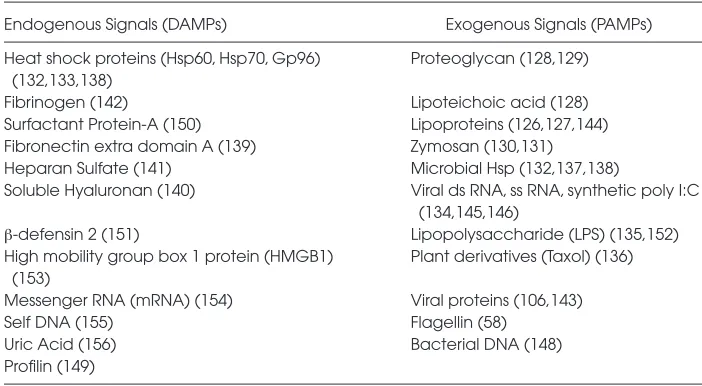
In seeking to answer this question, we and others have focused on the innate immune receptors that are present on the mucosa, and have examined the re-sponse of these receptors to known lig-ands in the development of mucosal in-flammatory disorders. Such ligands may be broadly grouped into two categories: the so called “danger signals,” a term used by Matzinger (24), also called damage-associated molecular patterns (DAMPs) ([25,26] provide recent re-views); and those ligands on the surface or interior of pathogen-associated mo-lecular patterns (PAMPs) (Table 2). Not surprisingly, there is a great deal of in-terest in identifying the important recep-tors for DAMPs and PAMPs expressed by various cell types, so as to accurately define their relative role in the develop-ment of inflammation. In this regard, several investigators have established that DAMPs and PAMPs are recognized by TLRs in many cells, including epithe-lial cells: a list of the TLRs and their cog-nate ligands appears in Table 1. Al-though current dogma suggests that circulating leukocytes play a central role in the coordination of the immune re-sponse, emerging evidence suggests that the epithelium also plays a key role in the recognition and response to various “danger molecules” (27–31). This review
will examine in detail the various roles of epithelial signaling via TLRs in the development of common, and often dev-astating, mucosal inflammatory condi-tions. Much of the focus will be on the TLR-initiated signaling in response to PAMPs, as the majority of work has been performed in this area.
RECOGNIZING DANGER: TLRS AND FRIENDS
Several recent investigators have shed light upon the important role of TLR sig-naling in the development of mucosal inflammation (31–34). To understand how TLRs may signal within the epithe-lium, information may be gained by ana-lyzing TLR signaling in other systems, primarily within leukocytes. A full de-scription of the molecular mechanisms by which TLR signaling occurs is be-yond the scope of this review; currently accepted concepts with respect to TLR signaling are described below (21,35 have recent reviews).
The structure of each member of the TLR family of receptors provides impor-tant clues to how they function. All cur-rently recognized TLRs are homologous with the interleukin-1 (IL-1) receptor, sharing an intracellular signaling domain, known as the Toll/IL-1R (TIR) domain (36). A model that depicts the currently accepted mode of TLR signaling in
leuko-cytes is shown in Figure 2, in which the interaction with TLR4 and its cognate lig-and lipopolysaccharide (LPS, endotoxin) is shown. The interaction of TLR4 with LPS leads to the activation of myeloid dif-ferentiation primary response protein 88 (MyD88)-dependent signaling, resulting in the induction of pro-inflammatory genes such as TNF-α, IL-1β, IL-6, and IL-10 (37–39) and MyD88-independent signaling cascades leading to activation of type-1 interferon (40 has a recent review) (38,41). MyD88-depedent signaling occurs as TIR domain-containing adapter protein (TIRAP/Mal) (42,43) and MyD88 (44–46) interact with TLR4 and recruit IL-1 recep-tor-associated kinase (IRAK) family mem-bers IRAK1 and IRAK4 to the signaling heterocomplex consisting of TLR4, MyD88, and TIRAP (46). Subsequent sig-naling occurs through tumor necrosis fac-tor recepfac-tor-associated facfac-tor 6 (TRAF6)-mediated (47) activation of transforming growth factor-β-activated protein kinase 1 (TAK1) (48). TAK1 forms a complex with TAK1 binding proteins (TAB), TAB1 (49), TAB2 (50), and TAB3 (51). The TAK1/ TAB1/TAB2/TAB3 complex formation leads to the phosphorylation of IκB by IκB kinase (IκK) (52), initiating nuclear factor-kappa B (NF-κB) signaling pathways and parallel activation of several mitogen-acti-vated protein kinases (MAP-kinases) in-cluding c-Jun N-terminal kinase (JNK) Table 2.A summary of known damage associated molecular patterns (DAMPs) and pathogen-associated molecular patterns (PAMPs)
Endogenous Signals (DAMPs) Exogenous Signals (PAMPs)
Heat shock proteins (Hsp60, Hsp70, Gp96) Proteoglycan (128,129) (132,133,138)
Fibrinogen (142) Lipoteichoic acid (128)
Surfactant Protein-A (150) Lipoproteins (126,127,144) Fibronectin extra domain A (139) Zymosan (130,131)
Heparan Sulfate (141) Microbial Hsp (132,137,138)
Soluble Hyaluronan (140) Viral ds RNA, ss RNA, synthetic poly I:C (134,145,146)
β-defensin 2 (151) Lipopolysaccharide (LPS) (135,152)
High mobility group box 1 protein (HMGB1) Plant derivatives (Taxol) (136) (153)
Messenger RNA (mRNA) (154) Viral proteins (106,143)
Self DNA (155) Flagellin (58)
Uric Acid (156) Bacterial DNA (148)
(53), p38 MAP-kinase (54), and extracellu-lar signal-regulated kinase (ERK) (55), ul-timately resulting in pro-inflammatory gene induction (56,57). It is noteworthy that MyD88-dependent signaling is thought to be the predominant signaling pathway for TLR2 (41), TLR5 (58), TLR7 (59), and TLR9 (60,61).
As shown in Figure 2, TLR4 also may signal in the absence of MyD88. Evi-dence for this was demonstrated by Kawai et al., who described that the acti-vation of NF-κB and MAP-kinases was reduced significantly, but not abolished, in MyD88-deficient mice (38). The MyD88-independent signaling pathway
proximally involves the activation of TRAM, a TIR-domain containing adapter molecule. TRAM associates with and ac-tivates TRIF, another TIR-domain con-taining adapter protein (62,63). TRIF then interacts with and activates TANK-binding kinase 1 (TBK1) and IKKε, two IκK homologs, which leads to the phos-phorylation of IRF3 (64,65) and translo-cation of IRF3 to the nucleus where it regulates the expression of various genes, including the type I IFN family of genes (66). TRIF also interacts with TRAF6 and receptor interacting protein 1 (RIP), leading to the activation of NF-κB (67,68). Importantly, TLR3 signals mainly through the MyD88-independent, TRIF-dependent pathway (62).
TLR-DEPENDENT SIGNALING IN THE INTESTINAL MUCOSA: A ROLE IN THE PATHOGENESIS OF INTESTINAL INFLAMMATION?
There is a wide and diverse spectrum of diseases that involves the develop-ment of inflammation of the intestinal mucosa. Such diseases include inflamma-tory bowel disease that is, Crohn’s dis-ease and ulcerative colitis, necrotizing enterocolitis (which is a leading cause of death and disability in newborn infants) and a variety of infectious causes of in-testinal dysfunction, due to entero-invasive organisms such as Salmonella
and Escherichia coli. As shown in Figure 1, signaling via TLRs could lead to the de-velopment of intestinal inflammation through direct interaction of TLRs with the intestinal epithelium, or through ef-fects on sub-epithelial and circulating leukocytes whose activation then leads to the initiation and propagation of mu-cosal inflammation. Although evidence exists to support this latter possibility, the expression of various TLRs in entero-cytes (Table 3) suggests the possibility that direct interaction of intestinal TLRs with cognate ligands (see Table 1) may occur. Enteric bacteria in general, and LPS in particular, have been shown to play a critical role in the development of many diseases of intestinal inflammation (69–72), further suggesting the possibility Figure 2.TLR4 signaling pathways. LPS binding to TLR4 requires binding protein (LBP),
that enterocyte TLR signaling may con-tribute directly to the development of these diseases.
To address the role(s), if any, of intes-tinal epithelial TLR signaling in the pathogenesis of intestinal inflammation, we, and others, have focused on TLR4, the receptor for LPS. Multiple enterocyte cell lines, including (IEC-6) rat entero-cytes (27,73), primary and cultured (HT-29 and T84) colonocytes (74–76) and (CMT93) mouse rectal cells (75) express TLR4, the TLR4 adapter protein MD-2, and MyD88. In these cell lines, activation by LPS leads to pro-inflammatory signal-ing (74–76) as well as changes in cellular processes including proliferation (73) and intracellular TLR4 trafficking (77). These findings provide evidence that en-terocytes may respond directly to LPS via TLR4, yet by no means prove the physiological relevance of such a re-sponse. However, clinical significance for TLR signaling in the pathogenesis of in-testinal mucosa is suggested as patients with inflammatory bowel disease dem-onstrate an increase in the expression of TLR4 and TLR2 in the intestinal mucosa (75,78), and we have found that TLR4 ex-pression is increased in experimental and
human necrotizing enterocolitis (32). Sensitization of the intestinal mucosa through upregulation of TLRs also oc-curs in other diseases of intestinal in-flammation, including inflammatory bowel disease and intestinal celiac dis-ease (75,79–81), suggesting a potential role in the injury response.
In seeking to further understand the role of enterocyte TLR4 in the pathogen-esis of intestinal inflammation, our labo-ratory recently has examined the role of enterocyte TLR4 activation in the patho-genesis of necrotizing enterocolitis (NEC) (32). NEC is the leading cause of death from gastrointestinal disease in preterm infants (71), and, currently, is one of the leading causes of death of newborns in the United States overall with a mortality rate of nearly 15% (82). We have established recently that entero-cyte TLR4 activation plays a critical role in the pathogenesis of NEC (32). Specifi-cally, we found that NEC in both mice and humans is associated with increased expression of TLR4 in the intestinal mu-cosa, and that physiological stressors as-sociated with NEC development, namely exposure to LPS and hypoxia, sensitize the murine intestinal
epithe-lium to LPS through upregulation of TLR4 (32). In support of a critical role for TLR4 in the development of NEC, TLR4-mutant C3H/HeJ mice were pro-tected from the development of NEC compared with wild-type C3H/HeOUJ littermates (32), a finding consistent with previous work by Caplan et al. (33). TLR4 activation in vitroled to increased enterocyte injury by induction of entero-cyte apoptosis and reduced epithelial healing, due to an inhibition of entero-cyte migration and proliferation. This latter finding suggests a role for entero-cyte TLR4 in the regulation of intestinal mucosal repair. In support of this possi-bility, increased NEC severity in wild-type C3H/HeOUJ mice resulted from in-creased enterocyte apoptosis and reduced enterocyte restitution and pro-liferation compared with TLR4-mutant mice. TLR4 signaling also led to in-creased serine-phosphorylation of intes-tinal focal adhesion kinase (FAK), a mol-ecule necessary for efficient enterocyte migration. Surprisingly, TLR4 co-immunoprecipitated with FAK in entero-cytes, and siRNA-mediated FAK inhibi-tion restored enterocyte migrainhibi-tion after TLR4 activation, demonstrating that the Table 3.The expression of Toll-like receptors within the gastrointestinal, pulmonary, and urinary mucosa.
Intestinal Epithelium Pulmonary Epithelium Urothelium
Small Intestine Large Intestine Airway Renal Epithelium Ureter and Bladder Epithelium
TLR1 RNA (157) RNA (158,159) RNA (99,160–163) RNA (110) Not determined
TLR2 RNA (81,164) RNA (80,88,158, RNA (99,100,160–163, RNA (110,112) RNA (171)
159,165,166) 167,168)
Protein (81,165) Protein (158,165) Protein (160,167,169) Protein (170)
TLR3 Protein (75,81) RNA (158, 165) RNA (160–162) RNA (110,171) RNA (171)
Protein (75) Protein (172)
TLR4 RNA (32,33,81) RNA (33,74,79,80, RNA (99,100,160–162, RNA (110,112) RNA (171)
88,165,166) 168,175)
Protein (27,32,75, Protein (75,79,88,158) Protein (160,175) Protein (111,112,176) Protein (176) 80,81,173,174)
TLR5 Protein (173) RNA (74,166) RNA (160–163) RNA (16,171) RNA (16,171)
Protein (75) Protein (160,163,169)
TLR6 Not determined RNA (158,159) RNA (99,160–163) RNA (110) Not determined
TLR7 RNA (177) RNA (158) RNA (160) Absent (178) Not determined
TLR8 Absent (177) RNA (158) RNA (160) Absent (180) Not determined
Protein (179)
TLR9 Protein (181,182) RNA (158,183) RNA (160,162) Neg (110,184) Not determined
TLR10 Not determined Absent (158,185) RNA (160) Not determined Not determined
FAK-TLR4 association regulates intes-tinal healing. Taken together, these find-ings demonstrate a critical role for TLR4 signaling in the intestinal epithelium in the development of NEC through effects on enterocyte injury and repair (32).
In addition to the effects of enterocyte TLR4 activation on the regulation of in-testinal injury and repair, our group also has demonstrated a surprising role for enterocyte TLR4 in the regulation of bac-terial translocation across the intestinal barrier (see Figure 1A). Translocation of bacteria across the intestinal barrier is important in the pathogenesis of not only intestinal inflammation, but also systemic sepsis, and may be a critical de-terminant of the development of multi-system organ dysfunction. We recently have shown that enterocyte TLR4 plays a key role in regulating the ability of ente-rocytes to internalize Gram-negative bac-teria into membrane-bound phagosomes. Further evidence that TLR4 signaling is both necessary and sufficient for phago-cytosis by epithelial cells was found as cultured enterocytes were able to inter-nalize LPS-coated but not uncoated latex particles, and MD2/TLR4-transfected HEK-293 cells acquired the capacity to in-ternalize E. coli, whereas non-transfected HEK-293 and HEK-293 transfected with dominant negative TLR4 bearing a P712H mutation did not. Strikingly, the internalization of Gram-negative bacteria into enterocytes in vivoand the transloca-tion of bacteria across the intestinal ep-ithelium to mesenteric lymph nodes were significantly greater in wild-type mice as compared with mice with muta-tions in TLR4 (27). These data suggest a novel mechanism by which bacterial translocation occurs, and suggest a criti-cal role for TLR4 in the phagocytosis of bacteria by enterocytes in this process.
The work reviewed above indicates that activation of TLR4 within the intes-tine is deleterious to the host, through ef-fects on intestinal barrier injury, repair, and bacterial translocation. The overrid-ing concept that enterocyte TLR4 activa-tion has negative effects on intestinal ho-meostasis is supported by work
demonstrating that TLR4 plays an im-portant role in protecting the host from the development of chemical-induced colonic inflammation through the main-tenance of intestinal homeostasis and the production of cytoprotective factors (83–85). However, subsequent studies have demonstrated that TLR4 may play a permissive role in the development of spontaneous colonic inflammation (86), suggesting either that the net effects of TLR4 on intestinal inflammation are de-pendent on the specific disease process examined, the anatomic location of the disease process, or that the interaction with various downstream effectors influ-ences the extent of intestinal inflamma-tion that develops. It is noteworthy that the inflammation observed in NEC is predominantly localized to the small in-testine as opposed to the colon (4,87), im-plying that the effects of TLR4 activation within small intestinal epithelial cells may lead to different effects than its role on the colonic epithelia. In support of this concept, it has been demonstrated previously that small intestinal entero-cytes are more responsive to LPS than colonic enterocytes, due in part to differ-ences in TLR4 expression and/or activity (88,89). Moreover, the increase in expres-sion of TLR4 within the ileum that we have observed after exposure to hypoxia and endotoxin suggests that TLR4-dependent signaling within the small bowel mucosa may be increased after ex-posure to these stressors. The combined effects of the enhanced baseline sensitiv-ity of the small intestine to LPS, and the upregulation of TLR4 expression in the intestine may partially explain the ob-served effects of enterocyte TLR4 in the induction of NEC. In support of this pos-sibility, Caplan et al. have recently demonstrated that TLR4 expressing mice are more susceptible to the development of NEC in a model of formula feeding and cold asphyxia through a mechanism involving the enhanced interaction with luminal bacteria (33).
In addition to TLR4, other TLRs have been shown to play a role in the patho-genesis of intestinal inflammation,
poten-tially via TLR-dependent signaling of the enterocytes themselves. For instance, both TLR2–/– and TLR9–/– mice were found recently to develop more severe intestinal inflammation compared with wild-type counterparts (90,91). Moreover, TLR5–/– mice have been found to de-velop spontaneous colitis (92) and the TLR5 ligand flagellin has been found to protect against enterocyte apoptosis (93). These findings indicate that TLR2, TLR5, and TLR9 may exert protective roles in the pathogenesis of intestinal inflamma-tion, or indeed may provide support for the maintenance of intestinal homeosta-sis. Since TLR2, TLR5, and TLR9 share the downstream mediator MyD88, it is possible that these studies provide mech-anistic insights into the protective role of MyD88 in the maintenance of intestinal homeostasis as identified by Medzhitov
et al. (83). Once again, though the story is more complicated than appears on first glance, as activation of TLR3, the only TLR family member that does not re-quire MyD88 to signal, with the specific ligand polyinosinic:polycytidylic acid (poly I:C) protected against the severity of DSS-induced colitis (94).
enterocytes. Additional studies designed to delineate the precise interaction be-tween the various enterocyte TLRs and their downstream receptors are required to resolve these possibilities.
Further insights regarding a potential role for TLR signaling in the pathogene-sis of intestinal inflammation may be learned from studying genetic polymor-phisms in humans with diseases of in-testinal inflammation and sepsis. The TLR4 Asp299Gly mutation is known to render TLR4 hyporesponsive to endo-toxin (99). This mutation has been asso-ciated with an increased incidence of in-flammatory bowel disease (ulcerative colitis and Crohn’s disease) (100,101). Furthermore, pancolitis, the most severe manifestation of ulcerative colitis, is more common in patients with the TLR1 Arg80Thr polymorphism and the TLR2 Arg753Gly polymorphism (102). In pa-tients with Crohn’s disease, the TLR1 Ser602Ile polymorphism is associated with a reduced risk of developing ileal disease (102). While no genetic polymor-phisms have been associated with NEC, further study is necessary as the current observations were made on small co-horts of patients (103).
TLR-DEPENDENT SIGNALING IN THE PULMONARY EPITHELIUM: A ROLE IN THE PATHOGENESIS OF PULMONARY INFLAMMATION?
Pulmonary inflammatory diseases rep-resent a broad spectrum of conditions that include allergic asthma, acute lung injury and acute respiratory distress syn-drome (ARDS), chronic obstructive pul-monary disease (COPD), and infectious pneumonia (96 has a recent review). Al-though these diseases traditionally have been considered to reflect the combined effects of activation of the adaptive im-mune system with the release of antibod-ies and mobilization of host immune cells, recent evidence has demonstrated an important role for the TLR family members of the innate immune system in their pathogenesis. And, in parallel with the mechanisms leading to the de-velopment of mucosal inflammation in
the intestine, an emerging body of litera-ture now provides evidence that epithe-lial TLR signaling plays a central role (15). Previous authors have shown that the pulmonary epithelium expresses a variety of TLRs (see Table 3), suggesting their role in the pathogenesis of pul-monary inflammation. In support of a role for TLR signaling in the pulmonary epithelium in the development of pul-monary inflammation, Noulin et al. have demonstrated that TLR4 and MyD88-dependent signaling are required for the bronchoconstriction, cytokine response, protein leak, and neutrophil recruitment observed in response to inhaled endo-toxin (28). Furthermore, using MyD88–/– bone marrow chimeras, Noulin et al. demonstrated that both resident and hematopoietic cells are necessary for the mucosal inflammatory response to in-haled endotoxin (28). Hajjar et al. demon-strated that MyD88-deficient mice transplanted with bone marrow from MyD88-expressing mice showed reduced chemokine production compared with MyD88 expressing mice that were trans-planted with MyD88-expressing bone marrow in a model of experimental
Pseudomonas aerogeninosapneumonia, in-dicating a requirement for resident pul-monary parenchymal cells in the re-sponse to experimental pneumonia. The local pulmonary cytokine response was predominately dependent on competent MyD88 signaling in bone-marrow de-rived cells, suggesting that collaboration between local parenchymal cells, includ-ing epithelial cells, and bone-marrow de-rived cells is required (29). In a model of bacterial pneumonia that utilizes inhaled LPS, the uptake of LPS was observed in bronchial epithelial cells and was associ-ated with increased TLR2 and TLR4 ex-pression in the bronchial epithelium (97). Similarly, in an equine model of recur-rent airway obstruction associated with inhaled endotoxrich stable dust, in-creased epithelial expression of TLR4 was observed and was associated with increased IL-8 expression by the airway epithelium (98). Taken together, these re-ports provide supportive evidence for an
important role for epithelial TLR signal-ing in the pathogenesis of mucosal in-flammation in the pulmonary system. Several groups have shown that air-way epithelial cells (AEC) express TLRs and secrete cytokines in response to TLR activation. AECs have been shown to ex-press TLR2 and TLR4 and release IL-8 in response to Streptococcus pneumoniae, lipoteichoic acid, and lipopolysaccharide (99,100). Furthermore, TLR9 activation in bronchial epithelial cells has been shown to potentiate IL-8 release from bronchial epithelial cells (101). Although it has been shown that multiple TLRs may sig-nal in the airway epithelium (15), micro-array analysis of the lung has revealed that TLR4 signaling accounts for 74% of the pulmonary response to experimental
Klebsiella pneumoniaepneumonia by com-paring the pulmonary response in wild-type mice to C3H/HeJ TLR4 mutant mice. The particular TLR4-dependent re-sponses included genes that are involved in cytokine and chemokine induction, neutrophil activation and recruitment, growth factor receptors, and TLR adap-tor molecules (102).
In addition to the evidence for TLR signaling in pulmonary epithelial cells in vitro, a variety of studies have shown that TLR activation may lead to the de-velopment of pulmonary inflammation
in vivo. For instance, the TLR4 mutant strains C3H/HeJ and C57BL/10ScCr showed reduced clearance of pulmonary
fluid in a murine model of influenza A virus infection (108).
Additional evidence implicating a role for TLR signaling in the development of pulmonary inflammation may be found in studies examining the development of pulmonary inflammation in human pa-tients with TLR polymorphisms. For in-stance, polymorphisms in TLR4 (Ala299Gly and Thr399Ile), which are known to lead to hyporesponsiveness to LPS (187), lead to a marked resistance to infection with Legionella pneumophila
(188). These TLR4 mutations have been correlated with the development of se-vere RSV infection in infants (120). An inactivating polymorphism in TLR5 (TLR5392STOP) that encodes a stop codon in the ligand binding domain of TLR5 is associated with an increased sus-ceptibility to infection with Legionella pneumophilacausing Legionnaire’s dis-ease (188). Taken in aggregate, the results of these in vitroand in vivostudies pro-vide epro-vidence for a role for TLR signal-ing in the pathogenesis of pulmonary in-flammation. Additional studies are required utilizing pulmonary-specific TLR deletions to further delineate the relative contributions of pulmonary ep-ithelial cell versus infiltrating leukocytes in the development of mucosal inflam-mation in the lung.
TLR-DEPENDENT SIGNALING IN THE UROEPITHELIAL TRACT: A ROLE IN THE PATHOGENESIS OF URINARY TRACT INFLAMMATION?
Akin to the gastrointestinal and pul-monary tracts, dysregulated epithelial signaling in the genitourinary system may lead to marked organ dysfunction. The expression of multiple TLRs within the urinary epithelium has now been es-tablished, suggesting the possibility that TLR signaling may regulate the interac-tion of the urinary epithelium with po-tential pathogens (see Table 3). TLR sig-naling within urinary tract epithelial cells leads to pro-inflammatory signaling in response to uropathogenic E. coli
(UPEC) and LPS (109,110). Furthermore, modulation of uroepithelial
inflamma-tion may be mediated by sensitizainflamma-tion of the uroepithelium through regulation of epithelial TLR expression in response to infection or injury. An increase in TLR4 expression in the urinary epithelium has been observed during systemic sepsis in a murine model of cecal ligation and puncture (111), and an increase in renal epithelial TLR2 and TLR4 expression has been observed in a murine model of local renal inflammation induced by ischemia (112). Further demonstrating a role for TLR activation in uroepithelial inflam-mation, TLR4 mutant C3H/Hej mice failed to clear uropathogenic E. coli
(UPEC) and showed reduced inflamma-tory mediator production compared with wild-type controls (109). TLR4 mutant C3H/Hej mice were resistant to LPS-induced renal failure, had less renal neu-trophilic infiltrate, and less renal cell apoptosis compared with wild-type con-trols (113). In addition to TLR4, other TLRs may participate in the develop-ment of uroepithelial inflammation. For instance, TLR5-deficient mice were found to be more susceptible to experi-mental UPEC urinary tract infection compared with wild-type counterparts (16), while mice with null mutations in TLR11, which is normally found to be strongly expressed in the bladder and kidney epithelium, developed markedly less severe kidney inflammation com-pared with wild-type counterparts (114). TLR2-deficient mice were protected from tubular injury and renal function deterio-ration in a model of kidney ischemia-reperfusion (115). The clinical signifi-cance of a role for TLR signaling in the pathogenesis of genitourinary inflamma-tion is found in clinical studies in which the incidence of acute rejection after kid-ney transplantation is reduced in pa-tients who received a graft heterozygous for either the TLR4 Asp299Gly or Thr399Ile polymorphism compared with grafts without these mutations (189), al-though conflicting results have been re-ported (190). Taken together, these stud-ies suggest an important role for TLR signaling in the development of urinary tract inflammation in a variety of models.
Which cells are required for the devel-opment of TLR-induced inflammation in the urinary tract? Evidence suggests that both epithelial and non-epithelial cell types may play a role. For instance, when TLR4 mutant C3H/Hej mice were transplanted with wild-type hematopoi-etic cells, the mice were unable to mount the necessary response to UPEC (30). By contrast, in a model of cisplatin-induced renal injury, the development of inflam-mation was dependent on competent TLR4 signaling in resident renal paren-chymal cells, as demonstrated in the study of TLR4–/– bone marrow chi-meras (31). Additional work is required to define more accurately the relative roles of TLR signaling within the epithe-lium versus the leukocytes in the devel-opment of mucosal inflammation in the epithelial tract.
PEACEFUL COEXISTENCE:
MECHANISMS ALLOWING EPITHELIAL CELLS TO INTERACT WITH BACTERIA WITHOUT INITIATING AN
EXAGGERATED INFLAMMATORY RESPONSE
The information reviewed above high-lights the important roles that TLRs play in the regulation of the inflammatory re-sponse at mucosal surfaces. However, it is well known that these mucosal sur-faces are constantly bathed in bacteria, and yet appear to mount little, if any, in-flammatory response. These observations lead to the question, “What controls the activation of TLRs during basal states, and what leads to their activation during inflammatory conditions?” While a com-plete answer to this question remains lacking, current evidence suggests that the regulation of TLR activity occurs through altering TLR or co-receptor ex-pression, TLR localization, TLR polarity, or signaling intermediate or negative regulatory protein expression, as de-scribed below.
states. For instance, low expression of TLR4, as has been observed in colonic biopsies from humans (75), has been sug-gested as a mechanism for colonocyte LPS hyporesponsiveness. Similar find-ings of low TLR4 expression have been observed in colonocyte cell lines (HT-29, SW480, colo205) and increased TLR4 ex-pression after IFN-γor TNF-αpriming, as may occur during inflammatory states, has been shown to enable LPS re-sponsiveness (88). Similarly, low expres-sion of TLR2 has been implicated in bronchial epithelial cell hyporesponsive-ness to Gram-positive bacteria (15).
Expression of TLR co-receptors in the epithelial cells also may play a role in ep-ithelial TLR responsiveness. Hypore-sponsiveness to LPS in colonic epithelial cell lines (Caco-2, T84, SW837, and HT-29) in the basal state is associated with low or absent expression of the TLR4 core-ceptor MD-2 (88,191) and priming of cul-tured colonocytes (HT-29) with IFN-γor TNF-αenabled LPS responsiveness in a mechanism that involved increased MD-2 expression (192). Recently, we also have demonstrated a transient increase in the expression of the LPS co-receptor CD14 in enterocytes after exposure to LPS (193). In the pulmonary system, ab-sent expression of the TLR2 coreceptor CD36 has been implicated in the hypore-sponsiveness of bronchial epithelial cell to Gram-positive bacteria (194).
Changes in the subcellular localization of TLRs also may play a role in their re-sponsiveness. Dissimilar to plasma mem-brane localized TLR4 in macrophages, TLR4 has been shown to be localized predominately in the Golgi apparatus in enterocytes (174), and TLR4 activation in enterocytes has been shown to require intracellular recognition of LPS in the Golgi apparatus and recruitment of IRAK-1 and MyD88 to the Golgi appara-tus (195). Furthermore, colonic HT-29 and colo205 cells express TLR4 predomi-nately in cytoplasmic fractions and are hyporesponsive to LPS in basal states (89). Intracellular TLR redistribution has been suggested as a mechanism for fla-gellin tolerance, as prolonged flafla-gellin
exposure resulted in redistribution of TLR5 to an intracellular location in T84 colonocytes. Increased cell surface TLR expression also has been suggested as a mechanism of increased TLR sensitivity. Colonic SW480 cells are LPS-responsive and express TLR4 on the cell surface, where LPS internalization is not neces-sary for TLR4-LPS interaction (89). Also, increased TLR9 surface expression was noted in response to DNA from patho-genic bacteria in HT-29 colonocytes (196).
Epithelial cell polarity and differential localization of TLRs on the apical and basolateral cell surface also has been shown to play a role in TLR sensitivity as the apical surface of epithelial cells is more likely to encounter bacteria in the normal state, whereas the basolateral surface of epithelial cells may be more likely to encounter TLR ligands only in states of disease. In support of this con-cept, TLR4 and TLR2 are expressed at the apical pole of T84 cells and redistrib-ute to a cytoplasmic compartment near the basal pole with activation (77). Fur-thermore, differential TLR9 signaling has been shown in colonic HCA-7 ep-ithelial cells. Apical TLR9 activation leads to attenuation of activation of NF-κB pathways, whereas basolateral TLR9 activation leads to activation of NF-κB signaling (91). In the pulmonary system, TLR2 was located at the apical pole of airway epithelial cells, and increased surface expression was observed in re-sponse to bacteria, whereas TLR4 was noted predominately in a basolateral lo-cation (160). In a recent finding by Soong et al., TLR2 became enriched in lipid rafts on the apical surface after bac-terial infection in airway epithelial cells, suggesting a role in the regulation of TLR sensitivity (197).
The regulation of signaling intermedi-ate molecules also may affect TLR sensi-tivity. For instance, although fetal intes-tinal cells are known to be responsive to LPS, postnatal endotoxin hyporespon-siveness of enterocytes has been ob-served, and recently shown to be due to a decrease in the expression of the TLR4 signaling intermediate, IRAK1 (198).
Negative regulatory molecules may play a role in regulating epithelial TLR signal-ing, including peroxisome proliferator-activated receptor-γ; the cytoplasmic zinc finger protein, A20; and the negative reg-ulator of TLR signaling, IRAK-M, as has been reviewed recently (199). The rele-vance of these molecules to signaling within epithelial cells remains to be defi-nitely demonstrated.
THERAPEUTIC MANIPULATION OF TLR SIGNALING IN THE SETTING OF MUCOSAL INFLAMMATION
Given the importance of TLR signaling to the development of mucosal inflam-mation, it is understandable that a great deal of interest exists in the development of agents that can interfere with TLR-signaling pathways. Such an anti-inflammatory approach may have partic-ular relevance in the case of epithelial inflammation, due to ready access of the gastrointestinal, pulmonary, and urinary mucosa through ingestion, inhalation, or instillation via catheter delivery meth-ods. Considerable attention has been placed on developing agents that are capable of modulation of the TLR4-mediated response, in particular through manipulation of the lipid A moiety of LPS. Such lipid A mimetics, termed aminoalkyl glucosaminide phosphates (AGPs), have been demonstrated to re-duce inflammation in experimental mod-els of systemic sepsis induced by intra-venous injection of Listeria monocytogenes
murine TLR4 (smTLR4) that, when trans-fected into murine macrophages, was se-creted and inhibited LPS-mediated macrophage NF-κB activation and TNF-αrelease in vitro(119). In addition, TLR-signaling intermediates have been targeted chemically to minimize the host inflammatory response. The synthetic peptide-mimetic compound ST2825 pre-vents MyD88 homodimer formation leading to an inhibition in MyD88-dependent signaling, and prevents TLR9-dependent inflammation in vivo(120). A cyclohexene derivative, TAK-242, pre-vents TLR4 activation and was found to reduce the cytokine response to endotox-emic shock in mice (121,122). The Vac-cinia virus protein A52R also reduces the TLR-mediated response by interacting with IRAK2 and TRAF6, and was found to reduce the cytokine response in an an-imal model of infectious otitis media (123,124) and to increase survival in models of endotoxemia (125).
As the effects of TLR signaling in hematopoietic cells, as well as epithelial cells, are more clearly defined, manipula-tion of TLR signaling may play a larger role in the treatment of patients with dis-eases of inflammation, and, as evidence continues to mount suggesting a protec-tive role for particular TLR signaling, di-rected and specific TLR activation may hold therapeutic promise.
PUTTING IT ALL TOGETHER: A MODEL FOR THE ROLE OF THE EPITHELIUM IN THE DEVELOPMENT OF MUCOSAL INFLAMMATION
Mucosal surfaces and the epithelial cells that line them are constantly ex-posed to potential pathogens. The evi-dence reviewed above suggests that the innate immune system, comprised of TLRs and their associated molecules, plays a pivotal role in the regulation of mucosal inflammation in response to in-vading pathogens. However, the very fact that these mucosal surfaces are bathed in potential pathogens as part of their daily existence and yet don’t de-velop inflammation under normal condi-tions raises an important scientific
ques-tion: When does epithelial TLR signaling within mucosal surfaces become patho-logical? Or stated differently, when is the balance tipped between “physiological” signaling and “pathological” signaling in favor of a pathological response? A de-finitive answer to this important ques-tion not only is necessary to fully eluci-date the steps required for the
development of mucosal inflammatory diseases, but is central for the design of effective anti-inflammatory strategies.
Our current thinking in this area based upon our work and the work of others is shown in Figure 3, in which the intensity of TLR signaling within the ep-ithelium varies depending upon the pre-vailing degree of systemic stress. Under basal conditions, epithelial-bacterial in-teractions that may occur via TLRs are likely to play roles in the regulation of processes that regulate barrier integrity, such as epithelial migration, prolifera-tion, and apoptosis (Figure 3A). How-ever, during states of systemic stress, such as hypoxia or remote infection, we submit that the extent of TLR signaling
within the epithelium becomes exagger-ated in response to PAMPS and DAMPS that are encountered. This “tips the bal-ance” in favor of mucosal barrier disrup-tion, and adversely affects mucosal re-pair while worsening mucosal injury (Figure 3B). The extent of inflammation that develops within the local microen-vironment likely is compounded further by the contribution of TLR activation on leukocytes, and the release of pro-inflammatory molecules. Under condi-tions in which the balance of TLR signal-ing within the epithelium can be “tipped back” to a homeostatic state, mucosal in-flammation may not develop. By con-trast, when the extent of TLR signaling is persistent, we propose that a “feed-forward” loop develops within the mu-cosa, resulting in persistent TLR signal-ing, cytokine release, and mucosal inflammation. The evaluation of the factors that maintain the degree of TLR signaling within the mucosa in the maintenance of homeostasis and the pathogenesis of disease is a topic of in-tensive investigation.
CONCLUSIONS AND DIRECTIONS FOR FURTHER RESEARCH
The importance of mucosal inflamma-tion as a clinical problem is well ac-cepted; however, the molecular and cel-lular signaling pathways that lead to its development remain incompletely un-derstood. Although much attention has been placed on the role of the epithelium as a targetin the mucosal inflammatory cascade, recent evidence has shed light upon the critical role that the epithelium itself, signaling in part through Toll-like receptors, may play in the initiation of a pro-inflammatory cascade in response to external stimuli. The field of mucosal in-flammation research is likely to be ad-vanced significantly through success in the following areas of study: 1) What are the relative roles of TLR signaling within the epithelium versus circulating leuko-cytes in the pathogenesis of mucosal in-flammation? 2) What is the precise trig-ger for TLR signaling within the epithelium that adversely affects the host, and what are the essential roles played by mucosal TLRs in the mainte-nance of mucosal homeostasis? 3) Are there TLR-signaling molecular interme-diates that differ between epithelial cells and leukocytes, and do such molecules confer epithelial-specific responses in the development of mucosal inflammation? 4) What regulates the interplay between the epithelium and the other cellular constituents of the mucosa, including neurons, endothelial cells, and endocrine cells during TLR activation? It is our be-lief that by addressing these important questions, one can be optimistic for the development of novel classes of anti-in-flammatory strategies aimed specifically at the treatment of these devastating dis-eases of mucosal inflammation.
ACKNOWLEDGMENTS DJH is supported by grant
R01GM078238-01 from the National In-stitutes of Health and the State of Penn-sylvania Tobacco Settlement Fund. SCG is supported in part by the Loan Repay-ment Program for Pediatric Research of the National Institutes of Health and a
Resident Research Award from the American College of Surgeons. WMR is supported in part by the Loan Repay-ment Program for Pediatric Research of the National Institutes of Health.
REFERENCES
1. Bibiloni R, Mangold M, Madsen KL, Fedorak RN, Tannock GW. (2006) The bacteriology of biopsies differs between newly diagnosed, un-treated, Crohn’s disease and ulcerative colitis pa-tients. J. Med. Microbiol. 55:1141–9.
2. Martinez-Medina M, Aldeguer X, Gonzalez-Huix F, Acero D, Garcia-Gil LJ. (2006) Abnormal mi-crobiota composition in the ileocolonic mucosa of Crohn’s disease patients as revealed by poly-merase chain reaction-denaturing gradient gel electrophoresis. Inflamm. Bowel Dis. 12:1136–45. 3. Xavier RJ, Podolsky DK. (2007) Unravelling the
pathogenesis of inflammatory bowel disease. Na-ture448:427–34.
4. Anand R, Leaphart CL, Mollen K, Hackam D. (2007) The role of the intestinal barrier in the pathogenesis of necrotizing enterocolitis. Shock 27:124–33.
5. Hippenstiel S, Opitz B, Schmeck B, Suttorp N. (2006) Lung epithelium as a sentinel and effector system in pneumonia—molecular mechanisms of pathogen recognition and signal transduction. Respir. Res. 7:97.
6. Holgate ST. (2007) Epithelium dysfunction in asthma. J. Allergy Clin. Immunol. 120:1233–44; quiz 1245–36.
7. Sigurs N, Bjarnason R, Sigurbergsson F, Kjellman B. (2000) Respiratory syncytial virus bronchiolitis in infancy is an important risk factor for asthma and allergy at age 7. Am. J. Respir. Crit. Care Med. 161:1501–7.
8. Bonventre JV, Zuk A. (2004) Ischemic acute renal failure: an inflammatory disease? Kidney Int. 66:480–5.
9. Parsons CL. (2007) The role of the urinary epithe-lium in the pathogenesis of interstitial cystitis/ prostatitis/urethritis. Urology69:9–16. 10. Haraoka M, et al. (1999) Neutrophil recruitment
and resistance to urinary tract infection. J. Infect. Dis. 180:1220–9.
11. Lewis K, et al. (2008) Decreased epithelial barrier function evoked by exposure to metabolic stress and nonpathogenic E. coli is enhanced by TNF-alpha. Am. J. Physiol. 294:G669–78.
12. Cleret A, et al. (2007) Lung dendritic cells rapidly mediate anthrax spore entry through the pul-monary route. J. Immunol. 178:7994–8001. 13. Frank JA, Wray CM, McAuley DF, Schwendener
R, Matthay MA. (2006) Alveolar macrophages contribute to alveolar barrier dysfunction in ven-tilator-induced lung injury. Am. J. Physiol. 291:L1191–8.
14. Rachmilewitz D, et al. (2004) Toll-like receptor 9 signaling mediates the anti-inflammatory effects
of probiotics in murine experimental colitis. Gas-troenterology126:520–8.
15. Mayer AK, et al. (2007) Differential recognition of TLR-dependent microbial ligands in human bronchial epithelial cells. J. Immunol. 178:3134–42. 16. Andersen-Nissen E, et al. (2007) Cutting edge:
Tlr5–/– mice are more susceptible to Escherichia coli urinary tract infection. J. Immunol. 178:4717–20.
17. Sappington P, et al. (2002) HMGB1 B box in-creases the permeability of Caco-2 enterocytic monolayers and impairs intestinal barrier func-tion in mice. Gastroenterology123:790–802. 18. Jiang D, et al. (2005) Regulation of lung injury
and repair by Toll-like receptors and hyaluronan. Nat. Med. 11:1173–9.
19. Boodoo S, Spannhake EW, Powell JD, Horton MR. (2006) Differential regulation of hyaluronan-induced IL-8 and IP-10 in airway epithelial cells. Am. J. Physiol. 291:L479–86.
20. Uematsu S, Akira S. (2008) Toll-Like receptors (TLRs) and their ligands. Handb. Exp. Pharmacol. 1–20.
21. Kawai T, Akira S. (2007) TLR signaling. Semin. Immunol. 19:24–32.
22. Kawai T, Akira S. (2006) TLR signaling. Cell Death Differ. 13:816–25.
23. Lee MS, Kim YJ. (2007) Signaling pathways downstream of pattern-recognition receptors and their cross talk. Ann. Rev. Biochem. 76:447–80. 24. Matzinger P. (1994) Tolerance, danger, and the
extended family. Ann. Rev. Immunol. 12:991–1045. 25. Bianchi ME. (2007) DAMPs, PAMPs and alarmins: all we need to know about danger. J. Leukoc. Biol. 81:1–5.
26. Miyake K. (2007) Innate immune sensing of pathogens and danger signals by cell surface Toll-like receptors. Semin. Immunol. 19:3–10. 27. Neal MD, et al. (2006) Enterocyte TLR4 mediates
phagocytosis and translocation of bacteria across the intestinal barrier. J. Immunol. 176:3070–9. 28. Noulin N, et al. (2005) Both hemopoietic and
resi-dent cells are required for MyD88-depenresi-dent pulmonary inflammatory response to inhaled en-dotoxin. J. Immunol. 175:6861–9.
29. Hajjar AM, et al. (2005) An essential role for non-bone marrow-derived cells in control of Pseudomonas aeruginosa pneumonia. Am. J. Respir. Cell. Mol. Biol. 33:470–5.
30. Schilling JD, Martin SM, Hung CS, Lorenz RG, Hultgren SJ. (2003) Toll-like receptor 4 on stromal and hematopoietic cells mediates innate resist-ance to uropathogenic Escherichia coli. Proc. Natl. Acad. Sci. U. S. A. 100:4203–8.
31. Zhang B, Ramesh G, Uematsu S, Akira S, Reeves WB. (2008) TLR4 signaling mediates inflamma-tion and tissue injury in nephrotoxicity. J. Am. Soc. Nephrol. 19:923–32.
33. Jilling T, et al. (2006) The roles of bacteria and TLR4 in rat and murine models of necrotizing enterocolitis. J. Immunol. 177:3273–82.
34. Phipps S, Lam CE, Foster PS, Matthaei KI. (2007) The contribution of toll-like receptors to the pathogenesis of asthma. Immunol. Cell Biol. 85:463–70.
35. Akira S, Takeda K. (2004) Toll-like receptor sig-nalling. Nat. Rev. Immunol. 4:499–511.
36. Medzhitov R, Preston-Hurlburt P, Janeway CA Jr. (1997) A human homologue of the Drosophila Toll protein signals activation of adaptive immu-nity. Nature388:394–7.
37. Hoshino K, et al. (1999) Cutting edge: Toll-like re-ceptor 4 (TLR4)-deficient mice are hyporespon-sive to lipopolysaccharide: evidence for TLR4 as the Lps gene product. J. Immunol. 162:3749–52. 38. Kawai T, Adachi O, Ogawa T, Takeda K, Akira S.
(1999) Unresponsiveness of MyD88-deficient mice to endotoxin. Immunity11:115–22. 39. Theiner G, et al. (2008) TLR9 cooperates with
TLR4 to increase IL-12 release by murine den-dritic cells. Mol. Immunol. 45:244–52
40. Pestka S, Krause CD, Walter MR. (2004) Interfer-ons, interferon-like cytokines, and their recep-tors. Immunol. Rev. 202:8–32.
41. Kawai T, et al. (2001) Lipopolysaccharide stimu-lates the MyD88-independent pathway and re-sults in activation of IFN-regulatory factor 3 and the expression of a subset of lipopolysaccharide-inducible genes. J. Immunol. 167:5887–94. 42. Horng T, Barton GM, Medzhitov R. (2001)
TIRAP: an adapter molecule in the Toll signaling pathway. Nat. Immunol. 2:835–41.
43. Fitzgerald KA, et al. (2001) Mal (MyD88-adapter-like) is required for Toll-like receptor-4 signal transduction. Nature413:78–83.
44. Burns K, et al. (1998) MyD88, an adapter protein involved in interleukin-1 signaling. J. Biol. Chem. 273:12203–9.
45. Muzio M, Natoli G, Saccani S, Levrero M, Manto-vani A. (1998) The human toll signaling path-way: divergence of nuclear factor kappaB and JNK/SAPK activation upstream of tumor necro-sis factor receptor-associated factor 6 (TRAF6). J.Exp. Med. 187:2097–101.
46. Wesche H, Henzel WJ, Shillinglaw W, Li S, Cao Z. (1997) MyD88: an adapter that recruits IRAK to the IL-1 receptor complex. Immunity7:837–47. 47. Cao Z, Xiong J, Takeuchi M, Kurama T, Goeddel DV. (1996) TRAF6 is a signal transducer for inter-leukin-1. Nature383:443–6.
48. Deng L, et al. (2000) Activation of the IkappaB ki-nase complex by TRAF6 requires a dimeric ubiq-uitin-conjugating enzyme complex and a unique polyubiquitin chain. Cell103:351–61.
49. Shibuya H, et al. (1996) TAB1: an activator of the TAK1 MAPKKK in TGF-beta signal transduction. Science272:1179–82.
50. Takaesu G, et al. (2000) TAB2, a novel adaptor protein, mediates activation of TAK1 MAPKKK by linking TAK1 to TRAF6 in the IL-1 signal transduction pathway. Mol. Cell. 5:649–58.
51. Ishitani T, et al. (2003) Role of the TAB2-related protein TAB3 in IL-1 and TNF signaling. EMBO J. 22:6277–88.
52. Wang Q, Wang X, Hernandez A, Kim S, Evers B. (2001) Inhibition of the phosphatidylinositol 3-kinase pathway contributes to HT29 and Caco-2 intestinal cell differentiation. Gastroen-terology120:1381–92.
53. Shirakabe K, et al. (1997) TAK1 mediates the ce-ramide signaling to stress-activated protein kinase/c-Jun N-terminal kinase. J. Biol. Chem. 272:8141–4.
54. Moriguchi T, et al. (1996) A novel kinase cascade mediated by mitogen-activated protein kinase ki-nase 6 and MKK3. J. Biol. Chem. 271:13675–9. 55. An H, et al. (2002) Involvement of ERK, p38 and
NF-kappaB signal transduction in regulation of TLR2, TLR4 and TLR9 gene expression induced by lipopolysaccharide in mouse dendritic cells. Immunology106:38–45.
56. Morse D, et al. (2003) Suppression of inflamma-tory cytokine production by carbon monoxide in-volves the JNK pathway and AP-1. J. Biol. Chem. 278:36993–8.
57. Zhang Y, et al. (2001) The regulatory effect of ERK1/2 signal pathway on production of TNFalpha induced by LPS in mice Kupffer cells. Chin. J. Traumatol. 4:139–42.
58. Hayashi F, et al. (2001) The innate immune re-sponse to bacterial flagellin is mediated by Toll-like receptor 5. Nature410:1099–103.
59. Hemmi H, et al. (2002) Small anti-viral com-pounds activate immune cells via the TLR7 MyD88-dependent signaling pathway. Nat. Im-munol. 3:196–200.
60. Hacker H, et al. (2000) Immune cell activation by bacterial CpG-DNA through myeloid differentia-tion marker 88 and tumor necrosis factor recep-tor-associated factor (TRAF) 6. J. Exp. Med. 192:595–600.
61. Schnare M, Holt AC, Takeda K, Akira S, Medzhi-tov R. (2000) Recognition of CpG DNA is medi-ated by signaling pathways dependent on the adaptor protein MyD88. Curr. Biol. 10:1139–42. 62. Yamamoto M, et al. (2003) Role of adaptor TRIF
in the MyD88-independent Toll-like receptor sig-naling pathway. Science301:640–3.
63. Hoebe K, et al. (2003) Identification of Lps2 as a key transducer of MyD88-independent TIR sig-nalling. Nature424:743–8.
64. Sharma S, et al. (2003) Triggering the interferon antiviral response through an IKK-related path-way. Science300:1148–51.
65. Fitzgerald KA, et al. (2003) IKKepsilon and TBK1 are essential components of the IRF3 signaling pathway. Nat. Immunol. 4:491–6.
66. Nakaya T, et al. (2001) Gene induction pathways mediated by distinct IRFs during viral infection. Biochem. Biophys. Res. Commun. 283:1150–6. 67. Sato S, et al. (2003) Toll/IL-1 receptor
domain-containing adaptor inducing IFN-beta (TRIF) as-sociates with TNF receptor-associated factor 6 and TANK-binding kinase 1, and activates two
distinct transcription factors, NF-kappa B and IFN-regulatory factor-3, in the Toll-like receptor signaling. J. Immunol. 171:4304–10.
68. Meylan E, et al. (2004) RIP1 is an essential media-tor of Toll-like recepmedia-tor 3-induced NF-kappa B activation. Nat. Immunol. 5:503–7.
69. Caplan MS, Kelly A, Hsueh W. (1992) Endotoxin and hypoxia-induced intestinal necrosis in rats: the role of platelet activating factor. Pediatr. Res. 31:428–34.
70. De Plaen IG, et al. (2002) Endotoxin, but not platelet-activating factor, activates nuclear factor-kappaB and increases IkappaBalpha and IkappaBbeta turnover in enterocytes. Immunol-ogy106:577–83.
71. Anand RJ, Leaphart CL, Mollen KP, Hackam DJ. (2007) The role of the intestinal barrier in the pathogenesis of necrotizing enterocolitis. Shock 27:124–33.
72. Gardiner KR, et al. (1995) Significance of systemic endotoxaemia in inflammatory bowel disease. Gut36:897–901.
73. Ruemmele FM, et al. (2002) Lipopolysaccharide modulation of normal enterocyte turnover by toll-like receptors is mediated by endogenously produced tumour necrosis factor alpha. Gut 51:842–8.
74. Otte J, Cario E, Podolsky D. (2004) Mechanisms of cross hyporesponsiveness to Toll-like receptor bacterial ligands in intestinal epithelial cells. Gas-troenterology126:1054–70.
75. Cario E, Podolsky DK. (2000) Differential alter-ation in intestinal epithelial cell expression of toll-like receptor 3 (TLR3) and TLR4 in inflam-matory bowel disease. Infect. Immun. 68:7010–7. 76. Funda DP, et al. (2001) CD14 Is Expressed and
Released as Soluble CD14 by Human Intestinal Epithelial Cells In Vitro: Lipopolysaccharide Ac-tivation of Epithelial Cells Revisited. Infect. Immun. 69:3772–81.
77. Cario E, et al. (2002) Commensal-associated mo-lecular patterns induce selective toll-like receptor-trafficking from apical membrane to cytoplasmic compartments in polarized intestinal epithelium. Am. J. Pathol. 160:165–73.
78. Szebeni B, et al. (2008) Increased expression of Toll-like receptor (TLR) 2 and TLR4 in the colonic mucosa of children with inflammatory bowel disease. Clin. Exp. Immunol. 151:34–41. 79. Gomariz RP, et al. (2005) Time-course expression
of Toll-like receptors 2 and 4 in inflammatory bowel disease and homeostatic effect of VIP. J.Leukoc. Biol. 78:491–502.
80. Ortega-Cava CF, et al. (2003) Strategic compart-mentalization of Toll-like receptor 4 in the mouse gut. J. Immunol. 170:3977–85.
81. Szebeni B, et al. (2007) Increased mucosal expres-sion of Toll-like receptor (TLR)2 and TLR4 in coeliac disease. J. Pediatr. Gastroenterol. Nutr. 45:187–93.
83. Rakoff-Nahoum S, Paglino J, Eslami-Varzaneh F, Edberg S, Medzhitov R. (2004) Recognition of commensal microflora by Toll-like receptors is re-quired for intestinal homeostasis. Cell118:229–41. 84. Araki A, et al. (2005) MyD88-deficient mice
de-velop severe intestinal inflammation in dextran sodium sulfate colitis. J. Gastroenterol. 40:16–23. 85. Fukata M, et al. (2005) Toll-like receptor-4 is re-quired for intestinal response to epithelial injury and limiting bacterial translocation in a murine model of acute colitis. Am. J. Physiol. Gastrointest. Liver Physiol. 288:G1055–65.
86. Rakoff-Nahoum S, Hao L, Medzhitov R. (2006) Role of toll-like receptors in spontaneous com-mensal-dependent colitis. Immunity25:319–29. 87. Kosloske AM, Musemeche CA. (1989)
Necrotiz-ing enterocolitis of the neonate. Clin. Perinatol. 16:97–111.
88. Abreu MT, et al. (2001) Decreased expression of Toll-like receptor-4 and MD-2 correlates with in-testinal epithelial cell protection against dysregu-lated proinflammatory gene expression in re-sponse to bacterial lipopolysaccharide. J. Immunol. 167:1609–16.
89. Suzuki M, Hisamatsu T, Podolsky DK. (2003) Gamma interferon augments the intracellular pathway for lipopolysaccharide (LPS) recogni-tion in human intestinal epithelial cells through coordinated up-regulation of LPS uptake and ex-pression of the intracellular Toll-like receptor 4-MD-2 complex. Infect. Immun. 71:3503–11. 90. Cario E, Gerken G, Podolsky DK. (2007) Toll-Like
receptor 2 controls mucosal inflammation by reg-ulating epithelial barrier function. Gastroenterol-ogy132:1359–74.
91. Lee J, et al. (2006) Maintenance of colonic ho-meostasis by distinctive apical TLR9 signalling in intestinal epithelial cells. Nat. Cell Biol. 8:1327–36.
92. Vijay-Kumar M, et al. (2007) Deletion of TLR5 re-sults in spontaneous colitis in mice. J. Clin. Invest. 117:3909–21.
93. Vijay-Kumar M, et al. (2006) Flagellin suppresses epithelial apoptosis and limits disease during en-teric infection. Am. J. Pathol. 169:1686–700. 94. Vijay-Kumar M, et al. (2007) Activation of
toll-like receptor 3 protects against DSS-induced acute colitis. Inflamm. Bowel Dis. 13:856–64. 95. Ortega-Cava CF, et al. (2003) Strategic
compart-mentalization of Toll-like Receptor 4 in the mouse gut. J. Immunol. 170:3977–85. 96. Chaudhuri N, Whyte MK, Sabroe I. (2007)
Re-ducing the toll of inflammatory lung disease. Chest131:1550–6.
97. Saito T, Yamamoto T, Kazawa T, Gejyo H, Naito M. (2005) Expression of toll-like receptor 2 and 4 in lipopolysaccharide-induced lung injury in mouse. Cell Tissue Res. 321:75–88.
98. Berndt A, et al. (2007) Elevated amount of Toll-like receptor 4 mRNA in bronchial epithelial cells is associated with airway inflammation in horses with recurrent airway obstruction. Am. J. Physiol. Lung Cell Mol. Physiol. 292:L936–43.
99. Schmeck B, et al. (2006) Pneumococci induced TLR- and Rac1-dependent NF-kappaB-recruitment to the IL-8 promoter in lung epithelial cells. Am. J. Physiol. Lung Cell. Mol. Physiol. 290: L730–7.
100. Armstrong L, et al. (2004) Expression of func-tional toll-like receptor-2 and -4 on alveolar ep-ithelial cells. Am. J. Respir. Cell. Mol. Biol. 31:241–5.
101. Parilla NW, Hughes VS, Lierl KM, Wong HR, Page K. (2006) CpG DNA modulates interleukin 1beta-induced interleukin-8 expression in human bronchial epithelial (16HBE14o-) cells. Resp. Res. 7:84.
102. Schurr JR, et al. (2005) Central role of toll-like receptor 4 signaling and host defense in experi-mental pneumonia caused by Gram-negative bacteria. Infect. Immun. 73:532–45.
103. Wang X, et al. (2002) Toll-like receptor 4 medi-ates innate immune responses to Haemophilus influenzae infection in mouse lung. J. Immunol. 168:810–5.
104. Lee JS, et al. (2005) TLR-4 pathway mediates the inflammatory response but not bacterial elimi-nation in E. coli pneumonia. Am. J. Physiol. Lung Cell Mol. Physiol. 289: L731–8.
105. Rodriguez N, et al. (2006) Differential involve-ment of TLR2 and TLR4 in host survival during pulmonary infection with Chlamydia pneumo-niae. Eur. J. Immunol. 36:1145–55.
106. Kurt-Jones EA, et al. (2000) Pattern recognition receptors TLR4 and CD14 mediate response to respiratory syncytial virus. Nat. Immunol. 1:398–401.
107. Knapp S, et al. (2004) Toll-like receptor 2 plays a role in the early inflammatory response to murine pneumococcal pneumonia but does not contribute to antibacterial defense. J. Immunol. 172:3132–8.
108. Le Goffic R, et al. (2006) Detrimental contribu-tion of the Toll-like receptor (TLR)3 to influenza A virus-induced acute pneumonia. PLoS Pathog. 2:e53.
109. Chassin C, et al. (2006) Renal collecting duct ep-ithelial cells react to pyelonephritis-associated Escherichia coli by activating distinct TLR4-de-pendent and -indeTLR4-de-pendent inflammatory path-ways. J. Immunol. 177:4773–84.
110. Tsuboi N, et al. (2002) Roles of toll-like receptors in C-C chemokine production by renal tubular epithelial cells. J. Immunol. 169:2026–33. 111. El-Achkar TM, et al. (2006) Sepsis induces
changes in the expression and distribution of Toll-like receptor 4 in the rat kidney. Am. J. Physiol. Renal. Physiol. 290: F1034–43. 112. Wolfs TG, et al. (2002) In vivo expression of
Toll-like receptor 2 and 4 by renal epithelial cells: IFN-gamma and TNF-alpha mediated up-regulation during inflammation. J. Immunol. 168:1286–93. 113. Cunningham PN, Wang Y, Guo R, He G, Quigg
RJ. (2004) Role of Toll-like receptor 4 in endo-toxin-induced acute renal failure. J. Immunol. 172:2629–35.
114. Zhang D, et al. (2004) A toll-like receptor that prevents infection by uropathogenic bacteria. Science303:1522–6.
115. Leemans JC, et al. (2005) Renal-associated TLR2 mediates ischemia/reperfusion injury in the kidney. J. Clin. Invest. 115:2894–903.
116. Cluff CW, et al. (2005) Synthetic toll-like recep-tor 4 agonists stimulate innate resistance to in-fectious challenge. Infect. Immun. 73:3044–52. 117. Fort MM , et al. (2005) A synthetic TLR4
antago-nist has anti-inflammatory effects in two murine models of inflammatory bowel disease. J. Immunol. 174:6416–23.
118. Brandl K, Gluck T, Hartmann P, Salzberger B, Falk W. (2005) A designed TLR4/MD-2 complex to capture LPS. J. Endotoxin Res. 11:197–206. 119. Iwami KI, et al. (2000) Cutting edge: naturally
occurring soluble form of mouse Toll-like re-ceptor 4 inhibits lipopolysaccharide signaling. J. Immunol. 165:6682–6686.
120. Loiarro M, et al. (2007) Pivotal Advance: Inhibi-tion of MyD88 dimerizaInhibi-tion and recruitment of IRAK1 and IRAK4 by a novel peptidomimetic compound. J. Leukoc. Biol. 82:801–10. 121. Ii M, et al. (2006) A novel cyclohexene
deriva-tive, ethyl (6R)-6-[N- (2-Chloro-4- fluorophenyl)sulfamoyl]cyclohex-1-ene-1-carboxylate (TAK-242), selectively inhibits toll-like receptor 4-mediated cytokine produc-tion through suppression of intracellular signal-ing. Mol. Pharmacol. 69:1288–95.
122. Yamada M, et al. (2005) Discovery of novel and potent small-molecule inhibitors of NO and cy-tokine production as antisepsis agents: synthesis and biological activity of alkyl 6- (N-substituted sulfamoyl) cyclohex-1-ene-1-carboxylate. J. Med. Chem. 48:7457–67.
123. Harte MT, et al. (2003) The poxvirus protein A52R targets Toll-like receptor signaling com-plexes to suppress host defense. J. Exp. Med. 197:343–51.
124. McCoy SL, Kurtz SE, Macarthur CJ, Trune DR, Hefeneider SH. (2005) Identification of a pep-tide derived from vaccinia virus A52R protein that inhibits cytokine secretion in response to TLR-dependent signaling and reduces in vivo bacterial-induced inflammation. J. Immunol. 174:3006–14.
125. Tsung A, et al. (2007) A novel inhibitory peptide of Toll-like receptor signaling limits
lipopolysaccharide-induced production of in-flammatory mediators and enhances survival in mice. Shock27:364–9.
126. Takeuchi O, et al. (2002) Cutting edge: role of Toll-like receptor 1 in mediating immune re-sponse to microbial lipoproteins. J. Immunol. 169:10–14.
127. Aliprantis AO, et al. (1999) Cell activation and apoptosis by bacterial lipoproteins through toll-like receptor-2. Science285:736–9.